Article

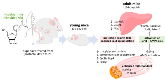
Nicotinamide Riboside Supplementation to Suckling Male Mice Improves Lipid and Energy Metabolism in Skeletal Muscle and Liver in Adulthood

1
Laboratori de Biologia Molecular, Nutrició i Biotecnologia (LBNB), Nutrigenomics, Biomarkers and Risk Evaluation Group, Universitat de les Illes Balears, 07122 Palma, Spain
2
CIBER de Fisiopatología de la Obesidad y Nutrición (CIBERobn), 07122 Palma, Spain
3
Institut d’Investigació Sanitària Illes Balears (IdISBa), 07120 Palma, Spain
*
Author to whom correspondence should be addressed.
Nutrients 2022, 14(11), 2259; https://doi.org/10.3390/nu14112259
Submission received: 25 April 2022 / Revised: 25 May 2022 / Accepted: 26 May 2022 / Published: 28 May 2022

Abstract

:
Nicotinamide riboside, an NAD+ precursor, has been attracting a lot of attention in recent years due to its potential benefits against multiple metabolic complications and age-related disorders related to NAD+ decline in tissues. The metabolic programming activity of NR supplementation in early-life stages is much less known. Here, we studied the long-term programming effects of mild NR supplementation during the suckling period on lipid and oxidative metabolism in skeletal muscle and liver tissues using an animal model. Suckling male mice received a daily oral dose of NR or vehicle (water) from day 2 to 20 of age, were weaned at day 21 onto a chow diet, and at day 90 were distributed to either a high-fat diet (HFD) or a normal-fat diet for 10 weeks. Compared to controls, NR-treated mice were protected against HFD-induced triacylglycerol accumulation in skeletal muscle and displayed lower triacylglycerol levels and steatosis degree in the liver and distinct capacities for fat oxidation and decreased lipogenesis in both tissues, paralleling signs of enhanced sirtuin 1 and AMP-dependent protein kinase signaling. These pre-clinical findings suggest that mild NR supplementation in early postnatal life beneficially impacts lipid and energy metabolism in skeletal muscle and liver in adulthood, serving as a potential preventive strategy against obesity-related disorders characterized by ectopic lipid accumulation.

Graphical Abstract

1. Introduction

Nicotinamide riboside (NR) is a form of vitamin B3 naturally contained in foods that functions as a precursor of NAD+ in mammals [1]. NAD+ is an essential redox cofactor for fuel oxidation and a rate-limiting substrate for sirtuins enzymes (SIRTs), which sense NAD+ levels to regulate multiple cellular processes, including energy metabolism. The NAD metabolome, particularly levels of NAD+ and/or NADP+ and NADPH, declines in target tissues in humans and rodents suffering from different forms of metabolic stress, such as aging, type 2 diabetes, and fatty liver (reviewed in [2,3,4]). Dietary NR supplementation appears to be beneficial in these contexts through replenishment of the NAD metabolome [2,3,4]. Among the NAD+ precursors, NR is of particular interest owing to its potency in vivo and lack of adverse clinical effects [2,4].
Supplementation with NR protects adult mice on obesogenic diets from weight gain and the development of insulin resistance and fatty liver [5,6,7]. NR supplementation favors liver health as it attenuates hepatic fat accumulation, fibrosis, low-grade inflammation, and liver damage in dietary animal models of hepatosteatosis [7,8], a genetic model of type 2 diabetes (KK/HIJ mice) [9], and in alcohol-fed mice [10,11]. Benefits of NR supplementation have also been described in animal models of muscle disorders, such as models of mitochondrial myopathies [12,13] and of muscular dystrophy characterized by muscle wasting [14,15]. Furthermore, NR treatment improves muscle quality and physical performance in middle-aged mice [16] and preserves muscle mass during weight loss in obese rats under calorie restriction [17]. The positive outcomes of these and other pre-clinical studies are prompting human trials assaying the impact of NR supplementation on a variety of metabolic and exercise-related end-points in healthy adults, old persons, and persons with obesity, with disparate results so far [18,19,20,21,22,23]. Mechanistically, the benefits of NR have been linked to increased mitochondria abundance and oxidative function in key metabolic tissues through NAD+ activation of SIRT1 [5,7,10,12,13], an enhancement of the differentiating capacity of adult progenitor cells [16], and effects on the gut microbiota [11,24].
NR is present in milk [1,25,26], which opens the door to programming effects on newborn development and future metabolism. Only recently, these aspects have begun to be explored [27,28,29]. Maternal NR supplementation (3 g/kg) during the lactation period enhances in mice maternal postpartum weight loss, milk production, and nursing behavior, resulting in weanlings with larger size and improved glycemic control, and it confers lasting advantages regarding behavioral/cognitive skills, physical performance, and body composition of offspring in adulthood [29]. Effects on offspring were largely attributed to increased transmission of macronutrients, micronutrients, and brain-derived neurotrophic factor (BDNF) into milk [29]. In a different approach, we have shown that direct, oral NR supplementation to suckling mice improves metabolic responses to high-fat diet (HFD) feeding in adulthood selectively in the male offspring, in which increases in lipolysis and the plasma leptin-to-adiponectin ratio following HFD feeding were blunted [27]. These beneficial effects are associated with a greater HFD-induced oxidative and thermogenic transcriptional response in brown and white adipose tissues (BAT and WAT) [27] and with changes in DNA methylation marks in brown fat-related genes in subcutaneous WAT [30]. Furthermore, early-life NR supplementation increased the commitment of progenitor cells resident in WAT of young male mice (but not young females) towards the beige (versus white) adipogenesis transcriptional program [28]. Collectively, these results indicated that NR supplementation during the suckling period programs for increased WAT energy metabolism in adulthood in male offspring. Whether early-life NR treatment could also influence skeletal muscle (SM) and liver metabolism features and responses to HFD feeding in the long-term in these animals remained to be studied. These aspects are addressed here.

2. Materials and Methods

2.1. Animal Experiment

The animal study protocols were reviewed and approved by the Bioethical Committee of the University of the Balearic Islands (Ref. 3513), and the handling of laboratory animals was according to international standards. The study design has been described in detail previously [27]. In brief, suckling NMRI mice in size-adjusted litters (12 pups/litter) received daily from postnatal day 2 to 20, with the aid of a pipette, a small volume (10–15 μL) of vehicle (water, control mice) or NR solution (Chemical Point, Desisenhofen, Germany) providing approximately 15-fold the total vitamin B3 ingested daily from maternal milk [27]. Weaning was on day 21, on a standard chow diet. The male offspring were used. From day 75 to day 90, the animals were habituated to a purified normal-fat diet (NFD, 10% energy as fat; Research Diets, New Brunswick, USA, ref. D12450J). On day 90, half of the animals in each treatment group (vehicle and NR) began receiving a purified high-fat diet (HFD, 45% energy as fat, mainly lard; Research Diets, ref. D12451) while the other half remained on the NFD, until euthanization on day 164 of age, making a total of 4 study groups (n = 5–6 animals/group, from 4–5 different litters: mice in each litter were split among the 4 study groups). The animals were euthanized by decapitation, under fed conditions, within the first 2 h of the light cycle. SM (gastrocnemius) and liver were dissected in their entirety, snap-frozen in liquid nitrogen, and stored at −80 °C until processing). Housing conditions were 2–3 animals per cage at 22 °C, with a 12 h light-dark cycle (lights on at 08:00) and ad libitum access to food and water. An independent experiment was conducted in which control and NR-treated male mice were euthanized at age 35 days (n = 5–6 animals/group). Early-life NR supplementation and the subsequent study protocol followed in this work were conducted in parallel with the study of long-term effects of early-life resveratrol supplementation [31] so that both works shared the control groups (i.e., the non-supplemented siblings studied after the NFD/HFD challenge in adulthood). Except for gastrocnemius muscle and liver weight of adult animals, other biometric parameters, as well as plasma and adipose tissue parameters of animal cohorts used in this work, have been reported elsewhere [27,28] (see also Supplementary Table S1).

2.2. RNA Isolation and Gene Expression Analysis

Total RNA was extracted from tissues using Tripure Reagent (Roche, Barcelona, Spain) following the supplier’s instructions. The RNA was purified with sodium acetate, quantified using a Nanodrop ND 1000 spectrophotometer (Nano-Drop Technologies Inc., Wilmington, NC, USA), and checked for integrity by means of agarose gel electrophoresis. Reverse transcription, PCR amplification of cDNAs of interest, and data analysis were conducted as previously described [32]. Sequences of primers used (from Sigma-Aldrich, St. Louis, MO, USA) are available upon request. Guanosine diphosphate dissociation inhibitor 1 (Gdi1), glyceraldehyde-3-phosphate dehydrogenase (Gapdh), or ribosomal protein, large, P0 (Rplp0) were used as reference genes for data normalization.

2.3. Mitochondrial DNA Content

Tissue total DNA was isolated with the DNeasy Blood & Tissue kit (Qiagen, Austin, TX, USA). qPCR quantification of a mitochondrial fragment DNA and a single-copy nuclear gene (beta-2-microglobulin) served to calculate the mitochondrial DNA to nuclear DNA ratio (mtDNA/nDNA), as previously described [33].

2.4. Lipid Extraction and Triacylglycerol Determination

Tissue lipids were extracted from 100 mg liver and 150 mg SM tissue, as previously described [34]. Triacylglycerol levels in the lipid extracts were analyzed using the Serum Triglyceride Determination Kit (Sigma-Aldrich).

2.5. Immunoblotting Analysis

Protein levels of total adenosine monophosphate-activated protein kinase (AMPK), phosphorylated AMPK (p-AMPK), and phosphorylated acetyl-CoA carboxylase (p-ACC) in SM were analyzed by immunoblotting as previously described [31], using the Odyssey Western blot kits (Li-Cor Biosciences, Lincoln, NE, USA). The signals of every protein of interest were normalized to the signal of heat shock protein 90.

2.6. Histological Analysis

Neutral lipid droplets in SM samples were detected by Oil Red O (ORO) staining of sections from frozen tissue, according to [35]. Freshly collected liver samples from 4 mice were fixed in 4% paraformaldehyde in 0.1 M sodium phosphate buffer, pH 7.4, overnight at 4 °C, dehydrated in a graded series of ethanol, and cleared in xylene before being embedded in paraffin blocks. Then, 5-micrometer-thick sections were cut with a microtome, mounted on slides, and stained with hematoxylin/eosin for light microscopy. Steatosis in histological liver sections was classified into grades according to Brunt et al. [36]. Grade 0 was assigned when no fat vacuoles were observed and grades 1, 2, and 3, when fat vacuoles were observed in less than 33%, between 33 to 66%, and more than 66% of hepatocytes, respectively.

2.7. Statistical Analysis

Data are expressed as mean ± SEM. Student’s t-test was used for single comparisons and two-way ANOVA to analyze the interaction of early-life treatment with the type of diet in adulthood (NFD or HFD). Statistical analyses were performed with IBM SPSS Statistics for Windows, version 27.0 (IBM Corp., Armonk, NY, USA). The threshold of significance was set at p < 0.05. Trends are highlighted when 0.05 < p < 0.1.

3. Results

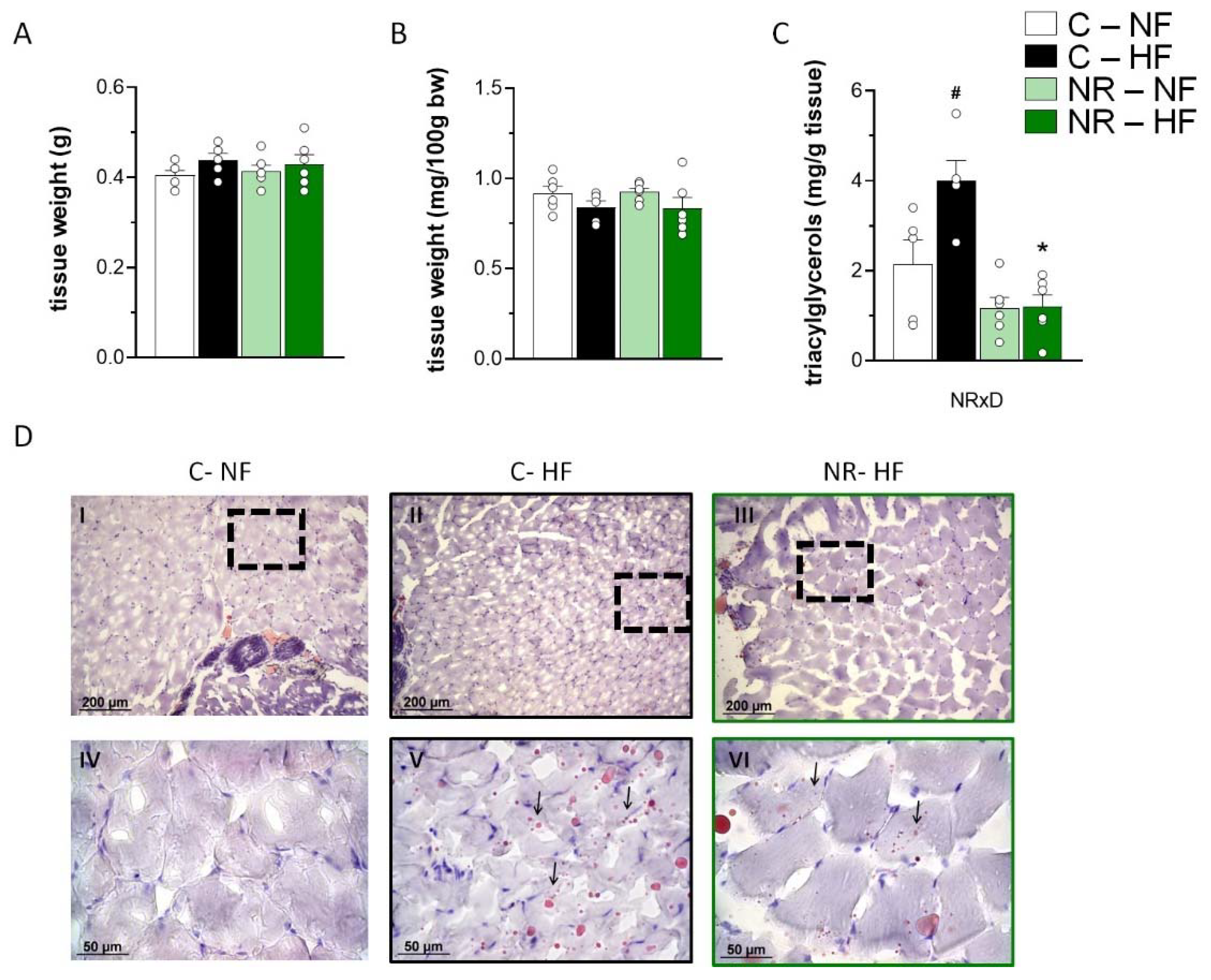
3.1. Early-Life NR Treatment Protected against HFD-Induced Triacylglycerol Accumulation in SM in Adulthood

To assess the long-term effects of early-life NR treatment on muscle features, we began by considering muscle mass and lipid content in control and treated adult mice on an NFD and after a 10-week HFD feeding period (Figure 1). Gastrocnemius muscle weight in adulthood was not affected by early-life NR treatment when expressed either as absolute value (mg) or relative to body weight (as %) (Figure 1A,B). The relative gastrocnemius muscle weight tended to be decreased by HFD feeding, irrespective of the neonatal NR treatment (p = 0.059 for diet effect, two-way ANOVA) (Figure 1B). Muscle triacylglycerol content, as determined by biochemical analyses (including both intramyocellular and extramyocellular triacylglycerol), was robustly increased after 10 weeks of HFD in the control mice but not the NR mice, who had lower content than controls after HFD (Figure 1C). Oil Red O staining of frozen muscle sections confirmed the presence of more intramyocellular neutral lipid droplets in SM of HFD-fed as compared to NFD-fed control mice and of fewer lipid droplets in the NR mice as compared to the control mice after HFD (Figure 1D). Thus, NR supplementation in the early stages of postnatal life suppressed the accumulation of triacylglycerols in SM induced by HFD feeding later in life.

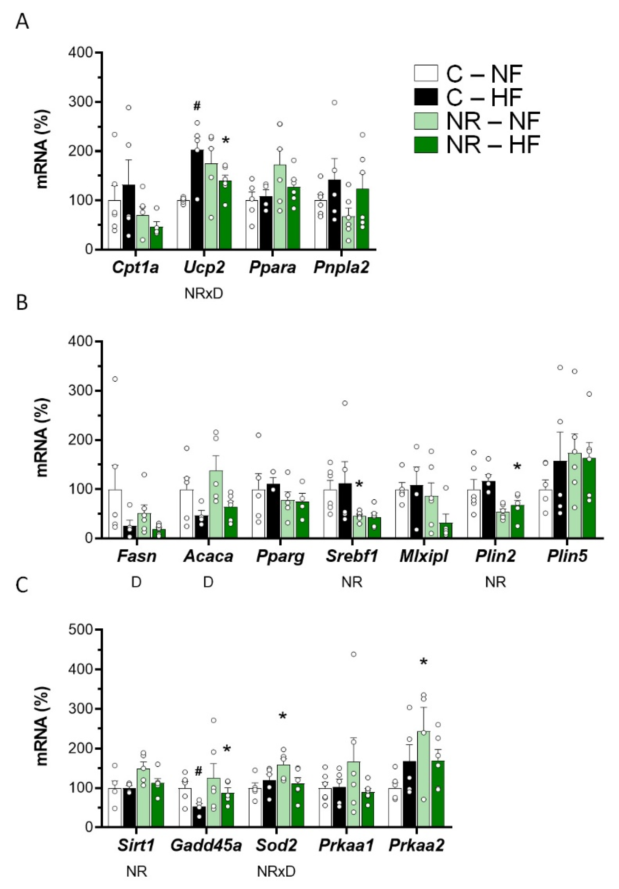
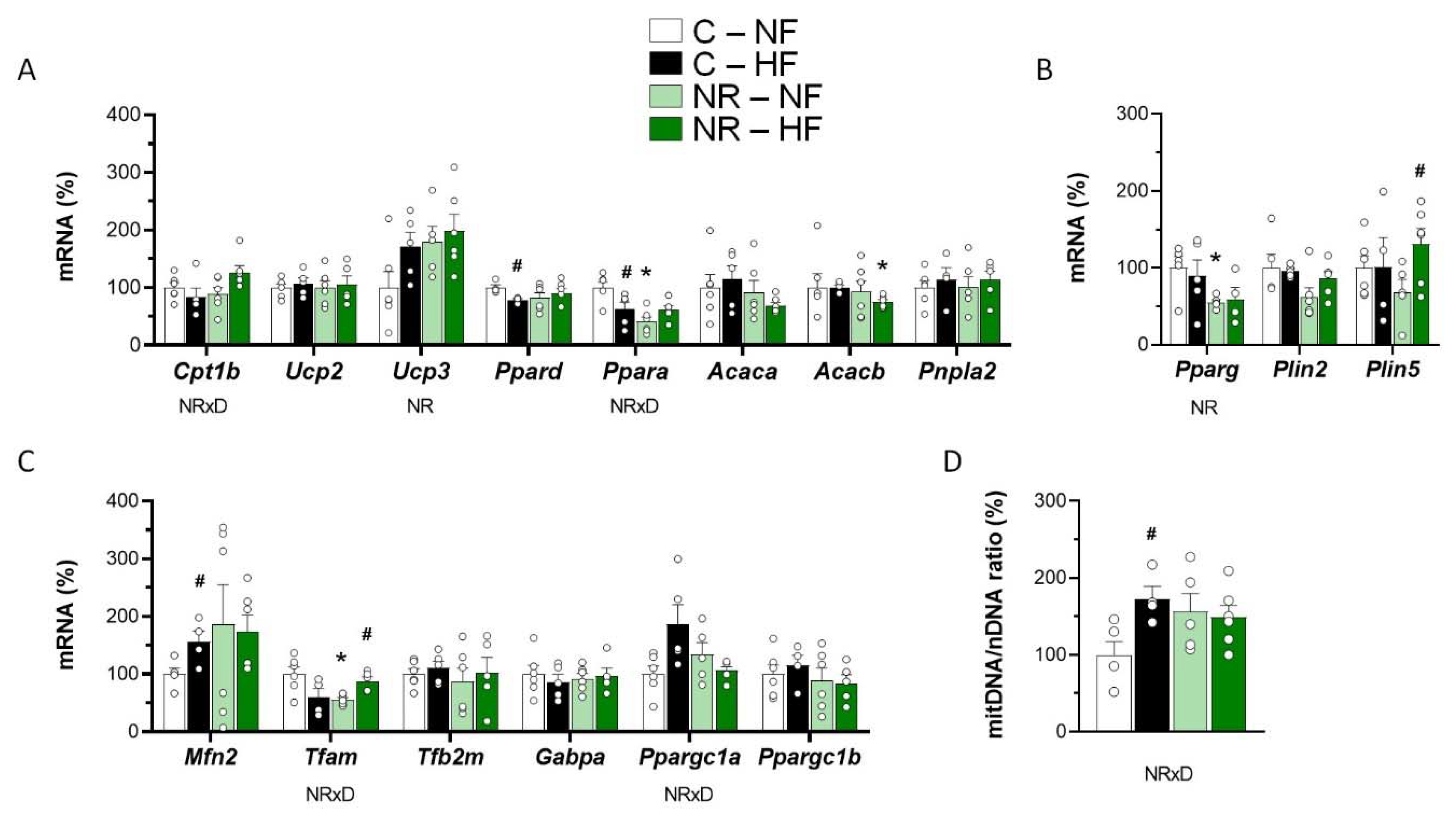
3.2. Early-Life NR Treatment Impacted Lipid Metabolism and Mitochondria Pathways in SM in Adulthood

Expression levels of genes related to fatty acid oxidation (FAO)/lipolysis (Figure 2A), lipogenesis/lipid droplet dynamics (Figure 2B), and mitochondria biogenesis/function (Figure 2C) were assessed in SM of control and NR mice after the 10-week NFD/HFD challenge. In good concordance with the muscle triacylglycerol content results, gene expression of Cpt1b—Encoding carnitine palmitoyl transferase 1 (CPT1), isoform b, the rate-limiting enzyme in muscle mitochondrial FAO—was increased by HFD feeding in the NR mice, but not the control mice (significant interactive treatment × diet effect, two-way ANOVA). Moreover, gene expression levels of Acacb—Encoding acetyl-CoA carboxylase (ACC, isoform b) producing the CPT1 inhibitor malonyl-CoA [37,38]—were lower in SM of NR mice compared with controls under HFD (p < 0.05, t-Student). Gene expression levels of Ucp3—encoding uncoupling protein 3, whose expression in SM highly correlates with the tissue FAO rate [21,22]—were greater in the NR mice, as indicated by two-way ANOVA. We also assessed the mRNA levels in SM of Ppara and Ppard, coding for peroxisome proliferator-activated receptor (PPAR) transcription factors involved in the transcriptional control of FAO, namely PPARɑ and PPARδ, respectively. NR mice displayed lower Ppara mRNA levels than controls under NFD, yet an HFD-induced decrease in Ppara mRNA levels present in the control mice was absent in the NR mice, resulting in a significant interactive treatment × diet effect (two-way ANOVA). Likewise, HFD feeding resulted in decreased Ppard mRNA levels in SM in control mice (p < 0.05, t-Student) but not in NR mice. Muscle gene expressions of Acaca (encoding ACC, isoform a), Ucp2 (encoding uncoupling protein 2), and Pnpla2 (encoding lipolytic adipose triglyceride lipase) were not affected by early-life NR treatment or the fat content of the diet (Figure 2A).
Observed differences in the expression levels in muscle of genes related to lipogenesis and lipid droplet metabolism were also in good concordance with the muscle triacylglycerol content results. Gene expression of Pparg, encoding PPARγ, was significantly decreased in SM of NR mice, and that of Plin2, encoding Perilipin2, trended lower as well in the NR mice (p = 0.057 for the NR effect, two-way ANOVA) (Figure 2B). PPARγ is a lipogenic transcription factor whose activity in SM has been related to fat infiltration in the tissue [39], and there is evidence that Perilipin2 participates in intramyocellular lipids synthesis and lipid droplet growth [40]. Interestingly, expression in SM of Plin5, encoding Perilipin5, was markedly and significantly induced after HFD feeding selectively in the NR mice but not the control mice. This is remarkable, considering that Perilipin5 anchors mitochondria to the lipid droplet membrane and that coupling via Perilipin5 augments mitochondria respiratory capacity [41].
The mtDNA/nDNA was used as a surrogate index of mitochondria content. HFD feeding increased this ratio in SM of control mice, in keeping with previous reports [42,43] but not further in the NR mice, which tended to have a higher mtDNA/nDNA ratio than controls under basal (NFD) conditions (p= 0.088, t-Student) (Figure 2D). Differences among groups in the SM mtDNA/nDNA ratio grossly mirrored differences in expression in SM of Mfn2 (encoding mitofusin 2) and Ppargc1a (encoding PPAR gamma coactivator 1 alpha) (Figure 2C). Strikingly, SM Tfam mRNA levels were lower in the NR mice than in the control mice under NFD, yet they were increased following HFD feeding in the NR mice only, resulting in a significant interactive treatment × diet effect (two-way ANOVA). Gene expression in SM of other genes related to mitochondria biogenesis and function analyzed (Tfb2m, Ppargc1b, Gabpa) was similar in all experimental groups (Figure 2D).

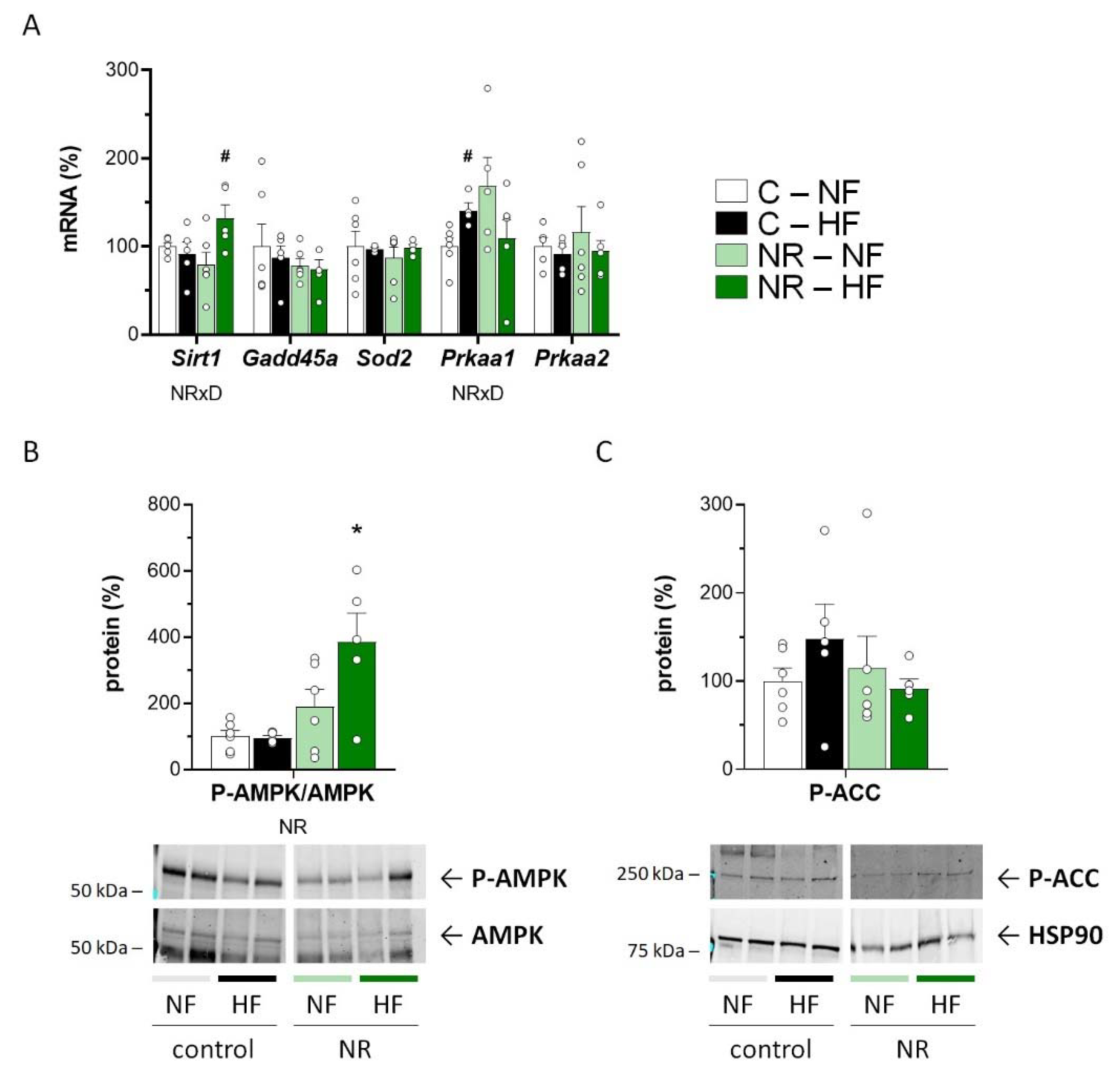
3.3. Early-Life NR Treatment Up-regulated AMPK and SIRT1 in SM in Adulthood

SIRT1 is a key mediator of the NR effects on energy metabolism [5], and SIRT1 and AMPK can positively feedback each other’s activity to activate FAO and mitochondrial biogenesis [44,45]. Interestingly, HFD feeding led to increased muscle Sirt1 mRNA levels selectively in the NR mice (Figure 3A), who also had a greater phospho-AMPK to AMPK ratio in muscle than control mice, especially following HFD feeding (Figure 3B). Additionally, muscle mRNA levels of Prkaa1, encoding one of two known AMPK catalytic subunit isoforms, were up-regulated in the NR mice compared with controls under NFD (p = 0.053, t-Student), albeit they were increased upon HFD in the control mice only (Figure 3A). These results are compatible with a relative up-regulation of the SIRT1/AMPK axis in SM of adult NR mice. However, known downstream transcriptional SIRT1 responses [46], namely Gadd45a and Sod2 expression, were not affected by the early-life NR treatment or the fat content of the diet (Figure 3A), and the same was true for the levels in muscle of the AMPK product phospho-ACC (Figure 3C).

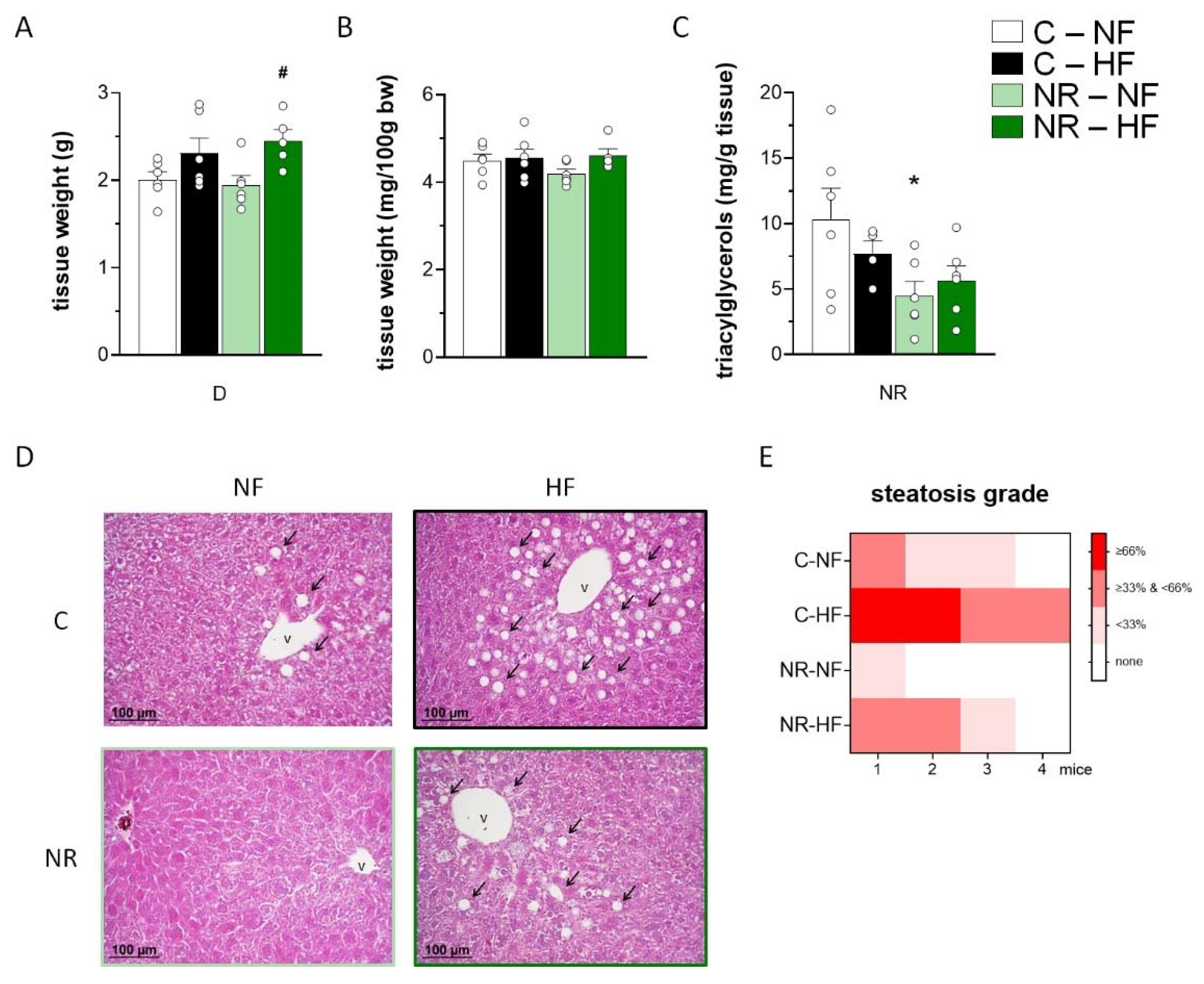
3.4. Early-Life NR Treatment Resulted in Decreased Liver Triacylglycerol Content and Modified Lipid Metabolism Capacities in Liver in Adulthood

Liver weight was increased by HFD feeding irrespective of NR treatment (Figure 4A), while the liver index (percentage of liver weight/body weight) was unaffected (Figure 4B). Liver triacylglycerol content, as measured through biochemical analysis, was unaffected by the HFD regimen imposed (Figure 4C), yet it was significantly lower in the NR mice than in the control mice both after NFD and HFD, as indicated by two-way ANOVA. Liver steatosis as a histological diagnosis was analyzed and semi-quantified (Figure 4D). Histological analyses of liver sections revealed an increased degree of steatosis in the HFD-fed animals as compared to the NFD-fed animals and a lower degree of steatosis in the NR mice as compared to the control mice, both after NFD and HFD feeding (Figure 4D,E).
Hepatic mRNA expression levels in the fed state of sets of genes related to FAO and lipolysis (Cpt1a, Ucp2, Ppara, Pnpla2), lipogenesis and lipid droplets (Fasn, Acaca, Pparg, Srebf1, Mlxipl, Plin2, Plin5), and the status of SIRT1 and AMPK pathways (Sirt1, Gadd45a, Sod2, Prkaa1, Prkaa2) are shown in Figure 5A–C, respectively. The lower triacylglycerol content found in the liver of adult NR mice paralleled significantly decreased expression levels of Srebf1 and Plin2 in the liver of these animals as compared with controls. Trends towards decreased expression of Mlxipl (p = 0.081 for treatment effect, two-way ANOVA, mainly under HFD) and increased expression of Ppara (p = 0.057 for treatment effect, two-way ANOVA) were also detected in the liver of NR animals. These results may be suggestive of decreased lipogenesis/lipid droplet formation and increased FAO in the liver of NR mice in the fed state. Nevertheless, a trend in reduction of liver Cpt1a expression levels was apparent in the NR mice (p = 0.071 for treatment effect, two-way ANOVA). Liver Ucp2 expression levels were increased after HFD in the control mice but not in the NR mice, although the NR mice tended to have increased basal levels than controls on the NFD (p = 0.075, t-Student). Hepatic gene expressions of Fasn and Acaca, encoding key enzymes for de novo synthesis of fatty acids in the liver, were suppressed in the HFD-fed animals regardless of the neonatal NR treatment, which had no effect on the expression of these two genes. Hepatic gene expressions of Pnpla2 and Pparg were unaffected by neonatal NR treatment or the fat content of the diet (Figure 5A,B). Regarding the genes related to the status of SIRT1 and AMPK pathways (Figure 5C), compared to their controls, adult NR mice had significantly increased hepatic Sirt1 mRNA expression levels regardless of the type of diet (as indicated by two-way ANOVA) and significantly increased Prkaa2 mRNA expression levels under NFD (p < 0.05, t-Student). Further, hepatic gene expression levels of SIRT1 transcriptional targets analyzed were greater in the NR mice as compared with the control mice under HFD (case of Gadd45a) or NFD (case of Sod2) feeding conditions.

4. Discussion

NR has been attracting a lot of attention in recent years owing to accumulated evidence of the beneficial effects of its contemporary administration against multiple metabolic complications and age-related disorders involving NAD+ decline in tissues [47]. The programming activity of NR supplementation in early-life stages has been much less studied, only recently, and so far mostly regarding long-term effects on adipose tissue features (our previous reports [27,28,30]) and cognitive/behavioral skills [29]. Using the same study design as in the present work, we previously reported sex-specific long-term effects of early life NR supplementation favoring better systemic responses to HFD feeding and thermogenic/oxidative capacities in WAT, specifically in the male offspring [27]. Here, we pursued a more comprehensive characterization of the programming activity of NR in the male progeny and reported novel results revealing long-term beneficial effects of direct NR supplementation to suckling mice on lipid and energy metabolism in SM and liver in adulthood. Results herein and in our previous works are noteworthy, considering the low NR dosage used and that treatment was limited to the suckling period, while the animals were challenged with an obesogenic diet and analyzed much later in life. Importantly, our study design allows studying direct programming effects of oral NR, independent of changes in milk composition that are inherent to (and demonstrated in) other designs, namely NR supplementation to the lactating dams [29].
Even if a non-obesity prone mouse strain was used in this work (NMRI mice), the HFD regimen applied (10 weeks duration, 45% calories as fat) did increase body weight, adiposity, and leptinemia (Supplementary Table S1 and [27]), as well as (this work) muscle triacylglycerol content and the degree of liver steatosis in our control mice, although it failed to alter insulin sensitivity or glucose control [27]. The observed HFD-induced increase in muscle triacylglycerol content is in good concordance with previous reports of this effect in multiple mouse strains after just 8 weeks of an HFD similar to the one used here ([48] and references therein). The fact that the HFD-induced increase in liver steatosis—which is a histological diagnosis of fat accumulation—was dissociated from an increase in the liver triacylglycerol content is not without precedent. For instance, in studies examining the timeline of the onset of non-alcoholic fatty liver disease in HFD-fed (60% energy as fat), obesity-prone (C57BL/6J) male mice, histological steatosis appeared well before a persistent, significant increase in liver triacylglycerol content could be detected (week 4 versus week 7–8) [49].
Interestingly, male mice supplemented with NR during the suckling period were completely protected against HFD-induced triacylglycerol accumulation in SM and had lower triacylglycerol content and steatosis in the liver after both NFD and HFD feeding compared to their control siblings. From the muscle results, protection against HFD-induced insulin resistance/prediabetes can be anticipated in the mice neonatally treated with NR, considering the close relationship between muscle (intramyocellular) lipid content and systemic insulin resistance, and that muscle lipids primarily reflect muscle triacylglycerol content (even if other lipid species, such as diacylglycerol or ceramides, mediate the unwanted effects) [50]. Likewise, protection against HFD-induced fatty liver progression is suggested by the liver results. Protective effects of early-life NR supplementation against metabolic complications under more severe models or pro-obesogenic feeding conditions in adulthood deserve further investigation, although it might be that, under such severe conditions, mild programming effects of low-dose neonatal nutritional treatments are blunted.
Protection against HFD-induced triacylglycerol accumulation in SM in the NR mice could be related to gene expression changes in the tissue observed selectively in these animals, but not in controls, following HFD, such as the HFD-induced increases in Plin5 and Cpt1b expression and decrease in Acacb expression. These changes in concert could favor increased dietary fat oxidation in muscle in the NR mice through increased Perilipin5-mediated enhancement of mitochondria function [41], increased CPT1 abundance, and increased CPT1 activity owing to lower levels of inhibitory malonyl-CoA. Furthermore, NR mice may be better prepared than controls to handle the excess dietary fat through oxidative metabolism in SM from the very beginning of the HFD challenge, as they displayed increased Ucp3 gene expression, as well as a trend to increased mitochondria content under basal (NFD) conditions. In good concordance with increased FAO in SM are also results herein suggesting a relative activation of SIRT1 and AMPK pathways in SM of NR mice as compared to controls. Dissociation of AMPK phosphorylation from ACC phosphorylation in the context of muscle FAO control, as observed here in the NR mice, has been reported previously in some settings [51,52]. In this context, it is to be noted that, besides acute stimulatory effects on FAO linked to AMPK-catalyzed ACC phosphorylation, active AMPK favors FAO through the induction of the expression of FAO-related genes, such as Cpt1b and Ucp3 [52,53,54]. The latter mechanisms appear to be preponderant in our experimental setting. Regarding the programming of liver metabolism, lower liver triacylglycerol content in the adult NR mice, as observed, could be related to increased FAO (up-regulated Ucp2 and Ppara expressions in NFD) and, more consistently, decreased lipogenesis and lipid droplet formation (down-regulated Srebf1, Mlxipl, and Plin2 expressions). As for muscle, results obtained are compatible with a relative activation of SIRT1 and AMPK pathways in the liver of adult NR mice as compared with their control siblings.
The study protocol used to assess long-term metabolic programming activity was conducted in parallel with early-life supplementation with NR (this work) and with the polyphenol resveratrol (RSV) [31]. NR and RSV have both been described as activators of SIRT1 [5,55,56], which is an important regulator of cellular energy metabolism [57] and a known epigenetic landscaper modulator [58]. In fact, early-life supplementation with NR (this work) or RSV [31] had similar effects on lipid metabolism in SM of male mice in adulthood and also effects in common in the liver. However, differences between NR and RSV programming effects were also noticed, especially regarding adult liver traits and responses. In particular, decreased liver triacylglycerol content and attenuation of liver steatosis were significant or apparent in the NR mice only. Differences were to be expected since NR and RSV have additional, non-overlapping biological targets for interaction besides SIRT1. In fact, the effects of the two neonatal treatments on WAT features in adulthood (including DNA methylation marks in browning-related genes in WAT) and the adipogenic fate of primary preadipocytes of young animals were similar but not identical [27,28,30]. For instance, long-term effects of early-life NR supplementation on WAT gene expression were seen mainly under HFD feeding conditions, which was not the case for early-life RSV supplementation [27].
The ultimate mechanisms explaining the distinct phenotypes observed in adulthood in neonatally NR-treated mice remain to be established. Strikingly, the early-life NR treatment applied had multiple effects on gene expression and responses to HFD feeding in SM and liver in adulthood, yet it did not affect the expression profile of studied genes in SM or liver of mice at a young age (35 days; Supplementary Figure S1 and S2). There is evidence that NR treatment can impact the cellular DNA methylation machinery, at least in adipocytes/adipose tissue [30], and it is conceivable that supplemental NR during the suckling period (or a stimulus subsequent to NR supplementation, such as SIRT1 activation) triggers an epigenetic effect or wave of effects in SM and/or liver tissues that is revealed, in terms of gene expression, later in life. However, early-life NR treatment in male mice leads to enhanced responses of WAT and BAT oxidative metabolism to HFD feeding in adulthood [27], and therefore, it cannot be overlooked that changes observed in muscle and liver of neonatally NR-treated mice might be secondary to the programming of more oxidative adipose tissues. Clearly, much more needs to be investigated regarding the mechanisms behind the metabolic programming of muscle and liver traits linked to early-life NR supplementation revealed, for the first time to our knowledge, in this work.
Taken together with our previous results [27], results herein suggest that, similar to early-life RSV supplementation, early-life NR supplementation could represent an adequate, multi-target-directed strategy to prevent obesity and metabolic disturbances related to ectopic fat accumulation in non-adipose tissues in adulthood in the male progeny. The sex-specificity of this recommendation should be stressed, particularly for NR, because our previous findings indicated not only a lack of induction of adult WAT browning but also alterations of concern in systemic glucose and lipid metabolism parameters in adulthood in the neonatally NR-supplemented female mice [27].

Supplementary Materials

The following supporting information can be downloaded at: https://www.mdpi.com/article/10.3390/nu14112259/s1, Table S1: Early-life nicotinamide riboside (NR) treatment attenuated high-fat (HF) diet-induced body fat accumulation and energy; Figure S1: Early-life nicotinamide riboside (NR) treatment did not significantly affect lipid metabolism capacities and mitochondria pathways in skeletal muscle of mice at a young age; Figure S2: Early-life nicotinamide riboside (NR) treatment did not significantly affect lipid metabolism capacities in liver of mice at a young age; Figure S3: Original images supporting western blot results shown in the histograms in Figure 3 panels B and C.

Author Contributions

M.L.B., J.R., and A.P. conceived the work. J.R. and M.L.B. designed the work. A.S. managed the animals and acquired data. M.L.B. and J.R. wrote the manuscript. All authors analyzed and interpreted data, revised the manuscript critically, and gave final approval of the version to be published. All authors have read and agreed to the published version of the manuscript.

Funding

This work was funded by the project PGC2018-097436-B-I00 financed by MCIN/AEI/10.13039/501100011033/ and “FEDER Una manera de hacer Europa” (to A.P.) and an XVI Concurso Nacional para la Adjudicación de Ayudas a la Investigación Científica y Técnica grant of the Fundación Ramón Areces (to M.L.B.).

Institutional Review Board Statement

The animal study protocol was approved by the Bioethical Committee of Universitat de les Illes Balears (protocol code 3513, approved 26/03/2012).

Data Availability Statement

All data collected are reported in the manuscript and the supplementary material.

Acknowledgments

The authors wish to thank the employees of the animal care unit of the Universitat de les Illes Balears and Enzo Ceresi for his excellent technical assistance with the histochemical analyses.

Conflicts of Interest

The authors declare no conflict of interest.

References

  1. Bieganowski, P.; Brenner, C. Discoveries of nicotinamide riboside as a nutrient and conserved NRK genes establish a Preiss-Handler independent route to NAD+ in fungi and humans. Cell 2004, 117, 495–502. [Google Scholar] [CrossRef] [Green Version]
  2. Yoshino, J.; Baur, J.A.; Imai, S.I. NAD(+) Intermediates: The Biology and Therapeutic Potential of NMN and NR. Cell Metab. 2018, 27, 513–528. [Google Scholar] [CrossRef] [PubMed] [Green Version]
  3. Okabe, K.; Yaku, K.; Tobe, K.; Nakagawa, T. Implications of altered NAD metabolism in metabolic disorders. J. Biomed. Sci. 2019, 26, 34. [Google Scholar] [CrossRef] [PubMed] [Green Version]
  4. Connell, N.J.; Houtkooper, R.H.; Schrauwen, P. NAD(+) metabolism as a target for metabolic health: Have we found the silver bullet? Diabetologia 2019, 62, 888–899. [Google Scholar] [CrossRef] [PubMed] [Green Version]
  5. Canto, C.; Houtkooper, R.H.; Pirinen, E.; Youn, D.Y.; Oosterveer, M.H.; Cen, Y.; Fernandez-Marcos, P.J.; Yamamoto, H.; Andreux, P.A.; Cettour-Rose, P.; et al. The NAD(+) precursor nicotinamide riboside enhances oxidative metabolism and protects against high-fat diet-induced obesity. Cell Metab. 2012, 15, 838–847. [Google Scholar] [CrossRef] [Green Version]
  6. Trammell, S.A.; Weidemann, B.J.; Chadda, A.; Yorek, M.S.; Holmes, A.; Coppey, L.J.; Obrosov, A.; Kardon, R.H.; Yorek, M.A.; Brenner, C. Nicotinamide Riboside Opposes Type 2 Diabetes and Neuropathy in Mice. Sci. Rep. 2016, 6, 26933. [Google Scholar] [CrossRef] [Green Version]
  7. Gariani, K.; Menzies, K.J.; Ryu, D.; Wegner, C.J.; Wang, X.; Ropelle, E.R.; Moullan, N.; Zhang, H.; Perino, A.; Lemos, V.; et al. Eliciting the mitochondrial unfolded protein response by nicotinamide adenine dinucleotide repletion reverses fatty liver disease in mice. Hepatology 2016, 63, 1190–1204. [Google Scholar] [CrossRef]
  8. Pham, T.X.; Bae, M.; Kim, M.B.; Lee, Y.; Hu, S.; Kang, H.; Park, Y.K.; Lee, J.Y. Nicotinamide riboside, an NAD+ precursor, attenuates the development of liver fibrosis in a diet-induced mouse model of liver fibrosis. Biochim. Biophys. Acta. Mol. Basis Dis. 2019, 1865, 2451–2463. [Google Scholar] [CrossRef]
  9. Lee, H.J.; Hong, Y.S.; Jun, W.; Yang, S.J. Nicotinamide Riboside Ameliorates Hepatic Metaflammation by Modulating NLRP3 Inflammasome in a Rodent Model of Type 2 Diabetes. J. Med. Food 2015, 18, 1207–1213. [Google Scholar] [CrossRef]
  10. Wang, S.; Wan, T.; Ye, M.; Qiu, Y.; Pei, L.; Jiang, R.; Pang, N.; Huang, Y.; Liang, B.; Ling, W.; et al. Nicotinamide riboside attenuates alcohol induced liver injuries via activation of SirT1/PGC-1alpha/mitochondrial biosynthesis pathway. Redox. Biol. 2018, 17, 89–98. [Google Scholar] [CrossRef]
  11. Yu, X.; Xue, M.; Liu, Y.; Zhou, Z.; Jiang, Y.; Sun, T.; Liang, H. Effect of nicotinamide riboside on lipid metabolism and gut microflora-bile acid axis in alcohol-exposed mice. Food Sci. Nutr. 2021, 9, 429–440. [Google Scholar] [CrossRef] [PubMed]
  12. Cerutti, R.; Pirinen, E.; Lamperti, C.; Marchet, S.; Sauve, A.A.; Li, W.; Leoni, V.; Schon, E.A.; Dantzer, F.; Auwerx, J.; et al. NAD(+)-dependent activation of Sirt1 corrects the phenotype in a mouse model of mitochondrial disease. Cell Metab. 2014, 19, 1042–1049. [Google Scholar] [CrossRef] [Green Version]
  13. Khan, N.A.; Auranen, M.; Paetau, I.; Pirinen, E.; Euro, L.; Forsstrom, S.; Pasila, L.; Velagapudi, V.; Carroll, C.J.; Auwerx, J.; et al. Effective treatment of mitochondrial myopathy by nicotinamide riboside, a vitamin B3. EMBO Mol. Med. 2014, 6, 721–731. [Google Scholar] [CrossRef] [PubMed]
  14. Ryu, D.; Zhang, H.; Ropelle, E.R.; Sorrentino, V.; Mazala, D.A.; Mouchiroud, L.; Marshall, P.L.; Campbell, M.D.; Ali, A.S.; Knowels, G.M.; et al. NAD+ repletion improves muscle function in muscular dystrophy and counters global PARylation. Sci. Transl. Med. 2016, 8, 361ra139. [Google Scholar] [CrossRef] [PubMed] [Green Version]
  15. Zhang, H.; Ryu, D.; Wu, Y.; Gariani, K.; Wang, X.; Luan, P.; D’Amico, D.; Ropelle, E.R.; Lutolf, M.P.; Aebersold, R.; et al. NAD(+) repletion improves mitochondrial and stem cell function and enhances life span in mice. Science 2016, 352, 1436–1443. [Google Scholar] [CrossRef] [PubMed] [Green Version]
  16. Seldeen, K.L.; Shahini, A.; Thiyagarajan, R.; Redae, Y.; Leiker, M.; Rajabian, N.; Dynka, A.; Andreadis, S.T.; Troen, B.R. Short-term nicotinamide riboside treatment improves muscle quality and function in mice and increases cellular energetics and differentiating capacity of myogenic progenitors. Nutrition 2021, 87–88, 111189. [Google Scholar] [CrossRef] [PubMed]
  17. de Castro, J.M.; Stein, D.J.; Medeiros, H.R.; de Oliveira, C.; Torres, I.L.S. Nicotinamide Riboside Neutralizes Hypothalamic Inflammation and Increases Weight Loss Without Altering Muscle Mass in Obese Rats Under Calorie Restriction: A Preliminary Investigation. Front. Nutr. 2021, 8, 648893. [Google Scholar] [CrossRef]
  18. Martens, C.R.; Denman, B.A.; Mazzo, M.R.; Armstrong, M.L.; Reisdorph, N.; McQueen, M.B.; Chonchol, M.; Seals, D.R. Chronic nicotinamide riboside supplementation is well-tolerated and elevates NAD(+) in healthy middle-aged and older adults. Nat. Commun. 2018, 9, 1286. [Google Scholar] [CrossRef]
  19. Dollerup, O.L.; Christensen, B.; Svart, M.; Schmidt, M.S.; Sulek, K.; Ringgaard, S.; Stodkilde-Jorgensen, H.; Moller, N.; Brenner, C.; Treebak, J.T.; et al. A randomized placebo-controlled clinical trial of nicotinamide riboside in obese men: Safety, insulin-sensitivity, and lipid-mobilizing effects. Am. J. Clin. Nutr. 2018, 108, 343–353. [Google Scholar] [CrossRef] [Green Version]
  20. Dolopikou, C.F.; Kourtzidis, I.A.; Margaritelis, N.V.; Vrabas, I.S.; Koidou, I.; Kyparos, A.; Theodorou, A.A.; Paschalis, V.; Nikolaidis, M.G. Acute nicotinamide riboside supplementation improves redox homeostasis and exercise performance in old individuals: A double-blind cross-over study. Eur. J. Nutr. 2019, 59, 505–515. [Google Scholar] [CrossRef]
  21. Elhassan, Y.S.; Kluckova, K.; Fletcher, R.S.; Schmidt, M.S.; Garten, A.; Doig, C.L.; Cartwright, D.M.; Oakey, L.; Burley, C.V.; Jenkinson, N.; et al. Nicotinamide Riboside Augments the Aged Human Skeletal Muscle NAD(+) Metabolome and Induces Transcriptomic and Anti-inflammatory Signatures. Cell Rep. 2019, 28, 1717–1728.e1716. [Google Scholar] [CrossRef] [PubMed] [Green Version]
  22. Stocks, B.; Ashcroft, S.P.; Joanisse, S.; Dansereau, L.C.; Koay, Y.C.; Elhassan, Y.S.; Lavery, G.G.; Quek, L.E.; O’Sullivan, J.F.; Philp, A.M.; et al. Nicotinamide riboside supplementation does not alter whole-body or skeletal muscle metabolic responses to a single bout of endurance exercise. J. Physiol. 2021, 599, 1513–1531. [Google Scholar] [CrossRef] [PubMed]
  23. Remie, C.M.E.; Roumans, K.H.M.; Moonen, M.P.B.; Connell, N.J.; Havekes, B.; Mevenkamp, J.; Lindeboom, L.; de Wit, V.H.W.; van de Weijer, T.; Aarts, S.; et al. Nicotinamide riboside supplementation alters body composition and skeletal muscle acetylcarnitine concentrations in healthy obese humans. Am. J. Clin. Nutr. 2020, 112, 413–426. [Google Scholar] [CrossRef] [PubMed]
  24. Lozada-Fernandez, V.V.; deLeon, O.; Kellogg, S.L.; Saravia, F.L.; Hadiono, M.A.; Atkinson, S.N.; Grobe, J.L.; Kirby, J.R. Nicotinamide Riboside-Conditioned Microbiota Deflects High-Fat Diet-Induced Weight Gain in Mice. mSystems 2022, 7, e0023021. [Google Scholar] [CrossRef] [PubMed]
  25. Trammell, S.A.; Yu, L.; Redpath, P.; Migaud, M.E.; Brenner, C. Nicotinamide Riboside Is a Major NAD+ Precursor Vitamin in Cow Milk. J. Nutr. 2016, 146, 957–963. [Google Scholar] [CrossRef] [PubMed] [Green Version]
  26. Ummarino, S.; Mozzon, M.; Zamporlini, F.; Amici, A.; Mazzola, F.; Orsomando, G.; Ruggieri, S.; Raffaelli, N. Simultaneous quantitation of nicotinamide riboside, nicotinamide mononucleotide and nicotinamide adenine dinucleotide in milk by a novel enzyme-coupled assay. Food Chem. 2017, 221, 161–168. [Google Scholar] [CrossRef] [PubMed]
  27. Serrano, A.; Asnani-Kishnani, M.; Rodriguez, A.M.; Palou, A.; Ribot, J.; Bonet, M.L. Programming of the Beige Phenotype in White Adipose Tissue of Adult Mice by Mild Resveratrol and Nicotinamide Riboside Supplementations in Early Postnatal Life. Mol. Nutr. Food Res. 2018, 62, e1800463. [Google Scholar] [CrossRef]
  28. Asnani-Kishnani, M.; Rodriguez, A.M.; Serrano, A.; Palou, A.; Bonet, M.L.; Ribot, J. Neonatal Resveratrol and Nicotinamide Riboside Supplementations Sex-Dependently Affect Beige Transcriptional Programming of Preadipocytes in Mouse Adipose Tissue. Front Physiol. 2019, 10, 83. [Google Scholar] [CrossRef] [Green Version]
  29. Ear, P.H.; Chadda, A.; Gumusoglu, S.B.; Schmidt, M.S.; Vogeler, S.; Malicoat, J.; Kadel, J.; Moore, M.M.; Migaud, M.E.; Stevens, H.E.; et al. Maternal Nicotinamide Riboside Enhances Postpartum Weight Loss, Juvenile Offspring Development, and Neurogenesis of Adult Offspring. Cell Rep. 2019, 26, 969–983.e964. [Google Scholar] [CrossRef] [Green Version]
  30. Serrano, A.; Asnani-Kishnani, M.; Couturier, C.; Astier, J.; Palou, A.; Landrier, J.F.; Ribot, J.; Bonet, M.L. DNA Methylation Changes are Associated with the Programming of White Adipose Tissue Browning Features by Resveratrol and Nicotinamide Riboside Neonatal Supplementations in Mice. Nutrients 2020, 12, 461. [Google Scholar] [CrossRef] [Green Version]
  31. Serrano, A.; Ribot, J.; Palou, A.; Bonet, M.L. Long-term programming of skeletal muscle and liver lipid and energy metabolism by resveratrol supplementation to suckling mice. J. Nutr. Biochem. 2021, 95, 108770. [Google Scholar] [CrossRef] [PubMed]
  32. Petrov, P.D.; Ribot, J.; Palou, A.; Bonet, M.L. Improved metabolic regulation is associated with retinoblastoma protein gene haploinsufficiency in mice. Am. J. Physiol. Endocrinol. Metab. 2015, 308, E172–E183. [Google Scholar] [CrossRef] [PubMed] [Green Version]
  33. Jokinen, R.; Marttinen, P.; Sandell, H.K.; Manninen, T.; Teerenhovi, H.; Wai, T.; Teoli, D.; Loredo-Osti, J.C.; Shoubridge, E.A.; Battersby, B.J. Gimap3 regulates tissue-specific mitochondrial DNA segregation. PLoS Genet. 2010, 6, e1001161. [Google Scholar] [CrossRef] [PubMed]
  34. Caimari, A.; Oliver, P.; Palou, A. Adipose triglyceride lipase expression and fasting regulation are differently affected by cold exposure in adipose tissues of lean and obese Zucker rats. J. Nutr. Biochem. 2012, 23, 1041–1050. [Google Scholar] [CrossRef]
  35. Mehlem, A.; Hagberg, C.E.; Muhl, L.; Eriksson, U.; Falkevall, A. Imaging of neutral lipids by oil red O for analyzing the metabolic status in health and disease. Nat. Protoc. 2013, 8, 1149–1154. [Google Scholar] [CrossRef] [Green Version]
  36. Brunt, E.M.; Janney, C.G.; Di Bisceglie, A.M.; Neuschwander-Tetri, B.A.; Bacon, B.R. Nonalcoholic steatohepatitis: A proposal for grading and staging the histological lesions. Am. J. Gastroenterol. 1999, 94, 2467–2474. [Google Scholar] [CrossRef]
  37. Abu-Elheiga, L.; Matzuk, M.M.; Abo-Hashema, K.A.; Wakil, S.J. Continuous fatty acid oxidation and reduced fat storage in mice lacking acetyl-CoA carboxylase 2. Science 2001, 291, 2613–2616. [Google Scholar] [CrossRef]
  38. O’Neill, H.M.; Lally, J.S.; Galic, S.; Thomas, M.; Azizi, P.D.; Fullerton, M.D.; Smith, B.K.; Pulinilkunnil, T.; Chen, Z.; Samaan, M.C.; et al. AMPK phosphorylation of ACC2 is required for skeletal muscle fatty acid oxidation and insulin sensitivity in mice. Diabetologia 2014, 57, 1693–1702. [Google Scholar] [CrossRef]
  39. Dammone, G.; Karaz, S.; Lukjanenko, L.; Winkler, C.; Sizzano, F.; Jacot, G.; Migliavacca, E.; Palini, A.; Desvergne, B.; Gilardi, F.; et al. PPARgamma Controls Ectopic Adipogenesis and Cross-Talks with Myogenesis During Skeletal Muscle Regeneration. Int. J. Mol. Sci. 2018, 19, 2044. [Google Scholar] [CrossRef] [Green Version]
  40. Morales, P.E.; Bucarey, J.L.; Espinosa, A. Muscle Lipid Metabolism: Role of Lipid Droplets and Perilipins. J. Diabetes Res. 2017, 2017, 1789395. [Google Scholar] [CrossRef]
  41. Kien, B.; Kolleritsch, S.; Kunowska, N.; Heier, C.; Chalhoub, G.; Tilp, A.; Wolinski, H.; Stelzl, U.; Haemmerle, G. Lipid droplet-mitochondria coupling via perilipin 5 augments respiratory capacity but is dispensable for FA oxidation. J. Lipid Res. 2022, 63, 100172. [Google Scholar] [CrossRef] [PubMed]
  42. Turner, N.; Bruce, C.R.; Beale, S.M.; Hoehn, K.L.; So, T.; Rolph, M.S.; Cooney, G.J. Excess lipid availability increases mitochondrial fatty acid oxidative capacity in muscle: Evidence against a role for reduced fatty acid oxidation in lipid-induced insulin resistance in rodents. Diabetes 2007, 56, 2085–2092. [Google Scholar] [CrossRef] [PubMed] [Green Version]
  43. Hancock, C.R.; Han, D.H.; Chen, M.; Terada, S.; Yasuda, T.; Wright, D.C.; Holloszy, J.O. High-fat diets cause insulin resistance despite an increase in muscle mitochondria. Proc. Natl. Acad. Sci. USA 2008, 105, 7815–7820. [Google Scholar] [CrossRef] [PubMed] [Green Version]
  44. Lan, F.; Cacicedo, J.M.; Ruderman, N.; Ido, Y. SIRT1 modulation of the acetylation status, cytosolic localization, and activity of LKB1. Possible role in AMP-activated protein kinase activation. J. Biol. Chem. 2008, 283, 27628–27635. [Google Scholar] [CrossRef] [Green Version]
  45. Canto, C.; Auwerx, J. PGC-1alpha, SIRT1 and AMPK, an energy sensing network that controls energy expenditure. Curr. Opin. Lipidol. 2009, 20, 98–105. [Google Scholar] [CrossRef] [Green Version]
  46. Aragones, G.; Suarez, M.; Ardid-Ruiz, A.; Vinaixa, M.; Rodriguez, M.A.; Correig, X.; Arola, L.; Blade, C. Dietary proanthocyanidins boost hepatic NAD(+) metabolism and SIRT1 expression and activity in a dose-dependent manner in healthy rats. Sci. Rep. 2016, 6, 24977. [Google Scholar] [CrossRef] [Green Version]
  47. Mehmel, M.; Jovanovic, N.; Spitz, U. Nicotinamide Riboside-The Current State of Research and Therapeutic Uses. Nutrients 2020, 12, 1616. [Google Scholar] [CrossRef]
  48. Montgomery, M.K.; Brown, S.H.J.; Mitchell, T.W.; Coster, A.C.F.; Cooney, G.J.; Turner, N. Association of muscle lipidomic profile with high-fat diet-induced insulin resistance across five mouse strains. Sci. Rep. 2017, 7, 13914. [Google Scholar] [CrossRef] [Green Version]
  49. Patton, A.K. Characterization of the Very Early Development of High Fat Diet-induced Non-alcoholic Fatty Liver Disease (NAFLD) and Efficacy of Novel Therapeutics for its Treatment. Ph.D. Thesis, Ohio University, Athens, OH, USA, May 2018. [Google Scholar]
  50. Samuel, V.T.; Shulman, G.I. The pathogenesis of insulin resistance: Integrating signaling pathways and substrate flux. J. Clin. Investig. 2016, 126, 12–22. [Google Scholar] [CrossRef] [Green Version]
  51. Wojtaszewski, J.F.; Mourtzakis, M.; Hillig, T.; Saltin, B.; Pilegaard, H. Dissociation of AMPK activity and ACCbeta phosphorylation in human muscle during prolonged exercise. Biochem. Biophys. Res. Commun. 2002, 298, 309–316. [Google Scholar] [CrossRef]
  52. Zordoky, B.N.; Nagendran, J.; Pulinilkunnil, T.; Kienesberger, P.C.; Masson, G.; Waller, T.J.; Kemp, B.E.; Steinberg, G.R.; Dyck, J.R. AMPK-dependent inhibitory phosphorylation of ACC is not essential for maintaining myocardial fatty acid oxidation. Circ. Res. 2014, 115, 518–524. [Google Scholar] [CrossRef] [PubMed] [Green Version]
  53. Canto, C.; Auwerx, J. AMP-activated protein kinase and its downstream transcriptional pathways. Cell Mol. Life Sci. 2010, 67, 3407–3423. [Google Scholar] [CrossRef] [PubMed] [Green Version]
  54. Amengual, J.; Garcia-Carrizo, F.J.; Arreguin, A.; Musinovic, H.; Granados, N.; Palou, A.; Bonet, M.L.; Ribot, J. Retinoic Acid Increases Fatty Acid Oxidation and Irisin Expression in Skeletal Muscle Cells and Impacts Irisin In Vivo. Cell Physiol. Biochem. 2018, 46, 187–202. [Google Scholar] [CrossRef] [PubMed] [Green Version]
  55. Lagouge, M.; Argmann, C.; Gerhart-Hines, Z.; Meziane, H.; Lerin, C.; Daussin, F.; Messadeq, N.; Milne, J.; Lambert, P.; Elliott, P.; et al. Resveratrol improves mitochondrial function and protects against metabolic disease by activating SIRT1 and PGC-1alpha. Cell 2006, 127, 1109–1122. [Google Scholar] [CrossRef]
  56. Price, N.L.; Gomes, A.P.; Ling, A.J.; Duarte, F.V.; Martin-Montalvo, A.; North, B.J.; Agarwal, B.; Ye, L.; Ramadori, G.; Teodoro, J.S.; et al. SIRT1 is required for AMPK activation and the beneficial effects of resveratrol on mitochondrial function. Cell Metab. 2012, 15, 675–690. [Google Scholar] [CrossRef] [Green Version]
  57. Canto, C.; Gerhart-Hines, Z.; Feige, J.N.; Lagouge, M.; Noriega, L.; Milne, J.C.; Elliott, P.J.; Puigserver, P.; Auwerx, J. AMPK regulates energy expenditure by modulating NAD+ metabolism and SIRT1 activity. Nature 2009, 458, 1056–1060. [Google Scholar] [CrossRef]
  58. Jing, H.; Lin, H. Sirtuins in epigenetic regulation. Chem. Rev. 2015, 115, 2350–2375. [Google Scholar] [CrossRef] [Green Version]
Figure 1. Early-life nicotinamide riboside (NR) treatment protected against high-fat (HF) diet-induced triacylglycerol accumulation in skeletal muscle in adulthood. Newborn male mice received NR or vehicle (water; control) from day 2 to 20 of age, were weaned onto a chow diet on day 21, and were assigned to either an HF or a normal-fat (NF) diet on day 90 for 10 weeks. Gastrocnemius muscle weight (A), weight as a percentage of body weight (B), and triacylglycerol content (C) at the end of the experiment. Data are the mean ± SEM of 5–6 animals per group. Circles correspond to individual data. Statistics (p < 0.05): NRxD treatment per diet interaction (two-way ANOVA); * NR vs. control, # HF vs. NF (t-Student). In (D), representative Oil Red O staining of Gastrocnemius muscle sections of indicated experimental groups are shown (panels I-III), together with corresponding magnification of indicated areas in boxes (panels IV-VI). Arrows in images denote intramyocellular neutral lipid droplets.
Nutrients 14 02259 g001
Figure 2. Early-life nicotinamide riboside (NR) treatment impacted lipid metabolism and mitochondria pathways in skeletal muscle in adulthood. Newborn male mice received NR or vehicle (water; control) from day 2 to 20 of age, were weaned onto a chow diet on day 21, and were assigned to either an HF or a normal-fat (NF) diet on day 90 for 10 weeks. mRNA levels of the indicated genes related to fatty acid oxidation/lipolysis (A), lipogenesis/lipid droplet (B), and mitochondria biogenesis and function (C) were analyzed in Gastrocnemius skeletal muscle at the end of the experiment. The mitochondrial DNA to nuclear DNA ratio of the same animals as an indicator of mitochondrial content is shown in (D). Data are the mean ± SEM of 5–6 animals per group and are expressed relative to the mean value of the NF-control group, which was set to 100. Circles correspond to individual data. Statistics (p < 0.05): NR treatment, and NRxD treatment per diet interaction (two-way ANOVA); * NR vs. control, # HF vs. NF (t-Student).
Nutrients 14 02259 g002
Figure 3. Early-life nicotinamide riboside (NR) treatment up-regulated AMPK and SIRT1 in skeletal muscle in adulthood. Newborn male mice received NR or vehicle (water; control) from day 2 to 20 of age, were weaned onto a chow diet on day 21, and were assigned to either an HF or a normal-fat (NF) diet on day 90 for 10 weeks. mRNA levels of the indicated genes related to SIRT1 and AMPK pathways (A), phospho-AMPK to AMPK ratio (B), and phospho-ACC protein levels (C) were analyzed in Gastrocnemius skeletal muscle at the end of the experiment. Data in the histograms are the mean ± SEM of 5–6 animals per group and are expressed relative to the mean value of the NF-control group, which was set to 100. Circles correspond to individual data. Illustrative Western blots are shown at the bottom of panels B and C (see also Supplementary Figure S3). Statistics (p < 0.05): NR treatment, and NRxD treatment per diet interaction (Two-way ANOVA); * NR vs. control, # HF vs. NF (t-Student).
Nutrients 14 02259 g003
Figure 4. Early-life nicotinamide riboside (NR) treatment resulted in decreased liver triacylglycerol content in the liver in adulthood. Newborn male mice received NR or vehicle (water; control) from day 2 to 20 of age, were weaned onto a chow diet on day 21, and were assigned to either an HF or a normal-fat (NF) diet on day 90 for 10 weeks. Liver weight (A), weight as a percentage of body weight (B), and triacylglycerol content (C) at the end of the experiment. Data are the mean ± SEM of 5–6 animals per group. Circles correspond to individual data. Statistics (p < 0.05): NR treatment, and D diet effect (two-way ANOVA); * NR vs. control, # HF vs. NF (t-Student). Representative micrographs (magnification × 10) of H&E stained liver sections (D) and heat map of hepatic steatosis grade in liver biopsies from four mice (E) of each experimental group are shown. Symbol images: arrows denote macrovesicular steatosis, v indicates central vein, p indicates portal vein.
Nutrients 14 02259 g004
Figure 5. Early-life nicotinamide riboside (NR) treatment modified lipid metabolism capacities in the liver in adulthood. Newborn male mice received NR or vehicle (water; control) from day 2 to 20 of age, were weaned onto a chow diet on day 21, and were assigned to either an HF or a normal-fat (NF) diet on day 90 for 10 weeks. mRNA levels of the indicated genes related to fatty acid oxidation/lipolysis (A), lipogenesis/lipid droplet (B), and SIRT1 and AMPK pathways (C) were analyzed in the liver at the end of the experiment. Data are the mean ± SEM of 5–6 animals per group and are expressed relative to the mean value of the NF-control group, which was set to 100. Circles correspond to individual data. Statistics (p < 0.05): NR treatment, D diet, and NRxD treatment per diet interaction (two-way ANOVA); * NR vs. control, # HF vs. NF (t-Student).
Nutrients 14 02259 g005
Publisher’s Note: MDPI stays neutral with regard to jurisdictional claims in published maps and institutional affiliations.

