Review

Lingonberries—General and Oral Effects on the Microbiome and Inflammation

by
Pirjo Pärnänen
1,*,
Hanna Lähteenmäki
1,
Taina Tervahartiala
1,
Ismo T. Räisänen
1 and
Timo Sorsa
1,2
1
Department of Oral and Maxillofacial Diseases, Head and Neck Center, University of Helsinki and Helsinki University Hospital, P.O. Box 63 (Haartmaninkatu 8), FI-00014 Helsinki, Finland
2
Division of Periodontology, Department of Dental Medicine, Karolinska Institutet, 14104 Huddinge, Sweden
*
Author to whom correspondence should be addressed.
Nutrients 2021, 13(11), 3738; https://doi.org/10.3390/nu13113738
Submission received: 4 August 2021 / Revised: 21 October 2021 / Accepted: 22 October 2021 / Published: 23 October 2021
(This article belongs to the Section Nutrition and Public Health)

Abstract

:
Lingonberry (Vaccinium vitis ideae L.) is a low-bush wild plant found in the northern hemisphere. The berries are used in traditional medicine in Finland to treat oral yeast infections. General and oral effects of lingonberries on the microbiome and inflammation are reviewed. A brief introduction to oral microbiome symbiosis and dysbiosis, innate and adaptive immunity and inflammation are included, and special features in microbe/host interactions in the oral environment are considered. In vitro anticancer, antimicrobial, antioxidant, anti-inflammatory, and in vivo mouse and human studies are included, focusing on the symbiotic effect of lingonberries on oral and general health.

1. Symbiosis and Dysbiosis

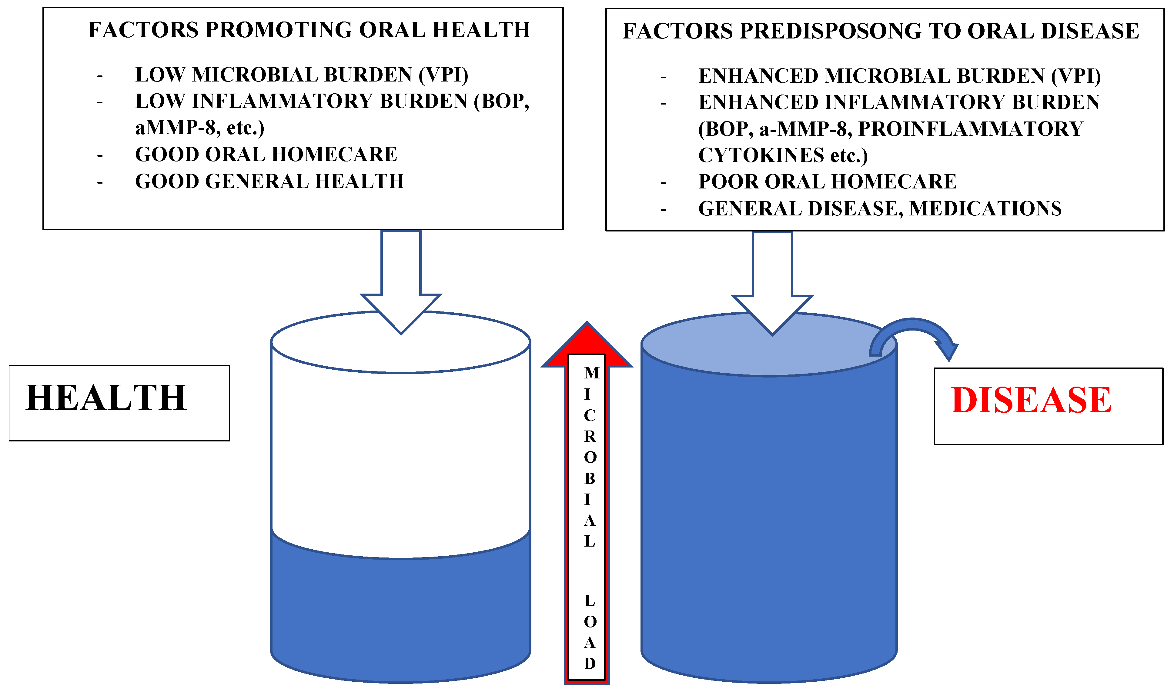
Microbiome research acknowledges the concept of a symbiotic/dysbiotic microbiome in health and disease [1]. The normal microbiome competes constantly with pathogenic or opportunistic bacteria, yeasts and viruses for nutrients and attachment sites on host cells. The main predisposing factors for a dysbiotic microbiome, and consequently predisposition to a diseased state in the oral cavity, are summarized in Figure 1. The microbial load could be seen as an indicator of the risk in a patient of developing a diseased state; initially, high counts of opportunistic pathogens and predisposing factors, e.g., systemic disease, medications and low or immune incompetent saliva, do not tend to overcome the “reservoir” or tolerance of the individual to stay healthy. In the case of, e.g., host immunodeficiency, a dysbiotic microbiome with opportunistic pathogen overgrowth may occur, resulting in a diseased state. Broad-spectrum antibiotics may also disturb and interfere with the normal microbiome long after treatment has ended.
After birth, the oral normal microbiome starts to build up with primary microbial species colonizers, which are mainly Viridans group streptococci [2]: Streptococcus salivarius (S. salivarius) and Streptococcus mitis (S. mitis), and subsequently the secondary colonizers, which tend also to favor anaerobic growth conditions. The periodontopathogenic bacteria belonging to the red triad (Porphyromonas gingivalis, Tannerella forsythia and Treponema denticola) are found and occur due to advanced periodontal disease. Local tissue oxidative, nutrient or pH conditions may also often favor the coaggregation tendency of certain bacterial species in biofilm formation. Carious, periodontal, candidal or viral infections may follow if defense mechanisms of the host are insufficient and defective. Infections may develop from low grade inflammation to acute infections and even proceed to life-threatening sepsis.

2. Innate and Adaptive Immunity and Inflammation

The first few hours of interaction with antigenic microbes trigger the host innate immunity. Monocytes differentiate to macrophages and their receptors recognize microbial surface components: e.g., mannose, glucan, or lipopolysaccharide. Pathogen recognition triggers inflammatory responses, i.e., “circulus vitious inflammationis”. Macrophages engulf microbes fusing with lysosomes to lyse microbes and release toxic and oxidative products (e.g., hydrogen peroxide, superoxide anion, nitric oxide) and at the same time, proinflammatory prostaglandins, leukotrienes and platelet-activating factor (PAF), proinflammatory cytokines (e.g., TNF-α, IL-1, IL-6, IL-12) and chemokines (e.g., monocyte chemoattractant protein (MCP-1)) to attract neutrophils to extravasate and migrate, in order to degranulate to sites of inflammation. Inflammation-related molecules have been extensively reviewed by Joseph et al. [3]. Natural killer (NK) cells contain and kill intracellular pathogens until cytotoxic T-cells are generated. Activation of the complement system occurs, and recognition of microbes follows three different pathways (classical, manna-binding -lectin, alternative). This allows opsonization and destruction of microbes directly or indirectly by the phagocytes. Complement components C3a, C4a and C5a are proinflammatory peptide mediators.
Subsequently, adaptive immunity is triggered if the pathogens overcome innate immunity targeting to defend the host from potential pathogen invasion and actions. Dendritic cells engulf and degrade pathogens and maturate to antigen-presenting cells. Cytosolic pathogens are eliminated by cytotoxic T-cells recognized via co-receptor CD8 (viruses and cytosolic bacteria). CD4 T-cells recognize pathogens and their products derived from the cellular vesicular compartment. CD4 T-cells are specialized to activate other cells and fall into two classes: T-helper cells (TH1), inflammatory cells, activate macrophages; and TH2 cells activate B cells to start producing antibodies called immunoglobulins (Ig). Main classes of immunoglobulins are IgG, IgM, and secretory IgA in the oral cavity; the infection is thus eliminated from the extracellular space (adapted from Janeway et al. [4]).

3. Special Features in Microbe/Host Interactions in the Oral Environment

The oral microbiome consists of hundreds of yet not completely identified microbial species. A symbiotic oral mucosal microbiome is essential for the host’s healthy state. The epithelial surface, trying to obstruct invasion of the microbes, is at constant alert by bombardment of microbes and their virulence factors, such as adhesins, invasins and proteases, which may degrade oral epithelial junctional proteins, e.g., laminin-332, fibronectin, E-cadherin, and claudin [5,6,7,8,9,10], allowing further promotion of the microbial burden’s invasive process through the epithelium or degradation of complement proteins of innate immunity. Gingival junctional epithelium is especially vulnerable as its main structural basement membrane protein is laminin-332.
Saliva and its antimicrobial substances, such as glycoproteins in mucus, lysozyme secreted by macrophages/neutrophils, β-defensins, lactoferrin, lactoperoxidase, statherin, histatins and secretory IgA [11] have an essential role in first line superficial defense. Matrix metalloprotease-8 (MMP-8), MMP-9, myeloperoxidase (MPO) and neutrophil (PMN) elastase have distinct roles in local and connective tissue collagen metabolism in the oral environment. The host exhibits housekeeping activity of MMP-1 and its inhibitors—tissue inhibitor of metalloproteases-1 and -2 (TIMP-1, TIMP-2)—as well as constant lysis and remodeling of collagenous structures. Oral microbes Treponema denticola [12,13] and Candida glabrata [14] have been shown to convert latent 75 kDa proMMP-8 to its lower molecular active and fragmented forms (aMMP -8), and elevated aMMP-8 activity in oral pathologies may especially cause undesirable and irreversible tissue damage. Tannerella forsythia, associated with severe periodontitis, is capable of degrading gelatin and type I collagen [15], and Porphyromonas gingivalis and Treponema denticola proteases, gingipain and dentisilin, respectively, as well as various other bacterial proteases activate human procollagenases [12,16]. Other non-matrix substrates of aMMP-8 are laminin-332 γ2- chain, insulin receptor, RANKL, TRAP, TREM-1: elevated concentrations of their proteolytic fragments may be found and detected during diseases and inflammations. Elevated levels of MMP-8 and MPO activity has been shown to correlate with poor prognosis of bacterial meningitis of childhood, pancreatitis, obesity, sepsis, and diabetes [17,18,19,20,21]. aMMP-8 may also be used as a biomarker of inflammation from oral rinse samples (PerioSafe®) or gingival crevicular fluid (GCF) in peri-implantitis (ImplantSafe®) [22]. If inflammation in periodontal tissues is prolonged, this low-grade inflammation, reflected by aMMP-8, may have also systemic effects on health, e.g., cardiovascular diseases, metabolic syndrome, Alzheimer’s disease, or autoimmune diseases [23,24].
Considering microbes and their infection/inflammation-inducing effects, proper management of oral health is crucial and exerts positive effects on general or systemic health. An acute infection’s spread may threaten general health. Low-grade inflammation induced and caused by persistent microbes is challenging because systemic antibiotics or local antiseptics may be used only for a limited time, and they abolish the normal microbiome alongside the pathogens. Natural products, and especially fermented plant foods, may eventually offer an alternative approach to novel anti-inflammatory and immunomodulatory pathways for controlling oral health [25].

4. Lingonberries

Lingonberry (Vaccinium vitis ideae L.) is a low-bush wild plant found in the northern hemisphere. The berries contain vitamins (A, B1, B2, B3, and C), potassium, calcium, magnesium, phosphorous and have a unique polyphenol composition [26], including flavonoids (anthocyanins, flavonols {e.g., quercetin}, flavanols {catechins])), phenolic acids, lignans, stilbenes (resveratrol), and phenolic polymers (e.g., proanthocyanidins)—of which anthocyanins, flavonols, and proanthocyanidines are the main constituents. Resveratrol may be extracted from berry peels and seeds. Lingonberries contain particularly high amounts of anthocyanins: cyanidin-3-galactoside (88%), cyanidin-3-arabinoside (10.6%) and cyanidin-3-glucoside (1.4%) [27]. Polyphenols from berries are bioavailable from diet [28] and they retain their biological activities in ileal samples [29]. Lingonberry bioactive molecules show anti-cancerous, antimicrobial, antioxidative and anti-inflammatory effects [30].
The following studies have used different fractions of lingonberries, depending on the extraction method used, and accordingly the antimicrobial effects seem to vary.

5. In Vitro Anticancer Studies

Hoornstra et al. [31] have demonstrated the inhibition of oral tongue squamous cell lines HSC-3 and SCC-25 carcinoma invasion and proliferation by fermented lingonberry juice similar that seen with curcumin. Lingonberries are known to exert ornithine decarboxylase inhibition, a key rate-limiting enzyme in polyamine synthesis, cell growth, DNA repair and carcinogenesis [32]. Lingonberries have additional antiproliferative effects against human breast, colon and cervical (HeLa) cancer growth [33,34,35].

6. In Vitro Antimicrobial Studies

There are limited amounts of studies on the effects of lingonberries against microbial growth; the most studied are intestinal pathogens and lactobacilli. Several antimicrobial mechanisms of polyphenols have been proposed [36,37,38]. Lingonberry polyphenols have also been proposed to act as antivirals [39], and in this regard, lingonberries have been shown to possess in vitro antiviral activity [40]. Other studies have also reported lingonberry’s antibacterial and antifungal activities: inhibition of growth of Candida, S. mutans, Porphyromonas gingivalis, Fusobacterium nucleatum, S. aureus, Salmonella enterica sv Typhimurium, S. epidermidis, P. gingivalis, P. intermedia, antiaggregation of S. mutans with Fusobacterium nucleatum or Actinomyces naeslundii, anti-adhesiveness of Neisseria meningitidis or oral streptococci in biofilm formation, and binding activity of Streptococcus pneumoniae, Streptococcus agalactiae and Streptococcus suis to berries and juices [36,41,42,43,44,45,46,47,48,49,50,51,52]. No effects of lingonberries on lactobacilli have been found in any of these studies.

7. In Vitro Antioxidant and Anti-Inflammatory Studies

Berries contain polyphenols which act as potent antioxidants and are neuroprotective, having beneficial effects on health [30,53]. Antioxidant activities of lingonberries have been assessed in vitro in several studies [41,54,55,56,57,58,59,60]. These studies report radical oxygen species scavenging by polyphenols and inhibition of oxidation of lipids and proteins.
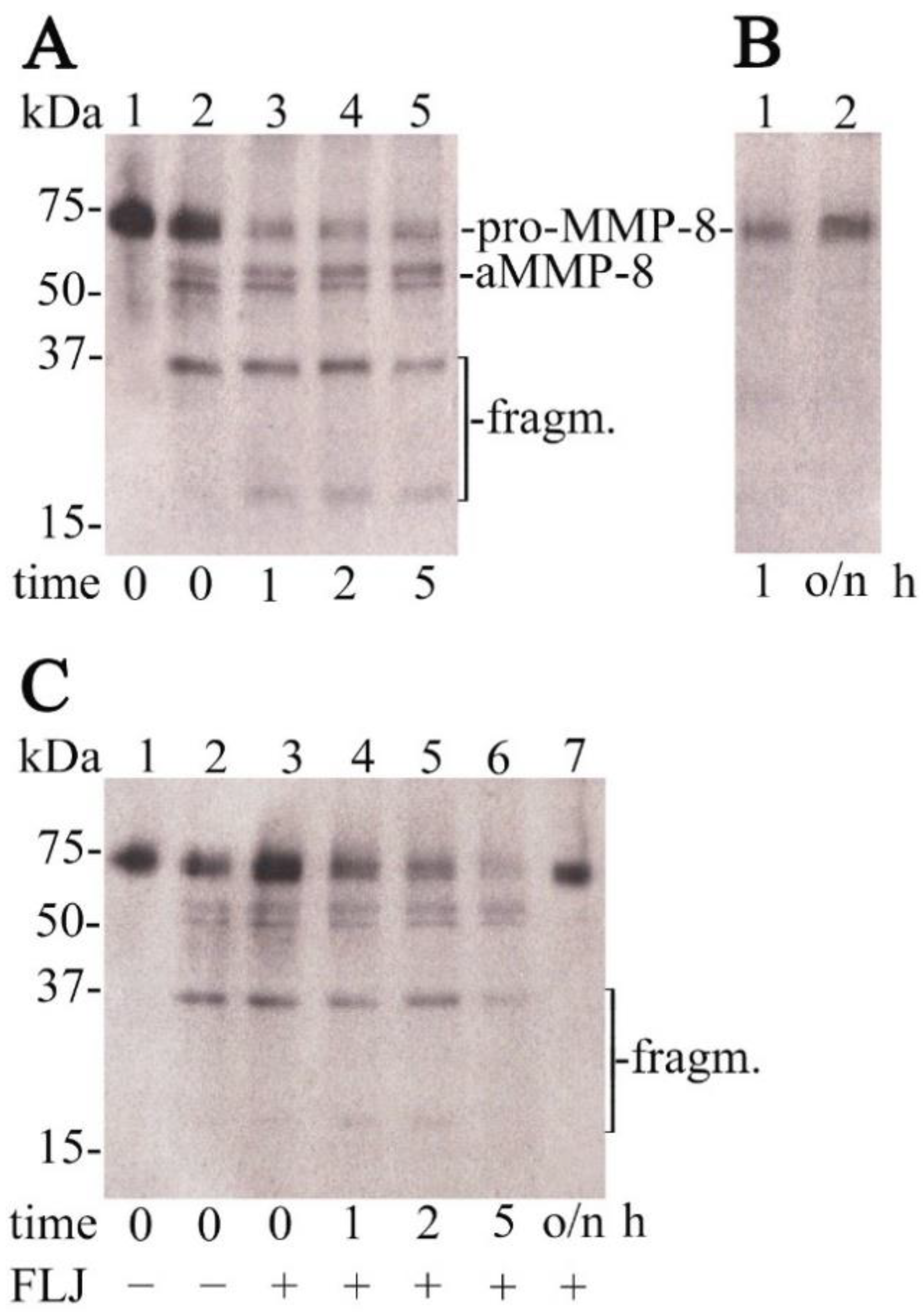
Several in vitro studies have also revealed anti-inflammatory mechanisms of lingonberries. Lingonberry cyanidin has been shown to inhibit in vitro p38 mitogen-activated protein kinase (MAPK) and c-Jun N-terminal kinase (JNK) phosphorylation, and reduce levels of protein kinase B (Akt) in UV-induced photoreceptor damage [61]. Lingonberry fruit extract has been shown in vitro to downregulate inflammatory mediators such as IL-6, TNF-α, IL-1β, MCP-1, COX-2 and iNOS in mouse adipocyte inflammation [62]. Lingonberry phenolic compounds suppress human THP-1 macrophage cells’ TNF-α and IL-6 production in vitro [41]. Candida glabrata’s cell-associated > 50 kDa proteolytic (gelatinolytic) fraction has been shown to activate human latent pro-MMP-8 [14], and this activation was time- and dose-dependently inhibited by FLJ (Figure 2). In vitro studies by Esposito et al. [63] revealed inhibition of reactive oxygen species (ROS) and NO production, and suppression of prostaglandin-endoperoxidase synthase 2 (COX-2) and inducible nitric oxid synthase (iNOS) expression in LPS-stimulated mouse macrophages. Additionally, human dermal fibroblast MMP2 and MCPI gene downregulation and upregulation of extracellular matrix protein-coding genes COLIA2 (pro-α2 chain of type I collagen), ITGB1 (integrin receptor subunit β1) and RHOA (guanosine triphosphate phosphatase) were detected, resulting in beneficial ECM remodeling, dermal wound healing and tissue repair based on earlier inflammatory resolution.
In vivo mouse studies have shown that lingonberry juice exerts anti-inflammatory and anti-atherothrombotic effects [64,65]. Lingonberries decrease glycaemia and hepatic triglyceride levels [66], reduce inflammation, high cholesterol, hyperglycaemia and obesity [67], and alter gut microbiota, improve metabolic/brain functions, and also reduce gut inflammatory properties [68] in mice fed a high-fat diet.

8. In Vivo Human Studies

In vivo human studies with lingonberries are scarce and few studies on consumption of mixtures of berries containing lingonberries have been conducted. One pilot intestinal study has been performed [29], which compared the bioavailability of polyphenols after in vitro digestion and following fecal fermentation of lingonberries in an in vivo digested lingonberry sample (ileal fluid sample of one male). The results showed that the (poly)phenol composition of lingonberry undergoes substantial but largely similar modifications in vivo and in vitro, retaining bioactivity in an anti-genotoxity model, in vitro samples also in a colorectal cancer model. It is proposed that the concentration rather than specific phenolic compounds is crucial to obtain effects.
Lingonberries or lingonberry nectar consumed with saccharose reduces postprandial glucose, insulin, and inflammation-related free fatty acid release responses, and lingonberry puree consumed with bread reduces insulin requirements, to maintain normal or slightly reduced postprandial glycaemia in healthy women [69,70].
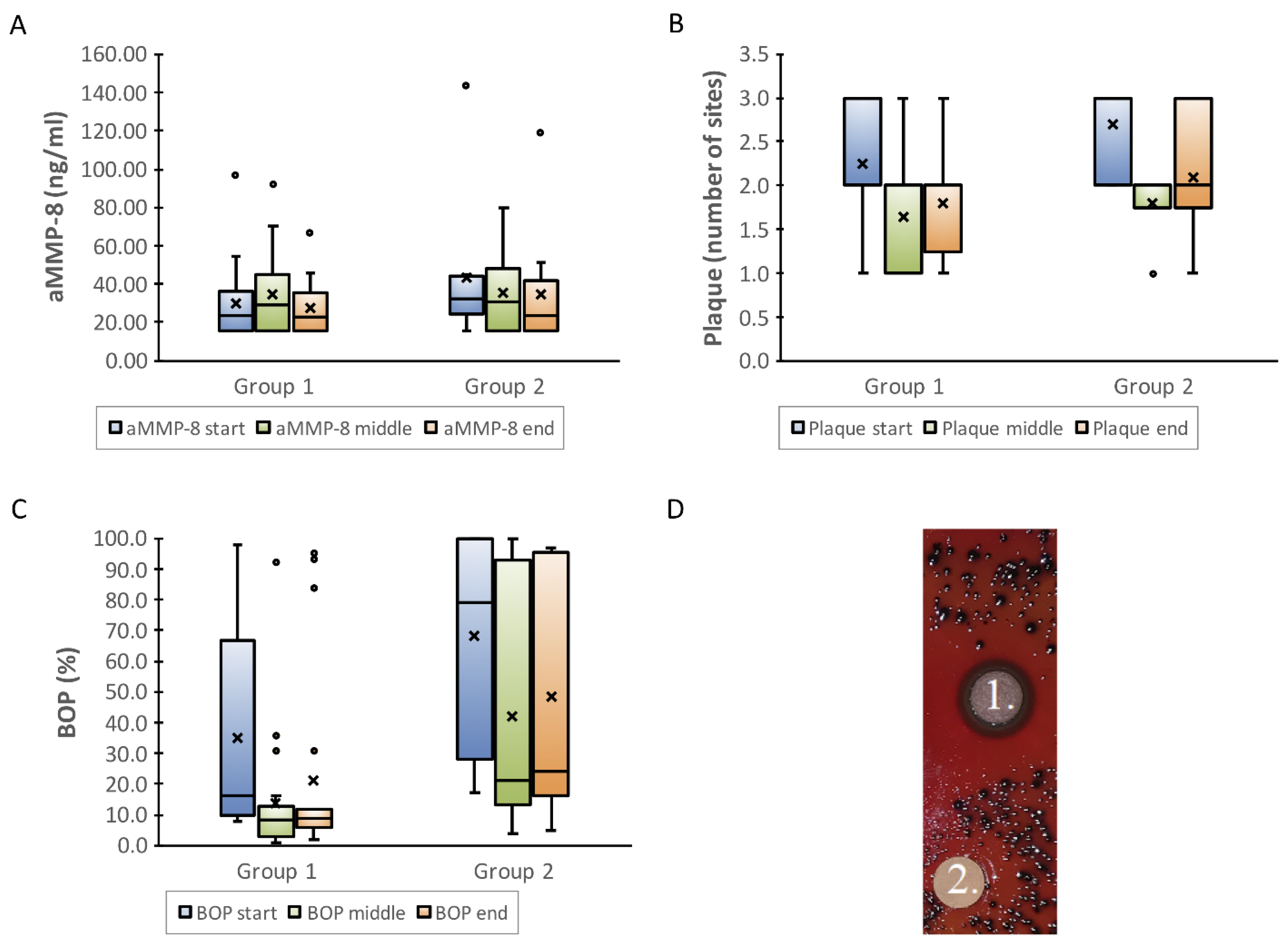
Originally, concentrated unfermented lingonberry juice was tested among 60 patients (Finnish Utility Model nro 8195, 71). The randomly chosen adult patients used 10 mL of the juice as a mouthwash twice daily for ten days, after which the trial had to be discontinued because of an unexpected Candida count increases in vivo due to the high sugar content of the juice. In the next clinical studies, the sugar content of the juice was reduced, and lingonberries were shown to have in vivo antimicrobial and anti-inflammatory properties in the form of fermented lingonberry juice (FLJ; Lingora®, Vantaa, Finland) in clinical human oral studies [44,71] on a total of 40 adults. Initially, FLJ was lyophilized and compressed into tablets [71]. Ten individuals were divided into two groups according to pre-scanned Candida counts: those with higher Candida counts (≥ 104 CFU/mL) took the lozenge three times daily, those with lower counts (< 104 CFU/mL) took it twice daily for ten days. The results showed promising antimicrobial effects in vivo. In the next study, 30 patients were recruited randomly. The group was divided into two subgroups to test the suitable amount of FLJ and minimum time of the mouthwash period needed to gain antimicrobial and anti-inflammatory effects: 20 patients with clinically normal levels of oral homecare/plaque used 10 mL of FLJ twice daily for 2 weeks (group 1), and 10 patients with apparent difficulties in maintaining proper oral homecare/higher amounts of plaque used 20 mL twice daily for 7 days (group 2) as a mouthwash for 30 s. The results showed similar antimicrobial effects in both groups. Additionally, the on-line and real-time analysis and recording of clinical periodontal parameters, such as bleeding on probing (BOP, gingival inflammation index) and visible plaque index (VPI, microbial burden index), and the oral periodontal tissue destruction biomarker, aMMP-8 [72,73,74], reflected and indicated beneficial antimicrobial and anti-inflammatory changes (Figure 3).
Lingonberries are a promising natural approach with regards to their studied and recorded beneficial oral and general health effects via antioxidant, anti-inflammatory, anti-proteolytic, anticancerous and antimicrobial effects. Lingonberries, in specially formulated FLJ, have thus been designed for safe oral use as a mouthwash, and have thus been shown to exert anti-cancerous, antimicrobial, anti-proteolytic and anti-inflammatory properties in in vitro and in vivo clinical human studies. The antimicrobial effects of FLJ have been assessed against the fifteen most common oral microbial strains, including Candida, Streptococci, and key dysbiotic periodontopathogens. The species monitered in oral in vivo studies were selected to represent key microbes known to cause disease, e.g., Candida in candidosis and S. mutans in dental caries. Excessive amounts of S. mutans co-associate with the severity of periodontal disease in older patients [75]; the numbers of these species declined statistically significantly. In comparison, lactobacilli count results show that FLJ did not inhibit their growth. In fact, their relative proportion in patient samples increased after FLJ intervention, as expected, and the microbial effects of FLJ lasted even after the discontinuation of the mouthwash regimen. The overall effect on the microbiota assessed by these cultivations has interesting consequences regarding the composition of the oral microbiota: the patient’s own lactobacilli were allowed to flourish, cutting off living space and nutrients from potentially pathogenic species. Lactobacilli are indeed known to inhibit S. mutans and Porphyromonas gingivalis growth [76]. Microbes of the normal microbiome, such as S. salivarius, may also produce beneficial antimicrobial substances [2]. This kind of shift in the composition of the microbiota from dysbiosis (disease) towards symbiosis (health) could be beneficial and defensive to control oral health.

9. Conclusions

FLJ would have a beneficial effect, particularly in individuals who have predisposing factors (age, medication, dry mouth) or difficulties in maintaining sufficient oral homecare. As lingonberries contain considerable amounts of natural sugars, only the tailored fermented juice with the majority of sugars removed exerts this effect optimally. The antimicrobial and anti-inflammatory effects seem to continue after use, seem to have a prolonged treatment effect, and shift the oral microbiome to a more symbiotic direction. Double-blinded randomized trials are needed to study the effects on saliva parameters, dental caries, periodontal disease, and general health—since FLJ may and can be conveniently as well as safely swallowed—focusing on anti-inflammatory, anti-oxidative, and systemic effects mediated by, e.g., cytokine and reactive oxygen species levels.

Author Contributions

Writing review and editing, P.P.; H.L.; T.T.; I.T.R.; T.S. Visualization P.P.; T.T.; I.T.R. All authors have read and agreed to the published verssion of the manuscript.

Funding

This work was supported by grants from University of Helsinki Research Foundation (TYH2016251, TYH2017251, TYH2018229, TYH2019319, TYH2022225, Y1014SL017, Y1014SL018, Y1014SULE1), Finland; Karolinska Institutet, Stockholm, Sweden; Finnish Dental Society Apollonia; The Minerva Foundation Selma and Maja-Lisa Selander’s Fund; The Yrjö Jahnsson Foundation sr; The Paulo Foundation; The Emil Aaltonen Foundation sr; The Juhani Aho Foundation for Medical Research sr; The Orion Research Foundation sr; and Helsingin Seudun Hammaslääkärit r.y. (Dentists of Helsinki Region Association), Finland.

Institutional Review Board Statement

The study was conducted according to the guidelines of the Declaration of Helsinki, and approved by the ethical committees of Stockholm Community, Sweden (2016/1410-31) and the Helsinki University Central Hospital, Finland (1271/2019 and 51/13/02/2009).

Informed Consent Statement

Informed consent was obtained from all subjects involved in the study.

Data Availability Statement

The data presented in this study are available on request from the corresponding author.

Acknowledgments

Open access funding provided by University of Helsinki.

Conflicts of Interest

Timo Sorsa is the inventor of U.S. patents 5652223, 5736341, 5866432, 6143476, 20170023571A1 (granted 6.6.2019), WO 2018/060553A1 (granted 31.5.2018), a co-inventor of the patent 10488415 B2, a Japanese Patent 2016-554676 and a South Korean patent 10-2016-7025378. Pirjo Pärnänen is the inventor of patent EP 2585087B1.

References

  1. Kilian, M.; Chapple, I.L.C.; Hannig, M.; Marsh, P.D.; Meuric, V.; Pedersen, A.M.L.; Tonetti, M.S.; Wade, W.G.; Zaura, E. The oral microbiome—An update for oral healthcare professionals. Br. Dent. J. 2016, 221, 657–666. [Google Scholar] [CrossRef]
  2. Dhotre, S.; Suryawanshi, N.; Selkar, S.; Nagoba, B. Viridans Group Streptococci and the Oral Ecosystem. Eur. J. Gen. Med. 2015, 13, 145–148. [Google Scholar] [CrossRef]
  3. Joseph, S.V.; Edirisinghe, I.; Burton-Freeman, B.M. Berries: Anti-inflammatory effects in humans. J. Agric. Food Chem. 2014, 62, 3886–3903. [Google Scholar] [CrossRef] [PubMed]
  4. Janeway, C.; Travers, P.; Walport, M.; Shlomchik, M. Immunobiology, The Immune System in Health and Disease 2001, 5th ed.; Garland Publishing: New York, NY, USA, 2001; pp. 1–91, 341–411. [Google Scholar]
  5. Pärnänen, P.; Kari, K.; Virtanen, I.; Sorsa, T.; Meurman, J. Human laminin-332 degradation by Candida proteinases. J. Oral. Pathol. Med. 2008, 37, 329–335. [Google Scholar] [CrossRef] [PubMed]
  6. Pärnänen, P.; Meurman, J.; Virtanen, I. Laminin-511 and fibronectin degradation with Candida yeast. J. Oral. Pathol. Med. 2009, 38, 768–772. [Google Scholar] [CrossRef]
  7. Pärnänen, P.; Meurman, J.; Samaranayake, L.; Virtanen, I. Human oral keratinocyte E-cadherin degradation by Candida albicans and Candida glabrata. J. Oral. Pathol. Med. 2010, 39, 275–278. [Google Scholar] [CrossRef] [PubMed]
  8. Pärnänen, P.; Meurman, J.; Sorsa, T. The effects of Candida proteinases on human proMMP-9, TIMP-1 and TIMP-2. Mycoses 2010, 54, 325–330. [Google Scholar] [CrossRef] [PubMed]
  9. Pärnänen, P.; Sorsa, T. Human Gingival Laminin-332 Degradation by Oral Bacteria. Laminins: Structure, Biological Activity and Role in Disease; Adams, D.C., Garcia, E.O., Eds.; Nova Science Publishers: Hauppauge, NY, USA, 2013; pp. 125–130. [Google Scholar]
  10. Nawaz, A.; Mäkinen, A.; Pärnänen, P.; Meurman, J. Proteolytic activity of non-albicans Candida and Candida albicans in oral cancer patients. New Microbiol. 2018, 41, 296–301. [Google Scholar]
  11. Lynge Pedersen, A.M.; Belstrøm, D. The role of natural salivary defences in maintaining a healthy oral microbiota. J. Dent. 2019, 80, S3–S12. [Google Scholar] [CrossRef]
  12. Sorsa, T.; Ingman, T.; Suomalainen, K.; Haapasalo, M.; Konttinen, Y.T.; Lindy, O.; Saari, H.; Uitto, V.-J. Identification of proteases from periodontopathogenic bacteria as activators of latent human neutrophil and fibroblast-type interstitial collagenases. Infect. Immun. 1992, 60, 4491–4495. [Google Scholar] [CrossRef] [Green Version]
  13. Nieminen, M.; Listyarifah, D.; Hagström, J.; Haglund, C.; Grenier, D.; Nordström, D.; Uitto, V.-J.; Hernandez, M.; Yucel-Lindberg, T.; Tervahartiala, T.; et al. Treponema denticola chymotrypsin-like proteinase may contribute to orodigestive carcinogenesis through immunomodulation. Br. J. Cancer 2018, 118, 428–434. [Google Scholar] [CrossRef] [PubMed] [Green Version]
  14. Pärnänen, P.; Sorsa, T.; Tervahartiala, T.; Nikula-Ijäs, P. Isolation, characterization and regulation of moonlighting proteases from Candida glabrata cell wall. Microb. Pathog. 2020, 149, 104547. [Google Scholar] [CrossRef]
  15. Hockensmith, K.; Dillard, K.; Sanders, B.; Harville, B.A. Identification and characterization of a chymotrypsin-like serine protease from periodontal pathogen, Tannerella forsythia. Microb. Pathog. 2016, 100, 37–42. [Google Scholar] [CrossRef] [PubMed]
  16. Okamoto, T.; Akaike, T.; Suga, M.; Tanase, S.; Horie, H.; Miyajima, S.; Ando, M.; Ichinose, Y.; Maeda, H. Activation of human matrix metalloproteinases by various bacterial proteinases. J. Biol. Chem. 1997, 272, 6059–6066. [Google Scholar] [CrossRef] [Green Version]
  17. Savonius, O.; Roine, I.; Alassiri, S.; Tervahartiala, T.; Helve, O.; Fernández, J.; Peltola, H.; Sorsa, T.; Pelkonen, T. The Potential Role of Matrix Metalloproteinases 8 and 9 and Myeloperoxidase in Predicting Outcomes of Bacterial Meningitis of Childhood. Mediat. Inflamm. 2019, 2019, 7436932. [Google Scholar] [CrossRef]
  18. Nukarinen, E.; Lindström, O.; Kuuliala, K.; Kylänpää, L.; Pettilä, V.; Puolakkainen, P.; Kuuliala, A.; Hämäläinrn, M.; Moilanen, E.; Repo, H.; et al. Association of Matrix Metalloproteinases-7, -8 and -9 and TIMP-1 with Disease Severity in Acute Pancreatitis. A Cohort Study. PLoS ONE 2016, 11, e0161480. [Google Scholar] [CrossRef] [PubMed]
  19. Turunen, A.; Kuuliala, K.; Kuuliala, A.; Tervahartiala, T.; Mustonen, H.; Puolakkainen, P.; Kylänpää, L.; Sorsa, T. Activated matrix metalloproteinase 8 in serum predicts severity of acute pancreatitis. Pancreatology 2021, 21, 862–869. [Google Scholar] [CrossRef]
  20. Lauhio, A.; Hästbacka, J.; Pettilä, V.; Tervahartiala, T.; Karlsson, S.; Varpula, T.; Varpula, M.; Ruokonen, E.; Sorsa, T.; Kolho, E. Serum MMP-8, -9 and TIMP-1 in sepsis: High serum levels of MMP-8 and TIMP-1 are associated with fatal outcome in a multicentre, prospective cohort study. Hypothetical impact of tetracyclines. Pharmacol. Res. 2011, 64, 590–594. [Google Scholar] [CrossRef] [PubMed]
  21. Lauhio, A.; Farkkila, E.; Pietilainen, K.H.; Astrom, P.; Winkelmann, A.; Tervahartiala, T.; Pirila, E.; Rissanen, A.; Kaprio, J.; Sorsa, T.A.; et al. Association of MMP-8 with obesity, smoking and insulin resistance. Eur. J. Clin. Investig. 2016, 46, 757–765. [Google Scholar] [CrossRef]
  22. Al-Majid, A.; Alassiri, S.; Rathnayake, N.; Tervahartiala, T.; Gieselmann, D.-R.; Sorsa, T. Matrix Metalloproteinase-8 as an Inflammatory and Prevention Biomarker in Periodontal and Peri-Implant Diseases. Int. J. Dent. 2018, 2018, 7891323. [Google Scholar] [CrossRef]
  23. Tuomainen, A.M.; Nyyssönen, K.; Laukkanen, J.A.; Tervahartiala, T.; Tuomainen, T.-P.; Salonen, J.T.; Sorsa, T.; Pussinen, P. Serum Matrix Metalloproteinase-8 Concentrations Are Associated With Cardiovascular Outcome in Men. Arter. Thromb. Vasc. Biol. 2007, 27, 2722–2728. [Google Scholar] [CrossRef]
  24. Kleinstein, S.E.; Nelson, K.E.; Freire, M. Inflammatory Networks Linking Oral Microbiome with Systemic Health and Disease. J. Dent. Res. 2020, 99, 1131–1139. [Google Scholar] [CrossRef]
  25. Shahbazi, R.; Sharifzad, F.; Bagheri, R.; Alsadi, N.; Yasavoli-Sharahi, H.; Matar, C. Anti-Inflammatory and Immunomodulatory Properties of Fermented Plant Foods. Nutrients 2021, 13, 1516. [Google Scholar] [CrossRef]
  26. Ek, S.; Kartimo, H.; Mattila, S.; Tolonen, A. Characterization of Phenolic Compounds from Lingonberry (Vaccinium vitis-idaea). J. Agric. Food Chem. 2006, 54, 9834–9842. [Google Scholar] [CrossRef]
  27. Andersen, Ø.M. Chromatographic Separation of Anthocyanins in Cowberry (Lingonberry) Vaccinium vites-idaea L. J. Food Sci. 1985, 50, 1230–1232. [Google Scholar] [CrossRef]
  28. Koli, R.; Erlund, I.; Jula, A.; Marniemi, J.; Mattila, P.; Alfthan, G. Bioavailability of various polyphenols from a diet containing moderate amounts of berries. J. Agric. Food Chem. 2010, 58, 3927–3932. [Google Scholar] [CrossRef] [PubMed]
  29. Brown, E.M.; Nitecki, S.; Pereira-Caro, G.; McDougall, G.J.; Stewart, D.; Rowland, I.; Crozier, A.; Gill, C.I. Comparison of in vivo and in vitro digestion on polyphenol composition in lingonberries: Potential impact on colonic health. Biofactors 2014, 40, 611–623. [Google Scholar] [CrossRef]
  30. Kowalska, K. Lingonberry (Vaccinium vitis-idaea L.) Fruit as a Source of Bioactive Compounds with Health-Promoting Effects—A Review. Int. J. Mol. Sci. 2021, 22, 5126. [Google Scholar] [CrossRef] [PubMed]
  31. Hoornstra, D.; Vesterlin, J.; Parnanen, P.; Al-Samadi, A.; Zlotogorski-Hurvitz, A.; Vered, M.; Salo, T. Fermented Lingonberry Juice Inhibits Oral Tongue Squamous Cell Carcinoma Invasion In Vitro Similarly to Curcumin. In Vivo 2018, 32, 1089–1095. [Google Scholar] [CrossRef] [PubMed] [Green Version]
  32. Bomser, J.; Madhavi, D.L.; Singletary, K.; Smith, M.A. In vitro anticancer activity of fruit extracts from Vaccinium species. Planta Med. 1996, 62, 212–216. [Google Scholar] [CrossRef] [PubMed]
  33. Olsson, M.E.; Gustavsson, K.-E.; Andersson, S.; Nilsson, Å.; Duan, R.-D. Inhibition of Cancer Cell Proliferation in Vitro by Fruit and Berry Extracts and Correlations with Antioxidant Levels. J. Agric. Food Chem. 2004, 52, 7264–7271. [Google Scholar] [CrossRef]
  34. McDougall, G.J.; Ross, H.A.; Ikeji, M.; Stewart, D. Berry Extracts Exert Different Antiproliferative Effects against Cervical and Colon Cancer Cells Grown in Vitro. J. Agric. Food Chem. 2008, 56, 3016–3023. [Google Scholar] [CrossRef] [PubMed]
  35. Onali, T.; Kivimäki, A.; Mauramo, M.; Salo, T.; Korpela, R. Anticancer Effects of Lingonberry and Bilberry on Digestive Tract Cancers. Antioxidants 2021, 10, 850. [Google Scholar] [CrossRef] [PubMed]
  36. Nohynek, L.J.; Alakomi, H.-L.; Kähkönen, M.P.; Heinonen, M.; Helander, I.M.; Oksman-Caldentey, K.-M.; Puupponen-Pimiä, R.H. Berry phenolics: Antimicrobial properties and mechanisms of action against severe human pathogens. Nutr. Cancer 2006, 54, 18–32. [Google Scholar] [CrossRef] [PubMed]
  37. Heinonen, M. Antioxidant activity and antimicrobial effect of berry phenolics—A Finnish perspective. Mol. Nutr. Food Res. 2007, 51, 684–691. [Google Scholar] [CrossRef] [PubMed]
  38. Riihinen, K.R.; Ou, Z.M.; Gödecke, T.; Lankin, D.C.; Pauli, G.F.; Wu, C.D. The antibiofilm activity of lingonberry flavonoids against oral pathogens is a case connected to residual complexity. Fitoterapia 2014, 97, 78–86. [Google Scholar] [CrossRef]
  39. Pärnänen, P.; Lähteenmäki, H.; Räisänen, I.; Tervahartiala, T.; Sorsa, T. Lingonberry polyphenols: Potential SARS-CoV-2 inhibitors as nutraceutical tools? Physiol. Rep. 2021, 9, e14741. [Google Scholar] [CrossRef]
  40. Nikolaeva-Glomb, L.; Mukova, L.; Nikolova, N.; Badjakov, I.; Dincheva, I.; Kondakova, V.; Doumanova, L.; Galabov, A.S. In vitro antiviral activity of a series of wild berry fruit extracts against representatives of Picorna-, Orthomyxo- and Paramyxoviridae. Nat. Prod. Commun. 2014, 9, 51–54. [Google Scholar] [CrossRef] [Green Version]
  41. Kylli, P.; Nohynek, L.; Puupponen-Pimiä, R.; Westerlund-Wikström, B.; Leppänen, T.; Welling, J.; Moilanen, E.; Heinonen, M. Lingonberry (Vaccinium vitis-idaea) and European Cranberry (Vaccinium microcarpon) Proanthocyanidins: Isolation, Identification, and Bioactivities. J. Agric. Food Chem. 2011, 59, 3373–3384. [Google Scholar] [CrossRef]
  42. Riihinen, K.; Ryynänen, A.; Toivanen, M.; Könönen, E.; Törrönen, R.; Tikkanen-Kaukanen, C. Antiaggregation potential of berry fractions against pairs of Streptococcus mutans with Fusobacterium nucleatum or Actinomyces naeslundii. Phytother. Res. 2011, 25, 81–87. [Google Scholar] [CrossRef] [Green Version]
  43. Pärnänen, P.; Nawaz, A.; Sorsa, T.; Meurman, J.; Nikula-Ijäs, P. The Effect of Fermented Lingonberry Juice on Candida glabrata Intracellular Protein Expression. Int. J. Dent. 2017, 2017, 6185395. [Google Scholar] [CrossRef] [PubMed]
  44. Pärnänen, P.; Nikula-Ijäs, P.; Sorsa, T. Antimicrobial and anti-inflammatory lingonberry mouthwash. a clinical pilot study in the oral cavity. Microorganisms 2019, 7, 331. [Google Scholar] [CrossRef] [PubMed] [Green Version]
  45. Puupponen-Pimiä, R.; Nohynek, L.; Meier, C.; Kähkönen, M.; Heinonen, M.; Hopia, A.; Oksman-Caldentey, K.-M. Antimicrobial properties of phenolic compounds from berries. J. Appl. Microbiol. 2001, 90, 494–507. [Google Scholar] [CrossRef]
  46. Puupponen-Pimiä, R.; Nohynek, L.; Hartmann-Schmidlin, S.; Kähkönen, M.; Heinonen, M.; Määttä-Riihinen, K.; Oksman-Caldentey, K.-M. Berry phenolics selectively inhibit the growth of intestinal pathogens. J. Appl. Microbiol. 2005, 98, 991–1000. [Google Scholar] [CrossRef] [PubMed]
  47. Puupponen-Pimiä, R.; Nohynek, L.; Alakomi, H.-L.; Oksman-Caldentey, K.-M. Bioactive berry compounds—Nnovel tools against human pathogens. Appl. Microbiol. Biotechnol. 2005, 67, 8–18. [Google Scholar] [CrossRef] [PubMed]
  48. Puupponen-Pimiä, R.; Nohynek, L.; Alakomi, H.-L.; Oksman-Caldentey, K.-M. The action of berry phenolics against human intestinal pathogens. Biofactors 2005, 23, 243–251. [Google Scholar] [CrossRef]
  49. Ho, K.Y.; Tsai, C.C.; Huang, J.S.; Chen, C.P.; Lin, T.C.; Lin, C.C. Antimicrobial activity of tannin components from Vaccinium vitis idaea L. J. Pharm. Pharmacol. 2001, 53, 187–191. [Google Scholar] [CrossRef] [PubMed]
  50. Toivanen, M.; Huttunen, S.; Duricova, J.; Soininen, P.; Laatikainen, R.; Loimaranta, V.; Haataja, S.; Finne, J.; Lapinjoki, S.; Tikkanen-Kaukanen, C. Screening of binding activity Streptococcus pneumoniae, Streptococcus agalactiae and Streptococcus suis to berries and juices. Phytother. Res. 2010, 24, S95–S101. [Google Scholar] [CrossRef] [PubMed]
  51. Toivanen, M.; Huttunen, S.; Lapinjoki, S.; Tikkanen-Kaukanen, C. Inhibition of adhesion of Neisseria meningitidis to human epithelial cells by berry juice polyphenolic fractions. Phytother. Res. 2011, 25, 828–832. [Google Scholar] [CrossRef] [Green Version]
  52. Kokubu, E.; Kinoshita, E.; Ishihara, K. Inhibitory effects of lingonberry extract on oral streptococci biofilm formation and bioactivity. Bull. Tokyo Dent. Coll. 2019, 60, 1–9. [Google Scholar] [CrossRef]
  53. Kelly, E.; Vyas, P.; Weber, J.T. Biochemical properties and neuroprotective effects of compounds in various species of berries. Molecules 2018, 23, 26. [Google Scholar] [CrossRef] [PubMed] [Green Version]
  54. Ho, K.Y.; Huang, J.S.; Tsai, C.C.; Lin, T.C.; Hsu, Y.F.; Lin, C.C. Antioxidant Activity of Tannin Components from Vaccinium vitis-idaea L. J. Pharm. Pharmacol. 1999, 51, 1075–1078. [Google Scholar] [CrossRef] [PubMed]
  55. Kähkönen, M.P.; Hopia, A.I.; Heinonen, M. Berry Phenolics and Their Antioxidant Activity. J. Agric. Food Chem. 2001, 49, 4076–4082. [Google Scholar] [CrossRef] [PubMed]
  56. Zheng, W.; Wang, S.Y. Oxygen radical absorbing capacity of phenolics in blueberries, cranberries, chokeberries, and lingonberries. J. Agric. Food Chem. 2003, 51, 502–509. [Google Scholar] [CrossRef]
  57. Viljanen, K.; Kylli, P.; Kivikari, R.; Heinonen, M. Inhibition of Protein and Lipid Oxidation in Liposomes by Berry Phenolics. J. Agric. Food Chem. 2004, 52, 7419–7424. [Google Scholar] [CrossRef]
  58. Määttä-Riihinen, K.R.; Kähkönen, M.P.; Törrönen, A.R.; Heinonen, M. Catechins and Procyanidins in Berries of Vaccinium Species and Their Antioxidant Activity. J. Agric. Food Chem. 2005, 53, 8485–8491. [Google Scholar] [CrossRef]
  59. Wu, C.-H.; Yen, G.-C. Inhibitory Effect of Naturally Occurring Flavonoids on the Formation of Advanced Glycation Endproducts. J. Agric. Food Chem. 2005, 53, 3167–3173. [Google Scholar] [CrossRef]
  60. Mane, C.; Loonis, M.; Juhel, C.; Dufour, C.; Malien-Aubert, C. Food Grade Lingonberry Extract: Polyphenolic Composition and In Vivo Protective Effect against Oxidative Stress. J. Agric. Food Chem. 2011, 59, 3330–3339. [Google Scholar] [CrossRef]
  61. Ogawa, K.; Tsuruma, K.; Tanaka, J.; Kakino, M.; Kobayashi, S.; Shimazawa, M.; Hara, H. The protective effects of bilberry and lingonberry extracts against UV light-induced retinal photoreceptor cell damage in vitro. J. Agric. Food Chem. 2013, 61, 10345–10353. [Google Scholar] [CrossRef] [PubMed]
  62. Kowalska, K.; Olejnik, A.; Zielińska-Wasielica, J.; Olkowicz, M. Inhibitory effects of lingonberry (Vaccinium vitis-idaea L.) fruit extract on obesity-induced inflammation in 3T3-L1 adipocytes and RAW 264.7 macrophages. J. Funct. Foods 2019, 54, 371–380. [Google Scholar] [CrossRef]
  63. Esposito, D.; Overall, J.; Grace, M.H.; Komarnytsky, S.; Lila, M.A. Alaskan Berry Extracts Promote Dermal Wound Repair Through Modulation of Bioenergetics and Integrin Signaling. Front. Pharmacol. 2019, 10, 1058. [Google Scholar] [CrossRef]
  64. Kivimäki, A.S.; Ehlers, P.I.; Siltari, A.; Turpeinen, A.M.; Vapaatalo, H.; Korpela, R. Lingonberry, cranberry and blackcurrant juices affect mRNA expressions of inflammatory and atherotrombotic markers of SHR in a long-term treatment. J. Funct. Foods 2012, 4, 496–503. [Google Scholar] [CrossRef]
  65. Kivimäki, A.S.; Siltari, A.; Ehlers, P.I.; Korpela, R.; Vapaatalo, H. Lingonberry juice negates the effects of a high salt diet on vascular function and low-grade inflammation. J. Funct. Foods 2014, 7, 238–245. [Google Scholar] [CrossRef]
  66. Eid, H.M.; Ouchfoun, M.; Brault, A.; Vallerand, D.; Musallam, L.; Arnason, J.T.; Haddad, P.S. Lingonberry (Vaccinium vitis-idaea L.) Exhibits Antidiabetic Activities in a Mouse Model of Diet-Induced Obesity. Evid. -Based Complementary Altern. Med. 2014, 2014, 645812. [Google Scholar] [CrossRef] [Green Version]
  67. Ryyti, R.; Hämäläinen, M.; Peltola, R.; Moilanen, E. Beneficial effects of lingonberry (Vaccinium vitis-idaea L.) supplementation on metabolic and inflammatory adverse effects induced by high-fat diet in a mouse model of obesity. PLoS ONE 2020, 15, e0232605. [Google Scholar] [CrossRef]
  68. Marungruang, N.; Kovalenko, T.; Osadchenko, I.; Voss, U.; Huang, F.; Burleigh, S.; Ushakova, G.; Skibo, G.; Nyman, M.; Prykhodko, O.; et al. Lingonberries and their two separated fractions differently alter the gut microbiota, improve metabolic functions, reduce gut inflammatory properties, and improve brain function in ApoE−/− mice fed high-fat diet. Nutr. NeuroSci. 2020, 23, 600–612. [Google Scholar] [CrossRef] [Green Version]
  69. Törrönen, R.; Kolehmainen, M.; Sarkkinen, E.; Mykkänen, H.; Niskanen, L. Postprandial glucose, insulin, and free fatty acid responses to sucrose consumed with blackcurrants and lingonberries in healthy women. Am. J. Clin. Nutr. 2012, 96, 527–533. [Google Scholar] [CrossRef] [PubMed]
  70. Törrönen, R.; Kolehmainen, M.; Sarkkinen, E.; Poutanen, K.; Mykkänen, H.; Niskanen, L. Berries reduce postprandial insulin responses to wheat and rye breads in healthy women. J. Nutr. 2013, 143, 430–436. [Google Scholar] [CrossRef] [PubMed] [Green Version]
  71. Pärnänen, P. A preparation for balancing the composition of the oral microbial flora. Europatent 2017, 2585087B1. [Google Scholar]
  72. Sorsa, T.; Alassiri, S.; Grigoriadis, A.; Räisänen, I.T.; Pärnänen, P.; Nwhator, S.O.; Gieselmann, D.R.; Sakellari, D. Active MMP-8 (aMMP-8) as a Grading and Staging Biomarker in the Periodontitis Classification. Diagnostics 2020, 10, 61. [Google Scholar] [CrossRef] [Green Version]
  73. Deng, K.; Pelekos, G.; Jin, L.; Tonetti, M.S. Diagnostic accuracy of a point-of-care aMMP-8 test in the discrimination of periodontal health and disease. J. Clin. Periodontol. 2021, 48, 1051–1065. [Google Scholar] [CrossRef]
  74. Raisanen, I.T.; Sorsa, T.; Tervahartiala, T.; Raivisto, T.; Heikkinen, A.M. Low association between bleeding on probing propensity and the salivary aMMP-8 levels in adolescents with gingivitis and stage I periodontitis. J. Periodontal. Res. 2021, 56, 289–297. [Google Scholar] [CrossRef]
  75. Contardo, M.S.; Diaz, N.; Lobos, O.; Padilla, C.; Giacaman, R.A. Oral colonization by Streptococcus mutans and its association with the severity of periodontal disease in adults. Rev. Clin. Periodoncia. Implantol. Rehabil. Oral. 2011, 4, 9–12. [Google Scholar] [CrossRef] [Green Version]
  76. Chen, L.-J.; Tsai, H.-T.; Chen, W.-J.; Hsieh, C.-Y.; Wang, P.-C.; Chen, C.-S.; Wang, L.; Yang, C.-C. In vitro antagonistic growth effects of Lactobacillus fermentum and Lactobacillus salivarius and their fermentatative broth on periodontal pathogens. Braz. J. Microbiol. 2012, 43, 1376–1384. [Google Scholar] [CrossRef] [PubMed] [Green Version]
Figure 1. Illustration of some factors affecting healthy and diseased state in the oral environment. VPI (visible plaque index), BOP (bleeding on probing), aMMP-8 (active matrix metalloprotease-8).
Nutrients 13 03738 g001
Figure 2. Candida glabrata T-1639 cell wall proteolytic fraction induced proMMP-8 activation, and inhibition of activation by fermented lingonberry juice (FLJ). (A). 1. proMMP-8 control, 2.–5. Candida glabrata T-1639 > 50 kDa fraction + proMMP-8, 0–5 h (incubated 37 °C). Fragmentation (activation) of proMMP-8 marked. (B). Lanes 1.–2. Candida glabrata T-1639 cell wall 10–50 kDa fraction + proMMP-8, 0 h-o/n (incubated 37 °C). No marked fragmentation of proMMP-8 is seen. (C). 1. proMMP-8 control, 2.–7. Candida glabrata T-1639 > 50 kDa fraction + proMMP-8 + FLJ, 0 h-o/n (incubated 37 °C). Inhibition of fragmentation of proMMP-8 can be seen. Molecular weight markers are indicated on the vertical axis. Performed according to Pärnänen et al. [14].
Nutrients 13 03738 g002
Figure 3. Inflammatory aMMP-8 and microbial visible plaque index (VPI), bleeding on probing (BOP) in vivo responses during fermented lingonberry juice (FLJ, Lingora®) mouthrinse trial. (A) aMMP-8, an oral fluid periodontal tissue destruction biomarker, represents and reflects inflammatory burden and periodontal disease activity, (B) VPI microbial burden and (C) BOP representing both inflammatory disease activity and microbial burden. Values of groups 1 and 2 at the start (0 d), middle (14 d, 7 d) and the end (28 d, 14 d). (D) Disc diffusion assay. Observed inhibition of P. gingivalis W50 growth with 100 µL FLJ (1.) compared to 10 µL 0.2% chlorhexidine (2.). Performed according to Pärnänen et al. [44]. The circles in the boxplots represent outlier values. “x” mean values.
Nutrients 13 03738 g003
Publisher’s Note: MDPI stays neutral with regard to jurisdictional claims in published maps and institutional affiliations.