Article

Effect of Heat-Killed Lactobacillus paracasei KW3110 Ingestion on Ocular Disorders Caused by Visual Display Terminal (VDT) Loads: A Randomized, Double-Blind, Placebo-Controlled Parallel-Group Study

1
Research Laboratories for Health Science & Food Technologies, Kirin Company, Ltd., 1-13-5 Fukuura Kanazawa-ku, Yokohama-shi, Kanagawa 236-0004, Japan
2
Technical Development Center, Koiwai Dairy Products Co. Ltd., Sayama, Saitama 350-1321, Japan
3
Inaba Ganka Eye Clinic, Osaka 530-0001, Japan
*
Author to whom correspondence should be addressed.
Nutrients 2018, 10(8), 1058; https://doi.org/10.3390/nu10081058
Submission received: 22 June 2018 / Revised: 30 July 2018 / Accepted: 7 August 2018 / Published: 9 August 2018

Abstract

:
Background: Visual display terminals (VDTs) emitting blue light can cause ocular disorders including eye fatigue. Some dietary constituents have been reported to be effective in improving ocular disorders while few clinical studies have been performed. We evaluated the effects of heat-killed Lactobacillus paracasei KW 3110 on improving ocular disorders and symptoms of eye fatigue among healthy human subjects with VDT loads. Methods: In vitro, the effect of L. paracasei KW3110 on blue light-induced human retinal pigment epithelial (ARPE-19) cell damage. For clinical studies, 62 healthy Japanese volunteers of 35 to 45 years of age who had experienced eye fatigue were randomized into two groups and given a placebo or L. paracasei KW3110-containing supplements for eight weeks. The primary endpoint was changes in VDT load-induced eye fatigue as determined by critical flicker frequency four and eight weeks after the start of supplementation. Results: In vitro, blue light-induced human retinal cell death was suppressed with the culture supernatants of cells treated with L. paracasei KW3110. In clinical study, the VDT load-induced reduction of critical flicker frequency tended to be milder in the L. paracasei KW3110 group when compared with the placebo group during the fourth week. Subgroup analysis classified by the degree of eye fatigue showed that the VDT load-induced reduction of critical flicker frequency was significantly better in the high-level eye fatigue subjects from the L. paracasei KW3110 group when compared with the placebo group during the fourth week (p = 0.020). Conclusions: L. paracasei KW3110 suppressed blue light-induced retinal pigment epithelial cell death. In the clinical study, ingestion of L. paracasei KW3110 had a potential to improve eye fatigue induced by VDT loads especially high levels of eye fatigue. However, further studies should be required to show more dependable clinical efficacy of L. paracasei KW3110.

1. Introduction

In recent years, the use of visual display terminals (VDTs) including computers, smart phones, and tablet devices has increased. Daily work performed using VDTs causes various subjective symptoms and discomfort related to eye fatigue [1,2,3,4]. The various symptoms of eye fatigue include ocular pain, blurred vision, dry eye sensation, and additional musculoskeletal complaints involving headaches, stiffness of the shoulders and neck, and low back pain [5,6,7,8,9]. Since these symptoms might lead to a lower quality of vision [10,11,12], the potential resolutions needed to mitigate these symptoms have raised concerns.
Some factors are known to cause eye fatigue [13] in which one has been intensively studied [7,14,15,16]. Continuous observations and results increased accommodations and decreased blink rates, which induce the oxidative stress and can lead to eye dryness and eye fatigue [14,17]. To prevent these problems, the effects of several dietary components such as anti-oxidants [18,19], carotenoids [20], polyphenols [21,22], and n-3 long-chain fatty acids [23,24,25] have been tested for improving eye functions and eye health in mice and humans.
Blue light emitted from LED displays is also a potential factor of eye fatigue. Blue light can damage retinal cells, which has been reported to be related to eye fatigue [6]. The retina is composed of not only ganglion cells and photoreceptor cells but also immune cells such as retinal macrophages. Retinal macrophages play a critical role in the response to retinal inflammation caused by blue light [26,27]. Although dietary nutrients or constituents having preventive effects on retinal inflammation have great potential in improving VDT-related eye fatigue, only a few dietary constituents have been studied [28].
Lactic acid bacteria are widely consumed as probiotics and para-probiotics to enhance gut barrier function and improve the immune system. Some strains have been reported to play other roles in humans such as preventing diarrhea, allergies, and metabolic disorders [29]. However, lactic acid bacteria have not yet been reported to mitigate blue light-induced retinal inflammation.
We previously reported that Lactobacillus paracasei KW3110 suppressed excessive inflammation including dermatitis in mice and humans [30,31,32,33]. In this study, in order to evaluate the preventive effects of L. paracasei KW3110 on blue light-induced retinal damages, we first investigated the efficacy of L. paracasei KW3110 on blue light-induced retinal cell damage in vitro in a human retinal cell line, ARPE-19. We then performed a randomized, double-blind, placebo-controlled parallel group study of 62 subjects using VDTs over eight weeks to evaluate the effects of micronutrients containing heat-killed L. paracasei KW3110 on VDT load-induced signs of ocular disorders and eye fatigue.

2. Materials and Methods

2.1. In Vitro Experiments

2.1.1. Preparation of Human PBMC-Derived M2 Macrophages and Culture Media

Human peripheral blood mononuclear cells (PBMCs) from healthy donors were purchased from Lonza (Basel, Switzerland). CD14+ cells were isolated from PBMCs by using a CD14+ MicroBead kit (Miltenyi Biotec, Sunnyvale, CA, USA). To generate M2 macrophages, CD14+ cells were cultured at a density of 8.0 × 10⁵ cells/mL for 6 days by using a Human M2 Macrophage Differentiation Kit (R&D Systems, Minneapolis, MN, USA). After human PBMC-derived M2 macrophage induction, L. paracasei KW3110 was added at a concentration of 10 μg/mL and culturing continued for 24 h. The culture supernatants were used for interleukin-10 (IL-10) levels and the effects on blue light-induced cell death.

2.1.2. Enzyme-Linked Immunosorbent Assay (ELISA)

The concentrations of human IL-10 were determined using Human Interleukin 10 ELISA Kit (BD Biosciences, San Jose, CA, USA).

2.1.3. Cell Culture

The human retinal pigment epithelial (RPE) cell line, ARPE-19, was obtained from the American Type Culture Collection (ATCC, Manassas, VA, USA). The cells were cultured in Dulbecco Modified Eagle’s Medium/F12 (DMEM/F12; Gibco, Carlsbad, CA, USA) with 10% fetal calf serum (Gibco) and 100 U/mL penicillin/streptomycin (Sigma-Aldrich, St. Louis, MO, USA).

2.1.4. Cell Death Analyses after Blue Light Exposure

ARPE-19 cells were pretreated with cultured supernatants from human PBMC-derived M2 macrophages stimulated with or without L. paracasei KW3110 at a concentration of 10 μg/mL. After 24 h cultivation, the cells were exposed to 1250 lux of blue light (LEDB-SBOXH; OptoCode Corp., Tokyo, Japan) at 470 nm for 50 min. In addition, ARPE-19 cells were also pretreated with phosphate-buffered saline (PBS) or IL-10 (10 ng/mL). After 24 h of cultivation, these cells were exposed to 1250 lux of blue light (LEDB-SBOXH, OptoCode Corp.) at 470 nm for 10 min. After 24 h of light exposure, the Hoechst 33342 stain (Dojindo, Tokyo, Japan) and propidium iodide (PI) (Dojindo) were added to the culture medium at final concentrations of 8.1 μM and 1.5 μM, respectively. PI-positive cells were determined as the ratio of PI-positive to Hoechst 33342-positive cells using an In Cell Analyzer2000 (GE Healthcare, Tokyo, Japan).

2.1.5. Cellular Metabolic Activity Assay

After 1250 lux of blue light exposure, the cellular metabolic activity was measured by using a water-soluble tetrazolium salt, 2-(2-methoxy-4-nitrophenyl)-3-(4-nitrophenyl)-5-(2,4-disulfophenyl)-2H-tetrazolium monosodium salt (WST-8). A total of 10 µL of the Cell Counting Kit-8 reagent (CCK-8; Dojindo Laboratories, Kumamoto, Japan) was added to each well and the cells were incubated at 37 °C for 3 h. The absorbance was measured at 492 nm (reference wave length, 660 nm).

2.2. Clinical Study

2.2.1. Ethics

This clinical study adhered to the ethical standards of the Declaration of Helsinki (revised version of 2013) and the Ethical Guidelines for Medical and Health Research Involving Human Subjects of the Ministry of Education, Culture, Sports, Science, and Technology of Japan and the Ministry of Health, Labor, and Welfare of Japan. This study obtained approval from the Ethical Committee for the Oriental Ueno Detection Center, General Incorporated Association Oriental Occupational Health Association Tokyo Branch. All subjects received the appropriate information related to the study and an informed consent was obtained in writing from each volunteer prior to enrollment in the study, which received no specific grant or funding from external organizations. The study was registered with the University Hospital Medical Information Network in Japan (UMIN) Clinical Trials Registry as UMIN000022586 and was conducted in compliance with the registered protocol.

2.2.2. Subjects

The study enrolled healthy Japanese male and female volunteers (35 to 45 years of age) who used VDTs daily and complained about VDT-induced eye fatigue. Figure 1 illustrates the participants’ flow through the study. At first, to determine whether the candidates met the inclusion criteria, blood tests (WBC, RBC, Hb, Ht, TP, Alb, TB, PLT, total-cholesterol, TG, LDL-cholesterol, HDL-cholesterol, UN, CRE, UA, AST (GOT), ALT (GPT), γ-GT (γ-GTP), LD, Na, K, CL, and GLU) and background questionnaires including data on age, sex, medical history, usual lifestyle, and history of VDT loads were collected. The subjects were selected by the inclusion and exclusion criteria. The inclusion criteria for this study were: (1) healthy men and women aged from 35 to 45 years, (2) individuals with subjective eye fatigue, (3) individuals who used a VDT for more than 6 h per day for more than 1 year, (4) individuals providing written informed consent after receiving enough explanation of the purpose and details of the study, understanding the study well, and deciding to attend the study of their own will, and (5) approval of admission to the study by the supervising physician. In addition, subjects were excluded from the study on the basis of the exclusion criteria listed in Table 1.

2.2.3. L. paracasei KW3110-Containing Capsules and Placebo Capsules

Subjects in the placebo group consumed hard capsules containing 200 mg non-genetically engineered cornstarch. Subjects in the L. paracasei KW3110 group consumed the same hard capsules containing 50 mg heat-killed L. paracasei KW3110 and 150 mg non-genetically engineered cornstarch for 8 weeks. The study controller confirmed that the two types of test supplements could not be distinguished based on taste, appearance, or smell.

2.2.4. Study Design

The present study was designed as a randomized, double-blind, placebo-controlled, parallel group study. Both the data monitor and data analysts were blinded to the treatment assignment until the analyses were completed. This study was planned and performed by a contract research organization, TTC Co., Ltd. (Tokyo, Japan) and the data were collected at Inaba Ganka Eye Clinic (Osaka, Japan) between June 2016 and November 2016. Overall, 62 of the 150 subjects (31 male and 31 female) met the above inclusion criteria and were enrolled. The male and female subjects were randomly assigned to the placebo group (n = 31) or the L. paracasei KW3110-treated group (n = 31) by allocating personnel using a previously reported random number [34] in a double-blind manner. The allocation personnel were not involved in determining subject eligibility, data collection, or analysis. The sample size was calculated in accordance with a previous report that evaluated the value of CS [35]. It was calculated that, for a sample size of 29 participants in each group, statistical differences would be observed between the two groups. Therefore, we set the number of subjects as 31 in each group. The personnel managing the test foods printed a mark (A or B) on the respective outer boxes enclosing the capsules and the assignment list was kept secret by an independent party until data analyses had been completed. As such, no one knew which type of capsule each subject took. Therefore, throughout this study, the subjects, site investigator, and raters involved in this study remained blinded to each subject’s assignment. Subjects ingested one capsule of either the placebo or L. paracasei KW3110-containing capsule per day after breakfast with a sufficient amount of water throughout the 8-week study period. Subjects were asked to maintain their usual lifestyle during the study period. During the study, the 62 subjects were instructed to visit the hospital to have an evaluation of their eye conditions by a physician three times during week 0, week 4, and week 8 throughout the ingestion period. The evaluations of eye conditions were carried out twice on the test day. Between the two evaluations, VDT loads were carried out. The VDT loads were imposed by having the subjects play a game (Where’s Wally? in Hollywood) by using an iPad Mini (Apple Inc., Cupertino, CA, USA) for 2 h during each test session. The distance between each subject and the display was kept within 45 cm. Compliance was monitored by interview and a subject diary.

2.2.5. Primary Outcome Measures

As primary outcomes, the critical flicker frequency (CFF) and eye conditions as measured by the visual analog scale (VAS) were obtained to evaluate the effect of L. paracasei KW3110 ingestion on eye fatigue. The CFF (Hz) was measured by using a Handy Flicker HF-II (Neitz Instruments Co., Ltd., Tokyo, Japan), according to the manufacturer’s instructions. A VAS was used to evaluate how the subjects felt about their own eye fatigue conditions. The VAS consisted of a 100 mm line drawn from “not at all” to “most” and subjects indicated the degree of each subjective eye fatigue symptom score at that moment on the line. The following criteria were evaluated by the subjects using the VAS: ocular pain, blurred vision, excess tearing, stiffness of waist or shoulders, ocular fatigue sensation, dazzled vision, double vision, frustration, stuffy head, eye redness, and headache. Constant sensitivity (CS) was also measured to evaluate the effect of L. paracasei KW3110 ingestion on visual function. CS was measured using a CS evaluation sheet, VTS-6000 (Vistech Inc., Hartford, CT, USA). The data were obtained before and after VDT loads at three time points, which were taken during week 0, week 4, and week 8 throughout the study.

2.2.6. Secondary Outcome Measures

As secondary outcomes, near point accommodation (NPA), tear production, spherical (SPH), and subjective symptoms of dry eye were measured to evaluate the effect of L. paracasei KW3110 ingestion on other eye conditions. NPA and SPH were determined using an NP Accomodometer (Kowa Co., Ltd., Nagoya, Japan) or the AutorefractometerRC-5000 (Tomey Co., Nagoya, Japan), respectively. Tear production was measured using tear production measuring strips, according to the Schirmer’s test. Subjective symptoms of dry eye were evaluated using the Dry Eye-Related Quality-of-Life Score (DEQS) questionnaire [36]. The DEQS questionnaire is comprised of 15 questions including six questions assessing the ocular symptoms and nine questions assessing the effects of dry eye disease on the quality of life.
The data for NPA, tear production, and SPH were obtained before and after acute VDT loads at three time points, which were taken during week 0, week 4, and week 8 throughout the study. The data of the DEQS questionnaire was obtained before the VDT loads at three time-points, which were taken during week 0, week 4, and week 8 after the beginning of the study.

2.2.7. Subgroup Analysis

Since the objective of this study was to evaluate the effects of L. paracasei KW3110 on VDT load-related eye disorders in the subjects who had subjective eye fatigue sensation, which is a high-level eye fatigue subgroup, the eye fatigue symptom score by VAS was greater than the average score of all subjects before the VDT load at week 0 was analyzed. Statistical analyses were performed as described in the statistical analysis section.

2.2.8. Statistical Analysis

Data analyses were performed using IBM SPSS Statistical software, version 23 (IBM, Armonk, NY, USA) and Microsoft Excel 2013 (Santa Clara, CA, USA). A paired Student’s t-test with the Holm correction for multiple comparisons was applied to evaluate for each group the change in values from when the test capsule ingestion began (week 0). An unpaired Student’s t-test with a Holm correction for multiple comparisons was applied to compare differences between the two groups (i.e., placebo versus L. paracasei KW3110 groups) at each test point and to compare differences between the two groups for changes in values starting when the test capsule ingestion began (week 0). A p value of <0.05 was considered to be statistically significant. Wilcoxon signed-rank tests with Holm corrections for multiple comparisons were performed for DEQS scores in each group. The Mann-Whitney U test with the Holm correction for multiple comparisons was performed for DEQS scores between the two groups. A p value of <0.05 was considered to be statistically significant.

3. Results

3.1. In Vitro Experiments

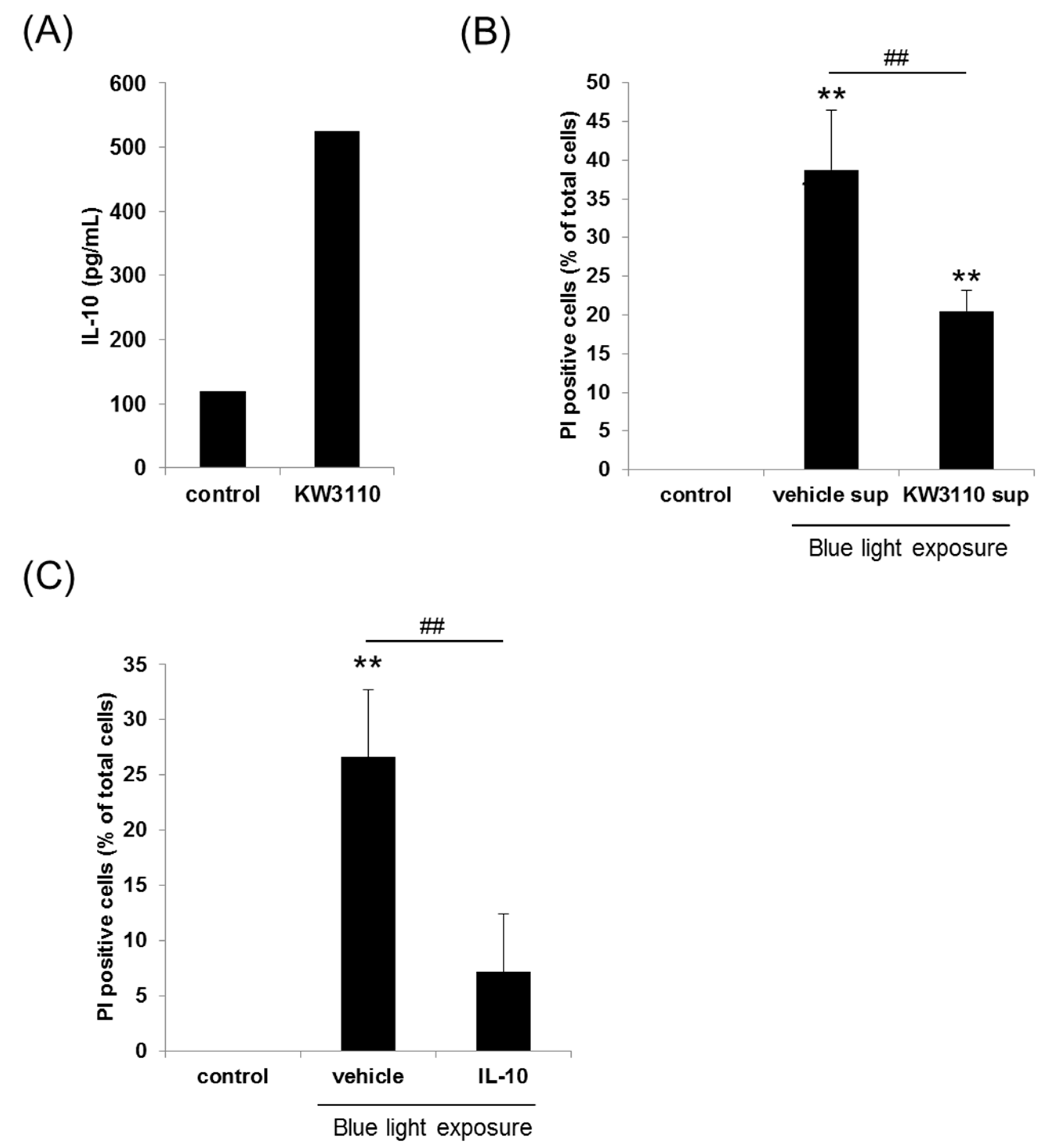
3.1.1. Effects of L. paracasei KW3110 on Human PBMC-Derived M2 Macrophage Activation

To determine the effects of L. paracasei KW3110 on human M2 macrophage activation, human PBMC-derived M2 macrophages were treated with L. paracasei KW3110. IL-10 levels as an index of M2 macrophage activity were measured from culture supernatants. L. paracasei KW3110 induced much higher IL-10 production when compared with the control cells (Figure 2A).

3.1.2. Effects of L. paracasei KW3110 on Blue Light-Induced Human Retinal Cell Death

We demonstrated blue light-induced human retinal pigment epithelial (ARPE-19) cell damage using a cell culture model in vitro. PI-positive dead cells, which were expressed as the ratio of PI-positive to Hoechst 33342-positive cells, were counted. To study the effect of blue light exposure toxicity on the ARPE-19 cell survival, ARPE-19 cells were incubated and exposed to 40, 50, 60, and 70 min of 1250 lux of blue light, respectively. There was a dose-dependent increase in PI-positive cells after blue light exposure (Figure S1). ARPE-19 cells that were exposed to 40 min and 50 min blue light exposure showed mild decrease in cell viability while cells that were exposed to 60 min and 70 min blue light exposure showed a marked decrease in cell viability to evaluate the effects of L. paracasei KW3110. Therefore, we performed further in vitro experiments under the 50 min of 1250 lux of the blue light exposure condition. Even though treatment with human PBMC-derived M2 macrophage supernatants (vehicle-sup) increased the number of PI-positive dead cells, treatment with L. paracasei KW3110-stimulated human PBMC-derived M2 macrophage supernatants (KW3110-sup) significantly inhibited blue light-induced cell death (Figure 2B and Figure S2). In the absence of blue light exposure, M2 macrophage supernatants, which were stimulated with or without L. paracasei KW3110, did not affect ARPE-19 cell survival (Figure S3). These results suggested that M2 macrophage supernatants, which were stimulated with L. paracasei KW3110, could suppress the blue light exposure-induced ARPE-19 cell death. To determine the inhibitory effects of KW3110-sup, we performed WST-8 assays. The results of the WST-8 assay indicated that blue light induced ARPE-19 cell death. KW3110-sup significantly inhibited blue light exposure-induced cell death (Figures S4 and S5). We also evaluated the effects of IL-10 on blue light-induced ARPE-19 cell death. Treatment with IL-10 significantly inhibited blue light-induced ARPE-19 cell death when compared with the vehicle control (Figure 2C).

3.2. Clinical Study

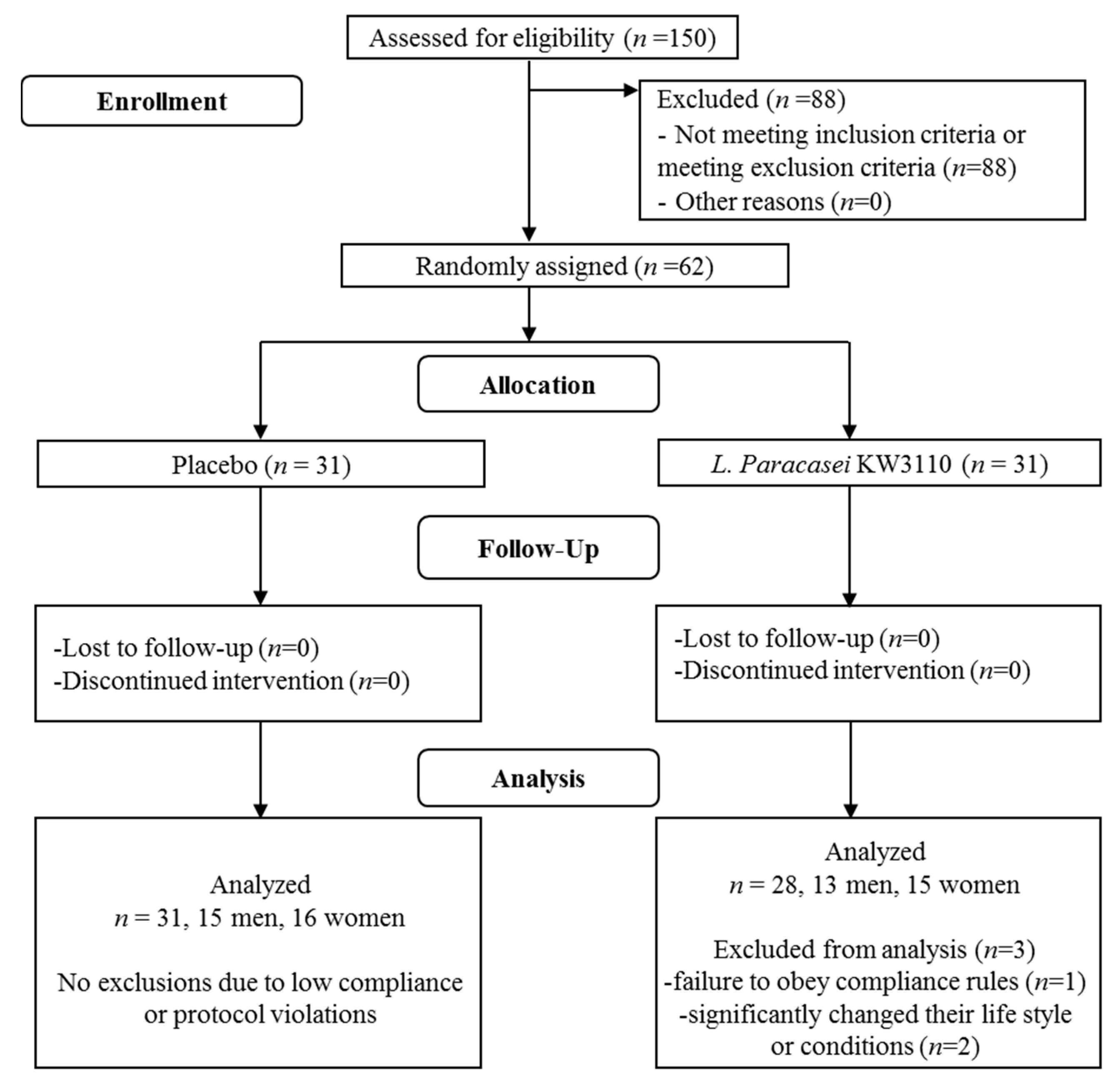
3.2.1. Background Information and Baseline Characteristics of the Subjects

No adverse events related to the test supplement were reported throughout the course of the study. The flow chart of the subjects enrolled in this study is shown in Figure 1. In accordance with the inclusion and exclusion criteria, 62 (male = 29, female = 33) of 150 participants were selected to be the subjects, which were judged eligible for this study by the supervising physician, and enrolled randomly into either the placebo group or the L. paracasei KW3110-treated group (n = 31 per group, respectively), which is described in the Materials and Methods section. Three subjects in the L. paracasei KW3110 group were excluded from data analyses before releasing the double-blind data due to failure to obey compliance rules (one subject) or the judgement by the supervising physician that the other two subjects had significantly changed their lifestyle or conditions. Consequently, the data analyses were carried out on 59 subjects (28 subjects in the L. paracasei KW3110 group and 31 subjects in the placebo group). The baseline characteristics of the 59 subjects are presented in Table 2. There were no significant differences in any characteristics between the placebo and L. paracasei KW3110 groups.

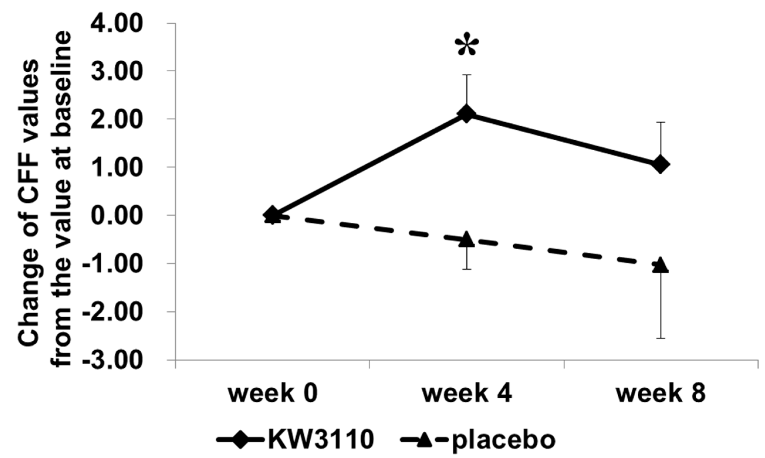
3.2.2. Ophthalmic Survey to Determine the Effects of L. paracasei KW3110 on VDT Load-Induced Eye Disorders

The results of the CFF tests are summarized in Table 3. As expected, VDT load-induced reductions in CFFs were observed at week 0 in both the placebo and the L. paracasei KW3110 group. There was no significant difference in the CFF values between the two groups. However, the variation, which is the difference of the values before and after the VDT load at each time point in the CFFs was significantly reduced at week 4 compared with week 0 only in the L. paracasei KW3110 group (p = 0.013) but not in the placebo group. Even though the changes from the values at week 0 in the CFFs were also evaluated (Table S1), there was no significant difference between the two groups.
In contrast, there was no effect of the VDT load on the variation of CS values in either of the two groups at any time points (Table S2). We could not detect significant differences in the variation ranges of CS between the two groups. Regarding the secondary outcomes such as other ophthalmic parameters, SPH, NPA, visual acuity, and the Schirmer test values, there were no significant differences in VDT load-induced variations in either of the two groups throughout the study (Table S3).

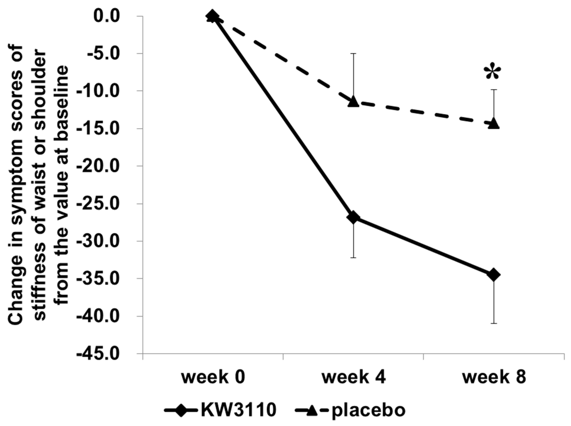
3.2.3. Questionnaire Survey to Determine the Effects of L. paracasei KW3110 on VDT Load-Induced Subjective Eye Conditions

The subjects recorded their symptoms of eye conditions subjectively by the VAS system using a questionnaire format. Symptom scores for stiffness of the shoulders or waist and ocular fatigue sensation were significantly lower at week 4 and week 8 than at week 0 in both groups (p < 0.01 or p < 0.05, Table 4). There were no significant differences in VDT load-induced variations of the symptoms and the changes from the values at week 0 in any symptom between the two groups (Table 4 and Table S4). While the DEQS scores were also significantly lower at week 4 and week 8 than at week 0 in both groups (p < 0.05, Table S4), there were no significant differences in VDT load-induced variations of the DEQS scores between the two groups.

3.2.4. Effects of L. paracasei KW3110 on VDT-Induced Eye Conditions in the Subjects Having a High-Level of Ocular Fatigue Sensation

Although the objective of this study was to evaluate the effects of L. paracasei KW3110 on VDT load-related eye disorders in the subjects who had subjective eye fatigue sensation. Eye fatigue symptom score levels by VAS differed among the subjects and a certain number of subjects had not been subjected to eye fatigue sensation. Therefore, we performed subgroup analysis. We analyzed the subjects with a high level of eye fatigue sensation whose ocular fatigue symptom score by VAS was greater than the average score of all subjects before VDT load during week 0. Consequently, the data analyses were carried out on 25 subjects (13 subjects in the L. paracasei KW3110 group and 12 subjects in the placebo group).
When the subjects with a high level of ocular fatigue sensation before VDT load were analyzed, the VDT load-induced variation in CFF as an indicator of eye fatigue was significantly reduced during week 4 compared with week 0 (Table 5) but only in the L. paracasei KW3110-treated group (n = 13, effect size, 1.087, 95% confidence interval, −0.059–3.124; p = 0.024) and not in the placebo group (n = 12). In addition, the change from the variation value at week 0 in CFF was greater in the L. paracasei KW3110 group than in the placebo group during week 4 (Figure 3, effect size, 1.614, 95% confidence interval, 0.519–4.686; p = 0.020). The subjective symptoms of eye fatigue including ocular pain, stiffness of the shoulders or waist, ocular fatigue sensation, and the stuffy head were also significantly improved in the L. paracasei KW3110 group (Table 6). The subjective symptom of eye redness before the VDT load was significantly reduced during week 4 in the L. paracasei KW3110-treated group when compared with the placebo group (Figure S6). Other subjective symptoms were not changed at any time point in both groups (data not shown). Furthermore, the changes from the values at week 0 in stiffness of the waist and/or shoulders were more reduced in the L. paracasei KW3110-treated group than in the placebo group during week 8 (Figure 4, effect size, −4.515; 95% confidence interval, −36.262–4.148; p = 0.019). This finding was inconsistent with the CFF data, which indicated that the VDT load-induced eye fatigue were more improved in the L. paracasei KW3110-treated group. There was no significant difference in other primary outcome (CS) and any secondary outcomes between the two groups.

4. Discussion

The purpose of the present study was to investigate the effects of L. paracasei KW3110 on VDT-induced eye disorders and eye fatigue in in vitro experiments and in a clinical trial. We demonstrated that L. paracasei KW3110 activated human M2 macrophages and significantly increased IL-10 production in cells. We also found that culture supernatants from cells treated with L. paracasei KW3110 suppressed blue light-induced retinal pigment epithelial cell death. In a randomized, double-blind, placebo-controlled parallel group study of humans with VDT load-induced eye disorders and eye fatigue, we observed that both objective parameters and subjectively quantified eye fatigue symptoms were alleviated with daily ingestion of heat-killed L. paracasei KW3110 supplementation. These results indicated that heat-killed L. paracasei KW3110 mitigated the VDT load-induced eye fatigue.
In this study, we showed that L. paracasei KW3110 had the potential of activating human PBMC-derived M2 macrophages and the potential of inducing the production of IL-10. In a previous study, Pam3Cys, which is a ligand for toll-like receptor 2 (TLR2), has been reported to induce IL-10 production by activating the ERK pathway in mouse dendritic cells [37]. Since lactic acid bacteria have TLR2 ligands like lipo-teichoic acid, the effect of L. paracasei KW3110 on IL-10 production in human PBMC-derived M2 macrophage might be mediated by the TLR2-dependent ERK pathway.
Macrophages play crucial roles in the central nervous system [38] and can be grouped into at least two subgroups including the classically activated M1 macrophages or the alternatively activated M2 macrophages [39,40]. Although M1 macrophages are pro-inflammatory and produce inflammatory cytokines such as IL-1β and tumor necrosis factor alpha (TNF-α, in contrast, M2 macrophages are associated with responses to anti-inflammatory reactions including tissue remodeling through the production of anti-inflammatory cytokines like IL-10) [41,42]. In the retina, activation of the M2 macrophage is also thought to promote retinal cell survival in several mouse models [27,43]. However, blue light exposure was reported to induce the inflammation due to the oxidative stress [44]. It is possible that M2 macrophage activation is stimulated by L. paracasei KW3110 and is important for the suppression of blue light-induced inflammation and human retinal pigment epithelial cell death in part through the production of IL-10 (Figure 2B,C).
VDTs including those emitting blue light may cause various visual problems and have adverse effects on the visual nervous system [6,7]. VDT load-induced visual disorders can be evaluated by objective ophthalmic parameters such as the CFF [6]. The CFF reflects neuronal impulse transmission from retinal ganglion cells to the primary visual cortex and has been used as an indicator of eye fatigue [6,45,46,47,48]. In the present study, in the L. paracasei KW3110-treated group with a high level of ocular fatigue sensation before VDT load, the change from the value at week 0 of VDT load-induced reduction in CFF was significantly mitigated when compared with the placebo group. These clinical results suggested that ingestion of L. paracasei KW3110 improved VDT load-induced eye fatigue. The initial in vitro results and a previous report also supported these clinical results. In a previous report, neural activity correlated with eye fatigue [49]. In our previous work, our group revealed that orally provided L. paracasei KW3110 interacted with the gut immune cells in mice and oral administration of L. paracasei KW3110 enhanced the cytokine levels in blood [33]. We also revealed L. paracasei KW3110 suppressed excessive inflammation including dermatitis in mice and humans. In this study, we showed that L. paracasei KW3110 could activate M2 macrophages and induce the anti-inflammatory cytokines like IL-10 (Figure 2A). We also showed that not only the M2 macrophage supernatants stimulated with L. paracasei KW3110, but IL-10 could significantly suppress light-induced retinal cell death (Figure 2B). Taken together, intake of L. paracasei KW3110 could suppress retinal inflammation in part through the anti-inflammatory cytokines in the blood, which is induced from the gut immune cells. In a previous report, a bilberry extract was also reported to protect retinal ganglion cells and also accompanied the mitigation of eye fatigue [50,51]. Therefore, the reduction of retinal cell death and inflammation might contribute to the beneficial effects of L. paracasei KW3110 in this study. Further clinical studies to evaluate the effects of L. paracasei KW3110 on inflammatory markers will be carried out to reveal the underlying mechanisms. While the relationship between retinal cell death and eye fatigue has been under investigation, the positive effects of L. paracasei KW3110 on VDT load-induced eye fatigue may be attributed to the inhibition of blue light-induced retinal cell death.
In our subjects, the subjective symptoms including stiffness of the waist or shoulders were mitigated by the ingestion of L. paracasei KW3110. The musculoskeletal discomfort including stiffness of the waist or shoulders are well-known to be associated with eye fatigue [1]. It was suggested that ingestion of L. paracasei KW3110 could improve musculoskeletal discomfort by reducing the symptoms of VDT load-induced eye disorders. However, the musculoskeletal symptoms were also reported to be related to blood flow [52]. Further studies are needed to delineate the mechanism underlying the effects of L. paracasei KW3110 on the mitigation of musculoskeletal discomfort.
In the present study, CFF and the subjective symptoms, which are the indicators of eye fatigue, were improved in the L. paracasei KW3110 group when compared with the placebo group. However, the improvement in CFF was significant four weeks after intake of L. paracasei KW3110 but was not significant during the eighth week. It is suggested that repeated eye fatigue evaluations might increase the learning effects during the eighth week, which resulted in the reduction of the difference between placebo and L. paracasei KW3110during the eighth week when compared with the fourth week. There was no significant difference between the L. paracasei KW3110 group and the placebo (control) group in other secondary outcomes including the Schirmer test results and DEQS as the indicators of dry eye. This may be because the VDT loads in this study were too mild to detect significant changes in dry eye symptoms. The results of this study were consistent with previous reports. In a previous study, VDT loads for more than eight hours could increase the risk of dry eye [53]. A previous study reported that VDT loads for two hours were too short to evaluate some parameters such as Schirmer’s test results [51]. Further studies involving longer VDT loads or brighter VDT loads might better evaluate the effect of L. paracasei KW3110 on VDT load-induced dry eye symptoms. However, in both CFF and VAS, which are indicators of eye fatigue, intake of L. paracasei KW3110 could mitigate eye fatigue when compared with the placebo group (Figure 3 and Figure 4). These results indicated that intake of L. paracasei KW3110 had positive effects on eye fatigue.
Since this study focused on evaluating the effects of L. paracasei KW3110 on VDT-induced eye disorders and eye fatigue, the clinical study enrolled only subjects who used VDT in their daily life activities. It might be necessary for the confirmation of clinical efficacy of L. paracasei KW3110 to carry out further studies and enrolling many more subjects. Nonetheless, there are many healthy people who have VDT work-related vision problems such as eye fatigue and the effects of L. paracasei KW3110 on improving VDT load-induced eye fatigue is, therefore, expected to be widely applicable.

5. Conclusions

In conclusion, the findings of this study indicated that L. paracasei KW3110 suppressed blue light-induced retinal pigment epithelial cell death in vitro and ingestion of L. paracasei KW3110 had positive effects for improving some objective and subjective parameters of eye disorders and eye fatigue induced by VDT loads. Further studies enrolling many more subjects should be carried out to more clearly reveal the clinical effects of L. paracasei KW3110 on ocular disorder including eye fatigue.

Supplementary Materials

The following are available online at https://www.mdpi.com/2072-6643/10/8/1058/s1. Figure S1: Dose-dependent effects of blue light exposure on ARPE-19 cell survival. Data are shown as the mean ± SE. Figure S2: Effects of M2 macrophage supernatants, stimulated with or without L. paracasei KW3110, on blue light-induced ARPE-19 cell survival. Representative fluorescence microscopy images of Hoechst 33342 (blue) and propidium iodide (PI) (red) after 12 h of blue light exposure. Figure S3: Effects of M2 macrophage supernatants, stimulated with or without L. paracasei KW3110, on ARPE-19 cell survival in the absence of blue light exposure. Data are shown as the mean ± SE. Figure S4: Effects of M2 macrophage supernatants, which are stimulated with or without L. paracasei KW3110, on ARPE-19 cell survival as determined by the WST-8 assay. The inhibitory effect of L. paracasei KW3110-stimulated human PBMC-derived M2 macrophage supernatants (KW3110-sup) or vehicle-treated supernatants (vehicle sup) on blue light-induced retinal cell death in ARPE-19 cells, which was measured by using WST-8 assays. Assays were performed in triplicate wells. The data show the mean ± SE for triplicate wells. ** p < 0.01 versus control, # p < 0.05 versus vehicle supernatants [one-way analysis of variance (ANOVA) followed by Tukey’s test]. Figure S5: Effects of IL-10 on blue light-induced ARPE-19 cell death. Representative fluorescence microscopy images of Hoechst 33342 (blue) and propidium iodide (PI) (red) after 12 hours of blue light exposure. Figure S6: Effects of L. paracasei KW3110 on eye redness for subjects with severe eye fatigue. The comparisons of eye redness in visual analog scale (VAS) values at each time point were performed by an unpaired Student’s t-test with a Holm correction for multiple comparisons (* p < 0.05 versus placebo). Data are shown as the means ± SE. KW3110 = L. paracasei KW3110-treated group, placebo = control group. Table S1: Changes of CFF from the value at week 0. Data are expressed as the mean ± SD. a Variation indicated the difference of the values before and after the VDT load at each point. VDT = visual display terminal. CFF = critical flicker frequency. KW3110 = L. paracasei KW3110-treated group. placebo = control group. Table S2: Effects of L. paracasei KW3110 on CS values. Data are expressed as the mean ± SD. The within-group comparisons between week 0 and week 4 or week 8 were carried out using a paired Student’s t-test with a Holm correction for multiple comparisons (** p < 0.01, * p < 0.05). a Variation indicated the difference of the values between before and after the VDT load at each point. VDT = visual display terminal. CS = contrast sensitivity. KW3110 = L. paracasei KW3110-treated group. placebo = control group. Table S3: SPH, Visual acuity, NPA, Schirmer’s tests, and DEQS questionnaire values. Data are expressed as the mean ± SD. The within-group comparisons between week 0 and week 4 or week 8 were carried out using a paired Student’s t-test with a Holm correction for multiple comparisons (** p < 0.01, * p < 0.05). a Variation indicated the difference of the values between before and after the VDT load at each point. VDT = visual display terminal. NPA, near point accommodation. DEQS = Dry Eye-Related Quality-of-Life Score. SPH = spherical and subjective symptoms. KW3110 = L. paracasei KW3110-treated group. placebo = control group. Table S4: Changes of 11 subjective symptoms of eye fatigue and related visual conditions from the values at week 0. Data are expressed as the mean ± SD. The within-group comparisons between week 0 and week 4 or week 8 were carried out using a paired Student’s t-test with a Holm correction for multiple comparisons (** p < 0.01, * p < 0.05). a Variation indicated the difference of the values between before and after the VDT load at each point. VDT = visual display terminal. KW3110 = L. paracasei KW3110-treated group. placebo = control group.

Author Contributions

Y.M., K.J., M.M., and O.K. designed this study. M.I. conducted this study as a principal investigator. Y.M. and O.K. wrote the manuscript and Y.M. assumed primary responsibility for the final content. All authors read and approved the final manuscript.

Funding

This study was funded by Kirin Company, Limited.

Acknowledgments

We thank Takayoshi Kirisako, Yasuyuki Tomita, and Masato Otake of Kirin Company for their valuable discussion.

Conflicts of Interest

The funder designed this study and supplied the test tablets and placebo. The funder had no role in data collection nor data analysis. This manuscript was prepared by the members of the funder. The funder decided to publish the results.

References

  1. Bergqvist, U. Possible health effects of working with vdus. Br. J. Ind. Med. 1989, 46, 217–221. [Google Scholar] [CrossRef] [PubMed]
  2. Knave, B.G.; Wibom, R.I.; Voss, M.; Hedstrom, L.D.; Bergqvist, U.O. Work with video display terminals among office employees. I. Subjective symptoms and discomfort. Scand. J. Work Environ. Health 1985, 11, 457–466. [Google Scholar] [CrossRef] [PubMed]
  3. Eltayeb, S.; Staal, J.B.; Hassan, A.; de Bie, R.A. Work related risk factors for neck, shoulder and arms complaints: A cohort study among dutch computer office workers. J. Occup. Rehabil. 2009, 19, 315–322. [Google Scholar] [CrossRef] [PubMed]
  4. Parihar, J.K.; Jain, V.K.; Chaturvedi, P.; Kaushik, J.; Jain, G.; Parihar, A.K. Computer and visual display terminals (vdt) vision syndrome (cvdts). Med. J. Armed Forces India 2016, 72, 270–276. [Google Scholar] [CrossRef] [PubMed]
  5. Aronsson, G.; Stromberg, A. Work content and eye discomfort in vdt work. Int. J. Occup. Saf. Ergon. 1995, 1, 1–13. [Google Scholar] [CrossRef] [PubMed]
  6. Murata, K.; Araki, S.; Kawakami, N.; Saito, Y.; Hino, E. Central nervous system effects and visual fatigue in vdt workers. Int. Arch. Occup. Environ. Health 1991, 63, 109–113. [Google Scholar] [CrossRef] [PubMed]
  7. Murata, K.; Araki, S.; Yokoyama, K.; Yamashita, K.; Okumatsu, T.; Sakou, S. Accumulation of vdt work-related visual fatigue assessed by visual evoked potential, near point distance and critical flicker fusion. Ind. Health 1996, 34, 61–69. [Google Scholar] [CrossRef] [PubMed]
  8. Goto, E.; Yagi, Y.; Matsumoto, Y.; Tsubota, K. Impaired functional visual acuity of dry eye patients. Am. J. Ophthalmol. 2002, 133, 181–186. [Google Scholar] [CrossRef]
  9. Uchino, M.; Schaumberg, D.A.; Dogru, M.; Uchino, Y.; Fukagawa, K.; Shimmura, S.; Satoh, T.; Takebayashi, T.; Tsubota, K. Prevalence of dry eye disease among japanese visual display terminal users. Ophthalmology 2008, 115, 1982–1988. [Google Scholar] [CrossRef] [PubMed]
  10. Thomson, W.D. Eye problems and visual display terminals—The facts and the fallacies. Ophthalmic Physiol. Opt. 1998, 18, 111–119. [Google Scholar] [CrossRef]
  11. Ye, Z.; Abe, Y.; Kusano, Y.; Takamura, N.; Eida, K.; Takemoto, T.; Aoyagi, K. The influence of visual display terminal use on the physical and mental conditions of administrative staff in japan. J. Physiol. Anthropol. 2007, 26, 69–73. [Google Scholar] [CrossRef] [PubMed]
  12. Friedman, N.J. Impact of dry eye disease and treatment on quality of life. Curr. Opin. Ophthalmol. 2010, 21, 310–316. [Google Scholar] [CrossRef] [PubMed]
  13. Vilela, M.A.; Pellanda, L.C.; Fassa, A.G.; Castagno, V.D. Prevalence of asthenopia in children: A systematic review with meta-analysis. J. Pediatr. 2015, 91, 320–325. [Google Scholar] [CrossRef] [PubMed]
  14. Collier, J.D.; Rosenfield, M. Accommodation and convergence during sustained computer work. Optometry 2011, 82, 434–440. [Google Scholar] [CrossRef] [PubMed]
  15. Kurimoto, S.; Iwasaki, T.; Nomura, T.; Noro, K.; Yamamoto, S. Influence of vdt (visual display terminals) work on eye accommodation. J. UOEH 1983, 5, 101–110. [Google Scholar] [CrossRef] [PubMed]
  16. Kajita, M.; Ono, M.; Suzuki, S.; Kato, K. Accommodative microfluctuation in asthenopia caused by accommodative spasm. Fukushima J. Med. Sci. 2001, 47, 13–20. [Google Scholar] [CrossRef] [PubMed]
  17. Portello, J.K.; Rosenfield, M.; Chu, C.A. Blink rate, incomplete blinks and computer vision syndrome. Optom. Vis. Sci. 2013, 90, 482–487. [Google Scholar] [CrossRef] [PubMed]
  18. Uchino, Y.; Uchino, M.; Dogru, M.; Fukagawa, K.; Tsubota, K. Improvement of accommodation with anti-oxidant supplementation in visual display terminal users. J. Nutr. Health Aging 2012, 16, 478–481. [Google Scholar] [CrossRef] [PubMed]
  19. Choi, W.; Kim, J.C.; Kim, W.S.; Oh, H.J.; Yang, J.M.; Lee, J.B.; Yoon, K.C. Clinical effect of antioxidant glasses containing extracts of medicinal plants in patients with dry eye disease: A multi-center, prospective, randomized, double-blind, placebo-controlled trial. PLoS ONE 2015, 10, e0139761. [Google Scholar] [CrossRef] [PubMed]
  20. Snodderly, D.M. Evidence for protection against age-related macular degeneration by carotenoids and antioxidant vitamins. Am. J. Clin. Nutr. 1995, 62, 1448S–1461S. [Google Scholar] [CrossRef] [PubMed]
  21. Lee, J.; Lee, H.K.; Kim, C.Y.; Hong, Y.J.; Choe, C.M.; You, T.W.; Seong, G.J. Purified high-dose anthocyanoside oligomer administration improves nocturnal vision and clinical symptoms in myopia subjects. Br. J. Nutr. 2005, 93, 895–899. [Google Scholar] [CrossRef] [PubMed] [Green Version]
  22. Wang, L.J.; Su, S.; Wu, J.; Du, H.; Li, S.S.; Huo, J.W.; Zhang, Y.; Wang, L.S. Variation of anthocyanins and flavonols in vaccinium uliginosum berry in lesser khingan mountains and its antioxidant activity. Food Chem. 2014, 160, 357–364. [Google Scholar] [CrossRef] [PubMed]
  23. Galbis-Estrada, C.; Pinazo-Duran, M.D.; Martinez-Castillo, S.; Morales, J.M.; Monleon, D.; Zanon-Moreno, V. A metabolomic approach to dry eye disorders. The role of oral supplements with antioxidants and omega 3 fatty acids. Mol. Vis. 2015, 21, 555–567. [Google Scholar] [PubMed]
  24. Neuringer, M. Infant vision and retinal function in studies of dietary long-chain polyunsaturated fatty acids: Methods, results, and implications. Am. J. Clin. Nutr. 2000, 71, 256S–267S. [Google Scholar] [CrossRef] [PubMed]
  25. Bhargava, R.; Kumar, P.; Phogat, H.; Kaur, A.; Kumar, M. Oral omega-3 fatty acids treatment in computer vision syndrome related dry eye. Contact Lens Anterior Eye 2015, 38, 206–210. [Google Scholar] [CrossRef] [PubMed]
  26. Rutar, M.; Natoli, R.; Provis, J.M. Small interfering rna-mediated suppression of ccl2 in muller cells attenuates microglial recruitment and photoreceptor death following retinal degeneration. J. Neuroinflamm. 2012, 9, 221. [Google Scholar] [CrossRef] [PubMed]
  27. Jiao, H.; Natoli, R.; Valter, K.; Provis, J.M.; Rutar, M. Spatiotemporal cadence of macrophage polarisation in a model of light-induced retinal degeneration. PLoS ONE 2015, 10, e0143952. [Google Scholar] [CrossRef] [PubMed]
  28. Miyake, S.; Takahashi, N.; Sasaki, M.; Kobayashi, S.; Tsubota, K.; Ozawa, Y. Vision preservation during retinal inflammation by anthocyanin-rich bilberry extract: Cellular and molecular mechanism. Lab. Investig. 2012, 92, 102–109. [Google Scholar] [CrossRef] [PubMed]
  29. Fontana, L.; Bermudez-Brito, M.; Plaza-Diaz, J.; Munoz-Quezada, S.; Gil, A. Sources, isolation, characterisation and evaluation of probiotics. Br. J. Nutr. 2013, 109 (Suppl. S2), S35–S50. [Google Scholar] [CrossRef] [PubMed]
  30. Ichikawa, S.; Fujii, R.; Fujiwara, D.; Komiyama, Y.; Kaisho, T.; Sakaguchi, M.; Konishi, Y. Myd88 but not tlr2, 4 or 9 is essential for il-12 induction by lactic acid bacteria. Biosci. Biotechnol. Biochem. 2007, 71, 3026–3032. [Google Scholar] [CrossRef] [PubMed]
  31. Wakabayashi, H.; Nariai, C.; Takemura, F.; Nakao, W.; Fujiwara, D. Dietary supplementation with lactic acid bacteria attenuates the development of atopic-dermatitis-like skin lesions in nc/nga mice in a strain-dependent manner. Int. Arch. Allergy Immunol. 2008, 145, 141–151. [Google Scholar] [CrossRef] [PubMed]
  32. Fujiwara, D.; Inoue, S.; Wakabayashi, H.; Fujii, T. The anti-allergic effects of lactic acid bacteria are strain dependent and mediated by effects on both th1/th2 cytokine expression and balance. Int. Arch. Allergy Immunol. 2004, 135, 205–215. [Google Scholar] [CrossRef] [PubMed]
  33. Ichikawa, S.; Miyake, M.; Fujii, R.; Konishi, Y. Orally administered lactobacillus paracasei kw3110 induces in vivo il-12 production. Biosci. Biotechnol. Biochem. 2009, 73, 1561–1565. [Google Scholar] [CrossRef] [PubMed]
  34. Moriguchi, S. “Nikkagiren Suchihyo” [Numerical Scheme]; JUSE Press: Tokyo, Japan, 1968; Volume 80, pp. 37–43. (In Japanese) [Google Scholar]
  35. Richer, S.; Stiles, W.; Statkute, L.; Pulido, J.; Frankowski, J.; Rudy, D.; Pei, K.; Tsipursky, M.; Nyland, J. Double-masked, placebo-controlled, randomized trial of lutein and antioxidant supplementation in the intervention of atrophic age-related macular degeneration: The veterans last study (lutein antioxidant supplementation trial). Optometry 2004, 75, 216–230. [Google Scholar] [CrossRef]
  36. Sakane, Y.; Yamaguchi, M.; Yokoi, N.; Uchino, M.; Dogru, M.; Oishi, T.; Ohashi, Y.; Ohashi, Y. Development and validation of the dry eye-related quality-of-life score questionnaire. JAMA Ophthalmol. 2013, 131, 1331–1338. [Google Scholar] [CrossRef] [PubMed]
  37. Kaji, R.; Kiyoshima-Shibata, J.; Nagaoka, M.; Nanno, M.; Shida, K. Bacterial teichoic acids reverse predominant il-12 production induced by certain lactobacillus strains into predominant il-10 production via tlr2-dependent erk activation in macrophages. J. Immunol. 2010, 184, 3505–3513. [Google Scholar] [CrossRef] [PubMed]
  38. Herz, J.; Filiano, A.J.; Smith, A.; Yogev, N.; Kipnis, J. Myeloid cells in the central nervous system. Immunity 2017, 46, 943–956. [Google Scholar] [CrossRef] [PubMed]
  39. Martinez, F.O.; Helming, L.; Gordon, S. Alternative activation of macrophages: An immunologic functional perspective. Annu. Rev. Immunol. 2009, 27, 451–483. [Google Scholar] [CrossRef] [PubMed]
  40. Biswas, S.K.; Mantovani, A. Macrophage plasticity and interaction with lymphocyte subsets: Cancer as a paradigm. Nat. Immunol. 2010, 11, 889–896. [Google Scholar] [CrossRef] [PubMed]
  41. Sica, A.; Mantovani, A. Macrophage plasticity and polarization: In vivo veritas. J. Clin. Investig. 2012, 122, 787–795. [Google Scholar] [CrossRef] [PubMed]
  42. Satoh, T.; Kidoya, H.; Naito, H.; Yamamoto, M.; Takemura, N.; Nakagawa, K.; Yoshioka, Y.; Morii, E.; Takakura, N.; Takeuchi, O.; et al. Critical role of trib1 in differentiation of tissue-resident m2-like macrophages. Nature 2013, 495, 524–528. [Google Scholar] [CrossRef] [PubMed]
  43. London, A.; Itskovich, E.; Benhar, I.; Kalchenko, V.; Mack, M.; Jung, S.; Schwartz, M. Neuroprotection and progenitor cell renewal in the injured adult murine retina requires healing monocyte-derived macrophages. J. Exp. Med. 2011, 208, 23–39. [Google Scholar] [CrossRef] [PubMed] [Green Version]
  44. Lau, L.I.; Chiou, S.H.; Liu, C.J.; Yen, M.Y.; Wei, Y.H. The effect of photo-oxidative stress and inflammatory cytokine on complement factor H expression in retinal pigment epithelial cells. Investig. Ophthalmol. Vis. Sci. 2011, 52, 6832–6841. [Google Scholar] [CrossRef] [PubMed]
  45. Lin, J.B.; Gerratt, B.W.; Bassi, C.J.; Apte, R.S. Short-wavelength light-blocking eyeglasses attenuate symptoms of eye fatigue. Investig. Ophthalmol. Vis. Sci. 2017, 58, 442–447. [Google Scholar] [CrossRef] [PubMed]
  46. Ayaki, M.; Hattori, A.; Maruyama, Y.; Nakano, M.; Yoshimura, M.; Kitazawa, M.; Negishi, K.; Tsubota, K. Protective effect of blue-light shield eyewear for adults against light pollution from self-luminous devices used at night. Chronobiol. Int. 2016, 33, 134–139. [Google Scholar] [CrossRef] [PubMed]
  47. Maeda, E.; Yoshikawa, T.; Hayashi, N.; Akai, H.; Hanaoka, S.; Sasaki, H.; Matsuda, I.; Yoshioka, N.; Ohtomo, K. Radiology reading-caused fatigue and measurement of eye strain with critical flicker fusion frequency. Jpn. J. Radiol. 2011, 29, 483–487. [Google Scholar] [CrossRef] [PubMed]
  48. Luczak, A.; Sobolewski, A. Longitudinal changes in critical flicker fusion frequency: An indicator of human workload. Ergonomics 2005, 48, 1770–1792. [Google Scholar] [CrossRef] [PubMed]
  49. Cai, T.; Zhu, H.; Xu, J.; Wu, S.; Li, X.; He, S. Human cortical neural correlates of visual fatigue during binocular depth perception: An fnirs study. PLoS ONE 2017, 12, e0172426. [Google Scholar] [CrossRef] [PubMed]
  50. Matsunaga, N.; Imai, S.; Inokuchi, Y.; Shimazawa, M.; Yokota, S.; Araki, Y.; Hara, H. Bilberry and its main constituents have neuroprotective effects against retinal neuronal damage in vitro and in vivo. Mol. Nutr. Food Res. 2009, 53, 869–877. [Google Scholar] [CrossRef] [PubMed]
  51. Ozawa, Y.; Kawashima, M.; Inoue, S.; Inagaki, E.; Suzuki, A.; Ooe, E.; Kobayashi, S.; Tsubota, K. Bilberry extract supplementation for preventing eye fatigue in video display terminal workers. J. Nutr. Health Aging 2015, 19, 548–554. [Google Scholar] [CrossRef] [PubMed]
  52. Matsumoto, H.; Kamm, K.E.; Stull, J.T.; Azuma, H. Delphinidin-3-rutinoside relaxes the bovine ciliary smooth muscle through activation of etb receptor and no/cgmp pathway. Exp. Eye Res. 2005, 80, 313–322. [Google Scholar] [CrossRef] [PubMed]
  53. Uchino, M.; Yokoi, N.; Uchino, Y.; Dogru, M.; Kawashima, M.; Komuro, A.; Sonomura, Y.; Kato, H.; Kinoshita, S.; Schaumberg, D.A.; et al. Prevalence of dry eye disease and its risk factors in visual display terminal users: The osaka study. Am. J. Ophthalmol. 2013, 156, 759–766. [Google Scholar] [CrossRef] [PubMed]
Figure 1. Summary flow diagram of this study.
Nutrients 10 01058 g001
Figure 2. Effects of Lactobacillus paracasei KW3110 on cell viability under blue light exposure conditions. (A) Lactobacillus paracasei LW3110 activated human M2 macrophages. Human peripheral blood mononuclear cells (PBMC)-derived M2 macrophages were treated with 10 μg/mL L. paracasei KW3110 or untreated (control) and cultured for 24 h. Interleukin 10 (IL-10) concentration of the culture medium was measured by ELISAs. (B) The inhibitory effect of L. paracasei KW3110-stimulated human PBMC-derived M2 macrophage supernatants (KW3110-sup) or vehicle-treated supernatants (vehicle sup) on blue light-induced retinal cell death in ARPE-19 cells as measured by propidium iodide (PI) staining. The number of cells exhibiting PI fluorescence was counted. PI positive cells were expressed as the ratio of PI-positive to Hoechst 33342-positive cells. Assays were performed in triplicate wells. The data shows the mean ± SD for triplicate wells. ** p < 0.01 versus control. ## p < 0.01 versus vehicle supernatants [one-way analysis of variance (ANOVA) followed by Tukey’s test]. (C) Inhibitory effects of IL-10 on blue light-induced retinal cell death in ARPE-19 cells. The number of cells exhibiting PI fluorescence was counted. PI-positive cells were expressed as the ratio of PI-positive to Hoechst 33342-positive cells. Assays were performed in quadruplicate wells. The data show the mean ± SD for triplicate wells. ** p < 0.01 versus the control. ## p < 0.01 versus the vehicle (one-way ANOVA followed by Tukey’s test). ELISA = enzyme-linked immunosorbent assay.
Nutrients 10 01058 g002
Figure 3. Changes of CFF values from the value during week 0 in subjects with severe ocular fatigue sensation before the VDT load. VDT = visual display terminal, CFF = critical flicker frequency. Data are calculated as the degrees of change from the values during week 0. The comparisons between two groups at each time point were performed by an unpaired Student’s t-test with a Holm correction for multiple comparisons (* p < 0.05 versus placebo). Data are shown as the means ± SE. KW3110 = L. paracasei KW3110-treated group, placebo = control group.
Nutrients 10 01058 g003
Figure 4. Changes in symptom scores of stiffness of the waist or shoulders from the values during week 0 in subjects with severe ocular fatigue sensation before the VDT load. VDT = visual display terminal. Data are calculated as the degrees of change from the values during week 0. The comparisons between two groups at each time point were performed by an unpaired Student’s t-test with a Holm correction for multiple comparisons (* p < 0.05 versus placebo). Data are shown as the means ± SE. KW3110 = L. paracasei KW3110-treated group, placebo = control group.
Nutrients 10 01058 g004
Table 1. Exclusion criteria for the study.
(1)Subjects who had amblyopia or strabismus
(2)Subjects who were diagnosed as presbyopia [the control width was less than 2.5 diopters (D) for the dominant eye]
(3)Subjects who had an uncorrected serious refractive error for the dominant eye
(4)Subjects who had undergone ophthalmic surgery for the dominant eye
(5)Subjects who had best-corrected visual acuity <1.0 for the dominant eye
(6)Subjects who had a serious eye disease
(7)Subjects who were under treatment for any chronic disease and/or used medicines continuously or medical supplies that were being commonly used
(8)Subjects who had the experience of a serious disease (e.g., diabetes, liver disease, kidney disease, and/or heart disease, thyroid gland disease, adrenal gland disease, digestive system disease, and/or metabolic disorder)
(9)Subjects who could not stop eating foods similar to the test foods and/or were taking drugs or health foods including lactic acid bacteria or Bifidobacterium
(10)Subjects with excessive alcohol-drinking behavior
(11)Subjects who regularly took drugs or health foods, which might have had effects on the eyes or were expected to be used during the study
(12)Subjects who could not stop taking drugs or health foods, which might have effects on immune functions
(13)Subjects with the possibility of drug and/or food allergy
(14)Subjects who could not stop drinking alcoholic beverages for 2 days until the pre-check day and check days
(15)Subjects who had a history of pollinosis
(16)Subjects who have a tendency to get diarrhea by eating dairy products
(17)Subjects diagnosed with drug or alcohol dependence
(18)Subjects who and/or whose family worked in a company selling or manufacturing health foods or foods with functional claims
(19)Subjects who could not execute a display work load test
(20)Subjects who were participating in other clinical studies or revoked an agreement acquisition day and/or participated in other clinical studies within 3 months or who were planning to participate in other clinical studies during this study
(21)Subjects who were pregnant, breastfeeding, planning to get pregnant, or breastfeeding during the study
(22)Subjects who were judged as unsuitable by the physician for laboratory data, anthropometric measurements, or physical examination values
(23)Subjects who were judged as unsuitable for the study as assessed by the background questionnaire
(24)Subjects who were judged as unsuitable by the supervising physician for other reasons
Table 2. Baseline characteristics of subjects analyzed in the study.
CharacteristicKW3110 Placebo p Values
Mean ± SDMean ± SD
Number of subjects (male/female)28 (13/15)31 (15/16)0.880
Age (years)40.3 ± 2.740.6 ± 2.80.651
Body weight (kg)58.27 ± 10.2959.34 ± 10.600.695
BMI (kg/m2)21.48 ± 2.5321.86 ± 2.400.564
Data are expressed as the mean ± standard deviation (SD) with the exception of sex. The comparisons between two groups were performed by an unpaired Student’s t-test except for the number of subjects (male/female). The number of subjects (male/female) was evaluated using a χ2 test. There was no significant difference in any parameters between the two groups. BMI = body/mass index. KW3110 = L. paracasei KW3110-treated group. placebo = control group.
Table 3. Effects of L. paracasei KW3110 on CFF values.
CFF (Hz)KW3110 Group (n = 28)Placebo Group (n = 31)
Week 0Week 4Week 8Week 0Week 4Week 8
Before VDT load39.13 ± 2.4739.57 ± 2.9539.98 ± 2.9439.03 ± 2.6239.45 ± 2.3639.77 ± 2.83
After VDT load37.68 ± 2.4039.87 ± 2.52 **39.56 ± 2.92 **38.10 ± 2.9739.24 ± 2.15 *39.95 ± 2.13 *
Variation a−1.45 ± 2.380.30 ± 2.70 *−0.42 ± 2.31−0.94 ± 2.04−0.22 ± 2.030.17 ± 2.97
Data are expressed as the mean ± SD. The within-group comparisons between week 0 and week 4 or week 8 were carried out using a paired Student’s t-test with a Holm correction for multiple comparisons (** p < 0.01, * p < 0.05). a Variation indicated the difference of the values before and after the VDT load at each point. CFF = critical flicker frequency. VDT = visual display terminal. KW3110 = L. paracasei KW3110-treated group, placebo = control group.
Table 4. The subjective symptoms of eye fatigue and related visual conditions.
Symptoms KW3110 Group (n = 28)Placebo Group (n = 31)
Week 0Week 4Week 8Week 0Week 4Week 8
Ocular painBefore VDT load15.0 ± 17.99.3 ± 13.57.4 ± 14.312.5 ± 20.27.8 ± 14.38.9 ± 18.2
After VDT load28.5 ± 28.723.4 ± 26.417.6 ± 22.725.5 ± 26.620.5 ± 25.516.7 ± 22.3
Variation a13.5 ± 20.814.1 ± 19.810.3 ± 16.613.0 ± 20.312.7 ± 21.47.8 ± 16.0
Blurred visionBefore VDT load15.6 ± 22.09.8 ± 16.98.1 ± 14.613.8 ± 15.810.2 ± 17.19.5 ± 15.5
After VDT load25.0 ± 26.319.6 ± 25.416.7 ± 24.526.3 ± 26.120.6 ± 24.319.8 ± 22.9
Variation a9.4 ± 16.49.8 ± 15.48.6 ± 17.512.5 ± 21.110.4 ± 17.810.4 ± 14.9
Excess tearingBefore VDT load8.8 ± 14.58.9 ± 15.73.5 ± 5.78.2 ± 13.77.9 ± 17.07.3 ± 17.3
After VDT load16.8 ± 22.013.6 ± 23.812.9 ± 19.217.8 ± 23.513.7 ± 21.411.4 ± 19.8
Variation a8.1 ± 18.84.7 ± 20.99.4 ± 15.59.6 ± 20.35.8 ± 12.84.1 ± 13.4
Stiffness of waist or shoulderBefore VDT load40.9 ± 24.529.9 ± 22.9 *21.1 ± 20.7 **31.7 ± 23.723.9 ± 24.9 *20.2 ± 22.0 **
After VDT load60.3 ± 21.844.3 ± 29.5 **42.1 ± 25.5 **48.0 ± 31.342.3 ± 31.435.5 ± 26.5 **
Variation a19.4 ± 16.314.4 ± 21.221.0 ± 19.116.3 ± 25.718.4 ± 24.815.3 ± 21.5
Ocular fatigue sensationBefore VDT load33.8 ± 25.623.0 ± 25.016.1 ± 20.6 **27.5 ± 23.916.5 ± 20.5 **15.0 ± 23.1 **
After VDT load56.7 ± 26.443.6 ± 29.7 **37.9 ± 27.8 **53.9 ± 26.142.5 ± 30.4 **38.2 ± 26.7 **
Variation a22.9 ± 18.320.6 ± 21.221.8 ± 23.426.4 ± 22.126.0 ± 24.223.2 ± 19.2
Dazzled visionBefore VDT load7.4 ± 14.97.9 ± 15.95.8 ± 14.15.9 ± 9.95.2 ± 11.62.6 ± 6.0
After VDT load17.6 ± 20.112.9 ± 20.713.9 ± 23.114.9 ± 22.411.2 ± 21.59.3 ± 19.8
Variation a10.2 ± 16.55.0 ± 13.88.2 ± 17.49.0 ± 19.66.0 ± 23.56.6 ± 16.9
Double visionBefore VDT load7.1 ± 14.17.5 ± 15.55.1 ± 13.24.2 ± 9.15.4 ± 11.53.1 ± 6.0
After VDT load12.8 ± 20.111.8 ± 19.911.4 ± 22.711.0 ± 22.09.4 ± 19.94.0 ± 7.4
Variation a5.7 ± 14.64.3 ± 12.06.3 ± 16.06.8 ± 19.54.0 ± 20.90.9 ± 4.1
FrustrationBefore VDT load11.3 ± 15.68.2 ± 12.07.4 ± 11.09.2 ± 16.69.1 ± 16.85.9 ± 10.2
After VDT load25.1 ± 27.721.4 ± 26.920.3 ± 27.118.7 ± 26.014.7 ± 17.714.5 ± 17.9
Variation a13.9 ± 19.213.3 ± 23.212.9 ± 21.09.5 ± 29.05.6 ± 18.98.5 ± 16.2
Stuffy headBefore VDT load18.0 ± 22.011.0 ± 14.37.8 ± 13.8 *11.9 ± 20.77.9 ± 12.97.8 ± 15.8
After VDT load31.8 ± 27.823.5 ± 28.720.3 ± 25.6 *19.3 ± 26.818.6 ± 28.717.5 ± 25.0
Variation a13.8 ± 22.312.6 ± 22.812.5 ± 19.67.4 ± 15.210.7 ± 22.69.6 ± 16.3
Eye rednessBefore VDT load12.2 ± 16.28.6 ± 11.66.9 ± 12.113.2 ± 22.113.6 ± 23.711.4 ± 25.6
After VDT load14.9 ± 21.615.4 ± 26.010.8 ± 20.417.0 ± 23.513.6 ± 22.310.3 ± 20.5
Variation a2.7 ± 17.26.9 ± 20.23.9 ± 16.33.8 ± 9.7−0.1 ± 11.2−1.1 ± 17.0
HeadacheBefore VDT load10.0 ± 17.47.8 ± 11.84.5 ± 9.77.5 ± 16.96.9 ± 15.26.5 ± 15.9
After VDT load26.9 ± 28.418.9 ± 24.014.6 ± 21.918.4 ± 25.715.4 ± 24.615.5 ± 23.4
Variation a16.9 ± 27.111.1 ± 20.110.0 ± 17.110.8 ± 19.58.4 ± 18.58.9 ± 15.1
Data are expressed as the mean ± SD. The within-group comparisons between week 0 and week 4 or week 8 were carried out using a paired Student’s t-test with a Holm correction for multiple comparisons (** p < 0.01, * p < 0.05). a Variation indicated the difference of the values before and after the VDT load at each point. VDT = visual display terminal. KW3110 = L. paracasei KW3110-treated group, placebo = control group.
Table 5. Effects of L. paracasei KW3110 on CFFs of subjects with severe eye fatigue.
KW3110 Group (n = 13)Placebo Group (n = 12)
Week 0Week 4Week 8Week 0Week 4Week 8
Before VDT load39.36 ± 2.3239.03 ± 2.0540.51 ± 3.1038.75 ± 2.7539.56 ± 2.3740.42 ± 3.46
After VDT load37.87 ± 2.2939.64 ± 1.91 *40.08 ± 2.66 *38.33 ± 3.3238.64 ± 2.0738.97 ± 1.79
Variation a−1.49 ± 2.350.62 ± 2.18 *−0.44 ± 2.47−0.42 ± 2.48−0.92 ± 1.75−1.44 ± 3.48
Data are expressed as the mean ± SD. The within-group comparisons between week 0 and week 4 or week 8 were carried out using a paired Student’s t-test with a Holm correction for multiple comparisons (* p < 0.05). a The variation indicated the difference of the values before and after the VDT load at each point. VDT = visual display terminal, CFF = critical flicker frequency. KW3110 = L. paracasei KW3110-treated group, placebo = control group.
Table 6. Effects of L. paracasei KW3110 on subjective symptoms of eye fatigue and related visual conditions for subjects with severe eye fatigue.
Symptoms KW3110 Group (n = 13)Placebo Group (n = 12)
Week 0Week 4Week 8Week 0Week 4Week 8
Ocular painBefore VDT load28.0 ± 18.07.7 ± 5.5 *11.5 ± 19.925.6 ± 26.517.4 ± 19.020.6 ± 25.2
After VDT load49.2 ± 26.731.2 ± 24.824.5 ± 22.838.4 ± 30.635.3 ± 29.632.2 ± 28.1
Variation a21.2 ± 25.923.5 ± 24.112.9 ± 16.412.8 ± 27.717.9 ± 28.611.6 ± 23.9
Stiffness of waist or shoulderBefore VDT load60.8 ± 13.833.9 ± 21.3 **26.2 ± 25.0 **50.4 ± 15.839.0 ± 21.836.1 ± 25.0 **
After VDT load74.5 ± 13.151.8 ± 27.8 *50.4 ± 26.2 **65.6 ± 24.160.5 ± 22.551.8 ± 22.6 *
Variation a13.7 ± 12.517.9 ± 26.824.2 ± 23.415.2 ± 18.521.5 ± 18.515.8 ± 14.3
Ocular fatigue sensationBefore VDT load59.1 ± 10.128.3 ± 23.8 **23.8 ± 26.0 **53.3 ± 15.234.5 ± 22.5 *35.8 ± 25.8 *
After VDT load74.8 ± 15.455.7 ± 24.7 *50.2 ± 25.0 **71.4 ± 17.462.6 ± 22.861.8 ± 18.7
Variation a15.7 ± 16.027.4 ± 22.626.5 ± 27.418.2 ± 16.628.1 ± 20.526.0 ± 19.4
Stuffy headBefore VDT load32.2 ± 24.612.3 ± 12.3 *10.8 ± 18.2 *21.5 ± 28.016.1 ± 16.615.8 ± 21.9
After VDT load43.5 ± 28.429.5 ± 28.225.1 ± 29.032.9 ± 30.431.0 ± 30.329.0 ± 28.2
Variation a11.4 ± 19.217.2 ± 28.514.3 ± 20.411.4 ± 19.114.9 ± 19.813.2 ± 18.0
Data are expressed as the mean ± SD. The within-group comparisons between week 0 and week 4 or week 8 were carried out using a paired Student’s t-test with a Holm correction for multiple comparisons (** p < 0.01, * p < 0.05). a Variations indicate the difference of the values before and after the VDT load at each point. VDT = visual display terminal. KW3110 = L. paracasei KW3110-treated group, placebo = control group.

