Article

Vitamin D Regulates Maternal T-Helper Cytokine Production in Infertile Women

1
Department of Obstetrics and Gynecology, Juntendo University, Faculty of Medicine, Tokyo 1138421, Japan
2
Center for Reproductive Medicine and Implantation Research, Sugiyama Clinic Shinjuku, Tokyo 1160023, Japan
*
Author to whom correspondence should be addressed.
Nutrients 2018, 10(7), 902; https://doi.org/10.3390/nu10070902
Submission received: 12 June 2018 / Revised: 2 July 2018 / Accepted: 10 July 2018 / Published: 13 July 2018

Abstract

:
Vitamin D (VD) deficiency is associated with reproductive failure. However, the relationship between VD and maternal immunity remains unclear. We investigated the clinical efficacy of VD in maternal T-helper (Th) cytokines in 276 infertile women and examined for Th1 and Th2 cells based on the deficient, insufficient, and sufficient serum 25-hydroxyvitamin D3 (25[OH]VD) levels (<12, 12–30, and >30 ng/mL, respectively). Most infertile women had a low-level of VD (87.3%). Immunological tests of pre-/post-VD supplementation were performed in patients who were deficient and insufficient in VD. Of 23 patients, 11 (47.8%) exhibited sufficient VD levels after supplementation. Th1/Th2 cell ratio in patients with insufficient VD was significantly decreased after supplementation (p = 0.004). After supplementation, serum 25(OH)VD levels of the patients: 11 in the sufficient group showed significant decreases in Th1 cell level and Th1/Th2 cell ratio (p = 0.032 and 0.010, respectively), whereas no significant differences in Th1/Th2 cell ratio were recognized in the insufficient group. Furthermore, mid-luteal endometrial biopsies (n = 18) were processed for primary cultures and measured interferon [IFN]-γ and interleukin [IL]-4 in condition media. Decidualizing cultures with 1,25-dihydroxvitamin D3 (1,25[OH]2VD) decreased IFN-γ. Sufficient VD supplementation in women with insufficient VD may optimize maternal T-helper cytokines during pregnancy via rebalancing the Th1/Th2 cell ratio.

Graphical Abstract

1. Introduction

Vitamin D (VD) is widely known as a steroid hormone associated with bone metabolism and calcium homeostasis [1]. VD also modulates immune function, such as T-helper (Th) cells, in a variety of organ systems [2]. As such, VD deficiency links with many autoimmune diseases that include systemic lupus erythematosus, type 1 diabetes, and autoimmune thyroid disease [3,4,5]. Insufficient serum VD is also associated with reproductive failure, which includes the implantation failure after in vitro fertilization (IVF) and embryo transfer (ET), and pregnancy complications, such as a recurrent miscarriage, preeclampsia, and gestational diabetes mellitus [6,7,8,9]. Rudick B. et al. [9] reported that recipient infertile patients with VD deficiency in an oocyte donation program had low implantation rates after IVF-ET.
Successful pregnancy requires an attachment of a semiheterograft embryo in the decidualized endometrium. Maternal immune tolerance in pregnancy is associated with balancing of Th cells, including Th1 (interleukin [IL]-2, intracellular interferon [IFN]-γ production), and Th2 (IL-4, IL-5, and IL-10 production) cells, in favor of Th2 cells [10,11]. Thus, impaired Th1- and Th2- cell balance can cause the reproductive failure, such as infertility and miscarriage.
The regulatory T (Treg) cell is known as an immune inhibitor of perturbed activation of immune responses against self-antigens. In human decidua, Treg cells are increased during the peri-implantation window locally because of a regulation of immune cytotoxicity against the conceptus antigen [12]. Human spontaneous abortion is involved with a decrease in Treg cells [13]. Besides, Th17 cells induce the chronic inflammatory processes with the production of IL-17 [14]. In human peripheral blood and decidua, the aberrant Th17 cell-mediated inflammatory response is related to a recurrent miscarriage [15]. As such, the optimal balance of Th1 and Th2 cells, as well as Treg and Th17 cells are important for successful pregnancy [10,11,16].
The storage form of VD; i.e., 25-hydroxyvitamin D3 (25[OH]VD), is converted to a requisite small amount of 1,25-dihydroxvitamin D3 (1,25[OH]2VD) by 25-hydroxyvitamin d-1α-hydroxylase (CYP27B1) at the target organ in vivo. The main cellular target for 1,25(OH)2VD is the vitamin D receptor (VDR), which is present in most tissues and cells in the body [17,18,19,20,21]. VDR-null mutant mice showed significant gonadal insufficiencies that led to hypergonadotropic hypogonadism with a reduction of activity of aromatase in the ovary [22]. Therefore, VD is an essential factor for a full gonadal function. In an in vitro study, VD treatment regulates the T-helper cell populations through an inhibition of cytotoxic Th1 cell proliferation, promotion of Th2 cells, suppression of Th17, and the induction of Treg cells in other tissues [23,24,25]. In the human uterus, local decidualized cells synthesize active 1,25(OH)2VD during early pregnancy [21]. Human decidua treated with 1,25(OH)2VD decreased and increased the expressions of Th1 cytokine (IFNG) and antimicrobial peptide (CAMP), respectively [21], suggesting that VD was an important suppressor of cytotoxic immune cytokines. However, the immunomodulatory effect of VD on pregnancy and the optimal preconception VD level remain poorly understood.
We evaluated the relationship between VD status and T-helper cells in infertile women and identified the role of VD in embryo receptivity via investigation of maternal T-helper cytokines.

2. Materials and Methods

2.1. Patient Selection

This study was approved by the local ethics committee of Juntendo University Faculty of Medicine (No. 14-103, Tokyo, Japan) and Sugiyama Clinic (No. 16-002; Tokyo, Japan). Blood and endometrial cell samples were collected after an informed written consent. Of 589 consecutive infertile women who underwent immunologic examinations as a test for the repeated reproductive failure between September 2014 and December 2017, we excluded 313 with potential impact factors on their immunity, including the use of immunosuppressive drugs (n = 4) and potential reproductive failure with immune abnormality, and recurrent miscarriage (n = 68) with a history of more than or equal to three times of clinical miscarriages and repeated implantation failure (n = 248), defined by more than or equal to four times of implantation failures after ET with morphologically good-quality embryos. Seven women had a history of recurrent miscarriage as well as repeated implantation failure. The storage form of VD, i.e., 25(OH)VD level is the best parameter to evaluate the VD status. In the final recruited 276 general infertile patients, we compared peripheral blood Th1 and Th2 cell levels and Th1/Th2 cell ratio among those with deficient, insufficient, and sufficient serum 25(OH)VD levels (<12, 12–30, and ≥30 ng/mL, respectively), according to a previous report [26].

2.2. Vitamin D Prospective Intervention Study

We analyzed alteration of T-helper cells in patients with VD deficiency before and after supplementation for 3 months. Of 28 infertile patients (age ≤40 years) with ≤30 ng/mL 25(OH)VD enrolled from the Fertility Outpatient Clinic, Department of Obstetrics and Gynecology, Juntendo University Hospital, five were excluded because one was absent during the follow-up, and four used immunosuppressive drugs because of post-liver transplantation or collagen disease. The remaining 23 patients received Vitamin D 1000 (Douglas Laboratories Company, Pittsburgh, PA, USA), which contained vitamin D3 (cholecalciferol), at a dose of 1000 IU per day for 3 months. Changes in serum VD status and various T-helper cell levels were analyzed before and after supplementation.

2.3. Analysis of T-helper Cells

Venous blood samples were obtained from the infertile patients for an evaluation of T-helper cells. Th1, Th2, Th17, and Treg cells were defined by measuring the IFN-γ, IL-4, IL-17, and the forkhead box P3 protein (FoxP3) production, respectively. We consigned flow cytometry to the company, SRL Inc., Tokyo, Japan. Blood samples were analyzed on the sampling day by laser flow cytometry (Fascinator II; BD Biosciences, Franklin Lakes, NJ, USA) using Phorbol 12-Myristate 13 Acetate, Ionomycin, Brefeldin-A (Sigma-Aldrich Corp., St. Louis, MO, USA), CD4 R-phycoerythrin-cyanine [PC]-5 (Immunotech, Oxford, UK), Fluorescence activated cell sorting [FACS] Lysing Solution (BD Biosciences), FACS Permeabilizing Solution 2 (BD Biosciences), Fastimmune IFN-γ, and Fluorescein isothiocyanate [FITC]/IL-4 PE (BD Biosciences). After surface staining of the activated whole blood samples with anti-CD4–PC5-conjugated monoclonal antibodies, red blood cell lysis and specific intracellular staining using FastImmuneTM IFN-γ-FITC/IL-4-PE (Becton Dickinson Biosciences, San Jose, CA, USA) were subsequently performed according to the manufacturer’s instructions. Th1 cells were determined as CD4+ T lymphocytes with IFN-γ without IL-4. Moreover, Th2 cells were CD4+ T lymphocytes with IL-4 without IFN-γ. The ratio of Th1/Th2 cell ratio was IFN-γ- to IL-4–positive T-helper cells. Th17 cells were CD4+ T lymphocytes with IL-17. In addition, Treg cells were CD4+, CD25+, and FoxP3+ T cells. Flow cytometry analysis was performed with FlowJo software (FlowJo ver.10; LLC, Ashland, OR, USA).

2.4. Analysis of Vitamin D

Serum 25(OH)VD and 1,25(OH)2VD levels were analyzed with double-antibody radioimmunoassay (SRL Inc, Tokyo, Japan) using the cryopreservation blood serum samples. 25(OH)VD concentration was measured by γ-counter (ARC-950; Hitachi-Aloka Medical, Tokyo, Japan) using the 25-Hydroxyvitamin D 125 I RIA Kit (Sceti Medical Labo K.K., Tokyo, Japan) and Acetonitrile 300 (Wako Pure Chemical Industries Ltd., Osaka, Japan). Moreover, 1,25(OH)2VD was measured by γ-counter (ARC-950, Hitachi-Aloka Medical) using the 1,25-Dihydroxy Vitamin D RIA (Immunodiagnostic Systems, East Boldon, UK).

2.5. Cytokine Analysis in Primary Human Endometrial Cell Culture

Endometrial samples were collected 7–11 days after luteinizing hormone surge from premenopausal infertile volunteers (n = 18) without endometriosis, intrauterine disorder, and any hormonal therapy. Human endometrial stromal cells (HESCs) were isolated, cultured, and maintained as described previously [27]. Primary cultures were passed once, allowed to grow to confluency and decidualized with 0.5 mM 8-bromoadenosine 3′5′-cyclic adenosine monophosphate (8-bromo-cAMP; B7880; SIGMA, Kanagawa, Japan), 1 μM progesterone (P4; P0130, SIGMA) with or without 1.2 × 10⁻⁷ M 1,25(OH)2VD (#C0145; LKT Labs, St. Paul, MN, USA) for 4 days. In accordance with the previous studies, the concentration of 1.0–1.2 × 10⁻⁷ M for 1,25(OH)2VD was selected as the appropriate physiologic level of circulating VD [28,29]. Secreted cytokine IFN-γ and IL-4 levels in culture media of HESCs with 8-bromo-cAMP and P4 with or without 1.2 × 10⁻⁷ M 1,25(OH)2VD were measured using the specific capture enzyme-linked immunosorbent assay (ELISA) sets (IFN-γ 550612, IL-4 550614; Human OptEIATMELISA Kit; BD, San Diego, CA, USA) according to the manufacturer’s instructions.

2.6. Immunohistochemical Staining for Vitamin D Receptor

HESCs were seeded in four-well chamber slides and cultured until confluency with 8-bromo-cAMP and P4 with or without 1.2 × 10⁻⁷ M 1,25(OH)2VD and then fixed with 4% paraformaldehyde. As such, the endogenous peroxide activity was inactivated with 1% H2O2 and blocked by 2% bovine serum albumin/Tris-buffered saline. After removing blocking buffer, the HESCs were stained with primary anti-VD receptor antibody (1:500, ab8756; Abcam, Inc., Cambridge, UK) at 4 °C overnight. After rinsing the cells, biotinylated rabbit anti-rat IgG (1:300, BA-4001; Vector Laboratories, Burlingame, CA, USA) was added and the cells were incubated at room temperature. VDR expression was detected using horseradish peroxidase-conjugated streptavidin (1:300, P0397; DAKO Japan, Tokyo, Japan). Staining was developed with 3,3′-diaminobenzidine (WAKO, Osaka, Japan), followed by counterstaining with hematoxylin and washing with cold running water. The primary antibody was omitted in the negative controls. Digital images were acquired using a section microscope scanner (BZ-X700, Keyence, Osaka, Japan).

2.7. Statistical Analysis

Categorical variables were evaluated with the Kruskal-Wallis test or chi-square test as appropriate. Spearman’s rank correlation coefficient was used to evaluate the correlation of 25(OH)VD and helper T cells. Student’s t-test was used to compare the differences between pre- and post-VD supplementation data. All statistical analyses were performed with GraphPad Prism ver.6.07 for Windows (GraphPad Software, San Diego, CA, USA). Statistical significance was defined as p < 0.05.

3. Results

3.1. Status of Helper T-Cell Immunity and Vitamin D in Infertile Women

The clinical characteristics of the infertile patients are shown in the Supplementary Table S1. Age and serum anti-Müllerian hormone level of the total study population were 36.0 ± 3.6 years and 3.7 ± 3.5 ng/mL, respectively. Of the patients, 18 (6.5%), 223 (80.8%), and 35 (12.7%) had VD deficiency, insufficiency, and sufficiency, respectively (Table 1). Most infertile women (87.3%) had 25(OH)VD level below 30 ng/mL. Immunologic profiles of the infertile women with VD deficiency, insufficiency, and sufficiency were shown in Table 1.
Th1/Th2 cell ratio in the sufficient group was relatively lower, but there was no significant difference among the three groups. There was no correlation between serum 25(OH)VD level and Th1 or Th2 cells, or Th1/Th2 cell ratio among the 276 general infertile patients (Figure 1). Nakagawa K. et al. [30] demonstrated that the aberrant elevated Th1 (CD4+/IFN-γ+)/Th2 (CD4+/IL-4+) cell ratio in women with a history of normal delivery was more than 10.3. Although there were no significant differences in Th1 cell level and Th1/Th2 cell ratio among sufficient, insufficient and deficient groups, our results showed that 41.9% (101/241 women) of patients with the VD insufficient and deficient groups had 10.3 or more in Th1/Th2 cell ratios, with a significantly higher rate comparing to that in the sufficient group (20.0%, 7/35 women, p = 0.046).

3.2. Immunological Impact of Preconception Vitamin D Supplementation

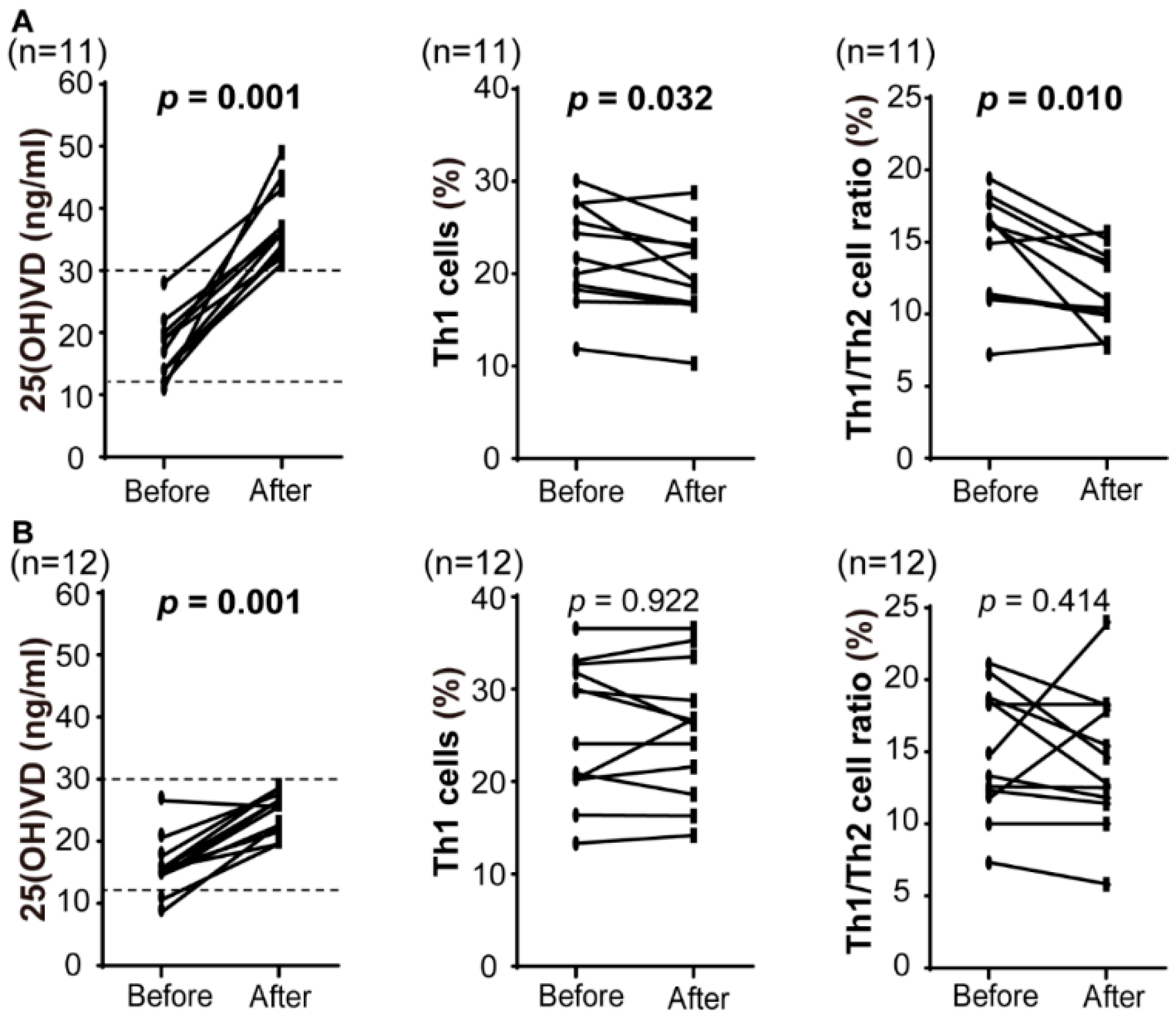
To identify the effect of the VD intervention on the systemic immune responses, we examined serum VD and T-helper cell levels before and after VD supplementation in patients with VD insufficiency or deficiency (Table 2). Intake of VD (cholecalciferol) at 1000 IU daily for 3 months significantly increased to 25(OH)VD levels (16.7 ± 4.7 to 31.0 ± 7.8 ng/mL, p < 0.0001). Of 23 patients, 22 (95.7%) recorded an increase of 25(OH)VD, and 11 (47.8%) reached sufficient VD levels (>30 ng/mL). VD supplementation relatively decreased and increased Th1 and Th2 cell levels, respectively, resulting in a significant inhibition of the Th1/Th2 cell ratio (14.8 ± 4.0 to 13.1 ± 4.1, p = 0.004). The biologically active form, 1,25(OH)2VD level was significantly increased (p = 0.019). Th17 and Treg cell levels were not changed significantly after VD supplementation (p = 0. 315 and p = 0.059, respectively; Table 2). We divided the patients into two groups with sufficient and insufficient groups after VD supplementation (Figure 2). Eleven patients in the sufficient group showed a significant decrease in Th1 cell level and Th1/Th2 cell ratio (p = 0.032 and 0.010, respectively), whereas no significant differences in Th1/Th2 cell ratio were recognized in the insufficient group. No complications or adverse effects of VD supplementation were identified in any patient.

3.3. Effect of Vitamin D on Decidualized Endometrial Stromal Cells

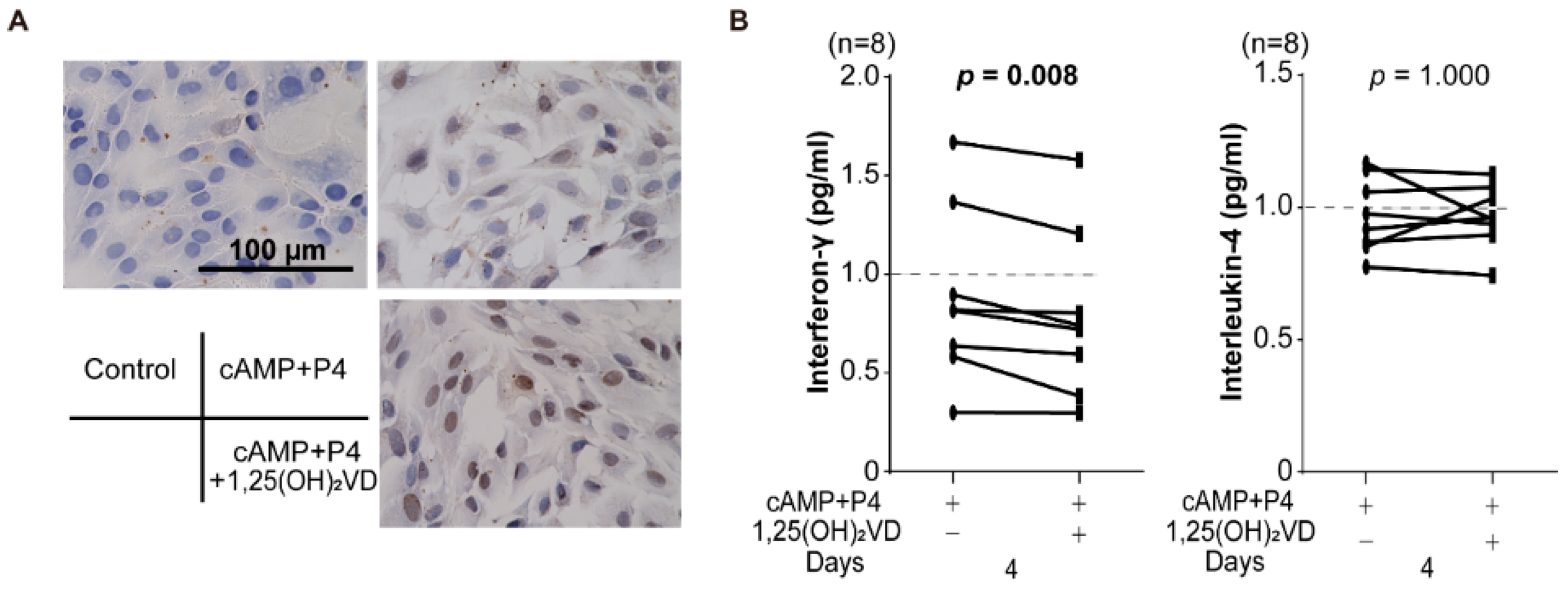
To identify the location of VDR, immunohistochemical staining for VDR in undifferentiated HESCs, and cells decidualized with 8-bromo-cAMP and P4 with or without 1,25(OH)2VD treatment for 4 days demonstrated that decidualization is associated with diffuse distribution of VDR in the cytoplasm in the cells. In decidualized cells with 1,25(OH)2VD treatment, VDR accumulated in the nucleus compared to the cells without 1,25(OH)2VD treatment (Figure 3A). Cytokine assay of condition media of decidualized HESCs with or without 1,25(OH)2VD demonstrated that IFN-γ, but not IL-4, levels were significantly reduced in decidualized cells compared to those without 1,25(OH)2VD treatment (p = 0.008; Figure 3B). We confirmed decidualization of HESCs using decidual markers, insulin-like growth factor-binding protein-1 [IGFBP1] and prolactin [PRL] mRNA expression (Supplementary Figure S1).

4. Discussion

Epidemiologically, the rate of reproductive-aged women with VD insufficiency and deficiency has been reported to be 20–90%, with a significantly higher rate between 2000 and 2004 compared with the 1990s rate [31]. Our study demonstrated that most infertile women had insufficient 25(OH)VD levels (Table 1). Indeed, average serum 25(OH)VD levels in subfertile women were reportedly lower than those in normal fertile women [32,33]. In northern European countries, seasonal variation in conception rates was attributed to changes in the VD levels with the sunlight exposure; as such, VD status was strongly associated with the conception rate [34]. VD deficiency is a global health issue in reproductive age women [35,36].
An aberrantly high Th1/Th2 cell ratio during the preconception period leads to the reproductive failure [30,37]. Our results showed that 41.9% of Japanese infertile patients with the VD insufficient and deficient groups had 10.3 or more in Th1/Th2 cell ratios, suggesting that nearly half of the infertile patients without sufficient VD levels had impaired immunologic tolerance. According to other reports, VD regulates T-helper cell populations through an inhibition of Th1 cell proliferation and promotion of Th2 cells [24]. In our data, VD supplementation decreased serum Th1/Th2 cell ratio; however, there was no significant change in each Th1 and Th2 cell level in infertile women with VD insufficiency or deficiency (Table 2). Nevertheless, when focusing on 11 patients who reached sufficient 25(OH)VD levels after supplementation, a decrease in serum Th1 cell level and Th1/Th2 cell ratio was recognized. Therefore, an optimal 25(OH)VD level for supporting pregnancy may be ≥30 ng/mL and may have a significant role in the regulation of immunologic embryo receptivity. In fact, VD replacement for patients with a history of recurrent pregnancy loss improves immune abnormality with insufficient or sufficient VD status, resulting in a prevention of pregnancy loss [38].
At local sites, 1,25(OH)2VD treatment promoted expression of VDR in the nucleus. Decidual change in HESCs did not alter secretion of IFN-γ and IL-4 significantly in the culture media. However, treatment of decidualized cells with 1,25(OH)2VD decreased the IFN-γ level, like the serum Th1 cell level. VD may have an important role in the regulation of not only systemic but also local Th1 immune response for optimization of maternal tolerance for pregnancy. Although VD has been reported to inhibit the response of Th17 cell levels and induce Treg cells [12,13,25], our results showed that Treg cells were relatively increased by VD supplementation. However, there was no significant difference in Th17 and Treg cell levels (Table 1).
In our study, 1000 IU VD replacement per day for 3 months resulted in sufficient VD levels (>30 ng/mL) in only half of the patients with VD deficiency/insufficiency. A dose of 1000 IU VD replacement per day may be insufficient as preconception care for normalization of maternal VD and immune status. Therefore, a proper dose of VD supplementation for the infertile women without VD sufficiency may be more than 1000 IU. And the infertile women with an elevated Th1/Th2 cell ratio after VD supplementation may need to receive immunotherapy including tacrolimus during IVF-ET treatment [30]. Taken together, proper VD replacement for infertile VD-insufficient patients may contribute to the maintenance of immune homeostasis by suppressing Th1 cells for a successful pregnancy.
This study has some limitations. First, VD is produced in the skin via sunlight exposure. Therefore, serum VD level varied according to the seasonal changes. In our data, no significant difference in the VD levels was recognized among the three groups (summer, winter, and both spring and autumn; Supplementary Table S2). Second, as regarding the experiments of the effect of VD intervention on T-helper cell levels, the number of the patients was 23, thus the study size was small to evaluate the effect of VD supplementation correctly.

5. Conclusions

Sufficient VD supplementation in women with insufficient or deficient VD may optimize maternal immune tolerance for pregnancy, via rebalancing the Th1/Th2 cell ratio, leading to prevention against proinflammatory obstetrical complications during pregnancy.

Supplementary Materials

The following are available online at https://www.mdpi.com/2072-6643/10/7/902/s1, Figure S1: RTQ-PCR analysis of IGFBP1 and PRL transcript levels in HESCs treated with 8-bromo-cAMP and P4 combined with or without 1,25(OH)2VD for 4 days, Table S1: Patient characteristics, Table S2: Relationship of 25(OH)VD and season.

Author Contributions

K.K. and Y.I. contributed to study conception and design; K K., Y.I., K.N., A.O., R.O., K.M. and M.J. dealt in the recruitment of subjects and procurement of samples; K.K., Y.I., K.N., A.O., R.O. and A.M. investigated samples; K.K. and Y.I. analyzed data; K.K. and Y.I. wrote the manuscript and R.S. and S.T. supervised the study.

Funding

Keiji Kuroda was supported by the Medical Research Encouragement Prize of the Japan Medical Association. Keisuke Murakami was supported by JSPS KAKENHI Grant Number 16K20208.

Acknowledgments

We wish to thank all participating patients. We also thank the Laboratory of Molecular and Biochemical Research, Research Support Center, Juntendo University Graduate School of Medicine, Tokyo, Japan, for technical assistance. And we thank Hisaya Akiba and Go Murayama in the Department of Immunology, Juntendo University, for valuable support and advice, and Mari Kitade and Ayako Masuda in the Department of Obstetrics and Gynecology, Juntendo University, for assisting with the collection of endometrial samples.

Conflicts of Interest

The authors declare no conflict of interest.

References

  1. Holick, M.F.; Binkley, N.C.; Bischoff-Ferrari, H.A.; Gordon, C.M.; Hanley, D.A.; Heaney, R.P.; Murad, M.H.; Weaver, C.M. Evaluation, treatment, and prevention of vitamin D deficiency: An Endocrine Society clinical practice guideline. J. Clin. Endocrinol. Metab. 2011, 96, 1911–1930. [Google Scholar] [CrossRef] [PubMed]
  2. Hewison, M. Vitamin D and the immune system: New perspectives on an old theme. Endocrinol. Metab. Clin. N. Am. 2010, 39, 365–379. [Google Scholar] [CrossRef] [PubMed]
  3. Bizzaro, G.; Shoenfeld, Y. Vitamin D and autoimmune thyroid diseases: Facts and unresolved questions. Immunol. Res. 2015, 61, 46–52. [Google Scholar] [CrossRef] [PubMed]
  4. El-Fakhri, N.; McDevitt, H.; Shaikh, M.G.; Halsey, C.; Ahmed, S.F. Vitamin D and its effects on glucose homeostasis, cardiovascular function and immune function. Horm. Res. Paediatr. 2014, 81, 363–378. [Google Scholar] [CrossRef] [PubMed]
  5. Terrier, B.; Derian, N.; Schoindre, Y.; Chaara, W.; Geri, G.; Zahr, N.; Mariampillai, K.; Rosenzwajg, M.; Carpentier, W.; Musset, L.; et al. Restoration of regulatory and effector T cell balance and B cell homeostasis in systemic lupus erythematosus patients through vitamin D supplementation. Arthritis Res. Ther. 2012, 14, R221. [Google Scholar] [CrossRef] [PubMed]
  6. Zhang, M.X.; Pan, G.T.; Guo, J.F.; Li, B.Y.; Qin, L.Q.; Zhang, Z.L. Vitamin D deficiency increases the risk of gestational diabetes mellitus: A meta-analysis of observational studies. Nutrients 2015, 7, 8366–8375. [Google Scholar] [CrossRef] [PubMed]
  7. Mirzakhani, H.; Litonjua, A.A.; McElrath, T.F.; O’Connor, G.; Lee-Parritz, A.; Iverson, R.; Macones, G.; Strunk, R.C.; Bacharier, L.B.; Zeiger, R.; et al. Early pregnancy vitamin D status and risk of preeclampsia. J. Clin. Investig. 2016, 126, 4702–4715. [Google Scholar] [CrossRef] [PubMed] [Green Version]
  8. Ota, K.; Dambaeva, S.; Han, A.R.; Beaman, K.; Gilman-Sachs, A.; Kwak-Kim, J. Vitamin D deficiency may be a risk factor for recurrent pregnancy losses by increasing cellular immunity and autoimmunity. Hum. Reprod. 2014, 29, 208–219. [Google Scholar] [CrossRef] [PubMed]
  9. Rudick, B.J.; Ingles, S.A.; Chung, K.; Stanczyk, F.Z.; Paulson, R.J.; Bendikson, K.A. Influence of vitamin D levels on in vitro fertilization outcomes in donor-recipient cycles. Fertil. Steril. 2014, 101, 447–452. [Google Scholar] [CrossRef] [PubMed]
  10. Raghupathy, R.; Makhseed, M.; Azizieh, F.; Omu, A.; Gupta, M.; Farhat, R. Cytokine production by maternal lymphocytes during normal human pregnancy and in unexplained recurrent spontaneous abortion. Hum. Reprod. 2000, 15, 713–718. [Google Scholar] [CrossRef] [PubMed] [Green Version]
  11. Ng, S.C.; Gilman-Sachs, A.; Thaker, P.; Beaman, K.D.; Beer, A.E.; Kwak-Kim, J. Expression of intracellular Th1 and Th2 cytokines in women with recurrent spontaneous abortion, implantation failures after IVF/ET or normal pregnancy. Am. J. Reprod. Immunol. 2002, 48, 77–86. [Google Scholar] [CrossRef] [PubMed]
  12. Aluvihare, V.R.; Kallikourdis, M.; Betz, A.G. Regulatory T cells mediate maternal tolerance to the fetus. Nat. Immunol. 2004, 5, 266–271. [Google Scholar] [CrossRef] [PubMed]
  13. Sasaki, Y.; Sakai, M.; Miyazaki, S.; Higuma, S.; Shiozaki, A.; Saito, S. Decidual and peripheral blood CD4+CD25+ regulatory T cells in early pregnancy subjects and spontaneous abortion cases. Mol. Hum. Reprod. 2004, 10, 347–353. [Google Scholar] [CrossRef] [PubMed]
  14. Crome, S.Q.; Wang, A.Y.; Levings, M.K. Translational mini-review series on Th17 cells: Function and regulation of human T helper 17 cells in health and disease. Clin. Exp. Immunol. 2010, 159, 109–119. [Google Scholar] [CrossRef] [PubMed]
  15. Wang, W.J.; Hao, C.F.; Yi, L.; Yin, G.J.; Bao, S.H.; Qiu, L.H.; Lin, Q.D. Increased prevalence of T helper 17 (Th17) cells in peripheral blood and decidua in unexplained recurrent spontaneous abortion patients. J. Reprod. Immunol. 2010, 84, 164–170. [Google Scholar] [CrossRef] [PubMed]
  16. Lee, S.K.; Kim, J.Y.; Hur, S.E.; Kim, C.J.; Na, B.J.; Lee, M.; Gilman-Sachs, A.; Kwak-Kim, J. An imbalance in interleukin-17-producing T and Foxp3(+) regulatory T cells in women with idiopathic recurrent pregnancy loss. Hum. Reprod. 2011, 26, 2964–2971. [Google Scholar] [CrossRef] [PubMed]
  17. Agic, A.; Xu, H.; Altgassen, C.; Noack, F.; Wolfler, M.M.; Diedrich, K.; Friedrich, M.; Taylor, R.N.; Hornung, D. Relative expression of 1,25-dihydroxyvitamin D3 receptor, vitamin D 1 alpha-hydroxylase, vitamin D 24-hydroxylase, and vitamin D 25-hydroxylase in endometriosis and gynecologic cancers. Reprod. Sci. 2007, 14, 486–497. [Google Scholar] [CrossRef] [PubMed]
  18. Perez-Fernandez, R.; Alonso, M.; Segura, C.; Munoz, I.; Garcia-Caballero, T.; Diguez, C. Vitamin D receptor gene expression in human pituitary gland. Life Sci. 1997, 60, 35–42. [Google Scholar] [CrossRef]
  19. Evans, K.N.; Bulmer, J.N.; Kilby, M.D.; Hewison, M. Vitamin D and placental-decidual function. J. Soc. Gynecol. Investig. 2004, 11, 263–271. [Google Scholar] [CrossRef] [PubMed]
  20. Vigano, P.; Lattuada, D.; Mangioni, S.; Ermellino, L.; Vignali, M.; Caporizzo, E.; Panina-Bordignon, P.; Besozzi, M.; Di Blasio, A.M. Cycling and early pregnant endometrium as a site of regulated expression of the vitamin D system. J. Mol. Endocrinol. 2006, 36, 415–424. [Google Scholar] [CrossRef] [PubMed] [Green Version]
  21. Evans, K.N.; Nguyen, L.; Chan, J.; Innes, B.A.; Bulmer, J.N.; Kilby, M.D.; Hewison, M. Effects of 25-hydroxyvitamin D3 and 1,25-dihydroxyvitamin D3 on cytokine production by human decidual cells. Biol. Reprod. 2006, 75, 816–822. [Google Scholar] [CrossRef] [PubMed]
  22. Kinuta, K.; Tanaka, H.; Moriwake, T.; Aya, K.; Kato, S.; Seino, Y. Vitamin D is an important factor in estrogen biosynthesis of both female and male gonads. Endocrinology 2000, 141, 1317–1324. [Google Scholar] [CrossRef] [PubMed]
  23. Guillot, X.; Semerano, L.; Saidenberg-Kermanac’h, N.; Falgarone, G.; Boissier, M.C. Vitamin D and inflammation. Jt. Bone Spine 2010, 77, 552–557. [Google Scholar] [CrossRef] [PubMed]
  24. Boonstra, A.; Barrat, F.J.; Crain, C.; Heath, V.L.; Savelkoul, H.F.; O’Garra, A. 1alpha,25-Dihydroxyvitamin d3 has a direct effect on naive CD4(+) T cells to enhance the development of Th2 cells. J. Immunol. 2001, 167, 4974–4980. [Google Scholar] [CrossRef] [PubMed]
  25. Cantorna, M.T.; Snyder, L.; Lin, Y.D.; Yang, L. Vitamin D and 1,25(OH)2D regulation of T cells. Nutrients 2015, 7, 3011–3021. [Google Scholar] [CrossRef] [PubMed]
  26. Ross, A.C.; Manson, J.E.; Abrams, S.A.; Aloia, J.F.; Brannon, P.M.; Clinton, S.K.; Durazo-Arvizu, R.A.; Gallagher, J.C.; Gallo, R.L.; Jones, G.; et al. The 2011 report on dietary reference intakes for calcium and vitamin D from the Institute of Medicine: What clinicians need to know. J. Clin. Endocrinol. Metab. 2011, 96, 53–58. [Google Scholar] [CrossRef] [PubMed]
  27. Ozaki, R.; Kuroda, K.; Ikemoto, Y.; Ochiai, A.; Matsumoto, A.; Kumakiri, J.; Kitade, M.; Itakura, A.; Muter, J.; Brosens, J.J.; et al. Reprogramming of the retinoic acid pathway in decidualizing human endometrial stromal cells. PLoS ONE 2017, 12, e0173035. [Google Scholar] [CrossRef] [PubMed]
  28. Tavakoli, M.; Jeddi-Tehrani, M.; Salek-Moghaddam, A.; Rajaei, S.; Mohammadzadeh, A.; Sheikhhasani, S.; Kazemi-Sefat, G.E.; Zarnani, A.H. Effects of 1,25(OH)2 vitamin D3 on cytokine production by endometrial cells of women with recurrent spontaneous abortion. Fertil. Steril. 2011, 96, 751–757. [Google Scholar] [CrossRef] [PubMed]
  29. Zeng, N.; Salker, M.S.; Zhang, S.; Singh, Y.; Shi, B.; Stournaras, C.; Lang, F. 1alpha,25(OH)2D3 induces actin depolymerization in endometrial carcinoma cells by targeting RAC1 and PAK1. Cell. Physiol. Biochem. 2016, 40, 1455–1464. [Google Scholar] [CrossRef] [PubMed]
  30. Nakagawa, K.; Kwak-Kim, J.; Ota, K.; Kuroda, K.; Hisano, M.; Sugiyama, R.; Yamaguchi, K. Immunosuppression with tacrolimus improved reproductive outcome of women with repeated implantation failure and elevated peripheral blood TH1/TH2 cell ratios. Am. J. Reprod. Immunol. 2015, 73, 353–361. [Google Scholar] [CrossRef] [PubMed]
  31. Looker, A.C.; Pfeiffer, C.M.; Lacher, D.A.; Schleicher, R.L.; Picciano, M.F.; Yetley, E.A. Serum 25-hydroxyvitamin D status of the US population: 1988–1994 compared with 2000–2004. Am. J. Clin. Nutr. 2008, 88, 1519–1527. [Google Scholar] [CrossRef] [PubMed] [Green Version]
  32. Al-Jaroudi, D.; Al-Banyan, N.; Aljohani, N.J.; Kaddour, O.; Al-Tannir, M. Vitamin D deficiency among subfertile women: Case-control study. Gynecol. Endocrinol. 2016, 32, 272–275. [Google Scholar] [CrossRef] [PubMed]
  33. Triggianese, P.; Watad, A.; Cedola, F.; Perricone, C.; Amital, H.; Giambini, I.; Perricone, R.; Shoenfeld, Y.; De Carolis, C. Vitamin D deficiency in an Italian cohort of infertile women. Am. J. Reprod. Immunol. 2017, 78. [Google Scholar] [CrossRef] [PubMed]
  34. Rojansky, N.; Brzezinski, A.; Schenker, J.G. Seasonality in human reproduction: An update. Hum. Reprod. 1992, 7, 735–745. [Google Scholar] [CrossRef] [PubMed]
  35. Hilger, J.; Friedel, A.; Herr, R.; Rausch, T.; Roos, F.; Wahl, D.A.; Pierroz, D.D.; Weber, P.; Hoffmann, K. A systematic review of vitamin D status in populations worldwide. Br. J. Nutr. 2014, 111, 23–45. [Google Scholar] [CrossRef] [PubMed]
  36. Holick, M.F. The vitamin D deficiency pandemic: Approaches for diagnosis, treatment and prevention. Rev. Endocr. Metab. Disord. 2017, 18, 153–165. [Google Scholar] [CrossRef] [PubMed]
  37. Nakagawa, K.; Kuroda, K.; Sugiyama, R.; Yamaguchi, K. After 12 consecutive miscarriages, a patient received immunosuppressive treatment and delivered an intact baby. Reprod. Med. Biol. 2017, 16, 297–301. [Google Scholar] [CrossRef] [PubMed] [Green Version]
  38. Chen, X.; Yin, B.; Lian, R.C.; Zhang, T.; Zhang, H.Z.; Diao, L.H.; Li, Y.Y.; Huang, C.Y.; Liang, D.S.; Zeng, Y. Modulatory effects of vitamin D on peripheral cellular immunity in patients with recurrent miscarriage. Am. J. Reprod. Immunol. 2016, 76, 432–438. [Google Scholar] [CrossRef] [PubMed]
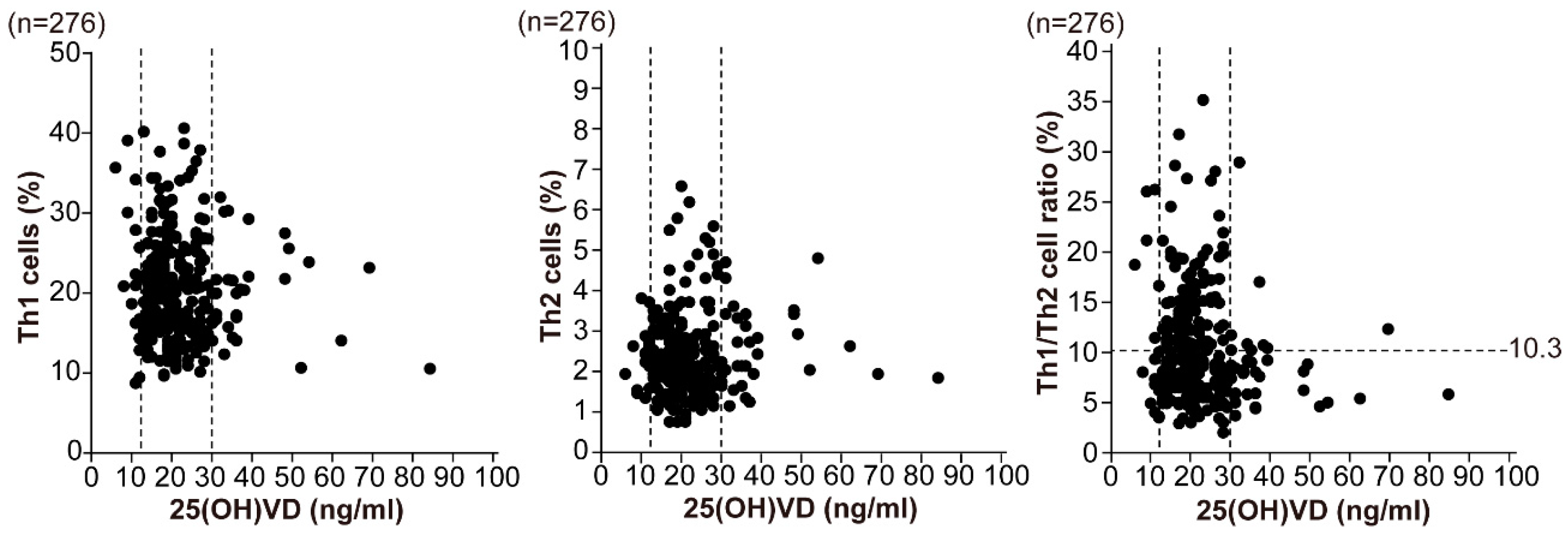
Figure 1. Relationship between serum 25(OH)VD and T-helper (Th) cells in infertile patients. The data demonstrated the relationships between 25(OH)VD and the Th1 and Th2 cell populations, and the Th1/Th2 cell ratio. All data were divided into three groups that were categorized as vitamin D deficiency, insufficiency, and sufficiency (25[OH]VD < 12, 12–30, and > 30 ng/mL respectively; dotted lines). Regarding the Th1 (CD4+/interferon [IFN]-γ+)/Th2 (CD4+/interleukin[IL]-4+) cell ratio, the normal range is <10.3 (dotted lines). Analysis of data from 276 general infertile patients showed a higher Th1/Th2 cell ratio in the VD-deficient and insufficient groups, but no correlation between serum 25(OH)VD and Th1 or Th2 cells or Th1/Th2 cell ratio. 25(OH)VD, 25-hydroxy vitamin D3; Th1 cell, IFN-γ–producing T-helper cell (CD4+/IFN-γ+); Th2 cell, IL-4–producing T-helper cell (CD4+/IL-4+).
Nutrients 10 00902 g001
Figure 2. Immunological alteration after VD supplementation in infertile women. A change in serum 25(OH)VD, Th1 cell, and Th1/Th2 cell ratio after intake of VD (cholecalciferol) at 1000 IU per day for 3 months in the infertile patients with VD insufficiency or deficiency. Normal range of Th1 (CD4+/IFN-γ+)/Th2 (CD4+/IL-4+) cell ratio is <10.3 (dotted lines). Of 23 patients, 11 reached above normal range after supplementation, whereas Th1 and Th1/Th2 ratio were significantly reduced (A). However, 12 patients who did not reach normal range showed no significant difference (B). 25(OH)VD, 25-hydroxyvitamin D3; Th1 cell, IFN-γ–producing T-helper cell (CD4+/IFN-γ+); Th2 cell, IL-4–producing T-helper cell (CD4+/IL-4+).
Nutrients 10 00902 g002
Figure 3. Role of vitamin D in decidualization of human endometrial stromal cell. (A) Immunohistochemical staining for vitamin D receptor (VDR) in human endometrial stromal cells [HESCs]. VDR was accumulated in the nucleus of HESCs cultured with 8-bromoadenosine 3′5′-cyclic adenosine monophosphate [8-bromo-cAMP] + P4 + 1,25(OH)2VD. (B) Cytokine assay of culture media of HESCs with 8-bromo-cAMP and P4 with or without 1,25(OH)2VD. IFN-γ levels were significantly reduced in decidualized cells with compared to those without 1,25(OH)2VD. * p < 0.05.
Nutrients 10 00902 g003
Table 1. Relationship between 25(OH)VD and T-helper (Th) cell populations in infertile women.
Deficiency (n = 18)Insufficiency (n = 223)Sufficiency (n = 35)p Value
Age (years)35.6 ± 4.636.1 ± 3.635.7 ± 3.50.899
25(OH)VD (ng/mL)10.7 ± 1.720.2 ± 4.439.3 ± 12.3<0.0001
Th1 cells (%)21.6 ± 8.920.8 ± 6.519.6 ± 5.60.748
Th2 cells (%)2.3 ± 0.72.3 ± 1.02.5 ± 1.00.621
Th1/Th2 cell ratio10.9 ± 7.510.6 ± 5.68.6 ± 4.50.411
Values are average ± standard deviation; 25(OH)VD, 25-hydroxyvitamin D3 (deficiency: <12 ng/mL, insufficiency: 12–30 ng/mL, sufficiency: >30 ng/mL); Th1 cell, interferon [IFN-γ]-producing T-helper cell (CD4+/IFN-γ+); Th2 cell, interleukin(IL)-4-producing T-helper cell (CD4+/IL-4+).
Table 2. Impact of vitamin D supplementation for infertile patients with vitamin D insufficiency and deficiency.
n = 23BeforeAfterp Value
25(OH)VD (ng/mL)16.7 ± 4.731.0 ± 7.8<0.0001
1,25(OH)2VD (pg/mL)49.9 ± 8.265.4 ± 16.80.019
Th1 cells (%)24.0 ± 6.723.0 ± 6.80.135
Th2 cells (%)1.7 ± 0.61.9 ± 0.60.141
Th1/Th2 cell ratio14.8 ± 4.013.1 ± 4.10.004
Th17 cells (%)1.65 ± 0.401.54 ± 0.630.315
Treg cells (%)5.74 ± 0.986.38 ± 1.030.059
Values are average ± standard deviation. 25(OH)VD, 25 hydroxyvitamin D3; 1,25(OH)2VDl 1,25 dihydroxyvitamin D3; Th1 cell, interferon [IFN]-γ-producing T-helper cell (CD4+/IFN-γ+/interleukin[IL]-4); Th2 cell, IL-4-producing T-helper cell (CD4+/IFN-γ-/IL-4+); Th17 cell, IL-17-producing T-helper cell (CD4+/IL-17+); Reguratory T [Treg] cell, CD4+/CD25+/FoxP3 T-helper cell. 1,25(OH)2VD, Th17 cells, and Treg cells were measured in eight patients.