Mineral Mapping and Ore Prospecting with HyMap Data over Eastern Tien Shan, Xinjiang Uyghur Autonomous Region

Abstract
:
1. Introduction
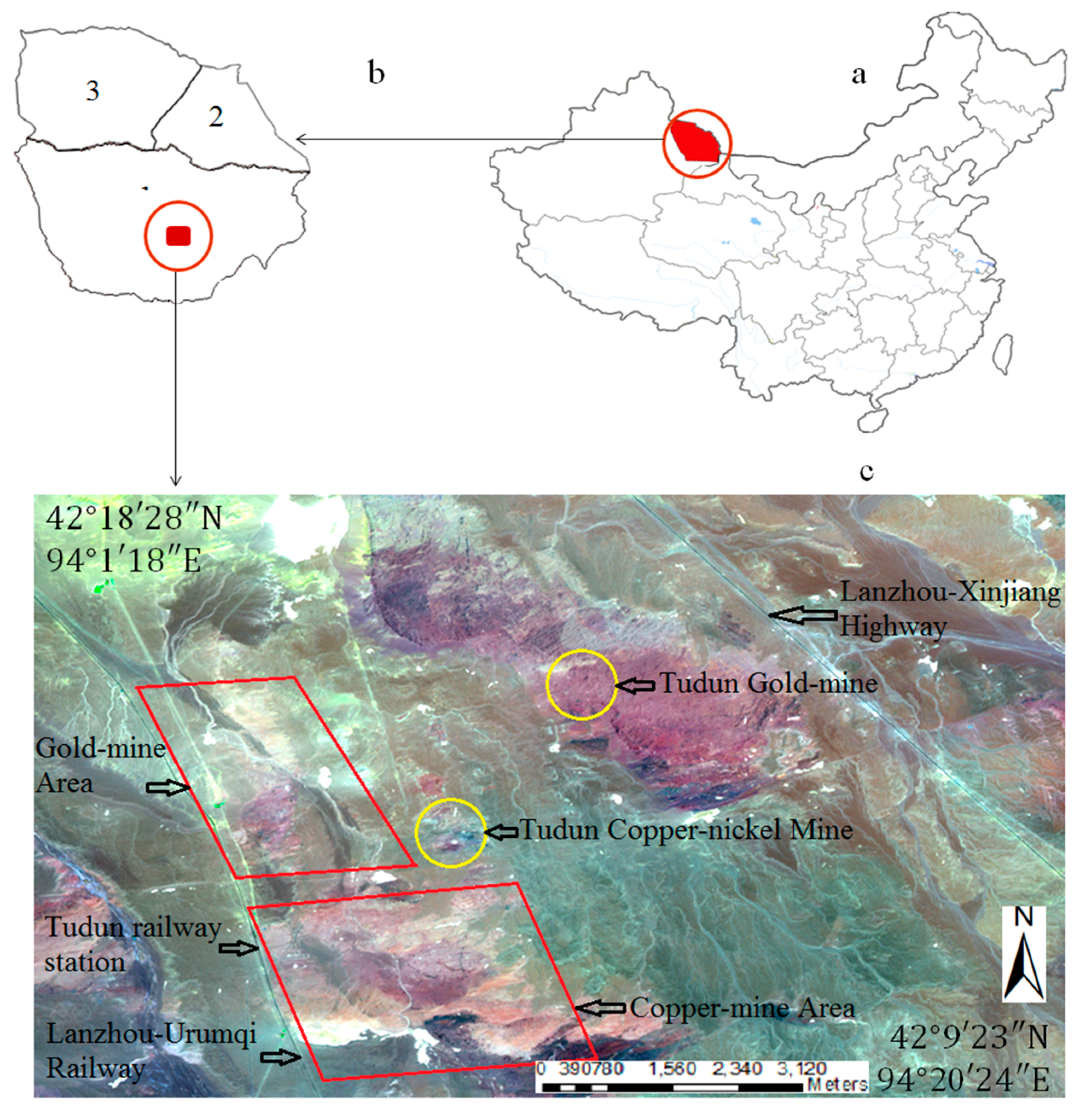
2. Geology of the Study Area

3. Data and Preprocessing
3.1. HyMap Dataset

| Wavelength (nm) | Bandwidth (nm) | Average Spectral Sampling Interval (nm) | Band Number |
|---|---|---|---|
| VIS:450–890 | 15–16 | 15 | 32 |
| NIR:890–1350 | 15–16 | 15 | 32 |
| SWIR1:1400–1800 | 15–16 | 13 | 32 |
| SWIR2:1950–2480 | 18–20 | 17 | 32 |
| IFOV | 2.5 mr along track, 2.0 mr across track | ||
| GIFOV | 3–10 m | ||
| Data coding | 16 bit | ||
| SNR | >500:1 | ||
3.2. PIMA
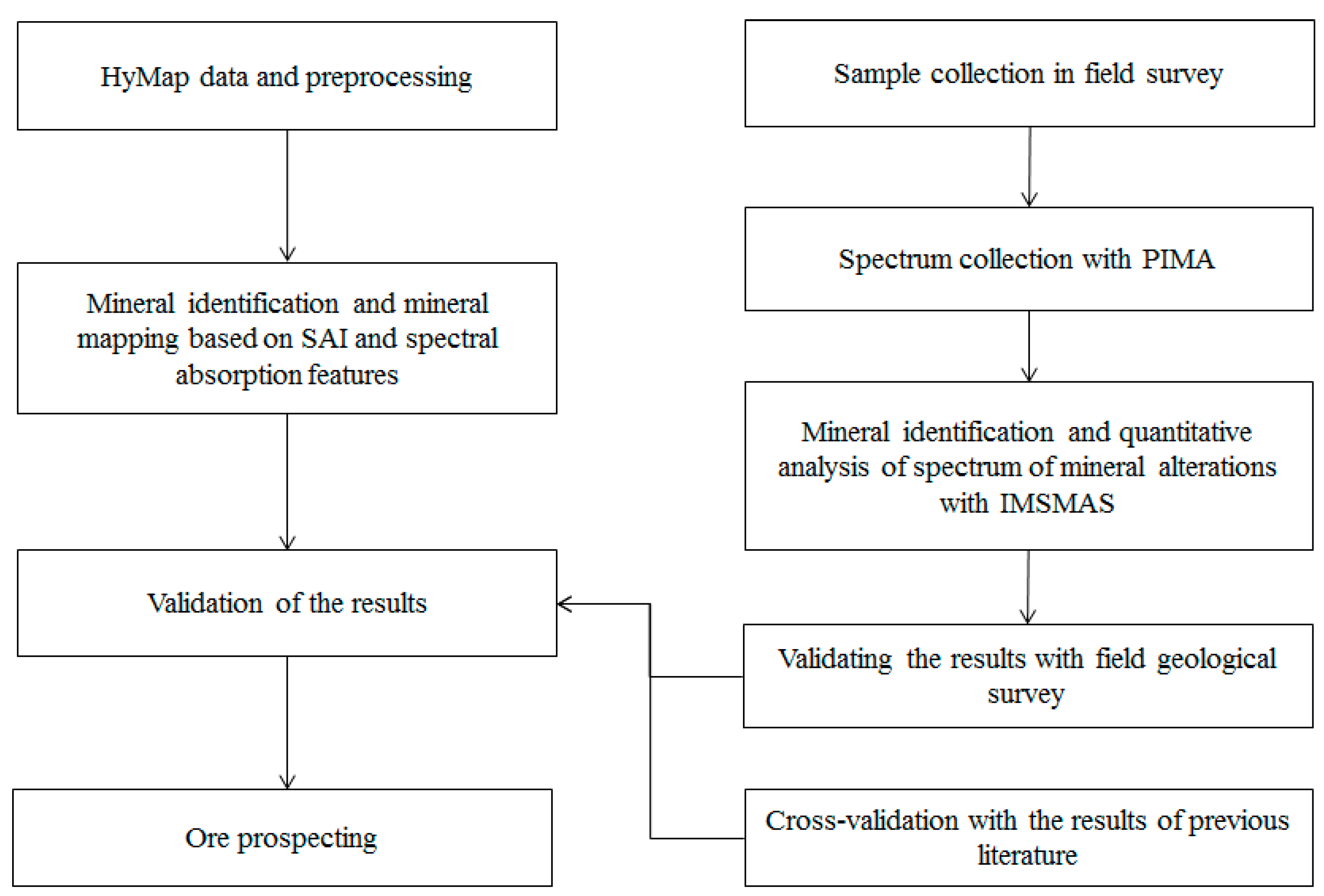
4. Methods
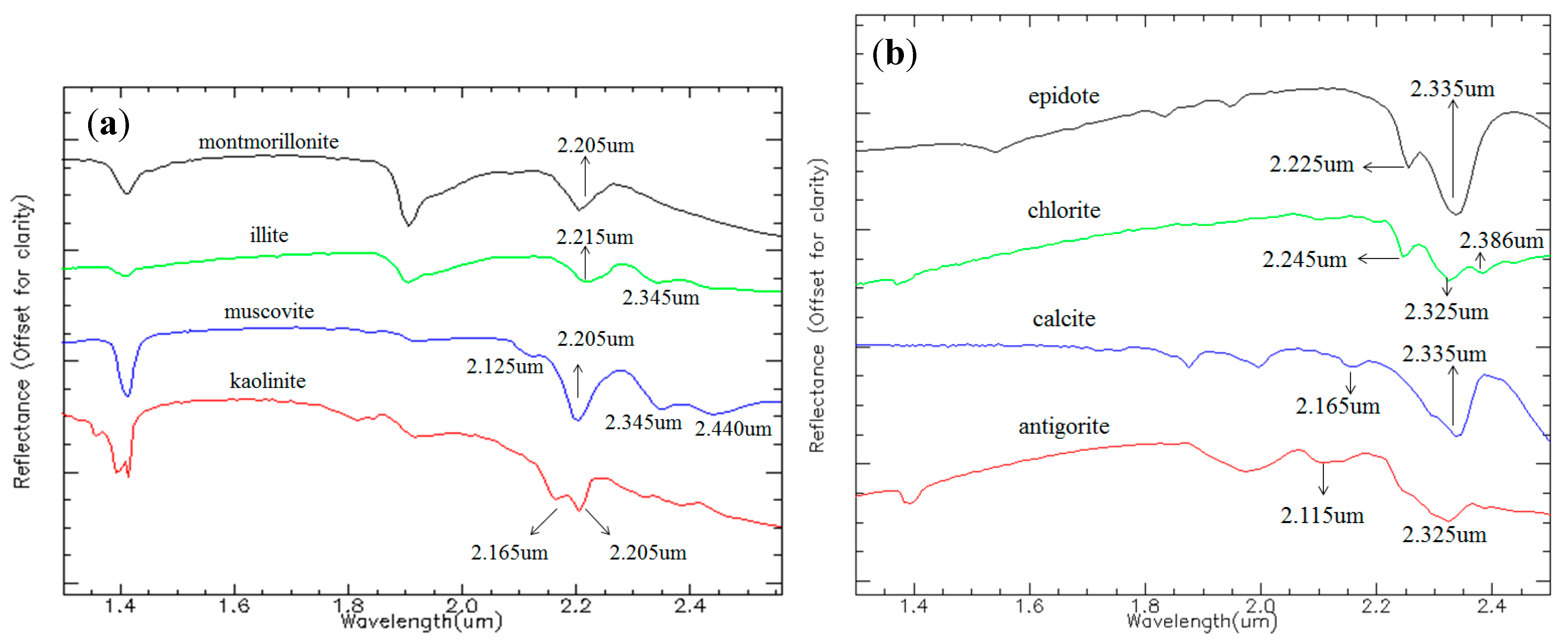
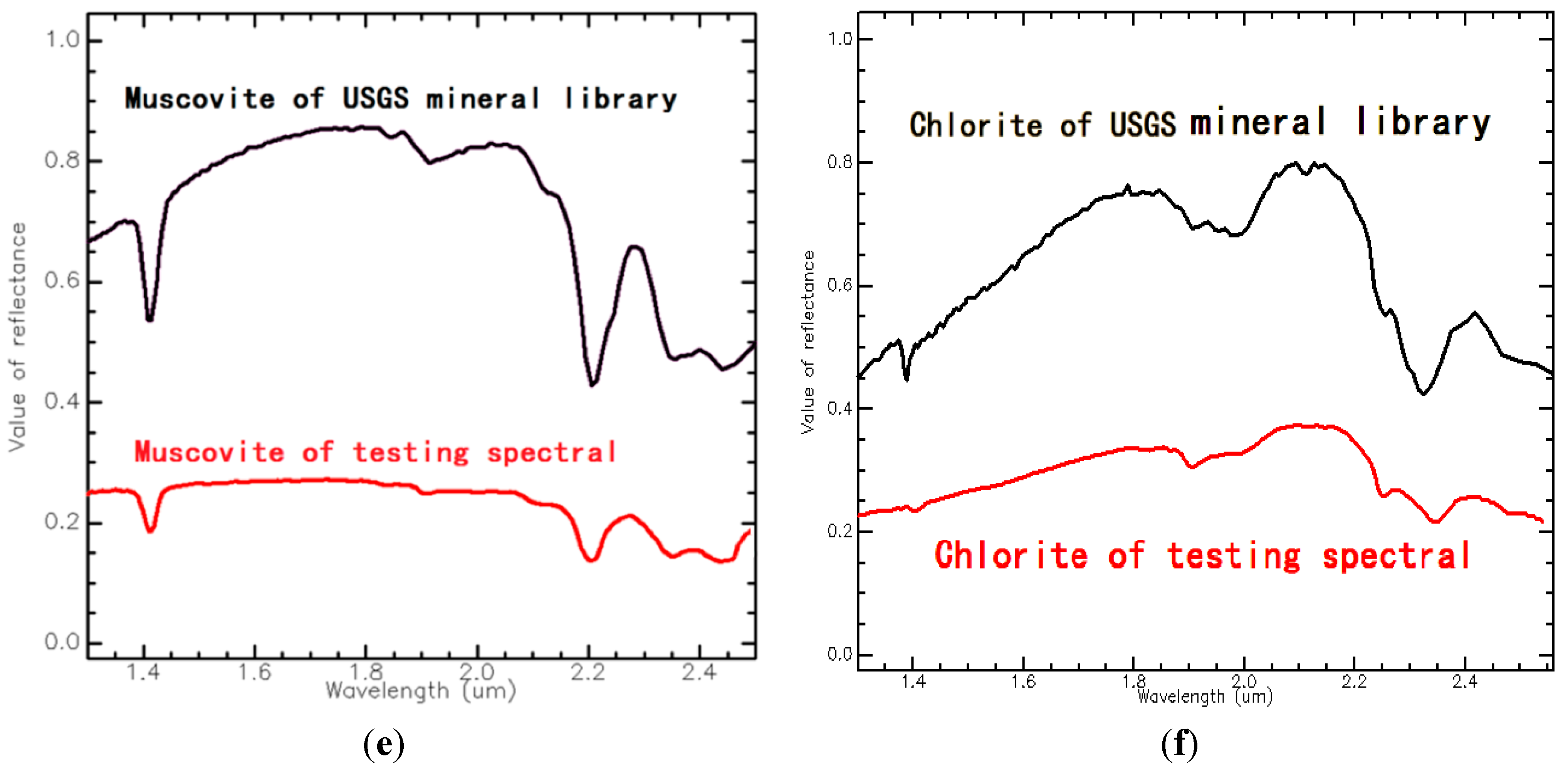
4.1. Spectral Absorption Features of Main Alteration Minerals
| Molecular Bond | Absorption Feature (μm) | Example of Mineral of SWIR |
|---|---|---|
| Al-OH | 2.160–2.220 | Montmorillonite, Illite, Muscovite, Kaolinite |
| Mg-OH | 2.300–2.360 | Epidote, Chlorite, Antigorite |
| Carbonate (CO3) | 2.300–2.350 | Calcite |
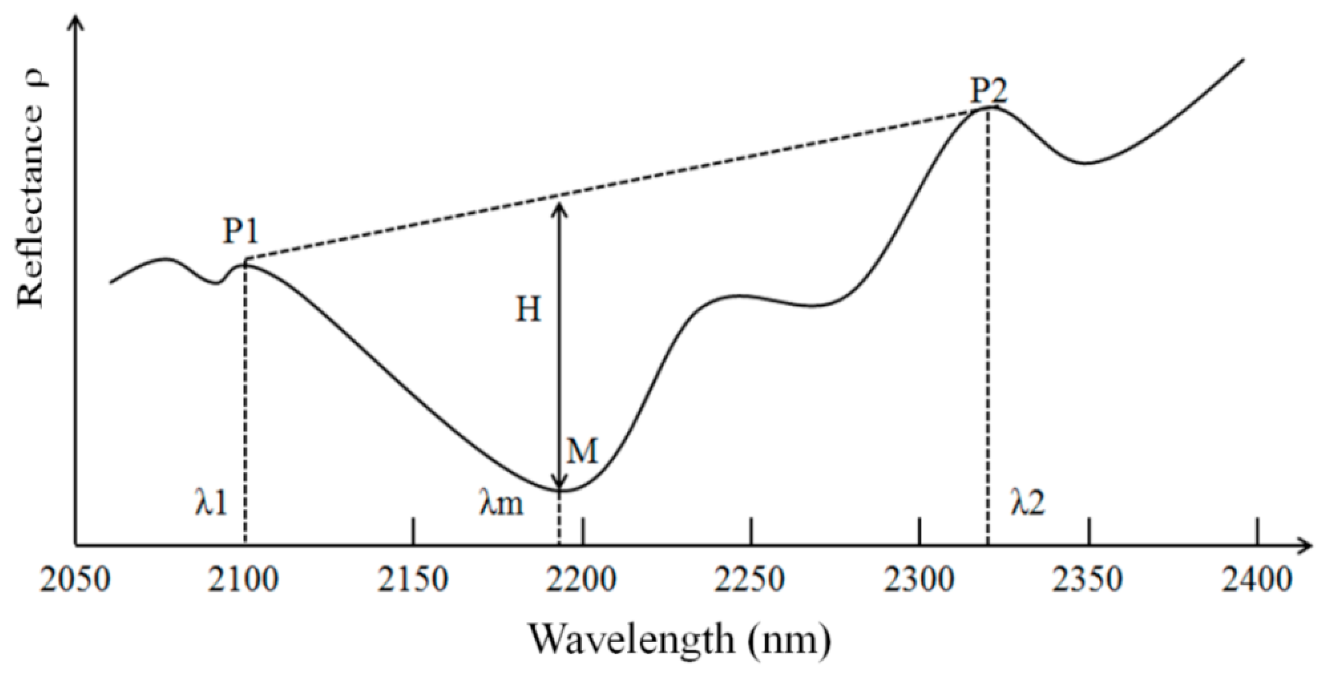
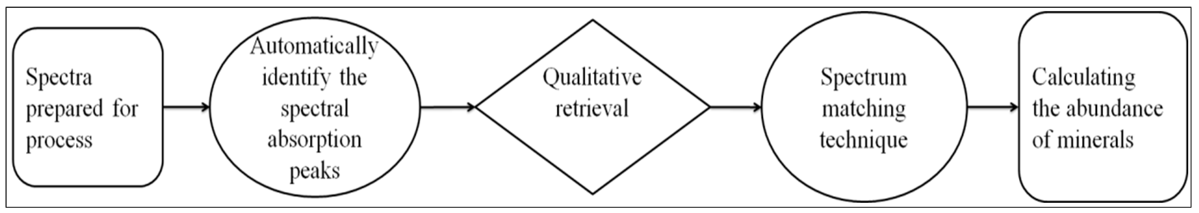
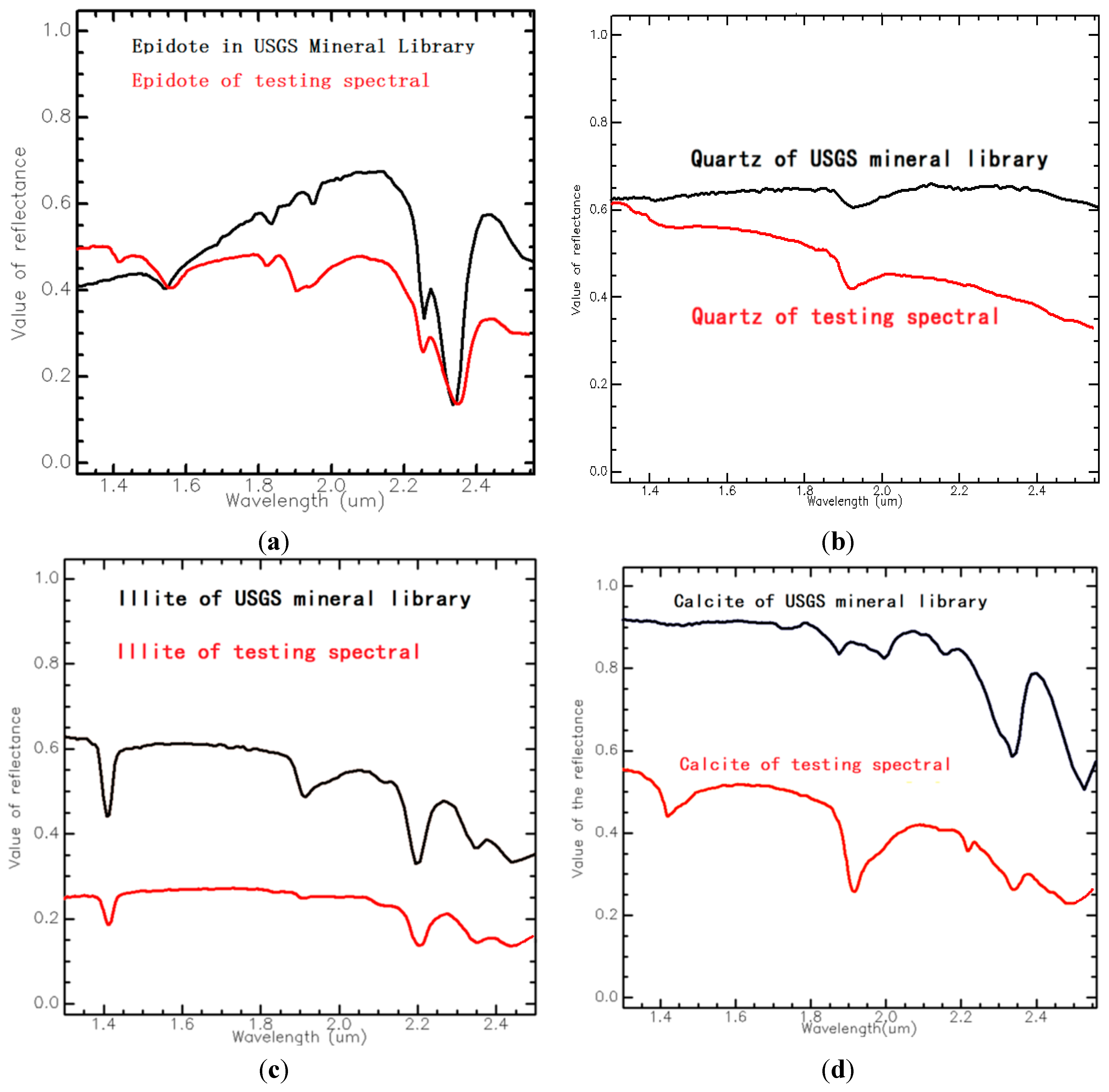
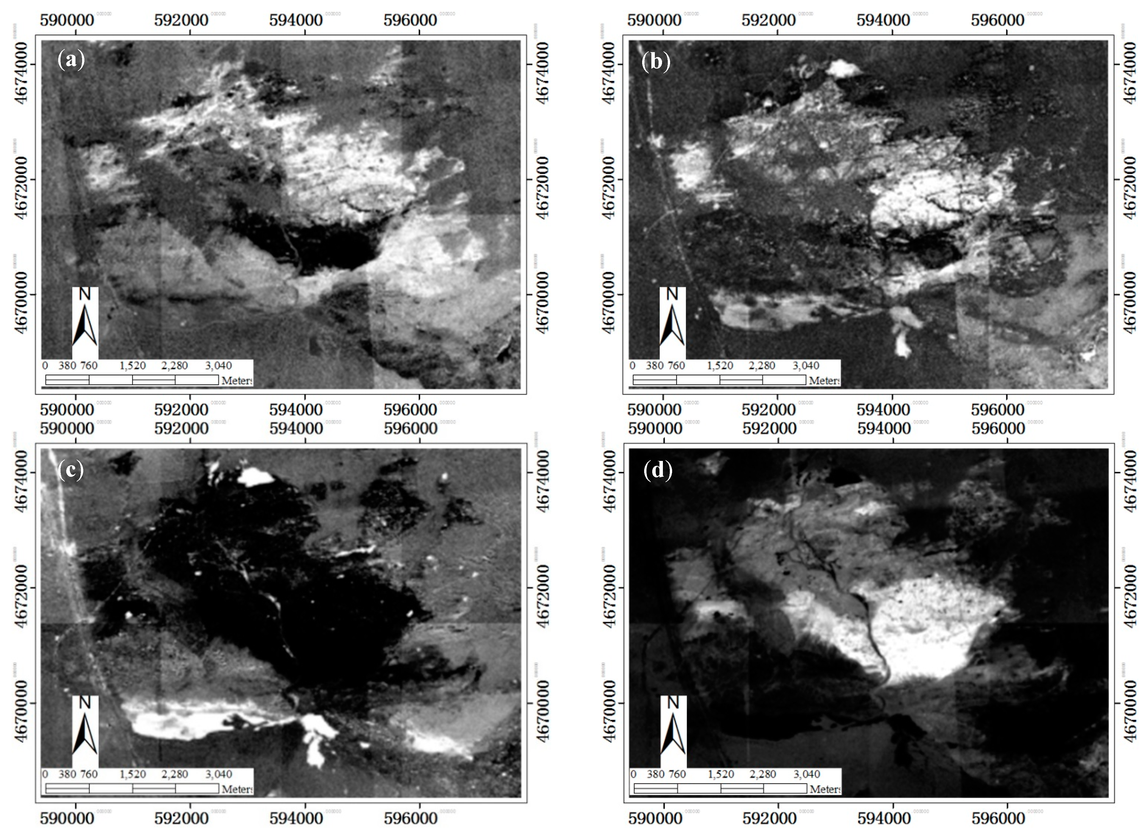
4.2. Spectral Absorption Features and the SAI Model

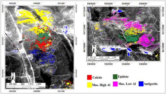
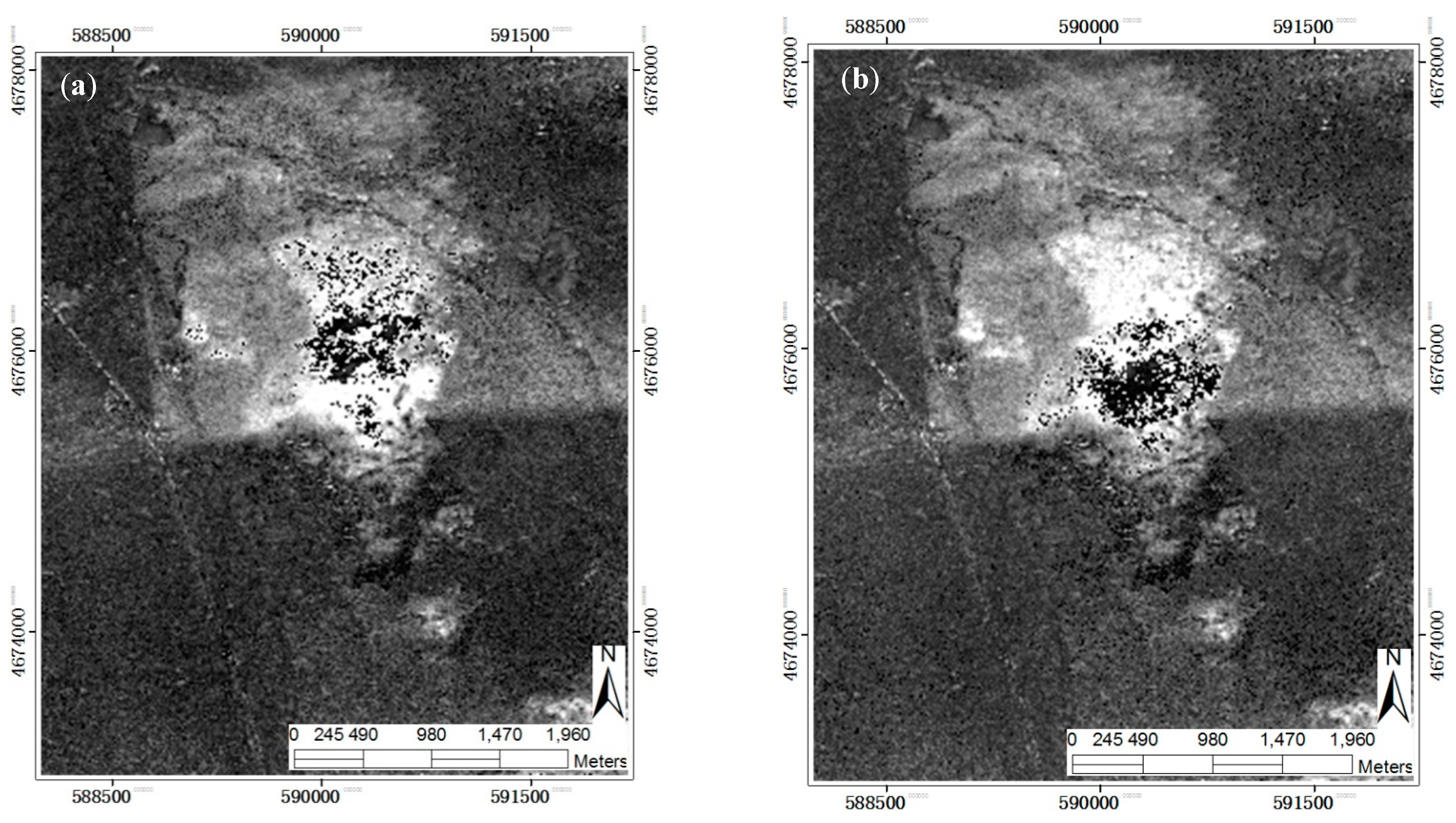
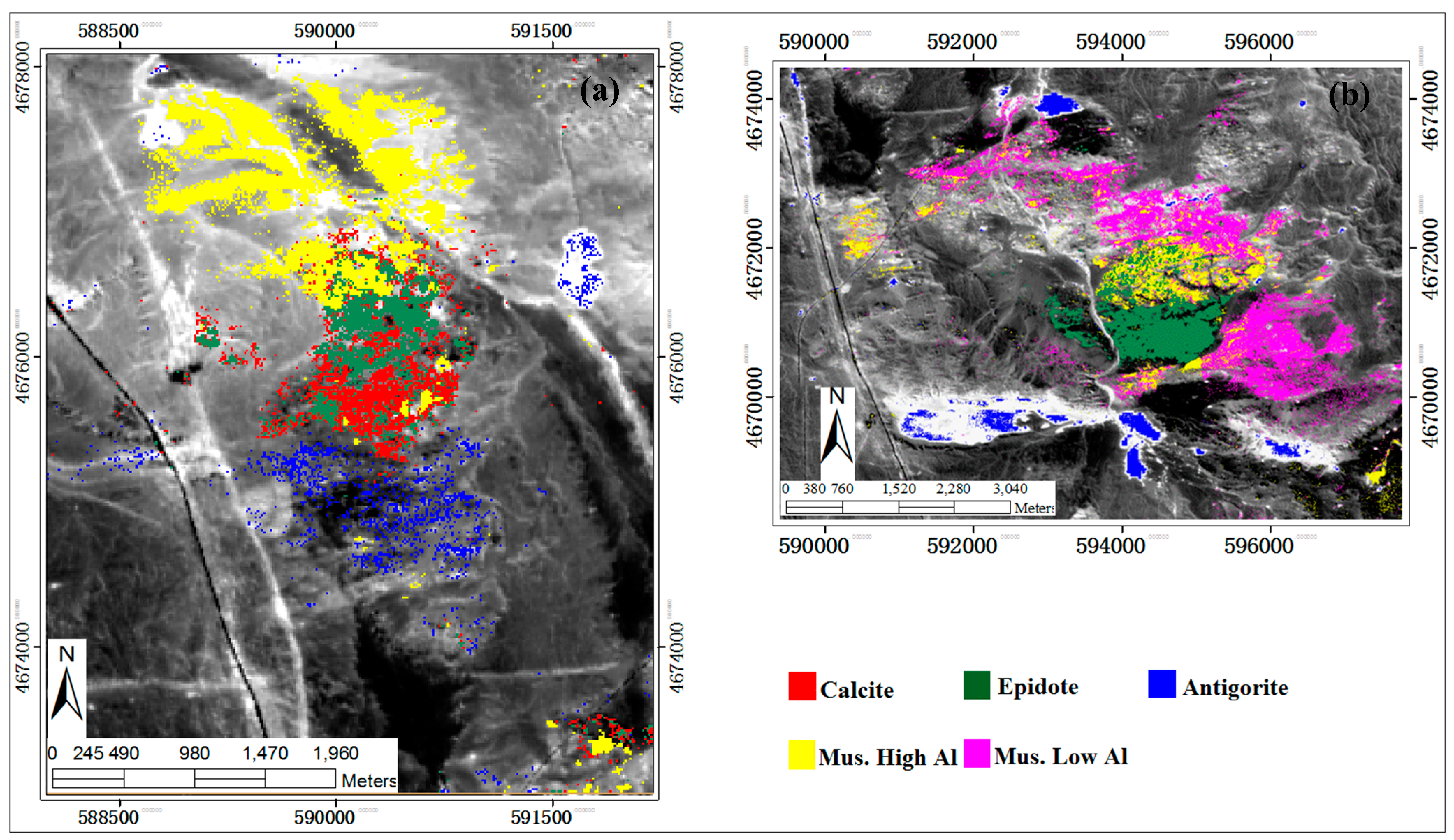
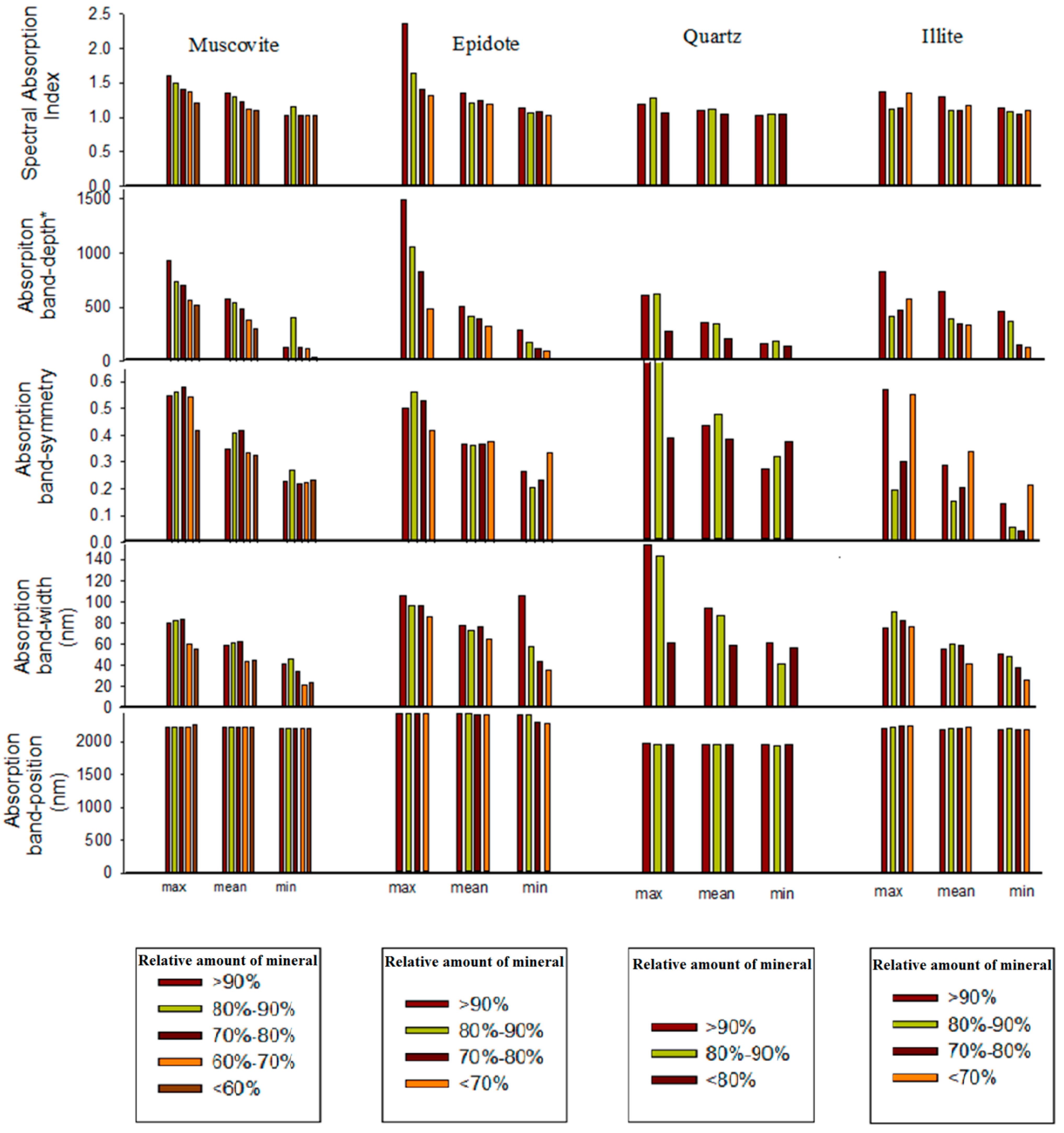
5. Results


Analysis of Alteration Mineral Information Extracted from Spectral Absorption Features
| Band-Position (nm) | Absorption Shoulders (nm) | Band-Width (nm) | Band-Symmetry | Major Minerals Extracted |
|---|---|---|---|---|
| 2330 | * b104 = 2192 b115 = 2379 | 187 | 0.173 | Calcite |
| 2210 | b103 = 2174 b108 = 2262 | 88 | 0.394 | Al-poor Muscovite |
| 2192 | b102 = 2156 b110 = 2297 | 141 | 0.677 | Al-rich Muscovite |
| 2335 | b109 = 2279 b119 = 2444 | 165 | 0.395 | Epidote |
| 2396 | b114 = 2363 b120 = 2460 | 97 | 0.335 | Antigorite |
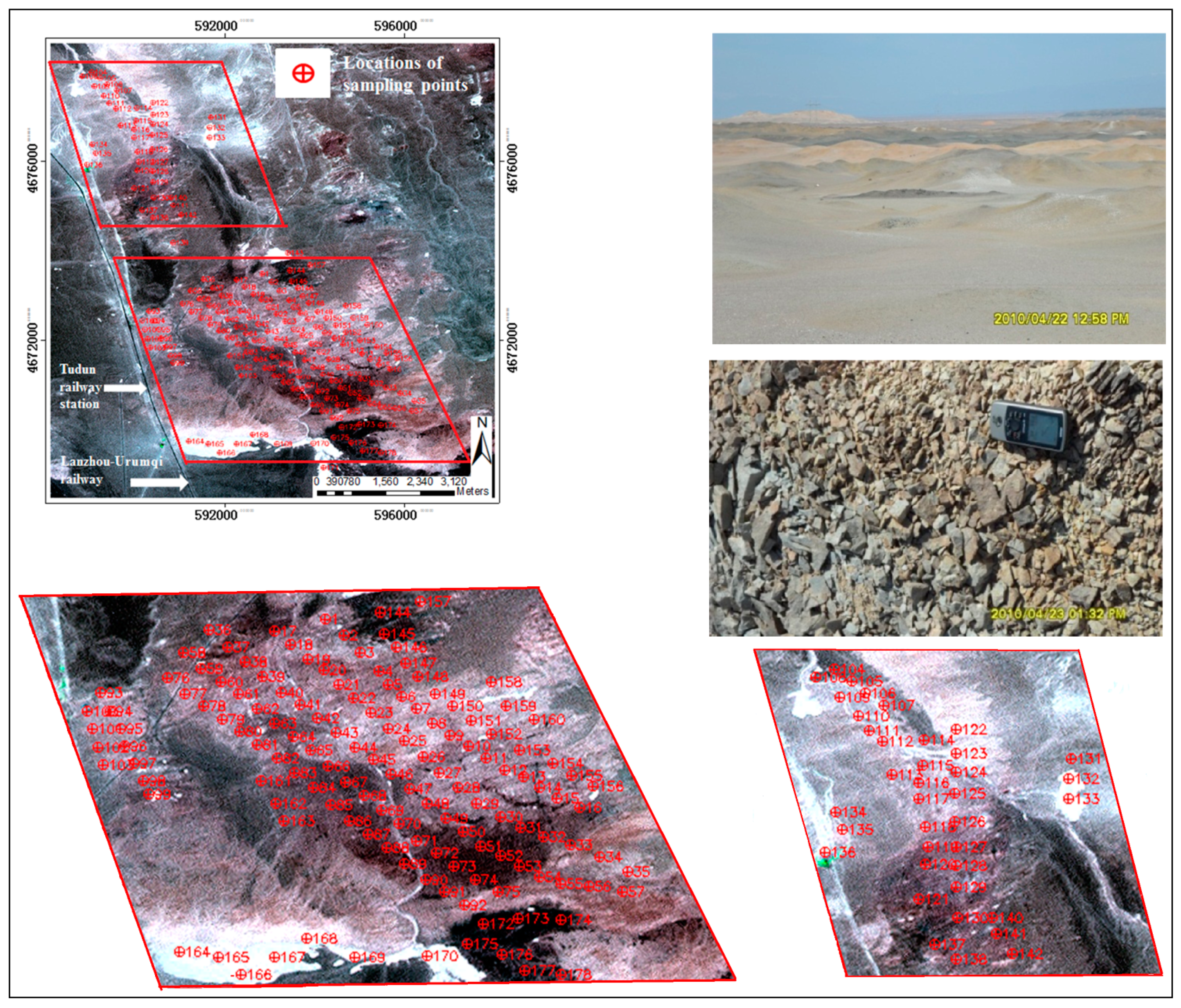
6. Validation of Results Using Field Survey Based on Spectral Knowledge
Measurement and Quantitative Analysis of Mineral Spectra in the Laboratory with a PIMA Spectrometer

| Mineral Components | Epidote | Chlorite | Muscovite | Illite | Calcite | Kaolinite | Quartz | |
|---|---|---|---|---|---|---|---|---|
| Relative Amount (%) | ||||||||
| 15–30 (%) | Number of Spectra | 120 | 51 | 136 | 21 | 11 | 14 | 45 |
| Number of Samples | 105 | 43 | 107 | 19 | 10 | 13 | 41 | |
| 31–45 (%) | Number of Spectra | 89 | 31 | 62 | 21 | 9 | 8 | 23 |
| Number of Samples | 81 | 28 | 55 | 19 | 9 | 8 | 22 | |
| 46–60 (%) | Number of Spectra | 77 | 11 | 50 | 23 | 7 | 8 | 41 |
| Number of Samples | 70 | 10 | 48 | 18 | 7 | 8 | 39 | |
| 61–75 (%) | Number of Spectra | 131 | 5 | 57 | 17 | 7 | 4 | 41 |
| Number of Samples | 116 | 4 | 50 | 13 | 7 | 4 | 39 | |
| 76–90 (%) | Number of Spectra | 172 | 1 | 36 | 12 | 18 | 5 | 36 |
| Number of Samples | 148 | 1 | 31 | 12 | 16 | 5 | 33 | |
| >90 (%) | Number of Spectra | 189 | 2 | 16 | 17 | 34 | 25 | 59 |
| Number of Samples | 161 | 2 | 14 | 15 | 26 | 25 | 51 | |
| Total Spectra | 778 | 101 | 357 | 111 | 99 | 64 | 245 | |
| Total Samples | 681 | 88 | 305 | 96 | 75 | 63 | 225 | |


7. Ore Prospecting

8. Conclusions
Acknowledgements
Author Contributions
Conflicts of Interest
References
- Duke, E.F. Near infrared spectra of muscovite, Tschermak substitution, and metamorphic reaction progress: implications for remote sensing. Geology 1994, 22, 621–624. [Google Scholar] [CrossRef]
- Green, R.O.; Eastwood, M.L.; Sarture, C.M.; Chrien, T.G.; Aronsson, M.; Chippendale, B.J.; Faust, J.A.; Pavri, B.E.; Chovit, C.J.; Solis, M.; et al. Imaging spectroscopy and the airborne visible/infrared imaging spectrometer (AVIRIS). Remote Sens. Environ. 1998, 65, 227–248. [Google Scholar] [CrossRef]
- Hubbard, B.E.; Crowley, J.K.; Zimbelman, D.R. Comparative alteration mineral mapping using visible to shortwave infrared (0.4–2.4 μm) Hyperion, ALI, and ASTER imagery. IEEE Trans. Geosci. Remote Sens. 2003, 41, 1401–1410. [Google Scholar] [CrossRef]
- Velasco, F.; Alvaro, A.; Suarez, S.; Herrero, J.M.; Yusta, I. Mapping Fe-bearing hydrated sulphate minerals with short wave infrared (SWIR) spectral analysis at San Miguel mine environment, Iberian Pyrite Belt (SW Spain). J. Geochem. Explor. 2005, 87, 45–72. [Google Scholar] [CrossRef]
- Petrovic, A.; Khan, S.D.; Thurmond, A.K. Integrated hyperspectral remote sensing, geochemical and isotopic studies for understanding hydrocarbon-induced rock alterations. Mar. Petrol. Geol. 2012, 35, 292–308. [Google Scholar] [CrossRef]
- Kruse, F.A.; Perry, S.L. Mineral mapping using simulated Worldview-3 short-wave-infrared imagery. Remote Sens. 2013, 5, 2688–2703. [Google Scholar] [CrossRef]
- Haest, M.; Cudahy, T.; Laukamp, C.; Gregory, S. Quantitative mineralogy from visible to shortwave infrared spectroscopic data: (II) 3D mineralogical characterisation of the Rocklea Dome channel iron deposit in Western Australia. Econ. Geol. 2012, 107, 229–249. [Google Scholar] [CrossRef]
- Haest, M.; Cudahy, C.; Rodger, A.; Laukamp, C.; Martens, E.; Caccetta, M. Unmixing vegetation from airborne visible-near to shortwave infrared spectroscopy-based mineral maps over the Rocklea Dome (Western Australia), with a focus on iron rich palaeochannels. Remote Sens. Environ. 2013, 129, 17–31. [Google Scholar] [CrossRef]
- Crowley, J.K.; Hook, S.J. Mapping playa evaporite minerals and associated sediments in Death Valley, California, with multispectral thermal infrared images. J. Geophys. Res. 1996, 101, 643–660. [Google Scholar] [CrossRef]
- Yamaguchi, Y.; Kahle, A.B.; Tsu, H.; Kawakami, T.; Pniel, M. Overview of advanced spaceborne thermal emission and reflection radiometer (ASTER). IEEE Trans. Geosci. Remote Sens. 1998, 36, 1062–1071. [Google Scholar] [CrossRef]
- Li, Z.-L.; Becker, F.; Stoll, M.P.; Wan, Z. Evaluation of six methods for extracting relative emissivity spectral from thermal infrared images. Remote Sens. Environ. 1999, 69, 197–214. [Google Scholar]
- Vaughan, R.G.; Hook, S.J.; Calvin, W.M.; Taranik, J.V. Surface mineral mapping at Steamboat Springs, Nevada, USA, with multi-wavelength thermal infrared images. Remote Sens. Environ. 2005, 99, 140–158. [Google Scholar] [CrossRef]
- Hecker, C.; Van der Meijde, M.; Van der Meer, F.D. Thermal infrared spectroscopy on feldspars—Successes, limitations and their implications for remote sensing. Earth-Sci. Rev. 2010, 103, 60–70. [Google Scholar] [CrossRef]
- Becker, F.; Li, Z.-L. Temperature independent spectral indices in thermal infrared bands. Remote Sens. Environ. 1990, 32, 17–33. [Google Scholar] [CrossRef]
- Li, Z.-L.; Becker, F. Properties and comparison of temperature independent thermal infrared spectral indices with NDVI for HAPEX data. Remote Sens. Environ. 1990, 33, 165–182. [Google Scholar] [CrossRef]
- Casey, K.A.; Kääb, A.; Benn, D.I. Geochemical characterization of supraglacial debris via in situ and optical remote sensing methods: A case study in Khumbu Himalaya, Nepal. The Cryosphere 2012, 6, 85–100. [Google Scholar] [CrossRef]
- Li, Z.-L.; Wu, H.; Wang, N.; Qiu, S.; Sobrino, J.A.; Wan, Z.; Tang, B.-H.; Yan, G.J. Land surface emissivity retrieval from satellite data. Int. J. Remote Sens. 2013, 34, 3084–3127. [Google Scholar] [CrossRef]
- Li, Z.-L.; Tang, B.-H.; Wu, H.; Ren, H.; Yan, G.J.; Wan, Z.; Trigo, I.F.; Sobrino, J. Satellite-derived land surface temperature: Current status and perspectives. Remote Sens. Environ. 2013, 131, 14–37. [Google Scholar] [CrossRef]
- Riley, D.N.; Hecker, C.A. Mineral mapping with airborne hyperspectral thermal infrared remote sensing at Cuprite, Nevada, USA. Therm. Infrared Remote Sens. 2013, 17, 495–514. [Google Scholar]
- da Silva, A.Q.; Paradella, W.R.; Freitas, C.C.; Oliveira, C.G. Evaluation of digital classification of polarimetric SAR data for iron-mineralized laterites mapping in the Amazon Region. Remote Sens. 2013, 5, 3101–3122. [Google Scholar]
- Ni, Z.Y; Huo, H.Y.; Gan, F.P.; Wang, Y.Q.; Li, X.W.; Liu, Z.G. Lithological discrimination based on the inversion of roughness in Hami Area, Xinjiang. IOP Conf. Ser.: Earth Environ. Sci. 2014. [Google Scholar] [CrossRef]
- Wang, Q.H.; Wang, R.S.; Guo, X.F. Application for discrimination of rock using hyperspectral remote sensing technique. Remote Sens. Land Resour. 2000, 4, 7. [Google Scholar]
- Laukamp, C.; Cudahy, T.; Caccetta, M.; Chia, J.; Gessner, K.; Haest, M.; Liu, Y.C.; Rodger, A. The uses, abuses and opportunities for hyperspectral technologies and derived geoscience information. Hyperspectral Technol. Deriv. Geosci. Inf. 2010, 51, 73–76. [Google Scholar]
- Laukamp, C.; Cudahy, T.; Thomas, M.; Jones, M.; Cleverley, J.; Oliver, N. Hydrothermal mineral alteration patterns in the Mount Isa Inlier revealed by airborne hyperspectral data. Aust. J. Earth Sci. 2011, 58, 917–936. [Google Scholar] [CrossRef]
- Laukamp, C.; Cudahy, T.; Cleverley, J.; Oliver, N.; Hewson, R. Airborne hyperspectral imaging of hydrothermal alteration zones in granitoids of the Eastern Fold Belt, Mount Isa Inlier, Australia. Geochem. Explor. Environ. Anal. 2011, 11, 3–24. [Google Scholar] [CrossRef]
- Rodger, A. SODA: A new method of in-scene atmospheric water vapor estimation and post-flight spectral recalibration for hyperspectral sensors: Application to the HyMap sensor at two locations. Remote Sens. Environ. 2011, 115, 536–547. [Google Scholar] [CrossRef]
- Rogge, D.; Rivard, B.; Segl, K.; Grant, B.; Feng, J. Mapping of NiCu–PGE ore hosting ultramafic rocks using airborne and simulated EnMAP hyperspectral imagery, Nunavik, Canada. Remote Sens. Environ. 2014, 152, 302–317. [Google Scholar] [CrossRef]
- Gan, F.; Wang, R.; Ma, A. Spectral identification tree (SIT) for mineral extraction based on spectral characteristics of minerals. Earth Sci. Front. 2003, 10, 445–454. [Google Scholar]
- Wang, R. Spectral identification and inversion of composition and component of objects with hyperspectral remote sensing. J. GeoInf. Sci. 2009, 11, 261–267. [Google Scholar]
- Fenstermaker, L.K.; Miller, J.R. Identification of fluvially redistributed mill tailings using high spectral resolution aircraft data. Photogramm. Eng. Remote Sens. 1994, 60, 989–995. [Google Scholar]
- Yuhas, R.H.; Goetz, A.F.; Boardman, J.W. Discrimination among semi-arid landscape endmembers using the spectral angle mapper (SAM) algorithm. In Proceedings of 1992 Summaries of the Third Annual JPL Airborne Geoscience Workshop, Pasadena, CA, USA, 1–5 June 1992; pp. 147–149.
- De Carvalho, O., Jr.; Meneses, P. Spectral correlation mapper (SCM): An improvement on the spectral angle mapper (SAM). In Proceedings of 2000 Summaries of the 9th Airborne Earth Science Workshop, Pasadena, CA, USA, 23–25 Februray 2000; pp. 65–74.
- Chang, C.-I. An information-theoretic approach to spectral variability, similarity, and discrimination for hyperspectral image analysis. IEEE Trans. Inform. Theory 2000, 46, 1927–1932. [Google Scholar] [CrossRef]
- Du, H.; Qi, H.; Peterson, G.D. Parallel ica and its hardware implementation in hyperspectral image analysis. Int. Soc. Opt. Photonics 2004. [Google Scholar] [CrossRef]
- Van der Meer, F. The effectiveness of spectral similarity measures for the analysis of hyperspectral imagery. Int. J. Appl. Earth Obs. Geoinf. 2006, 8, 3–17. [Google Scholar] [CrossRef]
- Van der Meer, F.; Bakker, W. CCSM: Cross correlogram spectral matching. Int. J. Remote Sens. 1997, 18, 1197–1201. [Google Scholar] [CrossRef]
- Clark, R.N.; Gallagher, A.J.; Swayze, G.A. Material absorption band depth mapping of imaging spectrometer data using a complete band shape least-squares fit with library reference spectra. In Proceedings of 1990 the Second Airborne Visible/Infrared Imaging Spectrometer (AVIRIS) Workshop, Denver, CO, USA, 4–5 July 1990; pp. 4–5.
- Abílio, O.; Júnior, C.; Meneses, P. R.; Guimarães, R.F. Analysis absorption band positioning: A new method for hyperspectral image treatment. In Proceedings of 2001 AVIRIS Workshops, Pasadena, CA, USA, 27 February–2 March 2001; pp. 59–65.
- Wang, J.-N.; Zheng, L.-F.; Tong, Q.-X. The spectral absorption identification model and mineral mapping by imaging spectrometer data. J. Remote Sens. 1996, 11, 20–31. (In Chinese) [Google Scholar]
- Rodger, A.; Laukamp, C.; Haest, M.; Cudahy, T. A simple quadratic method of wavelength tracking for absorption features in continuum removed spectra. Remote Sens. Environ. 2012, 118, 273–283. [Google Scholar] [CrossRef]
- Van Ruitenbeek, F.J.A.; Debba, P.; Van der Meer, F.D.; Cudahy, T.; Van der Meijde, M.; Hale, M. Mapping white micas and their absorption wavelengths using hyperspectral band ratios. Remote Sens. Environ. 2006, 102, 211–222. [Google Scholar] [CrossRef]
- Gabr, S.; Ghulam, A.; Kusky, T. Detecting areas of high-potential gold mineralization using ASTER data. Ore Geol. Rev. 2010, 38, 59–69. [Google Scholar] [CrossRef]
- Ranjendran, S.; Al-Khirbash, S.; Pracejus, B.; Nasir, S.; Al-Abri, A.H.; Kusky, T.M.; Ghulam, A. ASTER detection of chromite bearing mineralized zones in Semail Ophiolite Massifs of the northern Oman Mountains: Exploration strategy. Ore Geol. Rev. 2012, 44, 121–135. [Google Scholar] [CrossRef]
- Rajendran, S.; Nasir, S.; Kusky, T.M.; Ghulam, A.; Gabr, S.; Gali, M.E. Detection of hydrothermal mineralized zones associated with Listwaenites rocks in the Central Oman using ASTER data. Ore Geol. Rev. 2013, 53, 470–488. [Google Scholar] [CrossRef]
- Liu, L.; Zhou, J.; Jiang, D.; Zhuang, D.; Mansaray, L.R.; Zhang, B. Targeting mineral resources with remote sensing and field data in the Xiemisitai area, West Junggar, Xinjiang, China. Remote Sens. 2013, 5, 3156–3171. [Google Scholar] [CrossRef]
- Wang, R.; Liu, D.; Yin, D. Research over the copper and nickel sulfide deposits metallogenic control conditions and the ore prospecting in the area of Tudun-Huangshan mountain in Hami, Xinjiang Uyghur Autonomous Region. J. Mineral. Petrol. 1987, 7, 1–152. (In Chinese) [Google Scholar]
- Yong, W. Study over the characteristic of the basic-ultrabased rock and the ability of containing ore, in Tudun area, Hami City, Xinjiang Uyghur Autonomous Region. Xinjiang Nonferrous Metals 2002, 25, 1–6. (In Chinese) [Google Scholar]
- Cocks, T.; Jenssen, R.; Stewart, A.; Wilson, I.; Shields, T. The HyMap airborne hyperspectral sensor: The system, calibration and performance. In Proceedings of 1998 EARSeL Workshop on Imaging Spectroscopy, Zurich, Sweden, 6–8 October 1998; pp. 37–43.
- Notesco, G.; Kopačková, V.; Rojík, P.; Schwartz, G.; Livne, I.; Dor, E.B. Mineral classification of land surface using multispectral LWIR and hyperspectral SWIR remote-sensing data. A case study over the Sokolov lignite open-pit mines, the Czech Republic. Remote Sens. 2014, 6, 7005–7025. [Google Scholar] [CrossRef]
- Martin, G.; Plaza, A. Spatial-spectral preprocessing prior to end member identification and un-mixing of remotely sensed hyperspectral data. IEEE J. Sel. Top. Appl. Earth Obs. Remote Sens. 2012, 5, 380–395. [Google Scholar]
- Robben, M.R. NIR Sensor Sorting, Basic study for application of NIR spectroscopy for sorting minerals. Bachelor’s Thesis, Rheinisch-Westfälische Technische Hochschule Aachen, Aachen, Germany, 2008. [Google Scholar]
© 2014 by the authors; licensee MDPI, Basel, Switzerland. This article is an open access article distributed under the terms and conditions of the Creative Commons Attribution license (http://creativecommons.org/licenses/by/4.0/).
Share and Cite
Huo, H.; Ni, Z.; Jiang, X.; Zhou, P.; Liu, L. Mineral Mapping and Ore Prospecting with HyMap Data over Eastern Tien Shan, Xinjiang Uyghur Autonomous Region. Remote Sens. 2014, 6, 11829-11851. https://doi.org/10.3390/rs61211829
Huo H, Ni Z, Jiang X, Zhou P, Liu L. Mineral Mapping and Ore Prospecting with HyMap Data over Eastern Tien Shan, Xinjiang Uyghur Autonomous Region. Remote Sensing. 2014; 6(12):11829-11851. https://doi.org/10.3390/rs61211829
Chicago/Turabian StyleHuo, Hongyuan, Zhuoya Ni, Xiaoguang Jiang, Ping Zhou, and Liang Liu. 2014. "Mineral Mapping and Ore Prospecting with HyMap Data over Eastern Tien Shan, Xinjiang Uyghur Autonomous Region" Remote Sensing 6, no. 12: 11829-11851. https://doi.org/10.3390/rs61211829
APA StyleHuo, H., Ni, Z., Jiang, X., Zhou, P., & Liu, L. (2014). Mineral Mapping and Ore Prospecting with HyMap Data over Eastern Tien Shan, Xinjiang Uyghur Autonomous Region. Remote Sensing, 6(12), 11829-11851. https://doi.org/10.3390/rs61211829