Article

Biodegradation of Low-Density Polyethylene by Native Aspergillus Strains Isolated from Plastic-Contaminated Soil

by
Walter Rojas-Villacorta
1,*,
Magaly De La Cruz-Noriega
2,
Nélida Milly Otiniano
2,
Nicole Terrones-Rodríguez
2 and
Claudio Quiñones-Cerna
3
1
Grupo de Biotecnología Microbiana y Vegetal, Universidad César Vallejo, Trujillo 13001, Peru
2
Institutos y Centros de Investigación de la Universidad Cesar Vallejo, Universidad Cesar Vallejo, Trujillo 13001, Peru
3
Laboratorio de Biotecnología e Ingenieria Genética, Universidad Nacional de Trujillo, Trujillo 13001, Peru
*
Author to whom correspondence should be addressed.
Sustainability 2025, 17(20), 8983; https://doi.org/10.3390/su17208983
Submission received: 8 August 2025 / Revised: 3 October 2025 / Accepted: 4 October 2025 / Published: 10 October 2025
(This article belongs to the Special Issue Plastic Debris and Environmental Sustainability)

Abstract

Plastic pollution is a pressing global environmental challenge, and low-density-polyethylene (LDPE) is among the most persistent synthetic polymers. This study investigates the in vitro biodegradation of LDPE by native Aspergillus strains isolated from plastic-contaminated soils in Trujillo, Peru. Molecular techniques were used to identify the Aspergillus species. The LDPE strips were incubated for 50 days, and biodegradation was evaluated by weight loss (%), pH variation, Fourier transform infrared spectroscopy (FTIR), and scanning electron microscopy (SEM). Likewise, the reduction rate and half-life of the polymer (t1/2) were calculated. Three strains of AspergillusA. niger H1C, A. ochraceopetaliformis H3C, and A. tamarii H6C—were isolated and evaluated for their ability to LDPE under in vitro conditions. A. niger H1C exhibited the most weight reduction (4.25 ± 1.67%) and a polymer half-life of 897.89 days, while A. tamarii H6C demonstrated a comparable loss (3.79 ± 1.52%) with a half-life of 901.6 days. A. ochraceopetaliformis H3C exhibited a moderate degradation (1.98 ± 0.37%), with the longest half-life recorded at 1757.33 days. The process was supported by pH variations. Furthermore, FTIR and SEM analyses revealed structural modifications in LDPE including formation of hydroxyl, carbonyl, and ether groups, suggesting oxidative and enzymatic activity-possibly mediated by lipases induced under lipid-rich conditions. This is the first report of A. ochraceopetaliformis and A. tamarii, highlighting their potential in sustainable plastic bioremediation strategies aligned with SDG 13 (Climate Action).

1. Introduction

Plastics are high-molecular-weight synthetic polymers derived from petroleum and composed of elements such as carbon, hydrogen, nitrogen, oxygen, chlorine and bromine [1,2]. These polymers are widely used throughout the world, and their production has grown rapidly from 1.5 million megagrams (Mg) in 1950 to 367 million Mg in 2020 [3,4,5]. The production of plastics exceeds that of any other type of material due to their properties of durability and resistance to degradation. These are characteristics that give them the versatility to be used in different sectors of industry and in people’s daily lives [6]. It is estimated that 60–80% of waste is plastic, and the amount of plastic in municipal solid waste is increasing every year, with packaging and containers being the most prevalent [7,8]. Research suggests that around 1.7–1.9 billion Mg are generated globally each year, and this is projected to reach 27 billion Mg per year by 2050 [9]. This category includes products such as packaging, sacks, bags, polyethylene terephthalate (PET) jars, high-density polyethylene (HDPE) bottles and other containers [8].
Polyethylene (PE) is made up of ethylene monomers and comes in different types such as low-density PE (LDPE), high-density PE (HDPE) and ultra-high-molecular-weight PE (UHM-WPE). The difference between LDPE and HDPE lies in the branching of their macromolecules. Greater branching gives the polymer greater elasticity and lower crystallinity. PE polymers with the chemical formula CnH2n have a high molecular weight and are harder and less dense than PP (polypropylene). This characteristic allows them to be used in the manufacture of packaging materials, laboratory materials (pipette tips and microcentrifuge tubes, etc.) and others [10].
Plastic pollution affects the environment, with 35% entering aquatic ecosystems each year and causing significant damage. In 2018, 14.5 million Mg of plastic waste polluted the oceans [11,12,13,14,15]. Plastics found in aquatic systems include megaplastics, macroplastics, mesoplastic and microplastics. Microplastics (0.1 and 5000 µm) range from 0.001 to 140 particles·m−3 in water and 0.2–8766 particles·m−3 in sediments in different aquatic environments around the world. They can be ingested and alter the ecosystem, causing damage to animal health and biodiversity [7,14,16]. On the other hand, incineration of plastic waste pollutes the atmosphere by releasing toxic compounds such as furans, mercury, dioxins and polychlorinated biphenyls [17]. Scientific evidence shows that these toxic compounds can affect the nervous system, cause injuries, eyes and respiratory tract irritation, and are even linked to cancer [3,10].
In this context, recycling (chemical and mechanical) and microbial degradation have been proposed as other ways to manage these wastes [18,19]. Biodegradation is a slow and complex process but is a promising technology due to its low energy consumption, complete degradation and environmental friendliness [18,20]. The biodegradation of plastics depends on several factors such as the properties of the plastics themselves (mobility, crystallinity, molecular weight, type of functional groups and additives added to the polymers), environmental factors (temperature, humidity, oxygen, and UV radiation) and the type of microorganism [1,21]. They can be studied in situ by screening or isolating plastic-degrading microorganisms [22]. Plastics can biodegrade because their carbon–carbon structure provides an energy source for microbial growth. Studies mention enzymes like cutinases, lipases, and laccases that degrade plastics through hydrolysis or oxidation, releasing electrons used by microbes. Similarly, the formation of microbial biofilms on plastic surfaces is essential for biodegradation, as it promotes microbial adhesion and creates a localized environment enriched with extracellular enzymes capable of degrading polymeric structures [1,10,21,22]. Four stages of plastic biodegradation have been identified: biodeterioration, depolymerization, bioassimilation and mineralization [18]. The enzymatic machinery of microorganisms is important in the early stages of biodegradation. It has been demonstrated that bacteria (Pseudomonas, Sphingobacterium, Bacillus, and Citrobacter, among others.) and fungi are capable of degrading plastics in vitro. Of them, the fungi exhibiting the ability to degrade complex compounds due to the diversity of enzymes they produce [20,21,23].
Plastic-degrading fungi are divided into eleven classes of three fungal phyla, such as the Ascomycota (Dothideomycetes, Eurotiomycetes, Leothiomycetes, Saccharomycetes and Sordariomycetes), Basidiomycota (Agaricomycetes, Microbotryomycetes, Tremell mycetes and Ustilaginomycetes) and Mucoromycota (Mucoromycetes, Microbotryomycetes, Tremellomycetes, Tritirachiomycetes and Ustilaginomycetes) and Mucoromycota (Mucoromycetes), of which the class Eurotiomycetes is characterized by the highest number of species of plastic-degrading fungi [24]. Plastic-degrading fungi produce hydrophobins (surface-active proteins) that coat the hyphae on hydrophobic substrates. Fungal growth on plastics can then cause small swellings and cracks in the polymer as they can penetrate it. The enzymes involved in the biodegradation of plastics are cutinase, lipase and proteases, lignocellulolytic enzymes. The presence of some pro-oxidant ions can also cause effective polymer degradation [21]. A variety of fungi are capable of degrading plastic, including several species of Aspergillus, Phanerochaete chrysosporium, Cladosporium cladosporioides, Pleurotus abalones, Pleurotus ostreatus, Agaricus bisporus, and Pleurotus eryngii [21].
The genus Aspergillus consists of anamorphic (asexual) filamentous fungi that reproduce by spores or conidia [25]. There are more than 340 species in this genus, which are found in a variety of habitats, including soil, decaying vegetation, seeds and grains. They play an important role in ecosystems and industrial processes [26,27]. Some Aspergillus strains have demonstrated their potential as bioremediators of environmental contaminants, such as hydrocarbons, heavy metals, and textile waste, owing to their secretion of extracellular enzymes, including manganese peroxidase, lignin peroxidase, and laccase [28,29]. The metabolic versatility of A. niger, demonstrated in studies such as that by Pu et al. [30], supports its biotechnological potential for biodegradation processes such as LDPE treatment. The species Aspergillus nidulans, Aspergillus flavus, Aspergillus glaucus, Aspergillus oryzae, Aspergillus nomius have been studied due to their potential to degrade plastic [21]. Munir et al. [29] reported other filamentous fungal species, Trichoderma viride and A. nonius, in the degradation of LDPE. On the other hand, Ali et al. [31] studied the degradation of polyvinyl chloride (PVC) in soil for ten months. In addition, Phanerochaete chrysosporium PV1, Lentinus tigrinus PV2, Aspergillus niger PV3 and Aspergillus sydowii PV4 were molecularly identified using the 18S rRNA gene.
Previous studies have demonstrated the potential of fungi, both in pure culture and in consortia, to degrade various types of plastics using them as the sole carbon source [32,33,34,35]. This technology offers a promising alternative for plastic bioremediation [36]. Therefore, the search for new strains is important because it opens new doors for innovative technologies using more efficient microorganisms in plastic degradation. Likewise, the search for native strains may be better adapted to local ecosystems, thus improving the performance of the treatments. In this context, the present research aims to isolate native Aspergillus strains from plastic-contaminated soils in Trujillo (Peru), which also have the potential to biodegrade LDPE, providing a sustainable alternative. In addition, the results will contribute to the advancement of knowledge in this field and will be in line with Sustainable Development Goal 13 (SDG-13), Climate Action.

2. Materials and Methods

2.1. Sampling Site

The sampling site is located in Trujillo city (Peru), around an agricultural area that has the following coordinates: latitude -8.113504 and longitude -78.999258 (8°06′48.6″ S 78°59′57.3″ W) (Figure 1a). In the surroundings area, different types of plastic waste are discarded.

2.2. Sampling of LDPE Residues from Contaminated Soil

Plastic-contaminated soil samples were collected from surrounding sugarcane fields. The soil sample (500 g) and plastic residues were collected at a depth of 3–5 cm (Figure 1b) and placed in sterile polyethylene bags. The samples were duly labeled and taken to the laboratory of the Institute and Research Centers of the Cesar Vallejo University (Trujillo, Peru).

2.3. LDPE-Degrading Aspergillus Isolation and Preliminary Enzymatic Assessment

2.3.1. Isolation on Solid Medium

In a flask containing 50 mL of sterile distilled water, 1 g of soil and pieces of collected plastic debris were added. The flask was then shaken at 150 rpm for 30 min at 28 ± 0.1 °C. Then 0.1 mL was transferred to Sabouraud Dextrose Agar (SDA; HiMedia, Rajasthan, India) with chloramphenicol and incubated at 28 ± 0.1 °C for 7 days. Afterwards, fungi with features of the genus Aspergillus were selected and subcultured on SDA to analyze the macroscopic features (rapid-growing colonies with velvety to powdery texture, greenish-gray surface coloration, and a yellowish to light brown reverse). Microscopic characteristics (hyphal morphology, vesicle shape, phialide arrangement, and conidial characteristics) were observed using the slide cultures technique. Finally, pure cultures were grown on SDA [37].

2.3.2. Preliminary Enzymatic Assessment Using Model Substrates

A preliminary evaluation of extracellular enzyme activity was performed using solid media with PEG-400 and vegetal oil (to explore potential lipase activity and enzymatic mechanisms related to LDPE degradation). The PEG-400 at 0.2% (w/v) was incorporated as a soluble polymer analog (only for preliminary selection) [38,39]. Although PEG-400 is chemically distinct from LDPE, its use in microbial assays has been validated for detecting strains capable of secreting hydrolytic or oxidative enzymes relevant to plastic degradation [40]. The assay was performed using a mineral salt medium, Bushnell–Haas broth (BHb; HiMedia, Maharashtra, India). This medium was composed of MgSO4 (0.20 g/L), CaCl2 (0.02 g/L), KH2PO4 (1.00 g/L), K2HPO4 (1.00 g/L), NH4NO3 (1.00 g/L), FeCl3 (0.05 g/L), and agar (15.0 g/L). PEG-400 was added to the medium prior to sterilization. Fungal strains were inoculated by puncture into the solid medium and incubated at 28 ± 0.1 °C for seven days. After incubation, Lugol’s iodine solution was applied to the plates to visualize clear zones around the colonies, indicating possible extracellular enzyme activity. The same procedure was followed for the 0.1% vegetal oil.
Although this assay did not directly evaluate LDPE degradation, it served to characterize the enzymatic potential that may play a role in the decomposition of oxidized or pretreated polymeric substrates, which supported subsequent analyses performed on LDPE films under controlled degradation conditions [41].

2.4. Molecular Identification of Aspergillus spp.

Molecular identification was carried out at the IncaBiotec S.A.C. laboratories (Tumbes, Peru) according to the protocol of Machuca-Guevara et al. [42]. DNA was extracted from the subcultured pure cultures and the ITS 1 (TCCGTAGGTGAACCTGCGG) and ITS 4 (TCCTCCGCTTCTTATTGATATATGC) gene regions were amplified by PCR, followed by purification of the amplicon, and these samples were stored at −20 °C until sequencing. Finally, sequence analysis was performed in Mycobank database (accessed on 1 December 2024: https://www.mycobank.org/), by homology alignment [43]. Sequence editing was performed in MEGA 11 software and a phylogenetic tree was constructed to determine the evolutionary relationships between sequences obtained from Mycobank and NCBI databases (accessed on 1 December 2024: https://www.ncbi.nlm.nih.gov/) and to identify and classify fungal species.

2.5. In Vitro Fungal Systems for the Assessment of LDPE Biodegradation

LDPE Strips and Fungal Inoculum

LDPE strips (1 cm × 5 cm), obtained from a wash bottle, were weighed using an analytical balance model ES-1000HA (Belltronic, Shenzhen, China; readability: 0.01 g) to determine their initial weight (W0). To sterilize the surfaces, the strips were immersed in 70% ethanol for one hour, dried in a laminar flow oven for 15 min, and exposed to ultraviolet light (254 nm) for 12 h [44]. To simulate a lipid-rich environment that could favor lipase activity, the strips were fully submerged in a 1% (v/v) solution of commercial vegetable oil diluted in distilled water. The incubation was carried out for 24 h at room temperature under constant agitation (100 rpm). After immersion, the strips were aseptically dried for ten minutes to remove excess oil, gently blotted with sterile absorbent paper, and handled inside a biosafety cabinet to maintain sterility. The treated LDPE strips were used immediately in biodegradation assays, with four strips assigned per fungal isolate, including an untreated control.
Fungal inoculum was prepared from pure cultures using 0.1% Tween 80 to release the spores. From this suspension, two dilutions (10−2) were made with sterile distilled water. Finally, the inoculum was adjusted in a Neubauer chamber to approximately 107 spores·mL−1 for each fungus [45].

2.6. LDPE Biodegradation Assessment

2.6.1. Preparing Tube Systems to Evaluate LDPE Biodegradation

Systems were prepared in sterile 20 × 125 mm screw-capped test tubes. These systems contained 12 mL of BHb, a strip of LDPE (introduced aseptically) and 1 mL inoculum (107 spores. mL−1) of each fungus to be tested. A negative control (BHb + LDPE strip) was used for each fungus. Systems were placed on an OS-2 orbital shaker (Boeco, Hamburg, Germany) at 55 rpm at 28 ± 0.1 °C for 50 days.

2.6.2. Aspergillus Strains Growth on LDPE Strips

A qualitative analysis was carried out to demonstrate the growth of the strains on the LDPE strips. The development of mycelium was observed during (In Vitro Systems) and at the end of the evaluation period in order to maintain the sterility of the systems.

2.6.3. pH Measurement

To demonstrate the metabolic activity of LDPE degradation, a 0.5 mL sample was aseptically collected every 10 days [40]. This volume was placed in a 2 mL cryovial and the pH was measured using a pH Checker model HI 98103 pH meter (Hanna Instruments, Smithfield, RI, USA). Three measurements were taken for each system, including the control, to obtain a mean.

2.6.4. Determine Weight Loss and Half-Life (t1/2) of LDPE Strips

The percentage weight loss was measured after 50 days of incubation. To remove mycelium from the LDPE strips, shaking with 30% (w/v) SDS overnight was necessary. They were then washed four times with 70% alcohol and distilled water. They were then dried in an oven at 60 °C for 12 h and the final weight (W1) was recorded [41,45]. The percentage of weight loss was calculated using the following formula:
Percentage of Weight loss (%) = (Initial Weight (W0) − Final weight (W1)/Initial Weight (W0)) × 100
In addition, the reduction rate (k) of the LDPE strips was calculated using the following formula [46]:
K = −1/t × (ln(W1/W0))
t is the degradation time in days in the formula. The K value was used to calculate the residual half-life (t1/2) of the LDPE strip using Aspergillus as a biodegrader [45].
t½ = 0.693/k
Each experimental system was replicated three times for each strain. The weight loss values obtained from each replicate were used to calculate the reduction constant (k) and half-life (t1/2) of the LDPE. These values were then reported as the mean ± standard deviation per strain.

2.6.5. Fourier Transform Infrared (FTIR) and Scanning Electron Microscope (SEM) Analysis

The LDPE strips after 50 days treatment with the Aspergillus strains (H1C, H3C and H6C) were analyzed by FTIR-ATR using the Nicolet™ iS50 FTIR Spectrometer (Thermo Scientific, Waltham, MA, USA). The methodology was based on the reference standard ASTM E1252 Standard Practice for General Techniques for Obtaining Infrared Spectra for Qualitative Analysis, where the wave number range is from 500 cm−1 to 4000 cm−1, and peak readings were performed in Perkin Elmer Spectrum 10 software [47]. The evaluation of the biodegradation of LDPE involved the calculation of the carbonyl index (IC) and the double bonds index (DBI). These values were derived by calculating the relative intensity of the carbonyl band at 1715 cm−1 and the double bond band at 1650 cm−1 with respect to that of the methylene cleavage band at 1460 cm−1 [48].
IC = A1715/A1460
DBI = A1650/A1460
Analysis of the LDPE surface after treatment with the fungi was carried out by electron microscopy (FEI QuantaTM 650 FEG SEM, Hillsboro, OR, USA). The samples were previously washed with distilled water, dried at room temperature, and sent without metallic coating. The analysis was conducted by a specialized company as an external service under low-vacuum conditions, using a secondary electron detector and aceleration voltage of 10 kV [49].

2.7. Statistics Analysis

SPSS version 26 statistical software was used for statistical analysis. Means of pH values and percentage of weight loss were used and standard deviations were calculated. The assumption of normality was evaluated using the Shapiro–Wilk normality tests (n < 50), yielding p-values > 0.05, indicating a normal distribution. The homogeneity of variances was confirmed using Levene’s test (p > 0.05). Therefore, a one-way analysis of variance (ANOVA) was employed to compare weight loss between treatments, followed by Tukey’s HSD post hoc test to identify significant differences between pairs of groups. OriginPro 9.0 software was used for the graphics.

3. Results

The results of the isolates show that all three strains (H1C, H3C and H6C) belong to the genus Aspergillus (Figure 2). On Sabouraud agar, filamentous fungi were observed and the color of the colony varied between dark brown (H3C and H6C) and black (H1C). On the reverse of the colony, the strains are cream (H1C) and dark brown (H3C and H6C). A. niger H1C colonies also showed faster growth than A. ochraceopetaliformis H3C and A. tamarii H6C colonies. Hyaline hyphae and conidiophores characteristic of the genus were also observed in slide cultures (Figure 2a). A qualitative assessment of extracellular enzyme activity revealed the formation of clear zones around fungal colonies on BHb supplemented with model substrates. After 24 h of incubation, all three strains showed halo formation on plates containing 0.2% (w/v) PEG-400 and 0.1% (v/v) vegetable oil, indicating the possible secretion of hydrolytic and lipolytic enzymes, respectively (Figure 2b). In the PEG-400 medium, which was used only for preliminary selection, the mean halo diameters were 18.00 mm for strain H1C, 13.33 mm for strain H3C, and 22.67 mm for strain H6C. In the vegetable oil medium, the halos were 18.67 mm (A. niger H1C), 23.67 mm (H3C), and 28.33 mm (H6C). This suggests a differential secretion of lipolytic enzymes among the isolates.
The results of the molecular characterization were based on the ITS gene and the H1C, H3C and H6C strains were identified as Aspergillus niger, A. ochraceopetaliformis and A. tamarii with a similarity of 99.99, 99.78 and 99.99%, respectively. Their sequences have been deposited in GenBank (accessed on 19 December 2024: https://www.ncbi.nlm.nih.gov/genbank/) with the following accession numbers PQ764478 (A. niger H1C), PQ764479 (A. ochraceopetaliformis H3C) and PQ764480 (A. tamarii H6C). Figure 3 shows the phylogenetic tree constructed using the neighbor-joining algorithm from the sequences of the three fungal isolates and similar species. This confirms the phylogenetic relationship of the three isolates.
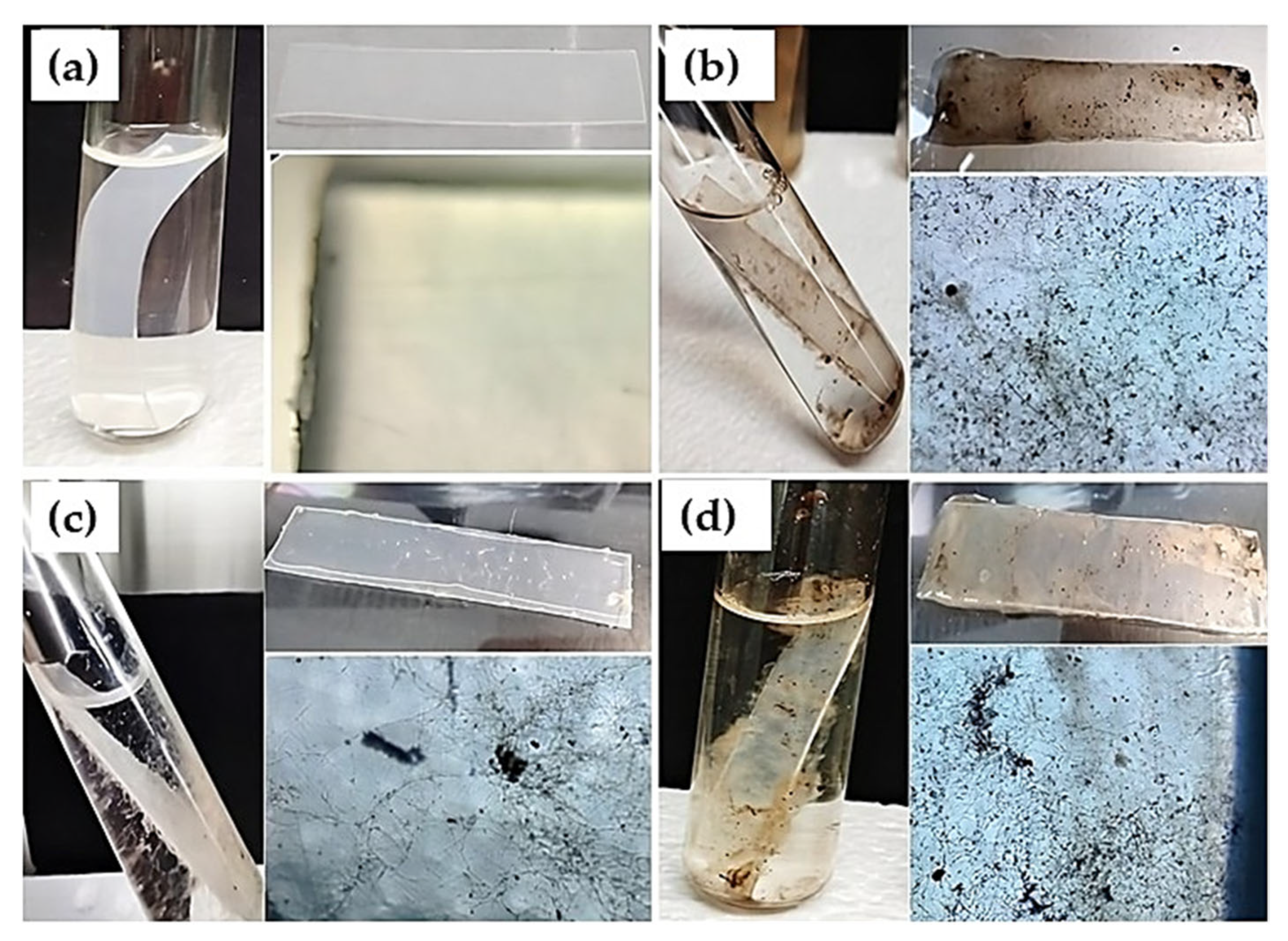
Figure 4 shows the growth of the three strains of Aspergillus on LDPE sheets. Greater growth was observed in A. niger H1C and A. tamarii H6C. The presence of hyphae and spores on the plastic surface is also observed at 40× magnification. The control shows no contamination of the system and no surface deterioration as confirmed by SEM.
Figure 5 shows the graph of pH variation in the fungal systems over 50 days. In the A. niger H1C system, the pH varied from slightly alkaline (7.41) to slightly acidic (6.46) over the 50-day evaluation period. Similarly, in the A. tamarii H6C system, a decrease in pH from 7.44 to 6.79 was observed. On the contrary, in the system with A. ochraceopetaliformis H3C, an increase in pH was observed over the 50 days, varying from 7.45 to an alkaline pH of 8.02. The control did not show any pH variations, remaining at slightly alkaline values (7.43–7.56). According to Tukey’s test, the pH variations were statistically different compared to the control (p < 0.05).
The results of the data regarding the weight loss of LDPE are shown in Table 1. The findings indicated that A. niger H1C and A. tamarii H6C resulted in the most significant weight reduction (p < 0.05), with values of 4.25 ± 1.67% and 3.79 ± 1.52%, respectively. On the other hand, the A. ochraceopetaliformis H3C exhibited a comparatively lesser effect (1.98 ± 0.37%). The control group exhibited no significant weight reduction. As detailed in Table 1, Tukey’s results indicated that A. niger H1C and A. tamarii H6C differed significantly from the control group (p = 0.008 and p = 0.015, respectively), while H3C showed no significant differences from the control (p = 0.220). No statistically significant differences were observed between the fungal strains themselves (p > 0.05). The reduction constant (k) of LDPE after treatments with the three Aspergillus strains is also presented. The A. niger H1C strain obtained a lower half-life value.
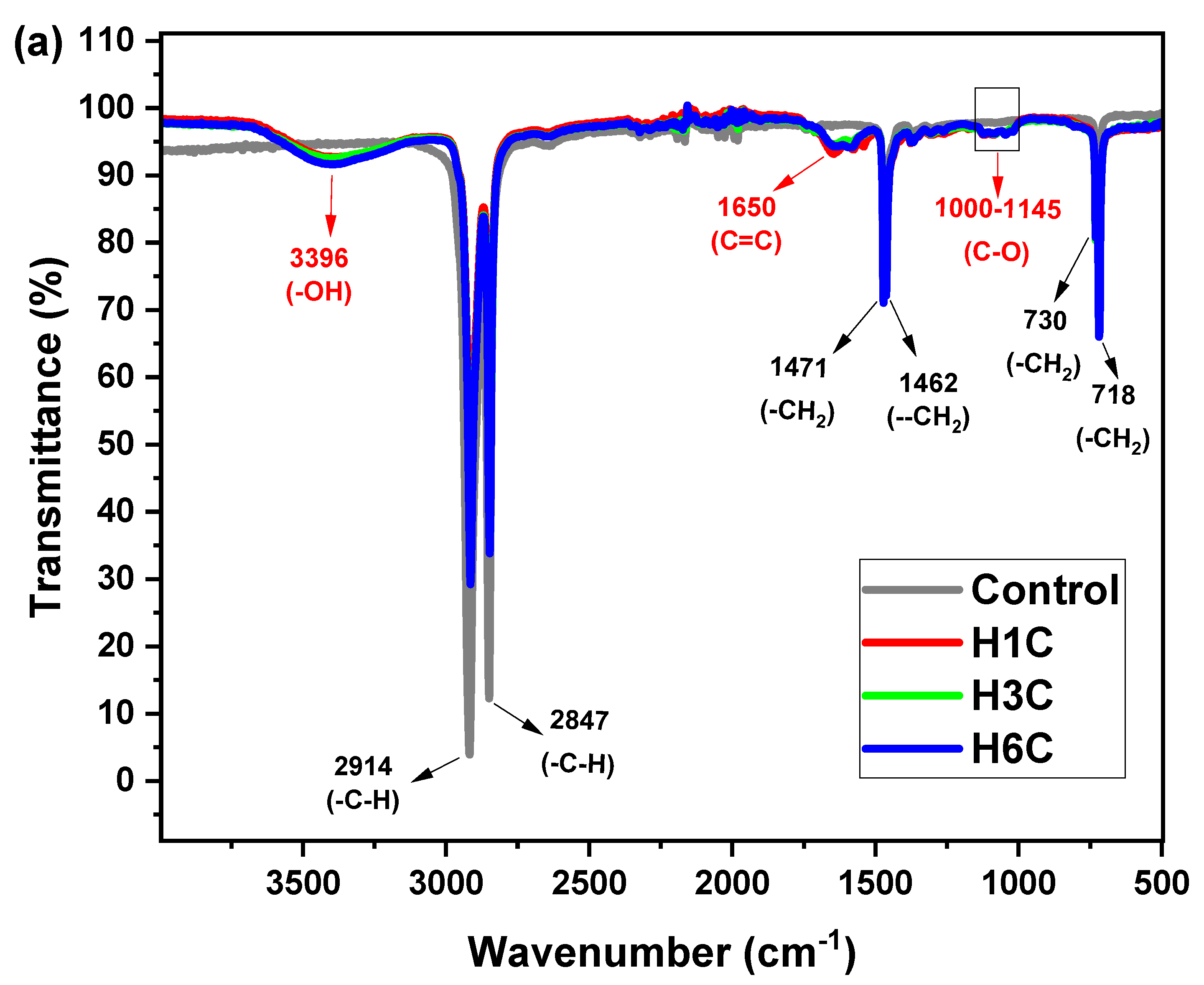
Figure 6a shows the FTIR spectra revealing structural modifications in LDPE after exposure to Aspergillus strains (H1C, H3C, and H6C), indicating an active biodegradation process. Increased intensity was observed in bands associated with hydroxyl groups (3396 cm−1), along with the appearance of characteristic signals for carbonyl groups (1650 cm−1) and C–O bonds (1000–1145 cm−1), indicative of oxidation and enzymatic activity. Additionally, a decrease in the intensity of bands (Figure 6b) corresponding to –CH bonds (2914 and 2847 cm−1) and alterations in the region of 730–718 cm−1 were recorded, suggesting a loss of polymer crystallinity. Collectively, these findings confirm the degradative potential of the evaluated strains, highlighting A. niger H1C as the most effective in LDPE breakdown. Figure 6c shows increases in the carbonyl (IC) and double bond (DBI) indices, which indicate the degradative action of microorganisms. A. niger H1C exhibited the greatest biodegradation, with IC and DBI values of 0.175 and 0.258, respectively, indicating substantial polymeric structure oxidation and breakdown. A. ochraceopetaliformis H3C and A. tamarii H6C exhibited degradation activity, albeit to a lesser extent (IC: 0.067; DBI: 0.158 and 0.223, respectively) compared to the control, which maintained values close to zero (IC: 0.025; DBI: 0.002). This confirms minimal structural alteration of the polymer by these strains.
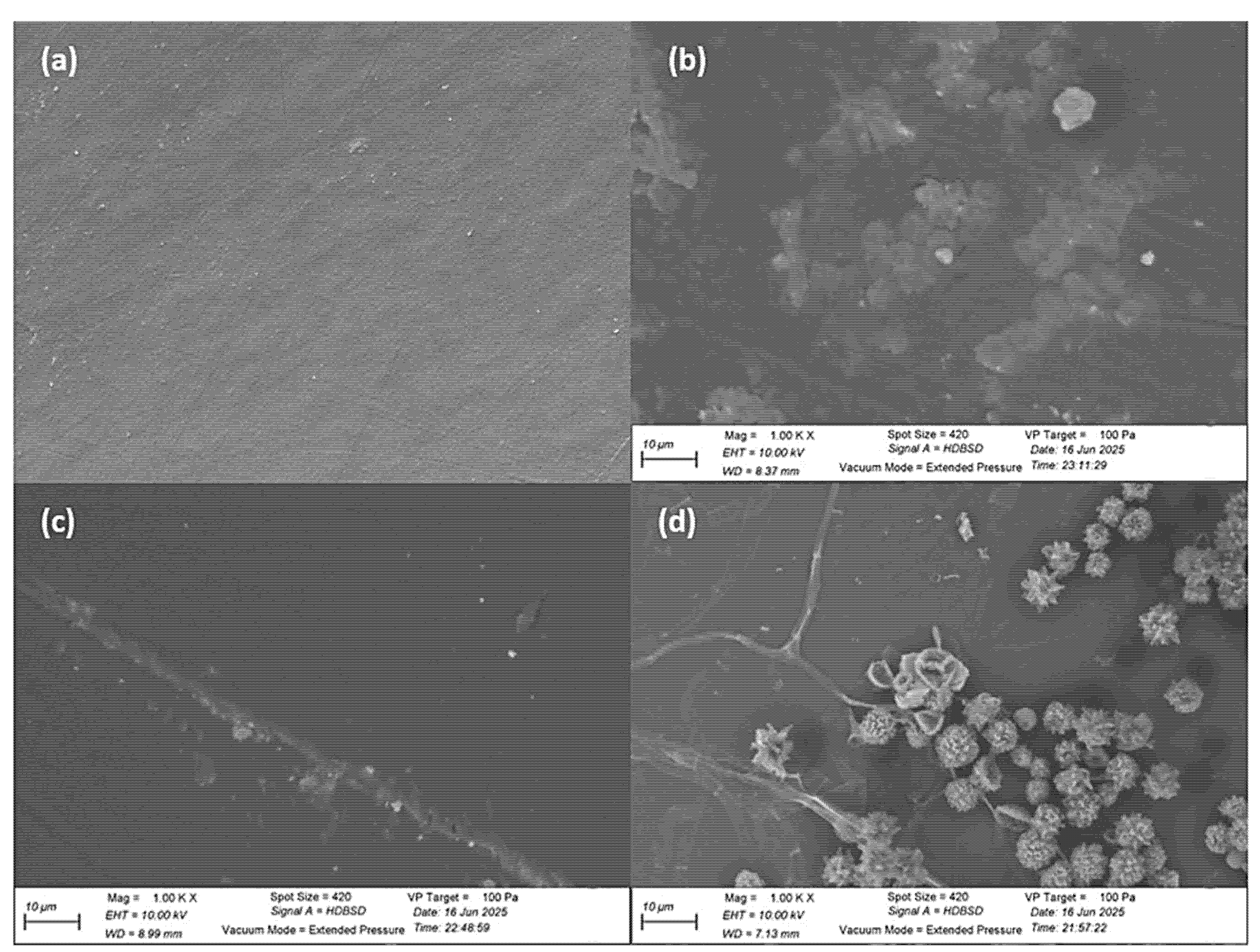
In Figure 7, the micrographs revealed surface alterations in LDPE sheets exposed to each fungal strain for 50 days. The sample treated with A. niger H1C showed microcracks, cracks, and areas of surface erosion, indicating clear modification of the polymer structure. The lamellae of sample treated with A. ochraceopetaliformis H3C showed less pronounced surface cavitations and a mostly homogeneous texture. In contrast, the sample exposed to A. tamarii H6C exhibited irregular grooves, surface detachments, and localized porosity. In the control sheet, surface modifications are not shown.

4. Discussion

The isolated fungi belong to the genus Aspergillus and grew on various culture media [50]. A. niger H1C exhibited accelerated growth on DPA agar, contrasting with the growth patterns observed in A. ochraceopetaliformis H3C and A. tamarii H6C. Its colony exhibited a white, felt-like appearance, accompanied by a dense layer of black conidia, while the reverse side displayed a pale-yellow color with radial cracks [51,52]. A. ochraceopetaliformis H3C manifests white colonies immersed in the culture medium, accompanied by brown conidia and a reverse side that exhibits a pale to brown pigmentation [53,54]. The A. tamarii H6C colony is distinguished by cinnamon-white mycelium and rough conidia, with a brown center in the colony that transitions to cream to deep yellow toward the periphery [52]. Microscopic characteristics also showed structures typical of the genus (conidia, conidiophores, hyaline and septate hyphae, and globular vesicles). The degradation of PEG-400 and vegetable oil resulted in the formation of clear zones. This qualitative assessment of enzymatic activity suggests the potential of the three Aspergillus strains to degrade plastic. It has been established that a number of enzymes, including peroxidases, esterases, ureases, and lipases, play a crucial role in the degradation of plastic [50]. The size of the halos (clear areas) varies depending on the enzymatic activity of each strain.
Molecular characterization demonstrated that Aspergillus strains exhibited high percentages of molecular similarity with A. niger CBS 101883, A. ochraceopetaliformis D11, and A. tamarii CBS 139350, respectively. A thorough analysis of the ITS sequence was conducted, revealing minimal variation within the established range of 421–452. This finding is consistent with the typical range of ITS sequence lengths observed in the fungal kingdom [55]. Phylogenetic analysis indicates that A. niger H1C exhibits a high degree of genetic similarity and a divergence of 0.00 with strains A. niger CBS 101883 and ATCC 16888. Furthermore, the genetic divergence of the species under consideration is minimal when compared with the species A. foetidus and A. neoniger, which are classified within the same clade. A. ochraceopetaliformis H3C demonstrates a divergence of 1.0 with A. ochraceopetaliformis D11, suggesting a high degree of genetic similarity and validating its classification within the species. A comparison of this species with the other groups (A. niger and A. tamarii) reveals a significantly higher divergence, with values of 62.00 and 60.00, respectively, thereby reinforcing its identity as a distinct species. A. tamarii H6C has been classified within the clade corresponding to A. tamarii (CBS 139350 and NRRL 20818), exhibiting a divergence of 0.00, thereby confirming its high degree of genetic similarity. By contrast, the external group Rhodotorula toruloides CBS 6016 exhibited the most significant divergence from the Aspergillus strain isolated with values of approximately 197, 199, and 199, respectively.
The growth of the three isolated Aspergillus strains on plastic shows one of the first steps in the biodegradation of plastic [2]. This increase in biomass indirectly indicates the degradation of LDPE, which is their only carbon source. The involvement of hydrophobins and enzymes is important in the colonization and depolymerization of plastics. Enzymes involved in oxidative degradation (laccases, hydroxylases, peroxidases and reductases) provide the fungus with carbon and energy for growth and development [56,57,58].
On the other hand, varying pH is another indicator of plastic degradation [10]. The systems with A. niger H1C and A. tamarii H6C showed a decrease in pH, reaching values of 6.46 and 6.79, respectively, whereas with A. ochraceopetaliformis H3C the pH increased to 8.02. Similarly, other studies show that some Aspergillus species can act at alkaline pH [59,60]. Aspergillus is known to grow in a pH range of 4 to 6, while some species grow in a range of 7 to 12 [48]. pH variation may also indirectly indicate LDPE biodegradation through metabolic activity, as pH affects the enzymatic activity responsible for such degradation [50,61].
The weight loss of LDPE strips after treatment with the three Aspergillus strains reached values of 4.25 ± 1.67% (A. niger H1C), 3.79 ± 1.50.2% (A. tamarii H6C) and 1.98 ± 0.37% (A. ochraceopetaliformis H3C). Research has demonstrated that certain strains of Aspergillus oryzae have been shown to achieve weight loss rates of 36.4 ± 5.53% and 40% within a 90-day period [62,63]. In similar studies, Aspergillus japonicus, A. niger, A. flavus and a consortium of Aspergillus achieved weight reductions of 10.2%, 8%, 19.4% and 26.15% (in 55 days), respectively [64,65,66]. With respect to other fungi, Cladosporium sp. CPEF-6 resulted in a 0.30 ± 0.06% reduction in weight loss [44]. A. niger H1C achieved the highest reduction rate and plastic half-life (k: 0.0009 ± 0.0035 g·day−1 and t1/2: 897.89 days). This means that it takes this number of days to halve the weight of the strip of LDPE. This value is slightly higher than other values obtained by a consortium of Bacillus bacteria (k: 0.00117 g·day−1, t1/2: 593.79 days) [67]. One of the highest values (k: 0.0224 g·day−1, t1/2: 30.93 days) was reported for Cladosporium sphaerospermum [46]. Another study using different species of microalgae reported a range of k values from 0.000123 g·day−1 (t1/2: 5655.594) to 0.005005 g·day−1 (t1/2: 138.4885) [56]. The observed variations in outcomes could be attributed to genetic variability and the physic-chemical conditions under which the experiments were conducted.
The FTIR graphs reveal structural changes attributable to biodegradation processes. The bands observed around the 3396 cm−1 peak are indicative of hydroxyl groups (–OH), suggesting the potential formation of alcohols, carboxylic acids, or amides during the degradation process [68]. The increase may be attributed to the oxidation of polymer chains and the appearance of terminal carboxylic groups [69], which are known to enhance the hydrophilicity of LDPE and facilitate enzymatic access. This phenomenon has been observed in lipase-mediate degradation of oxidized polyethylene, where ester bonds formed during oxidation are selectively cleaved by fungal lipases, resulting in reduced crystallinity and molecular weight [41]. On the other hand, a decline in the intensity of the spectral region encompassing the 2914 and 2847 cm−1 bands, corresponding to -CH group stretching vibrations in LDPE, was observed in all treated samples, suggesting backbone decomposition [70]. Furthermore, A. niger H1C exhibits a higher oxidation pattern, likely due to the organic acids it produces, which acidify the medium, thereby promoting oxidation and enzymatic cleavage by lipases [71,72]. Similar oxidative and hydrolytic modifications have been reported for A. niger lipase acting on PE, PET, and PS, where FTIR and SEM analyses confirmed the formation of alkoxy, hydroxyl, and carbonyl groups as indicators of enzymatic degradation [72]. The appearance of a band at 1650 cm−1 is attributed to carbonyl group formation, indicates structural changes under fungal treatment [50]. The presence of bands within the region of 1000 to 1145 cm−1 is indicative of the stretching vibrations of C-O groups in alcohols and ethers, which are byproducts of polyethylene degradation. Research has demonstrated that the degradation of polyethylene, triggered by the enzymatic activity of Aspergillus, results in the formation of functional groups [40,50,72]. Alterations in the 730 and 718 cm−1 regions, associated with CH2 rocking vibrations, suggest reduced crystallinity and increased polymer accessibility [73].
The increase in the double bond index (DBI) and the decrease in the carbonyl index (IC) during LDPE degradation by Aspergillus strains may reflect the transformation of carbonyl groups into other compounds and the formation of unsaturated structures. These results suggest differences in enzymatic capacity and biodegradative potential between strains. These modifications contribute to the overall susceptibility of the polymer to enzymatic attack, including by lipases [41,72]. These spectral changes were most pronounced in samples treated with A. niger H1C, which exhibited increased intensity at 3396 cm−1, a distinct carbonyl band at 1650 cm−1, and a marked reduction in –CH stretching signals, confirming its stronger oxidative effect.
The images obtained by SEM revealed significant morphological changes on the surface of the LDPE sheets treated with different fungal strains for 50 days. In the sample treated with A. niger H1C, microcracks, fissures, and areas with irregular surface erosion were observed, indicating significant structural deterioration of the polymer. Furthermore, the presence of structures that are consistent with mycelium suggests the occurrence of active colonization of the surface. These findings are consistent with previous studies highlighting the ability of A. niger to secrete oxidative enzymes, which can break polymer bonds and facilitate its degradation [72]. In the case of the sample treated with A. ochraceopetaliformis H3C, the LDPE surface exhibited minimal alteration, characterized by shallow surface cavitations and predominantly intact regions. The overall texture manifested as more homogeneous and smoother, thereby suggesting limited interaction of the fungus with the polymer substrate. This reduced degradative activity can be ascribed to either diminished enzyme production or to a diminished affinity of this strain for hydrophobic surfaces, such as those of LDPE. In contrast to the findings observed in A. niger H1C, there was an absence of mycelial colonization or deterioration in structural integrity. The sample that was treated with A. tamarii H6C exhibited intermediate degradation. The SEM images revealed the presence of surface detachment, irregular grooves, and localized porosity, which collectively suggest the occurrence of moderate enzymatic activity, accompanied by sporadic colonization of the polymer. Although A. ochraceopetaliformis and A. tamarii have not been specifically studied for LDPE degradation, other Aspergillus species have shown promising results. These findings suggest that Aspergillus species, in general, have the potential to degrade LDPE, making them valuable alternatives for bioremediation efforts to address plastic pollution [60,74]. These results underscore the potential of specific Aspergillus species in plastic biodegradation processes, particularly A. niger, whose structural action proved to be the most significant at the surface level.
The present study highlights the potential of native Aspergillus strains to induce structural changes in LDPE under controlled conditions, offering preliminary evidence of their biodegradation capacity. These findings contribute to the development of microbial strategies for plastic waste management. Moreover, this research aligns with SDG 13, which promotes alternative approaches for the treatment of xenobiotics, including plastics, in ways that are both environmentally sustainable and effective.

5. Research Limitations

Limitations are presented by this study. The low level of LDPE mass loss may be attributed to differences in fungal biodegradation capacity. The polymer did not undergo oxidative pretreatment, as UV radiation was applied solely for sterilization and did not alter its structure. Moreover, the extended half-life of LDPE (up to 897.89 days) suggests that time is a limiting factor for the practical implementation of fungal biodegradation strategies, particularly under natural conditions. Although the current analyses offer preliminary insights into the biodegradation potential of the strains, future studies could benefit from the incorporation of laccase and extended incubation periods to enhance degradation efficiency.

6. Conclusions

In summary, the native strains A. niger H1C, A. ochraceopetaliformis H3C, and A. tamarii H6C, isolated from plastic-contaminated soils, exhibited their ability to biodegrade LDPE under in vitro conditions. Among them, A. niger H1C exhibited the most significant degradative performance, with a weight loss of 4.25 ± 1.67%, a biodegradation constant (k) of 0.0009 ± 0.0035 g·day−1, and a residual polymer half-life of 897.89 days. The biodegradation process was supported by pH variation (from slightly acidic to alkaline) and confirmed through FTIR and SEM analyses, which revealed structural and oxidative modifications in the polymer matrix. The increase in carbonyl (IC) and double bond (DBI) indices confirms the oxidation and molecular cleavage process of the polymer, likely facilitated by fungal enzymatic activity including lipases induced under lipid-rich conditions. The molecular identification of A. ochraceopetaliformis and A. tamarii as species with LDPE biodegradation potential is reported for the first time. These fungi are positioned as promising candidates for biotechnological strategies for plastic waste remediation, in line with the principles of environmental sustainability and compliance with SDG 13.
Budling on these findings, further research is needed to fully understand the mechanisms involved and optimize biodegradation efficiency. Improving pretreatment methods, such as the use of chemical or thermal oxidation, could make the polymer more susceptible to microbial attack and improve degradation rates. In addition, the integration of enzymatic treatments such as laccase, the use of co-culture systems, and the extension of incubation periods could lead to scalable applications in waste management. These strategies could be applied in bioreactors or composting systems and adapted to local conditions, contributing to region-specific solutions in line with circular economy principles and environmental sustainability goals.

Author Contributions

Conceptualization, W.R.-V.; methodology, W.R.-V., M.D.L.C.-N. and N.T.-R.; software, C.Q.-C.; validation, W.R.-V.; formal analysis, W.R.-V., N.M.O. and N.T.-R.; investigation, W.R.-V., M.D.L.C.-N. and N.T.-R.; resources, N.M.O.; data curation, N.T.-R. and C.Q.-C.; writing—original draft preparation, W.R.-V.; writing—review and editing, N.T.-R.; visualization, W.R.-V. and M.D.L.C.-N.; supervision, W.R.-V.; project administration, W.R.-V.; funding acquisition, W.R.-V. All authors have read and agreed to the published version of the manuscript.

Funding

This research was funded by the Universidad Cesar Vallejo (Trujillo, Peru) and is a product of the Docente Research Project, with code P-2023-98, approved by Resolution N°184-2023-VI-UCV on 3 July 2023.

Institutional Review Board Statement

Not applicable.

Informed Consent Statement

Not applicable.

Data Availability Statement

The ITS sequences of the three Aspergillus isolates are deposited in GenBank under the following codes: A. niger H1C: PQ764478 (accessed on 19 December 2024: https://www.ncbi.nlm.nih.gov/nuccore/PQ764478); A. ochraceopetaliformis H3C: PQ764479 (accessed on 19 December 2024: https://www.ncbi.nlm.nih.gov/nuccore/PQ764479); A. tamarii H6C: PQ764480 (accessed on 19 December 2024: https://www.ncbi.nlm.nih.gov/nuccore/PQ764480).

Acknowledgments

The authors would like to thank the Universidad Cesar Vallejo (Trujillo, Peru) for allowing the development of the research project and for funding the publication of the manuscript. We also thank the project P-2023-113: Sustainable microbiology for the use of plastic waste in the generation of electricity, for collaborating in the analysis of LDPE by FTIR.

Conflicts of Interest

The authors declare no conflicts of interest.

References

  1. Bahl, S.; Dolma, J.; Jyot Singh, J.; Sehgal, S. Biodegradation of Plastics: A State of the Art Review. Mater. Today Proc. 2021, 39, 31–34. [Google Scholar] [CrossRef]
  2. Zeenat; Elahi, A.; Bukhari, D.A.; Shamim, S.; Rehman, A. Plastics Degradation by Microbes: A Sustainable Approach. J. King Saud Univ. Sci. 2021, 33, 101538. [Google Scholar] [CrossRef]
  3. Adeniran, A.A.; Shakantu, W. The Health and Environmental Impact of Plastic Waste Disposal in South African Townships: A Review. Int. J. Environ. Res. Public Health 2022, 19, 779. [Google Scholar] [CrossRef]
  4. North, E.J.; Halden, R.U. Plastics and Environmental Health: The Road Ahead. Rev. Environ. Health 2013, 28, 1–8. [Google Scholar] [CrossRef]
  5. Sa’adu, I.; Farsang, A. Plastic Contamination in Agricultural Soils: A Review. Environ. Sci. Eur. 2023, 35, 13. [Google Scholar] [CrossRef]
  6. Geyer, R.; Jambeck, J.R.; Law, K.L. Production, Use, and Fate of All Plastics Ever Made. Sci. Adv. 2017, 3, e1700782. [Google Scholar] [CrossRef]
  7. Kadac-Czapska, K.; Knez, E.; Gierszewska, M.; Olewnik-Kruszkowska, E.; Grembecka, M. Microplastics Derived from Food Packaging Waste-Their Origin and Health Risks. Materials 2023, 16, 674. [Google Scholar] [CrossRef] [PubMed]
  8. Us Epa, O. Plastics: Material-Specific Data. Available online: https://www.epa.gov/facts-and-figures-about-materials-waste-and-recycling/plastics-material-specific-data (accessed on 24 November 2024).
  9. Fayshal, M.A. Current practices of plastic waste management, environmental impacts, and potential alternatives for reducing pollution and improving management. Heliyon 2024, 10, e40838. [Google Scholar] [CrossRef]
  10. Kotova, I.B.; Taktarova, Y.V.; Tsavkelova, E.A.; Egorova, M.A.; Bubnov, I.A.; Malakhova, D.V.; Shirinkina, L.I.; Sokolova, T.G.; Bonch-Osmolovskaya, E.A. Microbial Degradation of Plastics and Approaches to Make It More Efficient. Microbiology 2021, 90, 671–701. [Google Scholar] [CrossRef]
  11. Shi, C.; Zhang, Y.; Shao, Y.; Ray, S.S.; Wang, B.; Zhao, Z.; Yu, B.; Zhang, X.; Li, W.; Ding, J.; et al. A review on the occurrence, detection methods, and ecotoxicity of biodegradable microplastics in the aquatic environment: New cause for concern. TrAC Trends Anal. Chem. 2024, 178, 117832. [Google Scholar] [CrossRef]
  12. Kumar, R.; Verma, A.; Shome, A.; Sinha, R.; Sinha, S.; Jha, P.K.; Kumar, R.; Kumar, P.; Shubham Das, S.; Sharma, P. Impacts of Plastic Pollution on Ecosystem Services, Sustainable Development Goals, and Need to Focus on Circular Economy and Policy Interventions. Sustainability 2021, 13, 9963. [Google Scholar] [CrossRef]
  13. Iroegbu, A.O.C.; Ray, S.S.; Mbarane, V.; Bordado, J.C.; Sardinha, J.P. Plastic Pollution: A Perspective on Matters Arising: Challenges and Opportunities. ACS Omega 2021, 6, 19343–19355. [Google Scholar] [CrossRef]
  14. Thushari, G.G.N.; Senevirathna, J.D.M. Plastic Pollution in the Marine Environment. Heliyon 2020, 6, e04709. [Google Scholar] [CrossRef]
  15. Wayman, C.; Niemann, H. The Fate of Plastic in the Ocean Environment—A Minireview. Environ. Sci. Process. Impacts 2021, 23, 198–212. [Google Scholar] [CrossRef] [PubMed]
  16. Watt, E.; Picard, M.; Maldonado, B.; Abdelwahab, M.A.; Mielewski, D.F.; Drzal, L.T.; Misra, M.; Mohanty, A.K. Ocean Plastics: Environmental Implications and Potential Routes for Mitigation—A Perspective. RSC Adv. 2021, 11, 21447–21462. [Google Scholar] [CrossRef]
  17. Evode, N.; Qamar, S.A.; Bilal, M.; Barceló, D.; Iqbal, H.M.N. Plastic Waste and Its Management Strategies for Environmental Sustainability. Case Stud. Chem. Environ. Eng. 2021, 4, 100142. [Google Scholar] [CrossRef]
  18. Zhang, F.; Zhao, Y.; Wang, D.; Yan, M.; Zhang, J.; Zhang, P.; Ding, T.; Chen, L.; Chen, C. Current Technologies for Plastic Waste Treatment: A Review. J. Clean. Prod. 2021, 282, 124523. [Google Scholar] [CrossRef]
  19. Damayanti, D.; Saputri, D.R.; Marpaung, D.S.S.; Yusupandi, F.; Sanjaya, A.; Simbolon, Y.M.; Asmarani, W.; Ulfa, M.; Wu, H.-S. Current Prospects for Plastic Waste Treatment. Polymers 2022, 14, 3133. [Google Scholar] [CrossRef]
  20. Mohanan, N.; Montazer, Z.; Sharma, P.K.; Levin, D.B. Microbial and Enzymatic Degradation of Synthetic Plastics. Front. Microbiol. 2020, 11, 580709. [Google Scholar] [CrossRef]
  21. Srikanth, M.; Sandeep, T.S.R.S.; Sucharitha, K.; Godi, S. Biodegradation of Plastic Polymers by Fungi: A Brief Review. Bioresour. Bioprocess. 2022, 9, 42. [Google Scholar] [CrossRef]
  22. Shilpa; Basak, N.; Meena, S.S. Microbial Biodegradation of Plastics: Challenges, Opportunities, and a Critical Perspective. Front. Environ. Sci. Eng. 2022, 16, 161. [Google Scholar] [CrossRef]
  23. Vaksmaa, A.; Guerrero-Cruz, S.; Ghosh, P.; Zeghal, E.; Hernando-Morales, V.; Niemann, H. Role of Fungi in Bioremediation of Emerging Pollutants. Front. Mar. Sci. 2023, 10, 1070905. [Google Scholar] [CrossRef]
  24. Ekanayaka, A.H.; Tibpromma, S.; Dai, D.; Xu, R.; Suwannarach, N.; Stephenson, S.L.; Dao, C.; Karunarathna, S.C. A Review of the Fungi That Degrade Plastic. J. Fungi 2022, 8, 772. [Google Scholar] [CrossRef]
  25. Malcolm, D.; Richardson, K.B.; Hope, W. Aspergillus. In Clinical Mycology; Elsevier: Amsterdam, The Netherlands, 2009; pp. 271–296. ISBN 9781416056805. [Google Scholar]
  26. Mohamed, A.H.; Balbool, B.A.; Abdel-Azeem, A.M. Aspergillus from Different Habitats and Their Industrial Applications. In Fungal Biology; Springer International Publishing: Cham, Switzerland, 2021; pp. 85–106. ISBN 9783030675608. [Google Scholar]
  27. Mousavi, B.; Hedayati, M.T.; Hedayati, N.; Ilkit, M.; Syedmousavi, S. Aspergillus Species in Indoor Environments and Their Possible Occupational and Public Health Hazards. Curr. Med. Mycol. 2016, 2, 36–42. [Google Scholar] [CrossRef] [PubMed]
  28. Mukherjee, A. Role of Aspergillus in Bioremediation Process. In New and Future Developments in Microbial Biotechnology and Bioengineering; Elsevier: Amsterdam, The Netherlands, 2016; pp. 209–214. ISBN 9780444635051. [Google Scholar]
  29. Munir, E.; Harefa, R.S.M.; Priyani, N.; Suryanto, D. Plastic Degrading Fungi Trichoderma Viride and Aspergillus Nomius Isolated from Local Landfill Soil in Medan. IOP Conf. Ser. Earth Environ. Sci. 2018, 126, 012145. [Google Scholar] [CrossRef]
  30. Pu, S.; Ma, H.; Deng, D.; Xue, S.; Zhu, R.; Zhou, Y.; Xiong, X. Isolation, Identification, and Characterization of an Aspergillus Niger Bioflocculant-Producing Strain Using Potato Starch Wastewater as Nutrilite and Its Application. PLoS ONE 2018, 13, e0190236. [Google Scholar] [CrossRef] [PubMed]
  31. Ali, M.I.; Ahmed, S.; Robson, G.; Javed, I.; Ali, N.; Atiq, N.; Hameed, A. Isolation and Molecular Characterisation of Polyvinyl Chloride (PVC) Plastic Degrading Fungal Isolates: Fungal Degradation of PVC Plastics. J. Basic Microbiol. 2014, 54, 18–27. [Google Scholar] [CrossRef] [PubMed]
  32. Guerrero Cabrera, J.P.; Cabello Torres, R.J.; Valdiviezo Gonzales, L.G.; Garzon Flores, A.; Reynoso Quispe, P. Application of Fungal Microorganisms for the Degradation “in Vitro” of Synthetic Polymers. In Proceedings of the 20th LACCEI International Multi-Conference for Engineering, Education, and Technology: “Education, Research and Leadership in Post-pandemic Engineering: Resilient, Inclusive and Sustainable Actions”, Hybrid Event, Boca Raton, FL, USA, 18–22 July 2022; Latin American and Caribbean Consortium of Engineering Institutions: Miami Lakes, FL, USA, 2022. [Google Scholar] [CrossRef]
  33. Russell, J.R.; Huang, J.; Anand, P.; Kucera, K.; Sandoval, A.G.; Dantzler, K.W.; Hickman, D.; Jee, J.; Kimovec, F.M.; Koppstein, D.; et al. Biodegradation of Polyester Polyurethane by Endophytic Fungi. Appl. Environ. Microbiol. 2011, 77, 6076–6084. [Google Scholar] [CrossRef]
  34. Méndez, C.R.; Vergaray, G.; Béjar, V.R.; Cárdenas, K.J. Aislamiento y Caracterización de Micromicetos Biodegradadores de Polietileno. Rev. Peru. Biol. 2006, 13, 203–205. [Google Scholar] [CrossRef]
  35. Uribe, D.; Giraldo, D.; Gutiérrez, S.; Merino, F. Biodegradación de Polietileno de Baja Densidad Por Acción de Un Consorcio Microbiano Aislado de Un Relleno Sanitario, Lima, Perú. Rev. Peru. Biol. 2011, 17, 133–136. [Google Scholar] [CrossRef]
  36. Ibrahim, S.S.; Ionescu, D.; Grossart, H.-P. Tapping into Fungal Potential: Biodegradation of Plastic and Rubber by Potent Fungi. Sci. Total Environ. 2024, 934, 173188. [Google Scholar] [CrossRef] [PubMed]
  37. Saira Abdullah Maroof, L.; Iqbal, M.; Farman, S.; Lubna Faisal, S. Biodegradation of Low-Density Polyethylene (LDPE) Bags by Fungi Isolated from Waste Disposal Soil. Appl. Environ. Soil Sci. 2022, 2022, 8286344. [Google Scholar] [CrossRef]
  38. Nademo, Z.M.; Shibeshi, N.T.; Gemeda, M.T. Isolation and Screening of Low-Density Polyethylene (LDPE) Bags Degrading Bacteria from Addis Ababa Municipal Solid Waste Disposal Site “Koshe”. Ann. Microbiol. 2023, 73, 6. [Google Scholar] [CrossRef]
  39. Gupta, K.K.; Devi, D.; Rana, D. Isolation And Screening of Low Density Polyethylene (Ldpe) Degrading Bacterial Strains from Waste Disposal Sites. World J. Pharm. Res. 2016, 5, 1633–1643. Available online: https://www.semanticscholar.org/paper/ISOLATION-AND-SCREENING-OF-LOW-DENSITY-POLYETHYLENE-Gupta-Devi/98c2a520cf88266f61f2fee8fe65c00df1f342e5 (accessed on 27 September 2024).
  40. Khan, S.; Ali, S.A.; Ali, A.S. Biodegradation of Low Density Polyethylene (LDPE) by Mesophilic Fungus “Penicillium citrinum” Isolated from Soils of Plastic Waste Dump Yard, Bhopal, India. Environ. Technol. 2023, 44, 2300–2314. [Google Scholar] [CrossRef]
  41. Kim, D.-W.; Lim, E.S.; Lee, G.H.; Son, H.F.; Sung, C.; Jung, J.-H.; Park, H.J.; Gong, G.; Ko, J.K.; Um, Y.; et al. Biodegradation of Oxidized Low Density Polyethylene by Pelosinus Fermentans Lipase. Bioresour. Technol. 2024, 403, 130871. [Google Scholar] [CrossRef]
  42. Machuca-Guevara, J.I.; Suárez-Peña, E.A.; Motte Darricau, E.; Mialhe-Matonnier, E.L. Caracterización Molecular de Los Microorganismos Presentes Durante El Proceso Fermentativo de Los Granos de Cacao (Theobroma cacao). Rev. Peru. Biol. 2019, 26, 535–542. [Google Scholar] [CrossRef]
  43. Kuchkarova, N.; Han, C.; Toshmatov, Z.; Chen, H.; Shao, H. Diversity of Fungal Endophytes Isolated from the Invasive Plant Solanum Rostratum. Acta Bot. Croat. 2024, 83, 76–79. [Google Scholar] [CrossRef]
  44. Gong, Z.; Jin, L.; Yu, X.; Wang, B.; Hu, S.; Ruan, H.; Sung, Y.-J.; Lee, H.-G.; Jin, F. Biodegradation of Low Density Polyethylene by the Fungus Cladosporium sp. Recovered from a Landfill Site. J. Fungi 2023, 9, 605. [Google Scholar] [CrossRef] [PubMed]
  45. Bustillo, A. Método para Cuantificar Suspensiones de Esporas de Hongos y Otros Organismos; Universidad Nacional de Colombia: Palmira, Colombia, 2010; Volume 10. [Google Scholar]
  46. Sathiyabama, M.; Boomija, R.V.; Sathiyamoorthy, T.; Mathivanan, N.; Balaji, R. Mycodegradation of Low-Density Polyethylene by Cladosporium Sphaerospermum, Isolated from Platisphere. Sci. Rep. 2024, 14, 8351. [Google Scholar] [CrossRef]
  47. E13 Committee Practice for General Techniques for Obtaining Infrared Spectra for Qualitative Analysis. Available online: https://store.astm.org/e1252-98r21.html (accessed on 20 December 2024).
  48. Esmaeili, A.; Pourbabaee, A.A.; Alikhani, H.A.; Shabani, F.; Esmaeili, E. Biodegradation of Low-Density Polyethylene (LDPE) by Mixed Culture of Lysinibacillus Xylanilyticus and Aspergillus Niger in Soil. PLoS ONE 2013, 8, e71720. [Google Scholar] [CrossRef]
  49. Mori-Mestanza, D.; Valqui-Rojas, I.; Caetano, A.C.; Culqui-Arce, C.; Cruz-Lacerna, R.; Cayo-Colca, I.S.; Castro-Alayo, E.M.; Balcázar-Zumaeta, C.R. Physicochemical Properties of Nanoencapsulated Essential Oils: Optimizing D-Limonene Preservation. Polymers 2025, 17, 348. [Google Scholar] [CrossRef]
  50. Nasrabadi, A.E.; Ramavandi, B.; Bonyadi, Z. Recent Progress in Biodegradation of Microplastics by Aspergillus Sp. in Aquatic Environments. Colloids Interface Sci. Commun. 2023, 57, 100754. [Google Scholar] [CrossRef]
  51. d’Halewyn, M.-A.; Chevalier, P. Aspergillus Niger. Available online: https://www.inspq.qc.ca/en/moulds/fact-sheets/aspergillus-niger (accessed on 21 January 2025).
  52. Nyongesa, B.W.; Okoth, S.; Ayugi, V. Identification Key for Aspergillus Species Isolated from Maize and Soil of Nandi County, Kenya. Adv. Microbiol. 2015, 05, 205–229. [Google Scholar] [CrossRef]
  53. Visagie, C.M.; Varga, J.; Houbraken, J.; Meijer, M.; Kocsubé, S.; Yilmaz, N.; Fotedar, R.; Seifert, K.A.; Frisvad, J.C.; Samson, R.A. Ochratoxin Production and Taxonomy of the Yellow Aspergilli (Aspergillus Section Circumdati). Stud. Mycol. 2014, 78, 1–61. [Google Scholar] [CrossRef]
  54. Akter, H.; Ara, I.; Alam, N.B.; Alam, N. The First Report of Aspergillus Ochraceopetaliformis Bat. & Maia from Small Indigenous Dry Fish Setipinna Phasa (Hamilton 1822) in Bangladesh. Jahangirnagar Univ. J. Biol. Sci. 2024, 12, 87–95. [Google Scholar] [CrossRef]
  55. Sharma, R.; Talukdar, D.; Bhardwaj, S.; Jaglan, S.; Kumar, R.; Kumar, R.; Akhtar, M.S.; Beniwal, V.; Umar, A. Bioremediation Potential of Novel Fungal Species Isolated from Wastewater for the Removal of Lead from Liquid Medium. Environ. Technol. Innov. 2020, 18, 100757. [Google Scholar] [CrossRef]
  56. Gowthami, A.; Syed Marjuk, M.; Raju, P.; Nanthini Devi, K.; Santhanam, P.; Dinesh Kumar, S.; Perumal, P. Biodegradation Efficacy of Selected Marine Microalgae against Low-Density Polyethylene (LDPE): An Environment Friendly Green Approach. Mar. Pollut. Bull. 2023, 190, 114889. [Google Scholar] [CrossRef]
  57. Zhang, J.; Gao, D.; Li, Q.; Zhao, Y.; Li, L.; Lin, H.; Bi, Q.; Zhao, Y. Biodegradation of Polyethylene Microplastic Particles by the Fungus Aspergillus Flavus from the Guts of Wax Moth Galleria Mellonella. Sci. Total Environ. 2020, 704, 135931. [Google Scholar] [CrossRef]
  58. Sánchez, C. Fungal Potential for the Degradation of Petroleum-Based Polymers: An Overview of Macro- and Microplastics Biodegradation. Biotechnol. Adv. 2020, 40, 107501. [Google Scholar] [CrossRef]
  59. Dharshni, S.; Kanchana, M. Kanchana Microbial Degradation of Low-Density Polyethylene (Ldpe) by Fungus Isolated from Landfill Soil. Plant Arch. 2021, 21, 809–815. [Google Scholar] [CrossRef]
  60. Das, M.P.; Kumar, S.; Das, J. Fungal-Mediated Deterioration and Biodegradation Study of Low-Density Polyethylene (LDPE) Isolated from Municipal Dump Yard in Chennai, India. Energy Ecol. Environ. 2018, 3, 229–236. [Google Scholar] [CrossRef]
  61. Temporiti, M.E.E.; Nicola, L.; Nielsen, E.; Tosi, S. Fungal Enzymes Involved in Plastics Biodegradation. Microorganisms 2022, 10, 1180. [Google Scholar] [CrossRef]
  62. Muhonja, C.N.; Makonde, H.; Magoma, G.; Imbuga, M. Biodegradability of Polyethylene by Bacteria and Fungi from Dandora Dumpsite Nairobi-Kenya. PLoS ONE 2018, 13, e0198446. [Google Scholar] [CrossRef]
  63. Jayaprakash, V.; Maheswari Devi Palempalli, U. Effect of Palmitic Acid in the Acceleration of Polyethylene Biodegradation by Aspergillus Oryzae. J. Pure Appl. Microbiol. 2018, 12, 2259–2268. [Google Scholar] [CrossRef]
  64. Raaman, N.; Rajitha, N.; Jayshree, A.; Jegadeesh, R. Biodegradation of Plastic by Aspergillus spp. Isolated from Polythene Polluted Sites around Chennai. J. Acad. Indus. Res. 2014, 16, 313–316. Available online: https://api.semanticscholar.org/CorpusID:3464885 (accessed on 25 December 2024).
  65. Makut, M.D.; Okey-Ndeche, N.F. Optimisation of Some Conditions for the Biodegradation of Low-Density Polyethylene Strips by Fungi Isolated from Parts of North Central Nigeria. Int. J. Latest Technol. Eng. Manag. Appl. Sci. 2024, 13, 35–42. [Google Scholar] [CrossRef]
  66. DSouza, G.C.; Sheriff, R.S.; Ullanat, V.; Shrikrishna, A.; Joshi, A.V.; Hiremath, L.; Entoori, K. Fungal Biodegradation of Low-Density Polyethylene Using Consortium of Aspergillus Species under Controlled Conditions. Heliyon 2021, 7, e07008. [Google Scholar] [CrossRef]
  67. Cabeza Vásquez, H.T.; Reaño Segundo, Y.J. Potencial En La Degradación de Polietileno de Baja Densidad En Consorcios de Bacterias Del Botadero de Residuos Sólidos “Pampas de Reque”, 2022; Universidad Nacional Pedro Ruíz Gallo: Lambayeque, Perú, 2024; Available online: https://hdl.handle.net/20.500.12893/13172 (accessed on 25 December 2024).
  68. Maan Shake, M.; Khlaif, A.T. Bioremediation of Low-Density Polyethylene (PE) by Fungi. J. Neonatal Surg. 2025, 14, 57–63. [Google Scholar] [CrossRef]
  69. Yang, S.; Yang, X.; Xie, J.; Hou, Q.; Liu, Y. Degradation Effect and Degradation Mechanism of Aspergillus niger on Polyethylene Components of Waste Cables. Trans. China Electrotech. Soc. 2024, 39, 76–87. [Google Scholar] [CrossRef]
  70. Sutkar, P.R.; Hadkar, S.S.; Kamble, S.A.; Dhulap, V.P. Biodegradation Potential of Low-Density Polyethylene (LDPE) Using Aspergillus Niger and Phanerochaete Chrysosporium. Discov. Environ. 2025, 3, 7. [Google Scholar] [CrossRef]
  71. Ahmed, M.; Iram, S.; Tabassum, N.; Sajid, M.; Paseutsakoun, K.; Aleksza, L.; Székács, A. Biodegradation Efficacy of Aspergillus niger and Trichoderma harzianum on Low-Density Polyethylene. Polymers 2025, 17, 1303. [Google Scholar] [CrossRef]
  72. Safdar, A.; Ismail, F.; Imran, M. Biodegradation of Synthetic Plastics by the Extracellular Lipase of Aspergillus Niger. Environ. Adv. 2024, 17, 100563. [Google Scholar] [CrossRef]
  73. Abraham, J.; Ghosh, E.; Mukherjee, P.; Gajendiran, A. Microbial Degradation of Low Density Polyethylene. Environ. Prog. Sustain. Energy 2017, 36, 147–154. [Google Scholar] [CrossRef]
  74. El-Sayed, M.T.; Rabie, G.H.; Hamed, E.A. Biodegradation of Low-Density Polyethylene (LDPE) Using the Mixed Culture of Aspergillus Carbonarius and A. Fumigates. Environ. Dev. Sustain. 2021, 23, 14556–14584. [Google Scholar] [CrossRef]
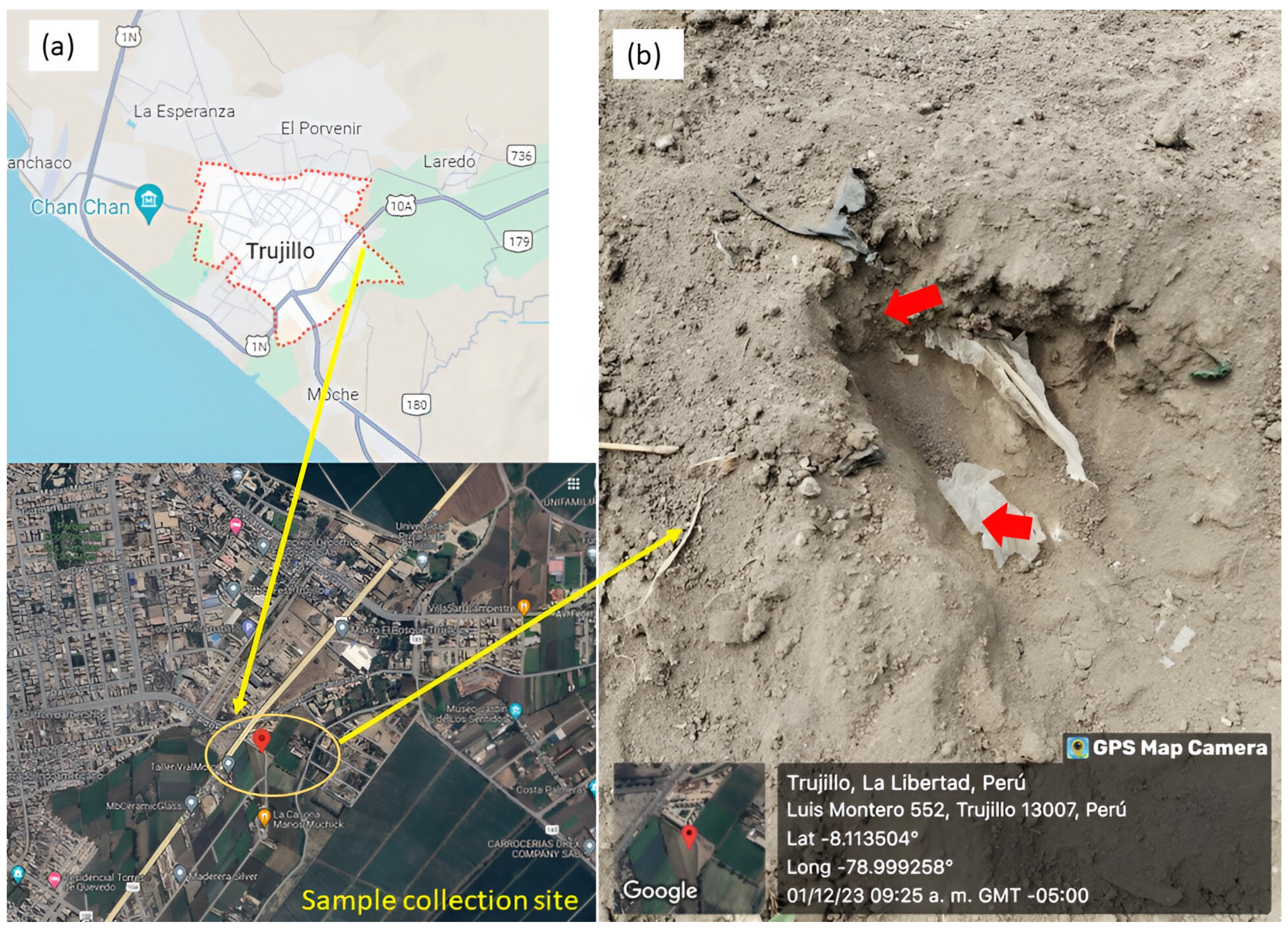
Figure 1. Location of the sampling (a). Plastic-contaminated soil showing signs of deterioration (b).
Sustainability 17 08983 g001
Figure 2. Aspergillus isolates: Morphology (a) and PEG/vegetable oil biodegradation zones (b).
Sustainability 17 08983 g002
Figure 3. Phylogenetic tree inferred by neighbor-joining with 400 bootstrap replicates. Explanations: Evolutionary distances were calculated using the number of differences method (units: base differences per sequence). Rhodotorula toruloides CBS 6016 was used as the outgroup.
Sustainability 17 08983 g003
Figure 4. Aspergillus strains growth on LDPE strips after 50 days of incubation: control (a), A. niger H1C (b), A. ochraceopetaliformis H3C (c) and A. tamarii H6C (d).
Sustainability 17 08983 g004
Figure 5. pH variation in in vitro systems during the 50-day treatment of LDPE strips with Aspergillus strains. Explains: Error bars show ±1 SD from three replicates.
Sustainability 17 08983 g005
Figure 6. Comparative FT-IR spectra of plastic samples treated with Aspergillus fungi (H1C, H3C and H6C) and control (a). Shortening of LDPE spectra (b). Carbonyl Index and Double Bound Index of three Aspergillus strains (c).
Sustainability 17 08983 g006aSustainability 17 08983 g006b
Figure 7. LDPE surface treated with Aspergillus strains for 50 days, as viewed by SEM. The control group consisted of untreated LDPE (a), as well as LDPE treated with A. niger H1C (b), A. ochraceopetaliformis H3C (c), and A. tamarii H6C (d).
Sustainability 17 08983 g007
Table 1. Percentage weight loss and half-life of LDPE due to biodegradation by Aspergillus fungi over 50 days.
Fungi StrainsW0 (g)W1 (g)% Loss of Weight ± SDReduction Rate Constant (k) of LDPE (g·day−1)Half-Life (t1/2) of the Treated LDPE (Days)
A. niger H1C0.200.194.25 ± 1.67b *0.0009 ± 0.0035897.89
A. ochraceopetaliformis H3C0.190.181.98 ± 0.37ab0.0004 ± 0.00021757.33
A. tamarii H6C0.250.243.79 ± 1.52b0.0008 ± 0.0004901.6
* Equal letters indicate no significant difference (p ≥ 0.05) by Tukey HSD test.
Disclaimer/Publisher’s Note: The statements, opinions and data contained in all publications are solely those of the individual author(s) and contributor(s) and not of MDPI and/or the editor(s). MDPI and/or the editor(s) disclaim responsibility for any injury to people or property resulting from any ideas, methods, instructions or products referred to in the content.