Article

Biological Colonization of Carolei’s Nymphaeum (Calabria, Italy)

Department of Biology, Ecology and Earth Science, University of Calabria, 87036 Rende, Italy
*
Author to whom correspondence should be addressed.
Sustainability 2025, 17(18), 8426; https://doi.org/10.3390/su17188426
Submission received: 17 July 2025 / Revised: 12 September 2025 / Accepted: 17 September 2025 / Published: 19 September 2025
(This article belongs to the Section Tourism, Culture, and Heritage)

Abstract

The nymphaeum originated as a monument dedicated to the nymphs and defined as a natural cave with a water source. Over time, it has been transformed into an artificial cave with the presence of fountains, statues and wall paintings. The nymphaeum is exposed to specific environmental conditions, leading to biodeterioration caused by vegetal organisms that find an ideal environment for their growth. This study aimed to document the vegetation present inside and outside the Carolei’s Nymphaeum, as well as the biofilm on the interior walls, particularly the painted walls. The biological work is part of a large-scale project involving building materials, thermo-hygrometric parameters, and partial pilot restoration work. Multiple approaches were used for biological analysis by combining microscopic, culture, and molecular techniques. We identified Pteridophytes, Angiosperms, and mosses, as well as fungal taxa, cyanobacteria, and chlorophytes in the biofilms. The results indicate that there is a very heterogeneous organism composition with significant biodeterioration potential. Biodeterioration is one of the major problems in the prevention, conservation, and restoration of cultural heritage, and the data gathered in this research may help to enhance the understanding of issues and develop suitable strategies for restoration, upkeep, and accessibility and usability.

1. Introduction

The nymphaeum in ancient Greece and Rome was a monument dedicated to the nymphs and, originally, was defined as a natural cave with a water source. However, it later became an artificial cave with various architectures, most of them round in shape, and featured fountains, statues, and wall paintings. In Italy, starting from the Renaissance, it became a characteristic architectural element of stately homes, where they almost became agrestic sanctuaries that also included plants and flowers.
The borderline between the cave and the nymphaeum is not well defined; the difference lies only in the fact that the nymphaeum places emphasize the sacredness of the place. Like any semi-hypogeal environment, the nymphaeum has high air relative humidity, considerable lighting, and poor ventilation (data not yet published) [1]. These factors, combined with the intrinsic properties of the materials that constitute the structure (stone material and wall painting) facilitate the growth of microorganisms and plants, which cause its biodeterioration over time, becoming visible both in the aesthetic aspect and in the loss of material [2].
The use of stone is common in the development of cultural heritage worldwide, regardless of its size, and it becomes significant in conserving artefacts over time [2].
Natural stone can be made in many different ways at the level of mineral composition, texture, and structure. Due to the variability in their physical and chemical properties, stones will possess different weather resistance capabilities. If we add to this the fact that, as described in literature, they also represent a natural habitat for a variety of microorganisms, these too can play a significant role in their alteration, inducing aesthetic, physical, and chemical changes [3,4,5].
Although stone decay is a natural phenomenon, it takes on another aspect and relevance when it concerns artefacts due to their loss of artistic and cultural value [2,6].
Among the main causes of stone deterioration, there are atmospheric agents such as temperature, relative humidity, and light, which can induce physical and chemical alteration processes, often followed by biological colonization [2,6].
There are therefore two fundamental concepts, which are biodeterioration, defined by Hueck [7,8] as “any undesirable change in the properties of a material caused by the vital activities of living organisms”, and bioreceptivity, defined by Guillitte [9] as “the aptitude of a material to be colonized by one or several groups of living organisms without necessarily undergoing any biodeterioration”, or as “the totality of material properties that contribute to the establishment, anchorage and development of fauna and/or flora” [9].
Bacteria, cyanobacteria, and microalgae are the pioneering colonizers of stone. This process results in the formation of a patina, which induces chemical and mechanical degradation and establishes the proper conditions for the growth of other organisms such as lichens and fungi. Microorganism activity can result, for example, in discoloration, formation of crusts, degradation of organic compounds, and biocorrosion [10].
Subsequently, the settlement of vascular plants can occur in cracks and fractures that create niches suitable for the accumulation of soil and organic matter.
Both the aerial parts and the roots can cause damage, but it is the latter that are responsible for the greatest damage, as they can penetrate inside the structure, grow in size, and cause both physical and chemical problems [11].
It is therefore essential to know the organisms involved in biodeterioration to better understand their role, taking into consideration both the intrinsic factors of the artefact material and environmental factors [12]. The biodeterioration level depends on the organism type, the material of the cultural item, and environmental factors such temperature, water, and nutrients. In this way, it becomes necessary to observe carefully the conditions of cultural resources, paying attention to the degree of manifestation of degradation and recognizing all the organisms that caused it.
Carolei’s Nymphaeum was included in a broader project born from researchers’ intention to restore the historical and artistic identity of a unique Calabrian site.
The entire project included diagnostic investigations of the constitutive materials and the construction techniques used, employing methods such as polarized light microscopy, energy dispersive spectroscopy (EDS) using a scanning electron microscope (SEM), X-ray diffractometry, and Raman spectroscopy. At the same time, thermo-hygrometric parameters (temperature and relative humidity) were monitored for the presence of water, a characteristic of the nymphaea (data not yet published).
The biological investigation aimed to gain a thorough understanding of the biodeteriorating agents that compromise the conservation of the mural paintings and the existing buildings.
Finally, once all the data had been collected, it was possible to establish an appropriate intervention methodology by selecting an area as a pilot site aimed at orienting an appropriate restoration and maintenance intervention methodology (data not yet published).
This set of knowledge is therefore essential for restorers and conservators in order to establish appropriate conservation procedures and evaluate which treatments are most suitable for restoration.
The examination of this monument, set in the magnificent environment of the historical park, where the naturalistic aspects around the Nymphaeum provide an additional stylistic mark, seeks to elevate and safeguard it through research and analysis that have directed techniques and technologies beneficial for securing the passage of this significant seventeenth-century artifact to future generations.
This work aims to show the effects of phototrophic microorganisms, fungi, mosses, and vascular plants on the deterioration of an artifact as particular as a nymphaeum. If we consider the continuous exchange with the external environment, we realize how these artifacts can be affected by biological deterioration, having the possibility of being colonized by many organisms. The cyanobacteria, algae, fungi, mosses, and vascular plants were then examined inside and outside the nymphaeum, combining microscopic, cultural, and molecular techniques and the use of Flores and guides for the recognition of the different organisms.

2. Materials and Methods

2.1. Site Description

The Nymphaeum historical park is located in Vadue, a hamlet part of the municipality of Carolei (Cosenza, Calabria, Italy). The locality’s name derives from the Latin word meaning “ford” and its origin is probably linked to the strategic position of the hamlet, located close to the affluent of Busento’s river.
The history of the Carolei, borne between legends and tales, tells of a territory that has seen many figures of ancient history pass through it over time. According to Bilotto [13], Carolei “stands on an ancient isthmian road that connected Cosenza with the Tyrrhenian Sea, as confirmed by the remains of a Roman settlement with a nymphaeum and a rural building where, according to tradition, Alaric, King of the Goths, died. He was later buried with his rich treasure, the fruit of the sack of Rome, in the bed of the Busento River, which was diverted for the occasion.”
The discovery of Carolei’s Nymphaeum was completely accidental after a conservative maintenance intervention carried out on the entire structure complex between 1890 and 1990. The monumental complex at the time was a cluster of buildings that bore similarities to an ancient rustic residence. The main structure is composed of the central inhabited area, which, in turn, includes several buildings placed on three sides, which form an internal courtyard protected on the fourth side by walling.
The residence appears, in fact, mutilated by its elements, which could have led one to think of a manor house of a very wealthy family in the 17th century; this is confirmed by the pictorial decorations with heraldic emblems on the walls of the main hall, elements that allow us to hypothesize the close link with the Civitella family, who arrived in Cosenza towards the end of the 16th century, as documented in the notarial deeds of the time [14,15,16].
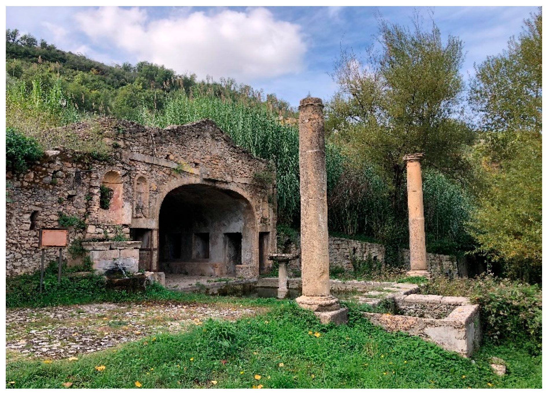
In fact, according to various historical studies, it is possible to trace the structure back to the 16th and 17th centuries [17]. Nearby, there is a small family church. To complete the monumental complex, we find the ruins of the nymphaeum, which recall the architecture of the seventeenth-century nymphaeum. It is characterized by a large frontal bath that, in turn, is connected to another smaller tub (Figure 1).
The Carolei’s Nymphaeum consists of a large square-shaped bath (5.30 m × 5.40 m), bounded by two outside columns in the “Tuscanico” style, and, on the opposite side, by a monumental front that frames the inner semi-underground hall, where the paintings are found. This last room is characterized by two entrances topped by two lintels that contain two niches with a shell-shaped cover, of which today only the left one remains. The bath is bounded by a large stone edging that allows one to walk comfortably around it, and, in the center, there is also a stone fountain. The frontal part is characterized by a large segmental arch that forms a kind of anomalous Venetian window with square shapes; the front side is characterized by calcarenite blocks, probably of local origin (Figure 2).
Each of the three inner walls is marked by three openings. The walls are painted with decorations of cornices, garlands, and mythological nature scenes. Today, due to the loss of many of the paintings, it is hard to decipher all of the scenes.
The shapes and dimensions of Carolei’s Nymphaeum are comparable to the archetypes of the same genre made from the late Renaissance to Baroque age, presenting many Roman Age elements [17].

2.2. Conservation Status

Carolei’s Nymphaeum is currently in a crumbling state of conservation due to the particular environmental conditions and poor restoration work performed in the past.
The structural stability is threatened by inadequate maintenance of the meteoric water disposal and the presence of herbaceous plants and root trees on the external walls and in the immediate vicinity, which compromise their conservation due to the action of the apparatus radical. The wall facings and the decorative elements are made up of very porous and heterogeneous limestone rocks. There are numerous fractures in the walls, phenomena of disintegration, erosion, and pulverization of the decorative elements, as well as widespread efflorescence and sub-efflorescence. The fruition of the nymphaeum’s wall paintings is strongly compromised by large gaps that affect 60% of the decorated surface, compromising the integrity and legibility of the decorative systems.
Finally, in different areas of the entire nymphaeum, the presence of a biological patina is evident, which also causes a chromatic alteration.

2.3. Sampling and Analysis

The biological sample collection phase is the first step, and it is essential to follow a correct methodological process. This step is important because analytical methodologies can vary based on the nature of the organisms to be sampled and based on the substrate in which the sampling will be carried out.
The sampling affected, in different ways, the phototrophic microorganisms, fungi, mosses, and vascular plants found both inside and outside the nymphaeum. The sampling of mosses and biofilms involved two areas: the painted front wall and the vault area. To take the samples, both sterile swabs and scalpels were used.

2.3.1. Vascular Plants

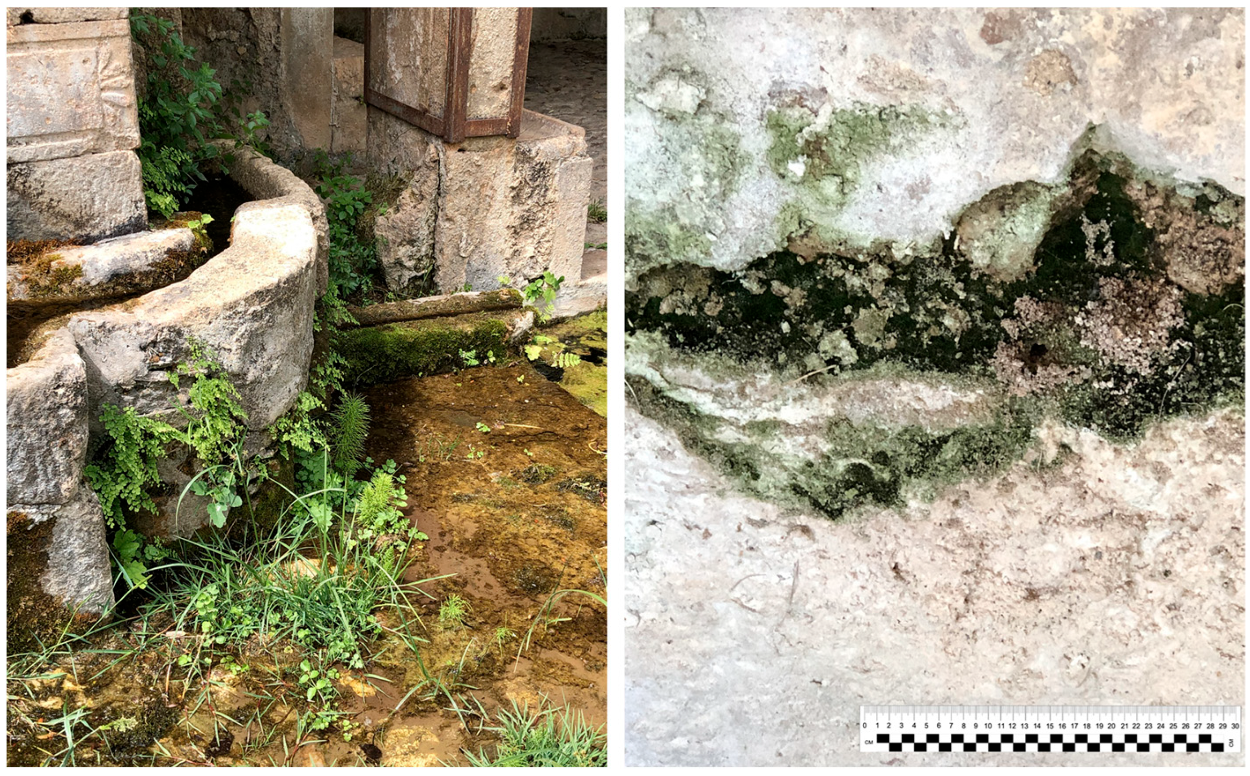
The sampling of vascular plants implicated both detailed photographic documentation on the internal and external walls of the nymphaeum and the collection of leaf material for those that did not present clear characteristics for their morphological recognition (Figure 3).
The morphological identification was carried out based on Pignatti [18], Pignatti et al. [19], and Tutin [20]. In addition, internet-based databases, such as Portal to the Flora of Italy (http://dryades.units.it/cercapiante/index.php (accessed on 12 June 2024)), were used.
Molecular techniques with ITS1 amplification were applied to the incomplete samples.
For each specimen, the family, identification (morphological or molecular), and plant life form were recorded according to Raunkiaer’s classification [21] and the Hazard Index (HI) proposed by Signorini [22,23]. This numerical index, ranging from 0 (minimal hazard) to 10 (high hazard), is based on specific morphological characteristics and on the plant life form (invasiveness, size, and shape of the root system).

2.3.2. Mosses

The presence of mosses was highlighted only inside the nymphaeum, both on the vault and on the front and side walls; in some cases, they appeared as isolated populations, and in others as part of biofilms (Figure 3).
The samples of mosses belonging to the populations were taken with the aid of sterile tweezers, taking care not to damage the substrate and for identification. Following microscopic examination, the database Mosses and Liverworts of Italy, Dryades project, was used (https://dryades.units.it/briofite/keys.html (accessed on 12 June 2024)).
For the samples present in the biofilms, collection with a scarification scalpel returned fragments that were not useful for recognition; therefore, molecular analyses were carried out.

2.3.3. Biofilm

Biofilms detected inside the nymphaeum, both on the vault (Figure 4a) and on the front and side walls (Figure 4b), were collected using sterile cotton swabs and a scalpel, scraping only the superficial microbial layer without affecting the substrate.
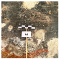
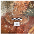
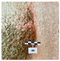
Careful observation of the biodeterioration of the walls and vault of the nymphaeum has allowed us to distinguish six different recurring morphological typologies, reported in Table 1.
Samples were stored at 4 °C and analyzed using optical microscopy on plate cultures and molecular techniques. To promote the growth of photoautotrophic microorganisms, BBM (Bold Basal Medium) and BG11 (Blue Green 11) were used, both in liquid and agar form. The growing conditions were as follows: temperature—22 °C, photoperiod—light 16 h, dark 8 h [24,25]. Then, several culture dilutions (from 10−1 to 10−5) were used to select phototrophic single-taxon cultures.
The morphological identification of cyanobacteria and algae was carried out using optical microscopy according to Anagnostidis and Komárek [26,27], Komárek and Anagnostidis [28,29], Komárek [30], and through the use of the CyanoDB site (www.cyanodb.cz (accessed on 12 June 2024)).
To characterize fungi, a solid MEA (Malt Extract Agar) was used as a selective medium for the detection, isolation, and enumeration of yeasts and molds.
Molecular techniques have been applied for both photoautotrophs and fungi.

2.4. Molecular Analysis

For molecular analysis, total DNA was extracted using the CTAB (cetyltrimethylammonium bromide) method [31]. PCR was performed using Readymix TAQ PCR (Merck Life Science) in a 25 μL volume (DNA template: 1 μL, buffer Master mix: 12.5 μL, primer: 0.125 μL each). The thermal cycler used was a Biometra Thermal Cycler (MJ Research Inc., Watertown, MA, USA).
The PCR primers were as follows: primer JK11 (5′-ATC CTG CAA TTC ACA CCA AGT ATC G-3′) and JK14 (5′-GGA GAA GTC GTA ACA AGG TTT CCG-3′) [32] for plants, primers 17SE (5′-ACG AAT TCA TGG TCC GGT GAA GTG TTC G-3′) and 26SE (5′-TAG AAT TCC CCG CTT CGC TCG CCG TTA C-3′) [33] for mosses, primers ITS1F (5′-CTT GGT CAT TTA GAG GAA GTA A-3′) and ITS4 (5′-CAG GAG ACT TGT ACA CGG TCC AG-3′) [34] for fungi and primer pairs 16S CYA359 (5′-GGG GAA TYT TCC GCA ATG-3′) CYA781 (5′-GAC TAC AGG GGT ATC TAA TCC CTT-3′) [35] for cyanobacteria.
The thermocycler programs used for each primer pair are described in Table 2.
The Genelute PCR Clean-up kit (Merck Life Science) was used for PCR product purification, and the sequencing reactions were performed at BMR Genomics in Padova (Italy). All sequences were edited using BioEdit version 7.7.1 software, and the nucleotide sequence similarity was determined using the BLAST 2.15.0 algorithm implemented in NCBI tools (www.ncbi.nlm.nih.gov/blast (accessed on 12 June 2024)), using 97% as a percentage similarity cut-off value.

3. Results

3.1. Vascular Plants

The nymphaeum is situated in a natural setting, and the selection of plants was restricted only to those present in the internal and external walls.
Ten plants were collected; some were determined morphologically, while others were subjected to molecular analysis because they did not have all the necessary characters for identification (Table 3).
The vascular flora include two Pteridophytes (Adiantum capillus-veneris and Equisetum arvense) and eight Angiosperms belonging to a vast variety of families, without any prevalence. The chorological spectrum reveals the presence of mainly Mediterranean species, while for the biological form, Therophytes, Phanerophytes, and Hemicryptophytes are equally present, with a single Chamaephyte.
The hazard index indicates that three species (Crepis neglecta, Polypogon viridis, and Veronica cymbalaria) are not very hazardous (HI 0–3), two species (Ficus carica and Quercus robur) are very hazardous (HI 7–10), and the remaining three species (Parietaria judaica, Samolus valerandi, and Trachelium caeruleum) are moderately hazardous (HI 4–6).

3.2. Mosses

On the front and side walls of the nymphaeum, populations of mosses were collected that, when looked at morphologically, were found to belong to the species Hypnum cupressiforme Hedw.
The two other mosses examined are part of sample A1 (green) and A2 (yellow), which were obtained using a scalpel on the painting of the vault. Observation using a stereomicroscope and optical microscope revealed fragments of mosses, which were fairly preserved in A1 and towards desiccation in A2. Both samples were analyzed molecularly, which allowed for the identification of the same species: Tortella tortuosa (Hedw.) Limpr.

3.3. Biofilms

Direct observation of the walls and vault of the nymphaeum showed the presence of green and black biofilms and the presence of pink to yellow stains (see Table 1).
Direct microscopical observation of raw samples and fresh fragments from cultures revealed that the collected biofilms were heterogeneous in species composition and mainly formed by fungi, Cyanobacteria, and Chlorophyta.

3.3.1. Fungi

The fungal community (Table 4) shows a total of 14 taxa, 10 Ascomycota, and 4 Basidiomycota. The most represented genus is Cladosporium (A1–A2–A6–A8), followed by Alternaria alternata (A6–A8), Aureobasidium pullulans (A1–A5), Penicillium chrysogenum (A2–A5), and Fusarium sp. (A2–A3).

3.3.2. Cyanobacteria and Chlorophyta

In the identification of cyanobacteria and algae in culture, the first difficulty is to reproduce the ideal conditions for growth in vitro. Since biofilms are formed by a consortium of microorganisms, morphological identification can also become problematic. Overall, cyanobacteria and green algae have been identified via direct microscopic observation and, for some, via application of molecular methodologies (Table 5).
Most photoautotrophic organisms are filamentous genera. Molecular analyses allowed for the identification of two genera of cyanobacteria, Leptolyngbya and Nostoc. The other genera, morphologically identified, are filamentous, except for Chroococcidiopsis, which is a coccoid genus.
There were five genera among the green algae that were isolated in pure culture and morphologically determined (Table 5).

4. Discussion

Studies have been underway for many years to understand the organisms responsible for biodeterioration, aiming to preserve and conserve sites of cultural interest. These investigations utilize different analytical approaches that complement new technologies.
During the investigation into the nymphaeum, all vegetal material was considered. The environment is particular because it is semiconfined, immersed in local vegetation, and is not adequately maintained.
The thermo-hygrometric parameters (temperature and relative humidity in the Mediterranean winter–spring) were measured with data loggers located both inside and at the boundary between inside and outside, revealing that while the temperature remained fairly constant (17–22 °C), the relative humidity consistently reached high values of up to 90%.
Limestone can be naturally degraded by atmospheric factors (e.g., rain and wind), which alone can induce chemical–physical deterioration such as erosion, expansion, fractures, and saline efflorescence. When the presence of biodeteriorating agents is added, the damage naturally increases and the presence of frescoes has a greater impact on the deterioration due to their dual nature, both organic (pigment binders) and inorganic (plaster or lime mortar) [36].
The distinctive feature of the Nymphaeum is the constant presence of water in the large basin situated at its entrance and wall paintings, largely illegible, which may be affected by biodeterioration. Both the high values of humidity and direct light providing nutrients are factors that influence biological growth [37,38].
From the scientific literature, it appears that organisms belonging to all domains (bacteria, algae, fungi, animals, lichens, mosses, ferns, and higher plants) have been isolated from the surfaces of the wall paintings [1,39].
Plants, mosses, and biofilms with heterogeneous compositions were collected and identified during the nymphaeum inspection. No lichens were found in the area.
Plants have been the subject of interest in recent years, especially for the flora of archaeological and historical sites present in the Mediterranean area, including Italy [40,41,42,43].
In Table 3, two Pteridophytes and eight Angiosperms are shown. Pteridophytes are represented by A. capillus-veneris and E. arvense, both of which are typically prevalent in humid regions. In particular, A. capillus-veneris, which is typical of caves, drips, and cemented and limestone walls, has proliferated significantly on both the internal and external walls of the nymphaeum.
Among the Angiosperms, T. caeruleum and V. cymbalaria are exclusively found only in the nymphaeum. Both are ruderal plants that grow on rocks and walls. Some plants that grow in the nymphaeum are very common in other Italian studies [44,45], with F. carica and P. judaica being the most classic and prevalent plants.
The presence of diverse plants inside is attributable to the various fractures and cracks in the walls, which have absorbed earthy material from outside. The wind or animals carried plant seeds that provide a suitable environment for germination and growth. The limestone’s water-retaining properties have also been beneficial to them.
Regarding plants, it is important to consider the Hazard Index (HI). Of all the species, the most dangerous species are F. carica (HI 10) and Q. robur (HI 9). The former is abundant and widespread on the external walls of the nymphaeum, while only a very small amount of Q. robur was found inside the nymphaeum, perhaps the seed, whose diffusion is mainly zoochorous, may have occurred by chance in an area where enough soil was deposited to allow for its germination. After a year, the growth was very slow because the conditions were insufficient for development.
The danger of a plant with a high HI is due to the vigorous roots that penetrate deep, which can cause breakage phenomena that can lead to the detachment of the material itself.
The mosses appeared as small populations in some areas inside the nymphaeum, whereas in others, they were a component of biofilms. The species Hypnum cupressiforme, identified as a population, grows in many different habitats and is usually found on walls and rocks. Its ability to grow on various surfaces and its adaptability explain its presence in different areas of the nymphaeum, regardless of environmental conditions. Furthermore, with the ability to retain liquids, it can easily serve as a substrate for other organisms, such as small plants. Grbić et al. [46] report its presence in a Serbian medieval tomb.
The other identified species is Tortella tortuosa, which prefers to grow on limestone rocks in areas that are shady or semi-shaded and range from cool to humid. It is very prevalent in archaeological areas, on castle walls, and at the entrance to quarries. This species has been found on the vault of the nymphaeum in two samples in the process of drying and in biofilms. Despite its recognition as a pioneer moss in the succession processes of other organisms, it has only utilized its ability to dry out and use a biofilm as a substrate.
Fungi, cyanobacteria, and green algae have been isolated in biofilms.
In biofilms, phototrophic organisms (algae and cyanobacteria) and heterotrophic organisms (fungi and bacteria) form extracellular polymeric substances (EPS), which protect the first colonizers and prepare the way for all the others. The first colonizers are pioneer organisms, such as cyanobacteria, which produce oxygen and organic substances, allowing for the adhesion of other organisms such as green algae and fungi.
Among the organisms recognized in this study, cyanobacteria such as Oscillatoria and Nostoc produce organic acids and retain salts, which affect the dissolution of limestone and cause green or black colorations. Chlorella and Stichococcus, both green algae, have the same characteristics that increase water retention and present specific problems related to limestone. Within a few years, a thin biofilm forms, which is then visible and enriched by cyanobacteria such as Leptolyngbya, Phormidium, and Nostoc [47].
Cyanobacteria such as Oscillatoria and Nostoc, among the identified organisms, produce organic acids, retain salts, and thus dissolve limestone, resulting in green or black discoloration. The green algae Chlorella and Stichococcus share the same characteristics, increasing water retention and creating specific problems for limestone.
Fungi, which are ubiquitous by nature, initially adhere to the biofilm with airborne conidia and then develop mycelium. The hyphae penetrate the limestone, entering the cracks and releasing both organic acids (oxalic, acetic, citric, etc.) and pigments, contributing to biodeterioration.
The fungal composition was dominated by species of the Ascomycota class, which are common in rock substrates. The presence of Basidiomycota is considered sporadic, potentially favored by root penetration, and the spores are probably transported not only by wind and animals but also by water infiltration, germinating using organic nutrients from the soil and/or phototrophic biofilms.
The Ascomycetes found on the Nymphaeum are among those that produce a high number of spores, and their hyphal growth is the basis of their ecological success, enabling them to better establish themselves better in the existing fractures. The damage, perpetuated over time, has led to structural alterations and various types of coloration, clearly visible and demonstrated by the collected samples.
Although Basiomycetes are not the most commonly cited fungi in stone biodeterioration, we have molecularly detected the presence of four genera, most of which are wood-decaying fungi, consistent with those present in the surrounding area, as the surrounding vegetation is abundant and abandoned to any control measures. Despite this, Basiomycetes can participate in microbial consortia that form biofilms on stone, although their detection occurs primarily through metagenomic and metabolomic analyses [48].
Filamentous fungi as genera Penicillium and Cladosporium have been isolated from the wall paintings of the parish church of Santo Aleixo (Portugal) [49] and from medieval wall paintings in Styria (Austria), forming spots of different colors [50]. They were also dominant on two deteriorated frescoes in the Refectory of Santa Chiara of the Monastery of San Damiano in Assisi [51].
Alternaria alternata, Engydontium album, and Penicillium chrysogenum are also present in the work of Mascaro et al. [43] inside a Gothic church in Calabria, which, due to its position, can be considered as a semi-confined environment that has deteriorated due to the presence of significant biofilm. Sarocladium subulatum is commonly found in soil and plant debris. Its presence in the nymphaeum is supported by the work of Soares et al. [52], where the same fungus was isolated from the “Roman Cryptoporticus of the National Museum Machado de Castro” in a very similar situation characterized by rather constant temperature and high humidity.
The identification of only a few genera belonging to Basidiomycetes may indicate that this group of organisms is not common in biofilms developing on limestone, as confirmed by Tang and Lian [53], who used culture-independent methods.
The few works dedicated to the study of nymphaea have focused their attention mainly on the chemical and physical deterioration of the constituent materials, leaving out biodeterioration. Thus, comparisons of the biofilm level can be made concerning similar environments, such as semi-confined spaces, crypts, and quarries, where conditions of high humidity, the presence of light, and the supply of nutrients allow for biological growth [37,38].
The present work carried out at Carolei’s Nymphaeum had precisely this objective and was intended to provide initial, fairly complete characterization of the biodeteriogens present, represented by vascular plants, mosses, fungi, cyanobacteria, and algae, which are favored in growth due to the microclimatic characteristics dominated, above all, by high humidity.
To address the issue of biodeterioration from plant organisms, it becomes necessary to determine which population has attacked a cultural asset and, above all, what its effects are on the growth surface.
Managing plants that are deteriorating historical sites poses a challenge for the preservation of cultural heritage, especially the growth of higher plants for which the risk factors in terms of danger and vulnerability should be thoroughly understood [54].
This work for plants is cited Signorini’s HI [22,23], which in recent years has shown some shortcomings in the evaluation of the damage itself. Different hazard indexes have been proposed by other authors [55,56,57].
In 2023, Caneva and collaborators [54] proposed a multifactorial index that, by taking into account different parameters, can better clarify the position of cultural heritage in the face of biodeterioration by higher plants. For a complete risk assessment, the authors suggest considering two factors linked to environmental elements (bioclimate and environmental context) and another four linked to the characteristics of the plant species (life forms, root system, ecology, and physiology). Each factor is divided into five hazard classes, from 1 (very low) to 5 (very high).
The bioclimatic conditions of the Nymphaeum are classified as class 3, and the potential for damage is contingent upon the colonizing species. Conversely, the environmental context suggests class 4, which is characterized by conditions that are conducive to biological growth and a non-negligible risk of biodeterioration. The other four factors consider values treated with the HI and are therefore in line with the data already reported.
Comparing the species of the Nymphaeum with those reported in Caneva’s work [54] and considering the six proposed values, we can affirm that F. carica is vulnerable and at high risk, P. judaica is vulnerable and at potential risk like the genus Adiantum, while Crepis should not represent a risk for the structure. They are certainly in line with the results obtained, but they become more complete because they are based on a greater number of characters, which allows us to develop a more valid method for concretely assessing the biodeterioration of higher plants on stone assets and, above all, how best to address it.
Most of the plants present inside the nymphaeum have a low HI; therefore, they cannot be considered dangerous and must be controlled to prevent them from becoming too invasive. For these plants, it is recommended to intervene by cutting or tearing or with appropriate biocide treatments. On the other hand, plants with a very high HI must necessarily be mechanically uprooted because they can potentially demonstrate the danger of their roots with the formation of fractures, which could be important for the stability of the structure.
The presence of biofilm and stains of varying colors in the vault and walls is the most reliable indicator of biodeterioration. As reported by Tomaselli et al. [58], the biodiversity of rupestral photosynthetic microorganisms appears to be quite extensive, especially considering that the habitats they colonize can be classified as extreme habitats. Photosynthetic microorganisms living in epilithic communities establish close relationships with lithotypes. The damage to stone monuments by epilithic biofilms includes a series of problems including discoloration, physical, chemical, and mechanical changes that cause different forms of damage on the surface of the limestone [59]. Biofilm removal involves both mechanical and chemical means, although the options for better solutions are still developing, progressing toward greater sustainability of the problem.

5. Conclusions

The biodeterioration of a cultural resource encompasses a variety of elements, including physical, chemical, environmental, and biological factors.
For Carolei’s Nymphaeum, macro- and microflora were detected inside and on the external walls. The surfaces of the painted walls of the nymphaeum and the entire structure showed exhibited a significant level of biodeterioration, but above all significant and notable diversity of biodeteriorating organisms.
Within the broader project (characterization of the substrate, thermo-hygrometric analysis, analysis of the frescoes, and first pilot project of a restored area), a series of considerations have emerged that could be suggested.
In the biological field, in addition to traditional techniques, the application of molecular biology methods, enhanced by new metagenomics and metabolomics techniques, could contribute significantly to a more comprehensive understanding, also including the bacteria involved in biodeterioration. These are currently the most widely used tools for profiling the entire microbial population in studies on the biodeterioration of cultural heritage, providing even more precise and timely indications for removal, especially concerning biofilms.
In fact, while with vegetation, it suffices to rely on HI by removing the most dangerous species and constantly controlling existing vegetation, different methods could be used for biofilms. There has recently been a trend toward introducing innovative methods, such as nanoparticles, biopolymers, and the use of microorganisms (bacteria, fungi) selected for their ability to degrade biodeteriogens without damaging the support material, as well as the use of essential oils, secondary metabolites produced by plants, which demonstrate biocidal properties. All of these methodologies are considered non-harmful to operators and would contribute to a more sustainable approach to both the environment and cultural heritage [60,61,62,63].
This research is the first to comprehensively characterize the vegetal and microbial community of Carolei’s Nymphaeum, which has never been the subject of a survey. Along with the other knowledge acquired, it is possible to formulate effective conservation strategies to prevent biodeterioration and create comprehensive and well-planned restoration plans among experts from all sectors involved. Currently, local authorities do not give the necessary attention to this cultural asset; we hope that the presentation of a comprehensive plan for the recovery and management of such a unique asset in the area will foster full interest and the possibility of recovering the area from both tourism and cultural perspectives.

Author Contributions

Conceptualization, A.M.P., R.G. and M.C.; methodology, A.M.P., R.G. and M.C.; software, A.M.P. and M.C.; validation, A.M.P.; formal analysis, A.M.P. and M.C.; investigation and data analysis, A.M.P., R.G. and M.C.; writing—original draft preparation, A.M.P. and M.C.; writing—review and editing, A.M.P., R.G. and M.C.; supervision, A.M.P.; project administration, A.M.P. All authors have read and agreed to the published version of the manuscript.

Funding

This research received no external funding.

Data Availability Statement

The data supporting the reported results are available upon request. DNA sequences of fungi and cyanobacteria were submitted to GenBank.

Conflicts of Interest

The authors declare no conflicts of interest.

References

  1. Caneva, G.; Nugari, M.P.; Salvadori, O. La Biologia Vegetale per i Beni Culturali. Biodeterioramento e Conservazione; Nardini: Firenze, Italy, 2005; Volume 1. [Google Scholar]
  2. Warscheid, T.; Braams, J. Biodeterioration of Stone: A Review. Int. Biodeterior. Biodegrad. 2000, 46, 343–368. [Google Scholar] [CrossRef]
  3. Krumbein, W.E. Microbial interactions with mineral materials. In Biodeterioration 7; Hougthon, D.R., Eggins, S., Eds.; Elsevier: New York, NY, USA, 1988; pp. 78–100. [Google Scholar]
  4. Saiz-Jimenez, C. Biodeterioration of stone in historic buildings and monuments. In Mycotoxins, Wood Decay, Plant Stress, Biocorrosion, and General Biodeterioration; Biodeterioration, Research; Llewellyn, G.C., Dashek, W.W., O’Rear, C.E., Eds.; Springer: Boston, MA, USA, 1994; Volume 4, pp. 587–603. [Google Scholar] [CrossRef]
  5. Gorbushina, A.A. Life on the Rocks. Environ. Microbiol. 2007, 9, 1613–1631. [Google Scholar] [CrossRef]
  6. Miller, A.Z.; Sanmartín, P.; Pereira-Pardo, L.; Dionísio, A.; Saiz-Jimenez, C.; Macedo, M.F.; Prieto, B. Bioreceptivity of Building Stones: A Review. Sci. Total Environ. 2012, 426, 1–12. [Google Scholar] [CrossRef]
  7. Hueck, H.J. The Biodeterioration of Materials as a Part of Hylobiology. Mater Org. 1965, 1, 5–34. [Google Scholar]
  8. Hueck, H. The Biodeterioration of Materials—An Appraisal. Int. Biodeterior. Biodegrad. 2001, 48, 5–11. [Google Scholar] [CrossRef]
  9. Guillitte, O. Bioreceptivity: A New Concept for Building Ecology Studies. Sci. Total Environ. 1995, 167, 215–220. [Google Scholar] [CrossRef]
  10. Cozzolino, A.; Adamo, P.; Bonanomi, G.; Motti, R. The Role of Lichens, Mosses, and Vascular Plants in the Biodeterioration of Historic Buildings: A Review. Plants 2022, 11, 3429. [Google Scholar] [CrossRef]
  11. Lisci, M.; Monte, M.; Pacini, E. Lichens and Higher Plants on Stone: A Review. Int. Biodeterior. Biodegrad. 2003, 51, 1–17. [Google Scholar] [CrossRef]
  12. Paiva, D.S.; Fernandes, L.; Trovão, J.; Mesquita, N.; Tiago, I.; Portugal, A. Uncovering the Fungal Diversity Colonizing Limestone Walls of a Forgotten Monument in the Central Region of Portugal by High-Throughput Sequencing and Culture-Based Methods. Appl. Sci. 2022, 12, 10650. [Google Scholar] [CrossRef]
  13. Bilotto, L. Le Serre Cosentine—Itinerari Culturali; Edizioni Santelli: Cosenza, Italy, 1994. [Google Scholar]
  14. Palmieri, L. Cosenza e le sue Famiglie Attraverso Testi Atti e Manoscritti; Tomi I–II; Pellegrini Editore: Cosenza, Italy, 1999. [Google Scholar]
  15. Addante, L. Cosenza e i Cosentini. Un Volo Lungo Tre Millenni; Rubbettino Editore: Soveria Mannelli, Catanzaro, Italy, 2001. [Google Scholar]
  16. De Rose, G. Da Ixia a Carolei Una Passeggiata Storica Ttraverso i Secoli. Cosenza, Italy, 1984. [Google Scholar]
  17. Cazzato, V.; Fagiolo, M.; Giusti, M.A. Atlante delle Grotte e dei Ninfei in Italia. Toscana, Lazio, Italia Meridionale e Isole; Electa Editore: Milano, Italy, 2001. [Google Scholar]
  18. Pignatti, S. Flora d’Italia; Edagricole: Bologna, Italy, 1982. [Google Scholar]
  19. Pignatti, S.; Guarino, R.; Rosa, M.L. Flora d’Italia; Flora d’Italia in 4 Volumi: Flora Digitale; Edagricole-New Business Media: Milano, Italy, 2017; ISBN 978-88-506-5243-3. [Google Scholar]
  20. Tutin, T.G.; Burges, N.A.; Chater, A.O.; Edmondson, J.R.; Heywood, V.H.; Moore, D.M.; Valentine, D.; Walters, S.; Webb, D. Flora Europaea, 2nd ed.; Cambridge University Press: Cambridge, UK, 1993; Volume 1. [Google Scholar]
  21. Raunkiaer, C. Life Forms of Plants and Statistical Plant Geography. In History of Ecology; Arno Press: New York, NY, USA, 1977; ISBN 978-0-405-10418-3. [Google Scholar]
  22. Signorini, M.A. Lo Studio e Il Controllo Della Vegetazione Infestante Nei Siti Archeologici. Una Proposta Metodologica. In L’Area Archeologica di Fiesole. Rilievi e Ricerche per la Conservazione; Marino, L., Nenci, C., Eds.; Alinea: Firenze, Italy, 1995; pp. 41–46. [Google Scholar]
  23. Signorini, M.A. L’indice Di Pericolosità: Un Contributo Del Botanico al Controllo Della Vegetazione Infestante Nelle Aree Monumentali. Inf. Bot. Ital. 1996, 28, 7–14. [Google Scholar]
  24. Castenholz, R.W. Culturing methods for cyanobacteria. In Methods in Enzymology; Elsevier BV: Amsterdam, The Netherlands, 1988; Volume 167, pp. 68–93. [Google Scholar]
  25. Preisig, H.R.; Andersen, R.A. Historical Review of Algal Culturing Techniques. Algal Cult. Tech. 2005, 65, 79–82. [Google Scholar]
  26. Anagnostidis, K.; Komárek, J. Modern Approach to the Classification System of Cyanophytes. 1—Introduction. Algol. Stud. 1985, 38, 291–302. [Google Scholar]
  27. Anagnostidis, K.; Komárek, J. Modern Approach to the Classification System of Cyanophytes. 3—Oscillatoriales. Arch. Hydrobiol. Algol. Stud. 1988, 50–53 (Suppl. 80), 327–472. [Google Scholar]
  28. Komárek, J.; Anagnostidis, K. Modern Approach to the Classification System of Cyanophytes. Chroococcales. Algol. Stud. Hydrobiol. 1986, 43, 157–226. [Google Scholar]
  29. Komárek, J.; Anagnostidis, K. Modern Approach to the Classification System of Cyanophytes 4—Noazstocales. Arch. Hydrobiol. Suppl. Monogr. Beitr. 1989, 82, 247–345. [Google Scholar]
  30. Komárek, J. Cyanoprokaryota Teil/3rd Part: Heterocytous Genera; Springer Spektrum: Berlin, Germany, 2013. [Google Scholar]
  31. Doyle, J.J.; Doyle, J.L. A Rapid DNA Isolation Procedure for Small Quantities of Fresh Leaf Tissue. Phytochem. Bull. 1987, 19, 11–15. [Google Scholar]
  32. Taberlet, P.; Gielly, L.; Pautou, G.; Bouvet, J. Universal Primers for Amplification of Three Non-Coding Regions of Chloroplast DNA. Plant Mol. Biol. 1991, 17, 1105–1109. [Google Scholar] [CrossRef] [PubMed]
  33. Sun, Y.; Skinner, D.Z.; Liang, G.H. Phylogenetic Analysis of Sorghum and Related Taxa Using Internal Transcribed Spacers of Nuclear Ribosomal DNA. Theor. Appl. Genet. 1994, 89, 26–32. [Google Scholar] [CrossRef]
  34. White, T.J.; Bruns, T.; Lee, S.; Taylor, J. Amplification and direct sequencing of fungal ribosomal RNA genes for phylogenetics. In PCR Protocols: A Guide to Methods and Applications; Innis, M.A., Gelfand, D.H., Sninsky, J.J., White, T.J., Eds.; Academic Press: New York, NY, USA, 1990; pp. 315–322. [Google Scholar] [CrossRef]
  35. Nubel, U.; Garcia-Pichel, F.; Muyzer, G. PCR Primers to Amplify 16S rRNA Genes from Cyanobacteria. Appl. Environ. Microbiol. 1997, 63, 3327–3332. [Google Scholar] [CrossRef]
  36. Pinna, D. Microbial Growth and its Effects on Inorganic Heritage Materials. In Microorganisms in the Deterioration and Preservation of Cultural Heritage; Joseph, E., Ed.; Springer: Cham, Switzerland, 2021. [Google Scholar] [CrossRef]
  37. Pietrini, A.M.; Ricci, S.; Nugari, M.P. Churches and crypts. In Plant Biology for Cultural Heritage. Biodeterioration and Conservation; Caneva, G., Nugari, M.P., Salvadori, O., Eds.; Getty Conservation Institute: New York, NY, USA, 2008; pp. 179–183. [Google Scholar]
  38. Roccardi, A.; Ricci, S.; Pietrini, A.M. Semienclosed environments. In Plant Biology for Cultural Heritage. Biodeterioration and Conservation; Caneva, G., Nugari, M.P., Salvadori, O., Eds.; Getty Conservation Institute: New York, NY, USA, 2008; pp. 206–210. [Google Scholar]
  39. Albertano, P.; Urzì, C. Structural Interactions Among Epilithic Cyanobacteria and Heterotrophic Microorganisms in Roman Hypogea. Microb. Ecol. 1999, 38, 244–252. [Google Scholar] [CrossRef]
  40. Motti, R.; Stinca, A. Analysis of the Biodeteriogenic Vascular Flora at the Royal Palace of Portici in Southern Italy. Int. Biodeterior. Biodegrad. 2011, 65, 1256–1265. [Google Scholar] [CrossRef]
  41. Motti, R.; Bonanomi, G.; Stinca, A. Deteriogenic Flora of the Phlegraean Fields Archaeological Park: Ecological Analysis and Management Guidelines. Nord. J. Bot. 2020, 38. [Google Scholar] [CrossRef]
  42. Dahmani, J.; Benharbit, M.; Fassar, M.; Hajila, R.; Zidane, L.; Magri, N.; Belahbib, N. Vascular Plants Census Linked to the Biodeterioration Process of the Portuguese City of Mazagan in El Jadida, Morocco. J. King Saud Univ. Sci. 2020, 32, 682–689. [Google Scholar] [CrossRef]
  43. Mascaro, M.E.; Pellegrino, G.; Palermo, A.M. Analysis of Biodeteriogens on Architectural Heritage. An Approach of Applied Botany on a Gothic Building in Southern Italy. Sustainability 2022, 14, 34. [Google Scholar] [CrossRef]
  44. Motti, R.; Bonanomi, G.; Stinca, A. Biodeteriogens at a Southern Italian Heritage Site: Analysis and Management of Vascular Flora on the Walls of Villa Rufolo. Int. Biodeterior. Biodegrad. 2021, 162, 105252. [Google Scholar] [CrossRef]
  45. Mascaro, M.E.; Pellegrino, G.; De Rose, I.; Palermo, A.M. Contribution to the Knowledge of Biodeteriogenic Flora on Three Historical Calabrian (Southern Italy) Churches. Open J. Ecol. 2021, 11, 287–300. [Google Scholar] [CrossRef]
  46. Grbić, M.L.; Simić, G.S.; Stupar, M.; Jelikić, A.; Sabovljević, M.; Dorđević, M.; Vukojević, J. Biodiversity’s Hidden Treasure: Biodeteriorated Archaeological Tombstones of Serbia. Curr. Sci. 2017, 112, 304–310. [Google Scholar] [CrossRef]
  47. Macedo, M.F.; Miller, A.Z.; Dionisio, A.; Saiz-Jimenez, C. Biodiversity of Cyanobacteria and Green Algae on Monuments in the Mediterranean Basin: An Overview. Microbiology 2009, 155, 3476–3490. [Google Scholar] [CrossRef] [PubMed]
  48. Li, Q.; Zhang, B.; Yang, X.; Ge, Q. Deterioration-Associated Microbiome of Stone Monuments: Structure, Variation, and Assembly. Appl. Environ. Microbiol. 2018, 84, e02680-17. [Google Scholar] [CrossRef]
  49. Rosado, T.; Gil, M.; Caldeira, A.T.; Martins, M.; Dias, C.; Carvalho, L.; Mirão, J.; Candeias, A. Material Characterization and Biodegradation Assessment of Mural Paintings: Renaissance Frescoes from Santo Aleixo Church, Southern Portugal. IJAH 2013, 8, 835–852. [Google Scholar] [CrossRef]
  50. Berner, M.; Wanner, G.; Lubitz, W. A Comparative Study of the Fungal Flora Present in Medieval Wall Paintings in the Chapel of the Castle Herberstein and in the Parish Church of St Georgen in Styria, Austria. Int. Biodeter. Biodegr. 1997, 40, 53–61. [Google Scholar] [CrossRef]
  51. Guglielminetti, M.; De Giuli Morghen, C.; Radaelli, A.; Bistoni, F.; Carruba, G.; Spera, G.; Caretta, G. Mycological and Ultrastructural Studies to Evaluate Biodeterioration of Mural Paintings. Detection of Fungi and Mites in Frescos of the Monastery of St Damian in Assisi. Int. Biodeter. Biodegr. 1994, 33, 269–283. [Google Scholar] [CrossRef]
  52. Soares, F.; Trovão, J.; Portugal, A. Phototrophic and Fungal Communities Inhabiting the Roman Cryptoporticus of the National Museum Machado de Castro (UNESCO site, Coimbra, Portugal). World J. Microbiol. Biotechnol. 2022, 38, 157. [Google Scholar] [CrossRef]
  53. Tang, Y.; Lian, B. Diversity on Endolithic Fungal Communities in Dolomite and Limestone Rocks from Nanjiand Canyon in Guizhou Larst Area, China. Can. J. Microbiol. 2012, 58, 685–693. [Google Scholar] [CrossRef]
  54. Caneva, G.; Hosseini, Z.; Bartoli, F. Risk, Hazard, and Vulnerability for Stone Biodeterioration due to Higher Plants: The Methodological Proposal of a Multifactorial Index (RHV). J. Cult. Herit. 2023, 62, 217–229. [Google Scholar] [CrossRef]
  55. Fitzner, B.; Heinrichs, K.; Kownatzki, R. Weathering Forms at Natural Stone Monuments—Classification, Mapping and Evaluation. Restor. Build. Monum. 1997, 3, 105–124. [Google Scholar] [CrossRef]
  56. Fitzner, B.; Heinrichs, K. Damage Diagnosis at Stone Monuments-Weathering Forms, Damage Categories and Damage Indices. Acta-Univ. Carol. Geol. 2001, 1, 12–13. [Google Scholar]
  57. Motti, R.; Bonanomi, G. Vascular Plant Colonization of Four Castles in Southern Italy: Effects of Substrate Bioreceptivity, Local Environment Factors and Current Management. Int. Biodeterior. Biodegrad. 2018, 133, 26–33. [Google Scholar] [CrossRef]
  58. Tomaselli, L.; Lamenti, G.; Bosco, M.; Tiano, P. Biodiversity of Photosynthetic Micro-Organisms Dwelling on Stone Monuments. Int. Biodeter. Biodegr. 2000, 46, 251–258. [Google Scholar] [CrossRef]
  59. Liu, X.; Qian, Y.; Wu, F.; Wang, Y.; Wang, W.; Gu, J.D. Biofilms on Stone Monuments: Biodeterioration or Bioprotection? Trends Microbiol. 2022, 30, 816–819. [Google Scholar] [CrossRef]
  60. Fidanza, M.R.; Caneva, G. Natural Biocides for the Conservation of Stone Cultural Heritage: A Review. J. Cult. Herit. 2019, 38, 271–286. [Google Scholar] [CrossRef]
  61. Becerra, J.; Mateo, M.P.; Ortiz, P.; Nicolás, G.; Zaderenko, A.P. Evaluation of the Applicability of Nano-biocide Treatments on Limestones Used in Cultural Heritage. J. Cult. Herit. 2019, 38, 126–135. [Google Scholar] [CrossRef]
  62. Ranalli, G.; Alfano, G.; Belli, C.; Lustrato, G.; Colombini, M.P.; Bonaduce, I.; Zanardini, E.; Abbruscato, P.; Cappitelli, F.; Sorlini, C. Biotechnology Applied to Cultural Heritage: Biorestoration of Frescoes Using Viable Bacterial Cells and Enzymes. J. Appl. Microbiol. 2005, 98, 73–83. [Google Scholar] [CrossRef] [PubMed]
  63. Palla, F.; Bruno, M.; Mercurio, F.; Tantillo, A.; Rotolo, V. Essential Oils as Natural Biocides in Conservation of Cultural Heritage. Molecules 2020, 25, 730. [Google Scholar] [CrossRef]
Figure 1. Geographical position of the Carolei’s Nymphaeum historical park (images from Google Earth, 36°16′34.65″ N 16°13′52.59″ E).
Sustainability 17 08426 g001
Figure 2. Study site: Carolei’s Nymphaeum (39°16′34.45″ N 16°13′52.40″ E).
Sustainability 17 08426 g002
Figure 3. Examples of the photo documentation related to plant and moss sampling inside and outside Carolei’s Nymphaeum.
Sustainability 17 08426 g003
Figure 4. Biofilm sampling points: (a) vault and (b) front wall.
Sustainability 17 08426 g004
Table 1. Samples collected of different biological alterations.
SamplingType
A1Dark green patinaSustainability 17 08426 i001
A2Yellow patinaSustainability 17 08426 i002
A3
A4
A7
Brilliant green patinaSustainability 17 08426 i003
A5Black patinaSustainability 17 08426 i004
A6Pink patinaSustainability 17 08426 i005
A8Green-pink patinaSustainability 17 08426 i006
Table 2. Thermocycler programs for different organisms. All programs were preceded by denaturation (3–5 min) followed by extension (5–10 min).
DenaturationAnnealingExtensionCycles
Plants30 s at 94 °C1 min at 55 °C1 min at 72 °C30
Mosses45 s at 94 °C45 s at 61 °C1 min at 72 °C30
Fungi30 s at 94 °C1 min at 55 °C45 s at 72 °C35
Cyanobacteria45 s at 94 °C45 s at 55 °C1 min at 72 °C30
Table 3. Inventory of the vascular plants in Carolei’s Nymphaeum. For each species, the name, family, Raunkiaer’s life form, Hazard Index (HI), wall position (I = Internal, E = External), and identification (M = Morphological, S = Sequence) are reported.
SpeciesFamilyLife FormHIPositionIdentification
Pteridophyta
Adiantum capillus-veneris L.PteridaceaeG rhiz3I/EM
Equisetum arvense L.EquisetaceaeG rhiz3I/EM
Angiospermae
Crepis neglecta L.AsteraceaeT scap2I/ES
Ficus carica LMoraceaeP scap10EM
Parietaria judaica L.UrticaceaeH scap5I/EM
Polypogon viridis (Gouan) Breistr.PoaceaeH caesp3I/ES
Quercus robur L. subsp. brutia (Ten.) O. SchwarzFagaceaeP scap9IM
Samolus valerandi L.PrimulaceaeH scap4I/ES
Trachelium caeruleum L.CampanulaceaeCh suffr4IS
Veronica cymbalaria BodardPlantaginaceaeT scap0IS
Table 4. Genus and species of fungi examined and sequenced in biofilm samples. GenBank accessions numbers, sequence length in base pair (bp), and the reference samples are reported.
PhylumGenus/SpeciesAccession NumberbpSample
Ascomycota
Alternaria alternataPP231814532A6/A8
Aureobasidium pullulansPP231815579A1
PP231816581A5
Cladosporium cladosporioidesPP231817517A6
PP231818547A1/A6
Cladosporium herbarumPP231819546A6
PP231820536A2
Parengyodontium albumPP231821562A8
Fusarium sp. PP231822526A2/A3
Penicillium chrysogenumPP231823575A5
PP231824580A2
Sarocladium subulatumPP231825553A8
Basidiomycota
Coprinellus sp.PP231826676A5
Hyphodermella rosaePP231827631A3
Phanerochaete martellianaPP231828681A1
Trametes sp.PP231829631A5
Table 5. Genus of Cyanobacteria and Chlorophyta examined and sequenced in biofilm samples. GenBank accessions numbers, sequences length in base pairs (bp), the reference samples, and the identification (M = Morphological, S = Sequence) are reported.
GenusAccession NumberbpSampleIdentification
Cyanobacteria
Chroococcidiopsis A7M
LeptolyngbyaPP854591379A7S
NostocPP854592409A4S
Phormidium A3M
Oscillatoria A7M
Chlorophyta
Chlorella A3–A7M
Microspora A3–A7M
Rhizoclonium A7M
Stichococcus A4–A7M
Ulothrix A4M
Disclaimer/Publisher’s Note: The statements, opinions and data contained in all publications are solely those of the individual author(s) and contributor(s) and not of MDPI and/or the editor(s). MDPI and/or the editor(s) disclaim responsibility for any injury to people or property resulting from any ideas, methods, instructions or products referred to in the content.