Article

Effect of Silk Fibroin as a Sustainable Solvent on the Extraction of Bixin from Annatto Seeds (Bixa orellana L.)

by
Swanny Ferreira Borges
1,2,
Fabricio H. e Holanda
2,3,
Kaio C. De Maria
2,
Sônia do Socorro do C. Oliveira
2,
David E. Q. Jimenez
2,
Celisnolia Morais Leite
4,
Valtencir Zucolotto
4 and
Irlon M. Ferreira
1,2,*
1
Programa de Pós-Graduação em Inovação Farmacêutica, Departamento de Ciências Biológicas e da Saúde, Curso de Farmácia, Universidade Federal do Amapá, Rod. JK, km 02, Macapá 68902-280, AP, Brazil
2
Laboratório de Biocatálise e Síntese Orgânica Aplicada, Departamento de Ciências Exatas, Curso de Química, Universidade Federal do Amapá, Rod. JK, km 02, Macapá 68902-280, AP, Brazil
3
Instituto Federal de Ensino Ciência e Tecnologia do Amapá, Rua Nilo Perçanha, Bairro Cajari, Laranjal do Jari 68920-000, AP, Brazil
4
Grupo de Nanomedicina e Nanotoxicologia, Instituto de Física de São Carlos, Universidade de São Paulo, São Carlos 13566-590, SP, Brazil
*
Author to whom correspondence should be addressed.
Sustainability 2025, 17(16), 7518; https://doi.org/10.3390/su17167518 (registering DOI)
Submission received: 2 June 2025 / Revised: 4 August 2025 / Accepted: 13 August 2025 / Published: 20 August 2025

Abstract

Bixin, an apocarotenoid from Bixa orellana seeds, is a valuable natural pigment with industrial and pharmacological applications. Traditional extraction methods rely on organic solvents, but eco-friendly alternatives like silk fibroin solution (SFS) are emerging. This study evaluated SFS for bixin extraction from annatto seeds, optimizing conditions using Box-Behnken Design (BBD). The optimal parameters 1.5% SFS, 60 °C, and 60 min yielded 10.87 mg/mL (liquid extract of annatto seeds, LEAS + SFS) and 150.72 mg/g (solid extract of annatto seeds, SEAS + SFS). Cell viability was assessed in human dermal fibroblasts (HDFn) and RAW 264.7 murine macrophages via MTT assay. After 24 and 72 h, LEAS + SFS, SEAS + SFS, purified bixin (PB), and SFS maintained >70% viability in HDFn cells. Similarly, RAW 264.7 cells showed >70% viability after 24 h, indicating low cytotoxicity. These results highlight the biocompatibility of SFS-extracted bixin, supporting its potential in food, cosmetics, and biomedicine. The study demonstrates that SFS is an effective, sustainable alternative to traditional solvents, offering high extraction efficiency and minimal toxicity. This method aligns with green chemistry principles, providing a promising solution for bixin production.

Graphical Abstract

1. Introduction

The seeds of Bixa orellana (annatto) hold significant economic value as a natural dye source, widely used in the textile, food, cosmetic, and pharmaceutical industries [1]. Furthermore, extracts from B. orellana seeds exhibit several pharmacological properties, including antiseptic [2], diuretic [3], hypoglycemic [4], antimicrobial [5,6], anti-inflammatory [7,8], and antioxidant effects [6].
The industrial applications and therapeutic properties of annatto seeds are primarily associated with the carotenoids present in the outer layer of the seeds, particularly an apocarotenoid known as bixin [1,9]. Bixin, [(2E,4E,6E,8E,10E,12E,14E,16Z,18E)-20-methoxy-4,8,13,17-tetramethyl-20-oxoicosa-2,4,6,8,10,12,14,16,18-nonenoic acid)], corresponds to 70–80% of the total pigments extracted from the seeds of B. orellana [10,11].
Several solvents and methods are employed to extract pigments from B. orellana seeds [1,5,6,7,8,10,12,13,14,15,16,17,18,19,20,21]. Although some methods improve extraction efficiency and yield, they often require highly toxic solvents, such as acetone, ethanol, ethyl acetate and methanol, that pose risks to human health and the environment or increase production costs [22]. Moreover, certain extraction methods may compromise specific properties of bixin or generate degradation products, as in the case of extraction with hot vegetable oil [12,22]. To overcome these challenges, a sustainable alternative involves replacing synthetic substances with natural compounds, such as silk fibroin, a biopolymer, in the carotenoid extraction process.
Silk is an animal fiber obtained from silkworms, particularly Bombyx mori. It is primarily composed of two proteins: fibroin and sericin. Fibroin is the main component of silk, constituting approximately 75% of its structure [23]. Its molecular configuration comprises three subunits: one heavy chain and two light chains [24,25]. This configuration imparts silk fibroin with properties such as biocompatibility, biodegradability, controllable degradation, ease of processability, elasticity, flexibility, and mechanical strength, making it ideal for applications in various sectors, including textiles, food, cosmetics, and biomedicine. The flexible nature of fibroin enables the formation of numerous nanostructures, such as films, fibers, micro- and nanoparticles, and hydrogels [23,24,26,27]. Furthermore, the amphiphilic nature of fibroin, characterized by the presence of hydrophilic and hydrophobic domains in its structure, favors self-organization in aqueous media and allows for versatile interactions with different types of molecules [24,28]. This property contributes to the solubilization of poorly water-soluble compounds, expanding its potential as a matrix for the incorporation and release of bioactive substances [29,30].
Recent studies have demonstrated that silk fibroin solution (SFS) may be used as an alternative emulsifying agent in the formulation of oil/water emulsions containing bioactive agents with larvicidal effects [31]; as controlled-release nanoparticles, obtained from the combination of fatty esters of Amazonian palm oils and SFS [32]; and as a surfactant in the development of polymeric nanoparticles containing SFS and naringenin for in vitro anti-LOX (lipoxygenases) applications [33].
However, there is a notable gap in the literature regarding extracting pigments from annatto seeds using an aqueous protein solution based on silk fibroin as a substitute for organic solvents. To address this gap, we propose optimizing the bixin extraction process using silk fibroin solution (SFS) as an innovative and sustainable alternative. Additionally, we evaluate the viability of HDFn and RAW 264.7 cells exposed to liquid extract of annatto seeds (LEAS + SFS) and solid extract of annatto seeds (SEAS + SFS), aiming to provide insights in human dermal fibroblast into the biocompatibility and potential applications of this method.

2. Materials and Methods

2.1. Reagents and Solvents

Silkworm cocoons were purchased from Bratac (São Paulo, Brazil). Sodium carbonate (99%) was purchased from Cromato Produtos Químicos and CaCl2 (99%) was purchased from Vetec. The solvents n-hexane (98.5%), methanol (99.8%) and ethyl acetate (99.5%) were purchased from Synth (São Paulo, Brazil). Chloroform (99.98%) was purchased from Reatec (Neuhausen am Rheinfall, Switzerland) and ethanol (99%) was purchased from Alcohols Commercial LTDA (São Paulo, Brazil). The DMEN culture medium used for cell viability testing was purchased from Vitrocell (Waldkirch, Germany). MTT (3-(4,5-dimethylthiazol-2-yl)-2,5-diphenyltetrazolium bromide) was purchased from Sigma (São Paulo, Brazil), and DMSO was purchased from Synth (São Paulo, Brazil).

2.2. Plant Material: Collection and Identification

In this study, the seeds of B. orellana were of the red phenotype, as described by Oliveira et al., 2022 [6]. The fruits of B. orellana were collected in the municipality of Macapá, Amapá, Brazil, in March 2023 (N 00° 15.918′ W 051° 08.141′). The dried plant specimen was stored at the Herbarium Amapaense (HAMAB) at the Institute of Scientific and Technological Research of the State of Amapá (IEPA) under registration number 019143. The biological and botanical nomenclature was described by Cantuária et al., 2022 [34].

2.3. Preparation of Silk Fibroin Solution

The silk fibroin solution (SFS) was prepared based on the method developed by Ferreira et al., 2014 [35] with adaptations. Silkworm cocoons (4.5 g) were chopped and degummed in Na2CO3 (2%) (w/v) at 50 °C for 24 h to remove sericin. The resulting silk fibers were filtered and washed with distilled water (3 × 500 mL) and dried at room temperature for 24 h. After this, the fibers were dissolved in a ternary solution (50 mL) of H2O: EtOH: CaCl2 (molar ratio 8:2:1) at 50 °C for 24 h. The final solution was dialyzed (cellulose tube with 16 kDa exclusion limit, from Viskase, Brazil) for 3 days at room temperature, with the dialysis water changed every 24 h. The fibroin solution was centrifuged (4000 rpm for 10 min) to remove impurities and larger particles. The SFS concentration was determined in w/w and adjusted according to experimental needs.

2.4. Experimental Design

The process parameters for the bixin extraction method were determined from the three-level, three-factor Box-Behnken (BBD) experimental design using the Design-Expert® Software statistical package (version 11, Stat-Ease Inc., Minneapolis, MN, USA) [33]. First, the process conditions were defined, and the three input factors or critical process parameters (CPPs) were selected: temperature (40, 50, and 60 °C), extraction time (30, 60, and 90 min), and SFS concentration (0.5, 1.0, and 1.5%). These input factors were designated as x1, x2, and x3, and defined in three levels, i.e., low, medium, and high values, conventionally indicated as −1, 0, and +1, respectively (Table 1). The design included five replicates of the central point and, therefore, consisted of a total of 15 experiments [33]. The measured responses or critical quality attributes (CQAs) were: bixin content in LEAS + SFS (mg/mL) and bixin content in SEAS + SFS (mg/mL).
For each CQA, the influence of input factors and their interactions on the responses could be described through the following nonlinear quadratic model generated by the design as:
Y = β0 + β1x1 + β2x2 + β3x3 + β11x12 + β22x22 + β33x32 + β12x1x2 + β13x1x3 + β 23x2x3
where: Y is the predicted response; b0 is a model constant; b1, b2, and b3 are the linear coefficients; b11, b22, and b33 are the quadratic coefficients; b12, b13, and b23 are the interaction coefficients; and x1, x2, and x3 are independent variables.

2.5. Bixin Extraction Process with Silk Fibroin Solution

The independent variables considered for extraction, defined by the experimental design, are presented in item 2.4, Table 1. Whole seeds of annatto in nature (0.5 g) were subjected to maceration with 5 mL of SFS in different concentrations (0.5, 1.0, and 1.5%), for 30, 60, and 90 min, under magnetic stirring (400 rpm) at temperatures of 40, 50 and 60 °C (Ika MAG HS 7). After maceration, the solution was vacuum filtered using a filter (Whatman paper No. 2, 90 mm, Cytiva, Marlborough, MA, USA). The seeds were separated, and the solid material retained in the filter was stored in amber glass, protected from light, and placed to dry in an oven (35 °C, 24h). After this, this sample was named a solid extract of annatto seeds from SFS (SEAS + SFS). The filtered solution was stored in amber glass, protected from light, and refrigerated (4 °C). This sample was named a liquid extract of annatto seeds from SFS (LEAS + SFS) (Figure 1). Subsequently, the samples were subjected to UV-Vis analysis to quantify the bixin content.

2.6. Purified Bixin

Purified bixin (PB), used in the cell viability assay (control group), was obtained as described by Oliveira et al., 2022 [6]. Briefly, 25 g of annatto seeds were washed twice with 100 mL of hexane. After discarding the solvent, the seeds were washed twice with 100 mL of methanol, followed by methanol removal. The seeds were then washed twice with ethyl acetate (100 mL). Each washing and extraction step was carried out under magnetic stirring for 15 min, and subsequently subjected to vacuum filtration using a Büchner funnel. Following filtration, the final ethyl acetate extract was dried under reduced pressure using a rotary evaporator. The concentrated extract was then transferred to a container placed in an ice bath. Chloroform (5 mL) was slowly added, followed by the gradual addition of ethanol (20 mL). The solution was then kept at 18 °C for 12 h to allow recrystallization. The resulting crystalline product was further purified by recrystallization in ethanol (50 mL) and filtered again using a Büchner funnel to obtain pure bixin. The purified bixin crystals were stored under protection from heat, moisture, and light. The chemical structure of the purified compound was subsequently verified through Fourier-transform infrared spectroscopy (FTIR), proton nuclear magnetic resonance (1H NMR), and carbon-13 nuclear magnetic resonance (13C NMR) analyses.

2.7. Chemical Characterization

2.7.1. Quantification of Bixin Content by UV-Vis Spectroscopy

The bixin content in each experimental combination (Section 2.4, Table 1, total = 30 samples) was quantified using a spectrophotometer (PerkElmer® Lambda 35 UV-Vis, Vaughan, Canada) equipped with 1 cm quartz cuvettes across a wavelength range of 300 to 600 nm. The standard bixin stock solution, prepared with 0.2 mg/L in ethanol and diluted to different concentrations (1.5, 2.0, 3.0 4.0, 6.0, 8.0, and 10.0 µg/mL), was used to construct the standard curve [y = 0.1216x + 0.0197 (R2 = 0.991)]. Sample analysis was conducted using stock solutions prepared from LEAS + SFS (0.1 µL of the sample diluted in 9.9 mL of ethanol) and SEAS + SFS (1 mg of the sample diluted in 10 mL of ethanol). Subsequently, 0.5 mL of stock solution (SEAS + SFS and LEAS + SFS) was diluted in 2.5 mL of ethanol for spectrophotometric analysis.

2.7.2. Fourier Transform Infrared (FTIR) Analysis

The best parameter identified by the BBD design (Run 2, Table 2) for bixin extraction was used to prepare the SEAS + SFS and LEAS + SFS samples evaluated in this analysis. A Fourier transform infrared spectrophotometer (Spectrum Two FT; PerkinElmer, Inc., Waltham, MA, USA), with an attenuated total reflection (ATR) sampling accessory, a diamond plate, and a deuterated triglycine sulfate detector was used to record the spectra of SEAS + SFS and LEAS + SFS, as well as SFS and PB. The spectral range was set between 350 and 4000 cm−1, and the resolution was adjusted to 0.5 cm−1 [36].

2.7.3. Analysis of Purified Bixin by 1H and 13C NMR

The 1H and 13C NMR spectra of purified bixin were recorded using an Agilent 400/54 Premium Shielded spectrometer (Agilent, Santa Clara, CA, USA) operating at 400 MHz (1H NMR) and 100 MHz (13C NMR). Deuterated chloroform (CDCl3) was employed as the solvent. Chemical shifts (δ) were expressed in parts per million (ppm) and referenced relative to the internal standard tetramethylsilane (TMS) and the deuterated solvent (CDCl3, δH 7.26; CDCl3, δC 77.16). Spectral data were analyzed using Master New version 9.0.

2.8. Cell Culture

Neonatal human dermal fibroblasts (HDFn cells) and macrophages (RAW 264.7 cells) were cultured in Dulbecco’s Modified Eagle’s Medium (DMEM, Vitrocell® Embriolife). The medium was supplemented with 10% fetal bovine serum (FBS) (Vitrocell® Embriolife, Brazil). The cultures were maintained in a humidified incubator at 37 °C with 5% CO2 in the Nanomedicine and Nano-toxicology Laboratory of the Physics Institute of São Carlos, University of São Paulo (São Carlos—SP), for kindly providing the HDFn and RAW 264.7 cell lines.

2.9. Cell Viability Assay

Bixin extracted under the optimal conditions identified by the BBD design (Run 2, Table 2) was utilized for the cell viability assay. The viability of HDFn and RAW 264.7 cells was assessed following treatment with SEAS + SFS, LEAS + SFS, PB, and SFS using the colorimetric 3-[4,5-dimethyl-thiazol-2-yl]-2,5-diphenyltetrazolium bromide (MTT) assay (Sigma-Aldrich, São Paulo, Brazil).
HDFn and RAW 264.7 cells were seeded in 96-well plates at a density of 1 × 10⁴ cells/well and incubated at 37 °C in a 5% CO2 atmosphere to allow cell adhesion. After 24 h, freshly prepared SEAS + SFS, LEAS + SFS, PB, and SFS samples at varying concentrations (0.016 to 2 μg/L) were added to the cell cultures and incubated for 24 and 72 h. Following the incubation period, the treatments were removed, and 200 μL of MTT solution (0.5 mg/mL in culture medium) was added to each well. The plate was then incubated for an additional 4 h. After this, the supernatant was discarded, and 100 μL of DMSO was added to solubilize the formazan crystals. The optical density of each well was measured at 570 nm using a multi-scanner automatic reader (SpectraMax M3, Molecular Devices, San Jose, CA, USA).
The solvent control consisted of cells treated with 0.1% and 0.5% DMSO, used to solubilize PB and SEAS + SFS, respectively. The negative control consisted of untreated cells maintained in culture medium (100% cell viability), while the positive control consisted of cells treated with 20% DMSO (100% cell death). Results were expressed as a percentage of cell viability relative to the controls, based on absorbance data from three independent experiments performed in triplicate (N = 9 assays per concentration) [37,38].

2.10. Statistical Analysis

The Design-Expert® Software (version 11, Stat-Ease Inc., Minneapolis, MN, USA) statistical package was used for BBD experimental design and data analysis. Analysis of variance (ANOVA) was used to evaluate the extraction efficiency in terms of bixin yield and to determine the significance, influence, and interactions of the independent variables, demonstrating the seed/SFS solution concentration ratio, temperature, and extraction time on bixin yield. All cell viability assays were analyzed in GraphPad Prism® (8.0) and Microsoft™ Excel 2016.

3. Results and Discussion

3.1. Analysis of Bixin Content by UV-Vis Spectroscopy and Optimization of Extraction Process Parameters Using the Box-Behnken Design

Conventionally, bixin extraction from annatto seeds occurs by indirect extraction using organic solvents, such as chloroform, ethanol, acetone and ethyl acetate, which is generally more effective and promotes a purer extraction of the pigment; and by direct extraction using hot vegetable oil (soybean, corn) or dilute alkaline solutions of potassium or sodium hydroxide. Industrially, the most widely used process is extraction with alkaline solution [21,39,40].
Parameters such as temperature, extraction time and seed/solvent ratio are applied in the different extraction methods. Regardless of the technique used, due to the instability of this pigment, its degradation by light is mainly taken into consideration, since bixin is photosensitive, in addition to the temperature and duration of the heating process, as well as oxidation in the presence of certain solvents [12,13].
However, bixin, due to its polarity, requires the use of organic solvents, which are related to human toxicity, environmental pollution, and high production costs, since they require additional separation techniques [7]. Therefore, it is important to develop methods based on “green” technologies for the extraction of bixin, which are efficient, environmentally safe, and economically viable [12,13,41,42].
In this sense, our work proposes the use of silk fibroin solution (SFS), a natural protein of animal origin, as a solvent for the extraction of bixin present in annatto seeds, as an alternative to traditional solvents.
Fibroin is amphiphilic in nature, due to the presence in its molecular structure of hydrophobic domains (heavy, major and nonpolar chain) and hydrophilic domains (light, minor and polar chain) in its molecular structure. The hydrophobic domain, which is crystalline in nature, contains organized and repeated sequences of amino acids (Gly-Ala-Gly-Ala-Ser), which are arranged in a β-sheet conformation. The hydrophilic region, which is amorphous in nature, is composed of amino acids distributed in a disordered manner, with no defined secondary structure [24,28]. Bixin is a predominantly lipid-soluble compound, with a greater affinity for nonpolar solvents, although its structure contains polar functional groups, such as carboxylic acid (polar head) and methyl ester (relatively nonpolar tail), which contribute to a certain degree of polarity [41]. Bixin can occur in cis (more unstable, soluble in moderately polar organic solvents) and trans (more stable configuration, with greater solubility in oils) forms. Approximately 80% of the bixin present in the seeds is in the cis form, which has low solubility in water due to the presence of the methoxycarbonyl group (–COOCH3) [43]. Furthermore, the presence of a triglyceride layer in the seeds hinders the penetration of solvents into the substrate, compromising extraction efficiency [6,44]. Removal of this lipid fraction favors the access of bixin to solvents, facilitating its extraction [14,44]. Nonpolar solvents are more effective in extracting lipids associated with bixin [6]. It is also observed that solvents of different polarities promote bixin extractions with different yields [12,13,41], which reinforces the notion that bixin has a moderate amphiphilic character, with distinct polarity regions capable of establishing different types of interactions with solvents. Thus, the presence of hydrophobic and hydrophilic domains in fibroin not only favored the removal of the lipid layer from the seeds, but also contributed to a more efficient extraction of bixin, as evidenced in the results presented (Figure 2, Table 2).
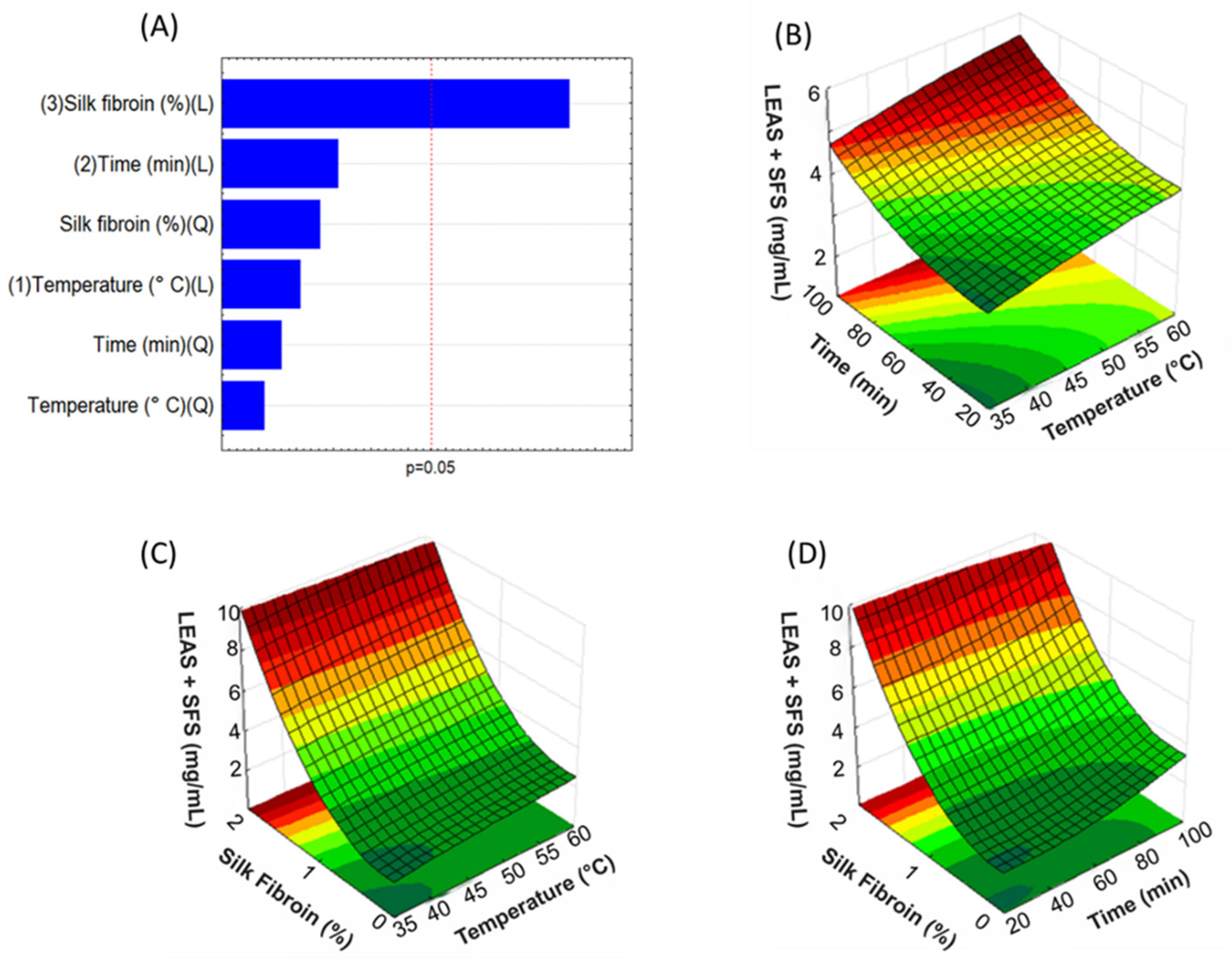
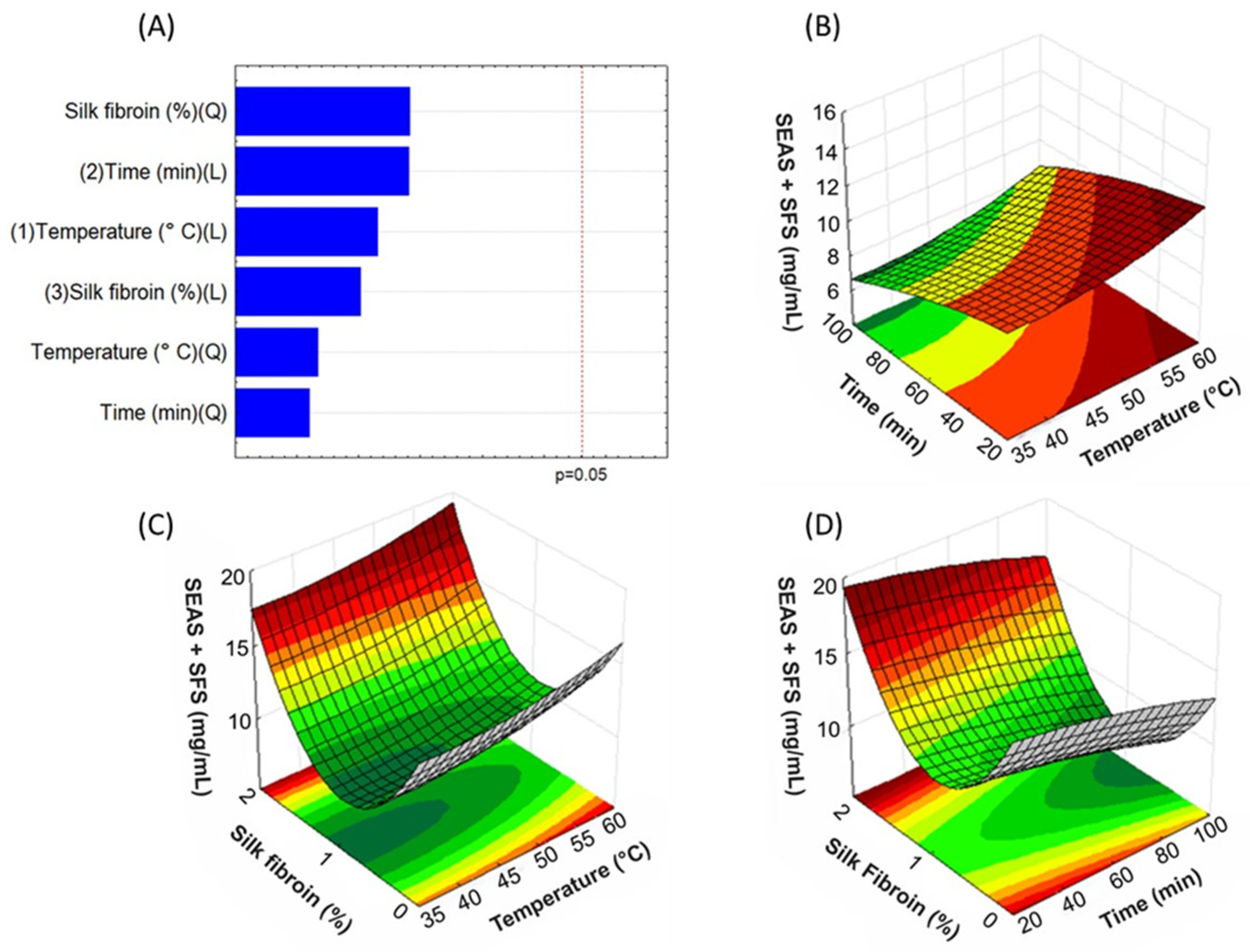
In addition to using SFS as a solvent to extract bixin, for the extraction method, we applied the Box-Behnken (BBD) experimental design, a statistical optimization of the process, with the aim of investigating the effect of the extraction variables (SFS concentration, temperature and time) on the bixin yield, and also identifying the best conditions for the process. Data analysis is performed based on the response surface methodology and Pareto chart (Figure 3 and Figure 4).
UV-Vis spectrophotometry is the most widely used method for determining dye concentration in plant extracts [45]. Therefore, the extraction efficiency of SFS on bixin content, in LEAS + SFS and SEAS + SFS, in each experimental combination (item 2.4, Table 1, total = 30 samples), was evaluated using the absorbance spectrophotometry technique in the UV-Vis region in a wavelength range of 300 to 600 nm, and the results are summarized in Table 2.
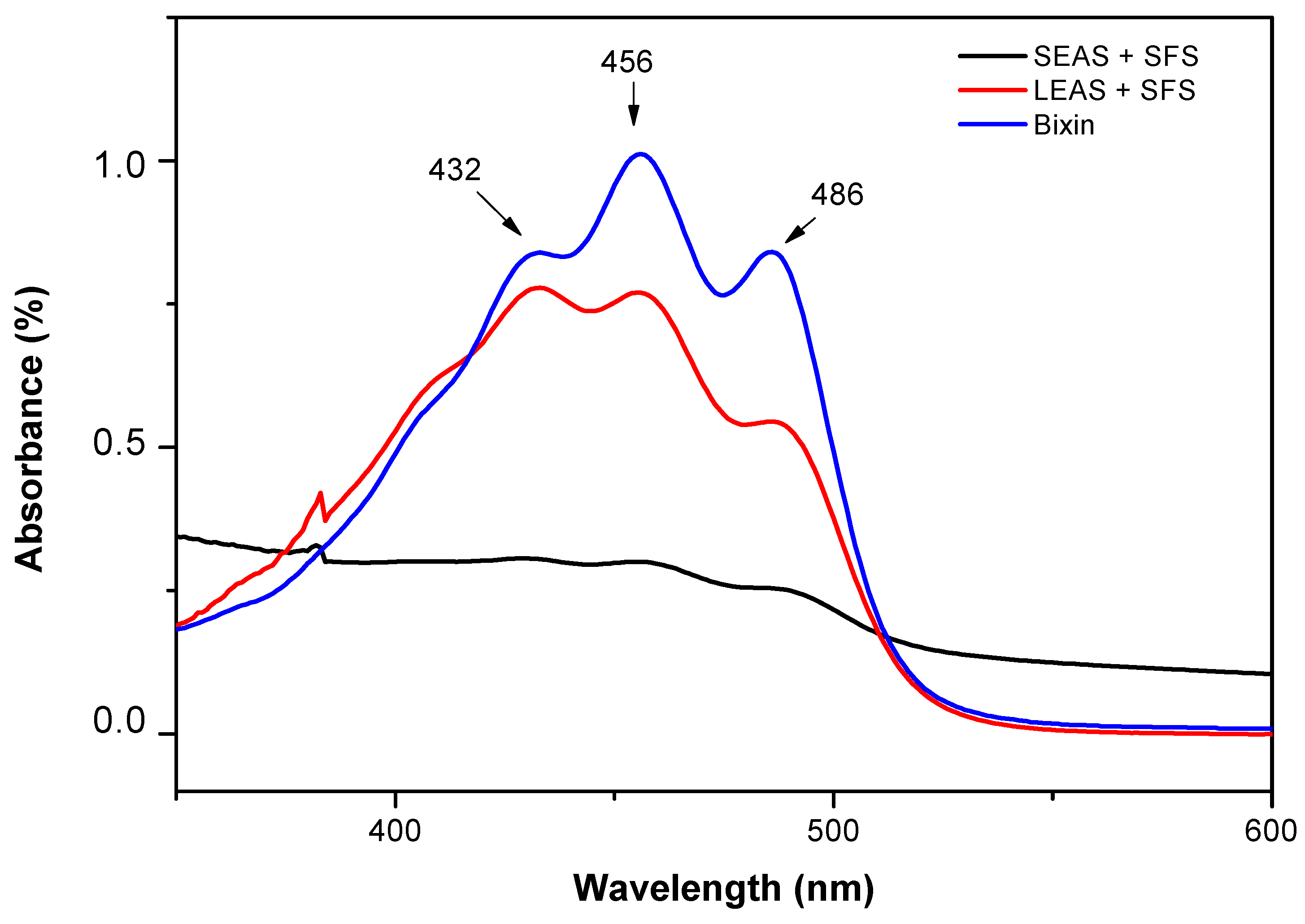
The conjugated double bond system in bixin constitutes the absorption chromophore above 400 nm (Figure 2). The absorbance spectrum for the standard bixin, solubilized in ethanol, showed peaks at 432, 456 and 486 nm. The SEAS + SFS sample showed an absorbance profile similar to that of the standard bixin (Figure 2), while the LEAS + SFS sample showed lower absorbance values at λ Max (Figure 2), which is in agreement with the results of [41], who reported that bixin has a higher affinity for polar solvents.
From the UV-Vis analysis results, the best parameter identified by the BBD design for bixin extraction was Run 2 (seed/concentration ratio 1.5% SFS at 60 °C for 60 min of magnetic stirring (Table 2), presenting a bixin content of 10.87 mg/mL in LEAS + SFS and 150.72 mg/g in SEAS + SFS (Table 2). This parameter was used to produce the LEAS + SFS and SEAS + SFS samples for infrared analysis and cell viability assay, and its UV-Vis spectrum is also shown in Figure 2.
According to the results, bixin contents in LEAS + SFS and SEAS + SFS ranged from 2.16 mg/mL (Run 8) to 10.87 mg/mL (Run 2) and 70.56 mg/g (Run 6) at 216.36 mg/g (Run 4), respectively (Table 2).
The maximum (10.87 mg/mL) and minimum (2.16 mg/mL) bixin content in LEAS + SFS were observed for the parameter combinations, with a seed/SFS concentration ratio of 1.5% at 60 °C for 60 min of agitation (Run 2) and a seed/SFS concentration ratio of 0.5% at 40 °C for 60 min of agitation (Run 8), respectively (Table 2). In SEAS + SFS, the maximum (216.36 mg/g) and minimum (70.56 mg/g) bixin content was observed for the parameter combinations, with a seeds/SFS concentration ratio of 1.0% at 60 °C for 30 min of agitation (Run 4) and a seed/SFS concentration ratio of 1.0% at 40 °C for 90 min of agitation (Run 6), respectively (Table 2).
The results of SEAS + SFS showed the highest levels of bixin, compared to LEAS + SFS, mainly in Run 4, with a maximum level of 216.36 mg/g. It is suggested that this result is due to a higher bixin content in the aril of B. orellana seeds [41]. The aril present on the outer surface of B. orellana seeds is rich in carotenoids, of which 80% is represented by the main carotenoid bixin [46].
However, although the yield of LEAS + SFS is lower than that of SEAS + SFS, the results are still comparable, since bixin in LEAS + SFS could be quantified by UV-Vis analysis. This finding indicates that SFS can extract a considerable amount of bixin. Thus, SFS may serve as a viable green alternative to the organic solvent extraction process, albeit with a slightly reduced bixin yield.
To enable factor-response analysis and understand the relationship between the variables in the data set, from the collected results, a Pareto chart and response surface were generated for all variables, to determine the significance of the influence of the factors (SFS concentration, temperature, and time) on the bixin content in LEAS + SFS (Figure 3) and SEAS + SFS (Figure 4).
Figure 3A shows that SFS concentration was the most influential factor on bixin content in the LEAS + SFS sample, with a statistically significant effect (p < 0.05).
Regarding SEAS + SFS, none of the factors (SFS concentration, temperature and time) significantly influenced the 95% confidence level for the bixin content response (Figure 4A).
According to the Pareto chart (Figure 3A), SFS was the factor that most significantly influenced the bixin content for the LEAS + SFS sample. Given this, we can conclude that increasing the SFS concentration could increase the extracted bixin levels, since the highest bixin level in LEAS + SFS was observed for the parameter combinations with a seed/SFS concentration ratio of 1.5% (Runs 2, 12, 13 and 14), that is, with SFS at its highest concentration used in this study.
To establish the significance of the adjusted model, analysis of variance (ANOVA) was used (Tables S1 and S2 of Supplementary Materials). A smaller p-value (p < 0.05) indicates that an individual factor is significant in the model [13]. In the case of LEAS + SFS, the SFS concentration had a more significant effect on the bixin content, with a p-value < 0.05 (Table S1 of Supplementary Materials). As for SEAS + SFS, none of the factors had a lower p-value (p < 0.05), and consequently did not have a significant effect on the bixin content (Table S2 of Supplementary Materials).
The concentration of bixin in B. orellana seeds varies according to environmental factors, such as crop variety, soil, climate, cultivation, growth conditions and post-harvest practices. Meanwhile, the extraction method, extraction conditions and parameters, and mainly, the polarity of the solvent used, significantly impact the extraction of bixin, and consequently its final content [6,12,13].
The impact of different organic solvents and their polarities on bixin yield, and the optimization of process parameters such as time, temperature and seed/solvent ratio were investigated in various/different studies.
Jayakumar et al., 2023 [13] showed that extraction with ethyl acetate (medium-nonpolar solvent) and methanol (polar solvent) produced the highest bixin contents, furthermore, bixin content increased with time (10, 20, and 30 min) and temperature (50, 60, and 70 °C) in the extracts in ethyl acetate, methanol and ethanol. Erdawati et al., 2021 [47] also found that ethyl acetate produced high bixin content.
David et al., 2022 [15] observed that increases in temperature and seed/solvent ratio increased bixin yield even in polar solvents, such as ethanol. According to Quiroz et al., 2019 [18], a higher seeds:solvent ratio associated with sufficient contact time promotes an increase in the affinity of the compounds with the solvents, resulting in greater bixin extraction.
Sudha et al., 2023 [12] observed that increasing the extraction time increased the bixin content, but after 30 min of extraction, it started to decrease, due to the thermal degradation effect of bixin pigment under high temperature for a longer time, since rapid increase in temperature facilitates the migration of pigments into the extraction medium [12,46].
In addition to organic solvents, researchers have studied the use of “generally recognized as safe” (GRAS) solvents, such as water, and green solvents, such as PEG and ethyl lactate for bixin extraction. These techniques are being studied with the aim of developing clean extraction technologies with environmental benefits [21].
Handayani et al., 2024 [21] extracted bixin by maceration using distilled water at various pH (4, 7 and 9) and extraction times (5, 7.5 and 10 min). The results showed that the variation in the pH of distilled water and extraction time influences the bixin levels. The highest levels of bixin (61.06%) were extracted in distilled water (pH 7, extraction time 7.5 min). These findings validate the use of distilled water as an alternative solvent to extract annatto pigment.
It was reported that the aqueous extract obtained at 50 °C for 30 min at a concentration ratio of 1:30 (1 g of seeds per 30 mL of water) under magnetic stirring (150 rpm), observed that the bixin content in the extract increased significantly in the extraction time of up to 30 min and remained approximately constant after this period [42]. The maximum bixin content was 5.80 ± 0.32%, which is equivalent to a bixin concentration of 72.21 ± 2.82 mg L−1; therefore, the aqueous extraction showed adequate efficiency, with the advantages of using a safe and environmentally friendly solvent.
In parallel with the use of water as a solvent, compounds such as polyethylene glycols (PEG) have been used as a green solvent for bixin extraction. PEGs have amphiphilic characteristics, which provide an increase in the aqueous solubility of some compounds with low polarity. Another particularity of PEGs is their acceptance in the food area, which has stimulated their use for the extraction of bioactive compounds with applicability in food, becoming an environmentally friendly alternative for bixin extraction [15,48,49].
David et al., 2022 [15] concluded that PEG-400 could produce 40% more dye (2.7 mgBix/gExt) than ethanol in times longer than 50 min, while ethanol achieved the maximum bixin concentration (1.6 mgBix/gExt) in just 34 min. PEG showed slower extraction than ethanol, but with a higher final bixin yield. This result demonstrates that amphiphilic solvents can be a viable alternative for bixin extraction.
Silk fibroin (SF), like PEG, has amphiphilic characteristics and application in food preservation, according to results shown by [50], who developed an edible coating material based on SF for preserving perishable fruits. The results suggested that SF coatings prolonged freshness and firmness, delayed respiration and prevented dehydration of perishable fruits.
Ethyl lactate is a green solvent that in terms of polarity is considered comparable to acetonitrile (polar). In addition, it forms different types of chemical interactions, such as hydrogen bonds and Van der Waals bonds, providing miscibility with polar/hydrophilic and nonpolar/hydrophobic carotenoids [41,51].
Recently, ref. [41] used ethyl lactate to extract bixin from annatto seeds (crushed and whole). The results indicated that the ethyl lactate extracts yielded 14.62% and 16.5% bixin for the whole and crushed seeds, respectively. This finding indicates that ethyl lactate can extract a considerable amount of bixin.

3.2. Chemical Characterization

3.2.1. Fourier Transform Infrared (FTIR) Analysis

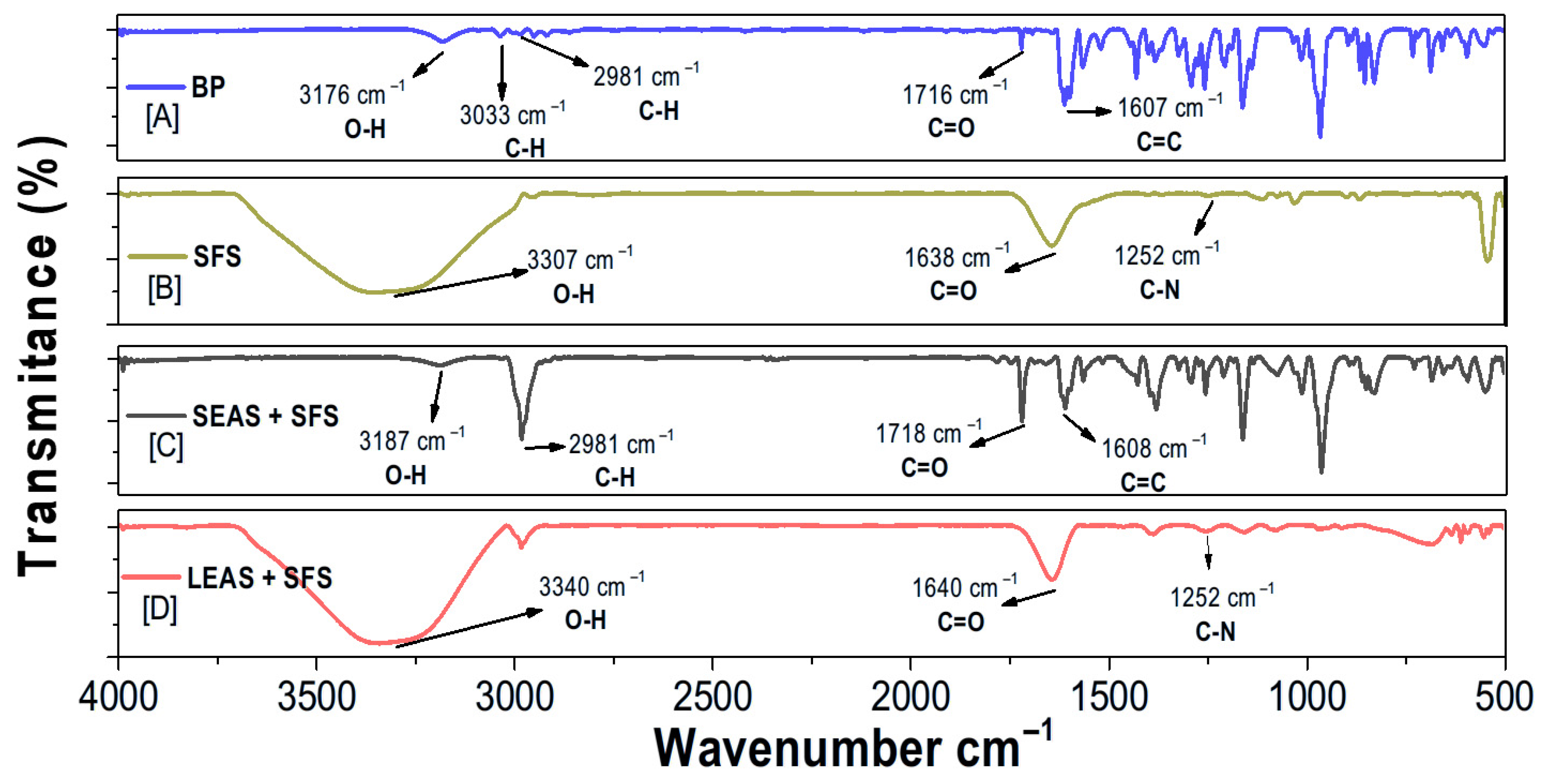
FTIR analysis identified the spectral profile of SEAS + SFS, LEAS + SFS, PB, and SFS, complementing the data obtained from the UV-Vis analysis. The FTIR spectrum for purified bixin (PB) (Figure 5A) displays characteristic vibration bands of the main functional groups. These include bands at 3176 cm−1 (O-H carboxylic acid), at 3033 cm−1 (C-H olefinic), at 2981 cm−1 (C-H methyl group), at 1716 cm−1 (C=O ester group), and 1607 cm−1 (C=C conjugated alkenes), consistent with the findings of Bitencourt et al. (2018) [52].
The FTIR spectrum of the silk fibroin solution (SFS) (Figure 5B) exhibited characteristic vibration bands corresponding to the main functional groups in fibroin. These included 3307 cm−1 (O-H group), 1638 cm−1 (C=O from amide group), and 1252 cm−1 (C-N from amide), as reported by Marinho et al. (2023) [32] (Figure 5B).
The SEAS + SFS spectrum (Figure 5C) exhibited vibration bands similar to those in the PB spectrum, such as 3187 cm−1 (O-H of the COOH group), 2981 cm−1 (CH of the CH3 group), 1718 cm−1 (C=O of the ester), and 1608 cm−1 of C=C (conjugated alkenes). These bands align with the UV-Vis analysis results, indicating that SEAS + SFS contained the highest bixin levels. On the other hand, the LEAS + SFS spectrum (Figure 5D) displayed only the characteristic vibration bands of fibroin, including 3340 cm−1 (O-H group), 1640 cm−1 (C=O group), and 1252 cm−1 (C-N of the amide group).

3.2.2. Analysis of Purified Bixin by 1H and 13C NMR

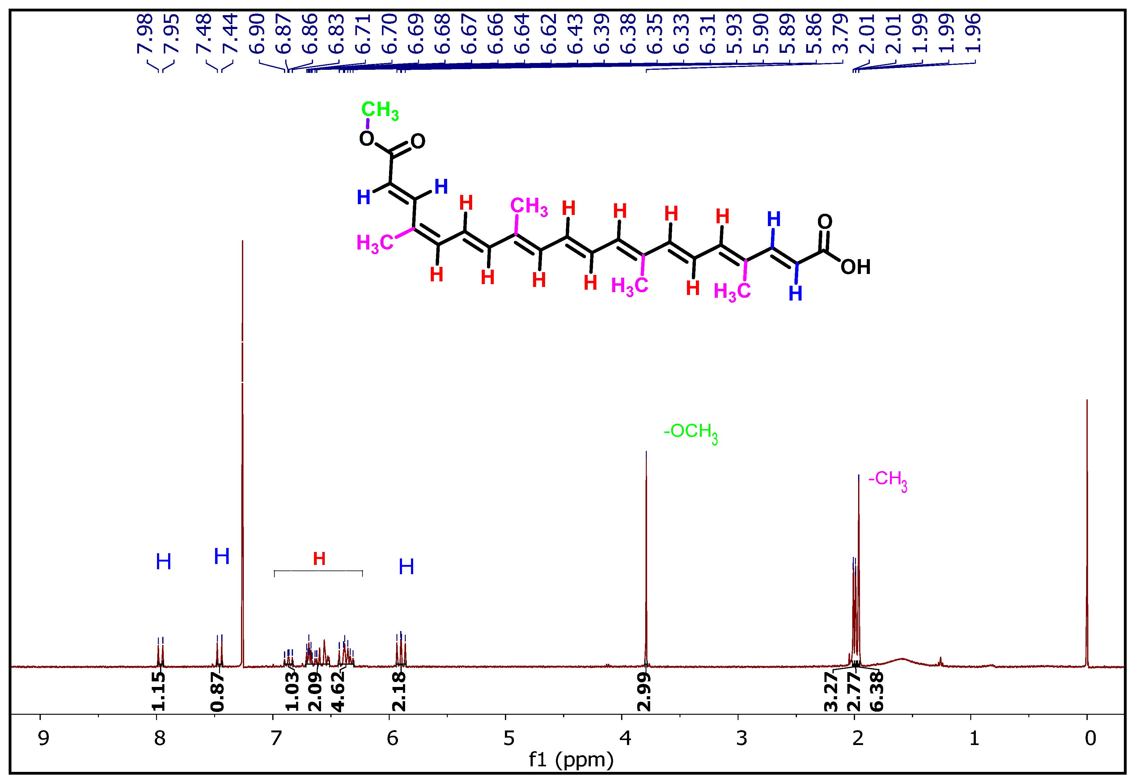
Figure 6 shows the results of 1H and 13C NMR analysis to identify signals from PB functional groups and confirm its structure.
We identified the main signals related to the bixin functional groups by 1H NMR (Figure 6). These include signals from olefinic hydrogens at 7.96 ppm (J = 15.4 Hz, 1H); 7.48 ppm (J = 15.5 Hz, 1H), and 5.90 ppm (J = 15.5, 13.6 Hz, 2H), which correspond to the double bonds adjacent to the carboxylic acid and ester groups, respectively. Signals olefinic hydrogens of the aliphatic chain, with peaks ranging from 6.71–6.30 ppm (multiplets for 10 hydrogens). A singlet at 3.79 ppm corresponds to the three hydrogens of the methoxyl group of the ester group, and the signals from the 12 hydrogens of the CH3 groups of the aliphatic structure of the molecule that appear in the 2.01–1.96 ppm range. The signal from the hydrogen of the hydroxyl group corresponding to the carboxylic acid does not appear, likely due to isotopic exchange with the deuterated solvent.
13C NMR spectra exhibited carbon signals corresponding to functional groups that support bixin’s structural composition (Table 3). The most prominent signals included the -C=O resonances at 170.75 and 167.97 ppm, associated with the carboxylic acid and ester carbonyl groups. Within the 151.0–114.8 ppm range, 17 signals of olefinic carbons appeared. A characteristic signal identified as of the carbon in the -OCH3 group of the ester appeared at 51.58 ppm. Additionally, four signals characteristic of -CH3 carbons appeared between 20.26 and 12.62 ppm (Table 3). So, the analyses of 1H and 13C NMR provided evidence of the main signals from the functional groups in bixin, confirming the structure of the compound, according to the analyses by Patra Soumen et al., 2021 [53] (Table 3).

3.3. Cell Viability Assay

The potential pharmaceutical applications of LEAS + SF and SEAS + SFS extracts are contingent upon various factors, including the biocompatibility of these materials. One key parameter for assessing biocompatibility is their impact on cell viability. Therefore, in this study, the viability of HDFn cells and RAW macrophages was evaluated following treatment with these materials using the MTT assay. This method relies on the metabolic activity of viable cells, as their mitochondrial function facilitates the conversion of tetrazolium salt into formazan crystals. The resulting intense purple coloration is directly proportional to cell viability [54,55].
Neonatal human dermal fibroblasts (HDFn cells) and macrophages (RAW 264.7 cells) were selected for the cell viability assay considering the potential biomedical applications of silk fibroin [56,57,58,59,60,61,62,63,64], annatto seed extract [41,65,66] and bixin [41,67], specifically in the area of tissue repair, with a biological approach to wound healing.
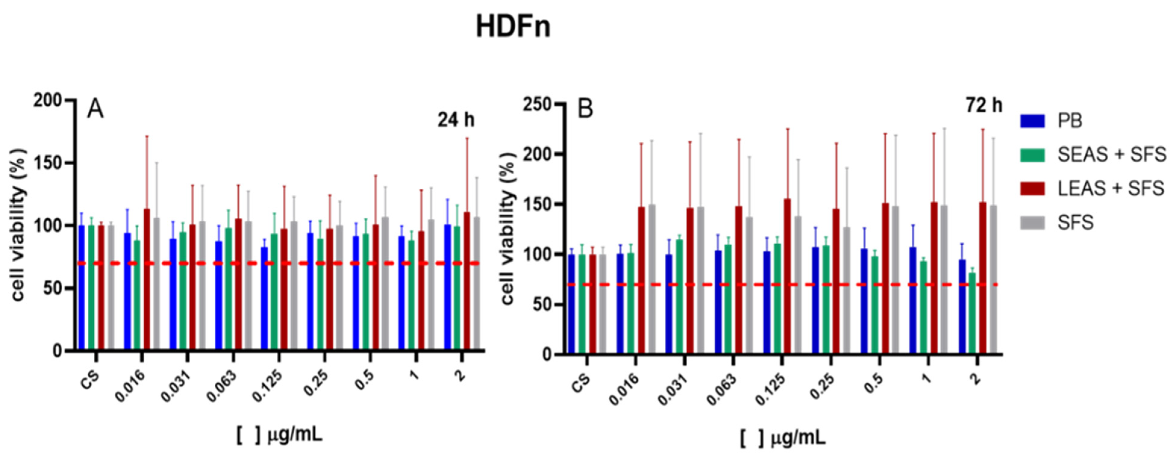
The results demonstrated that exposure of HDFn cells to LEAS + SFS, SEAS + SFS, PB, and SFS for 24 h (acute exposure) or 72 h (long-term effect following acute exposure) (ISO 10993-5, 2009 [68,69]) did not reduce cell viability at any of the tested concentrations compared to the control group (p < 0.05), with viability levels remaining within the 70–100% range (Figure 7A). According to the ISO 10993-5 [69] standard, cell viability below 70% indicates cytotoxic potential; therefore, LEAS + SFS, SEAS + SFS, PB, and SFS did not exhibit cytotoxicity at the tested concentrations in HDFn cells. Moreover, the results suggest that LEAS + SFS and SFS promoted HDFn cell proliferation, as cell viability exceeded 100% (Figure 7B).
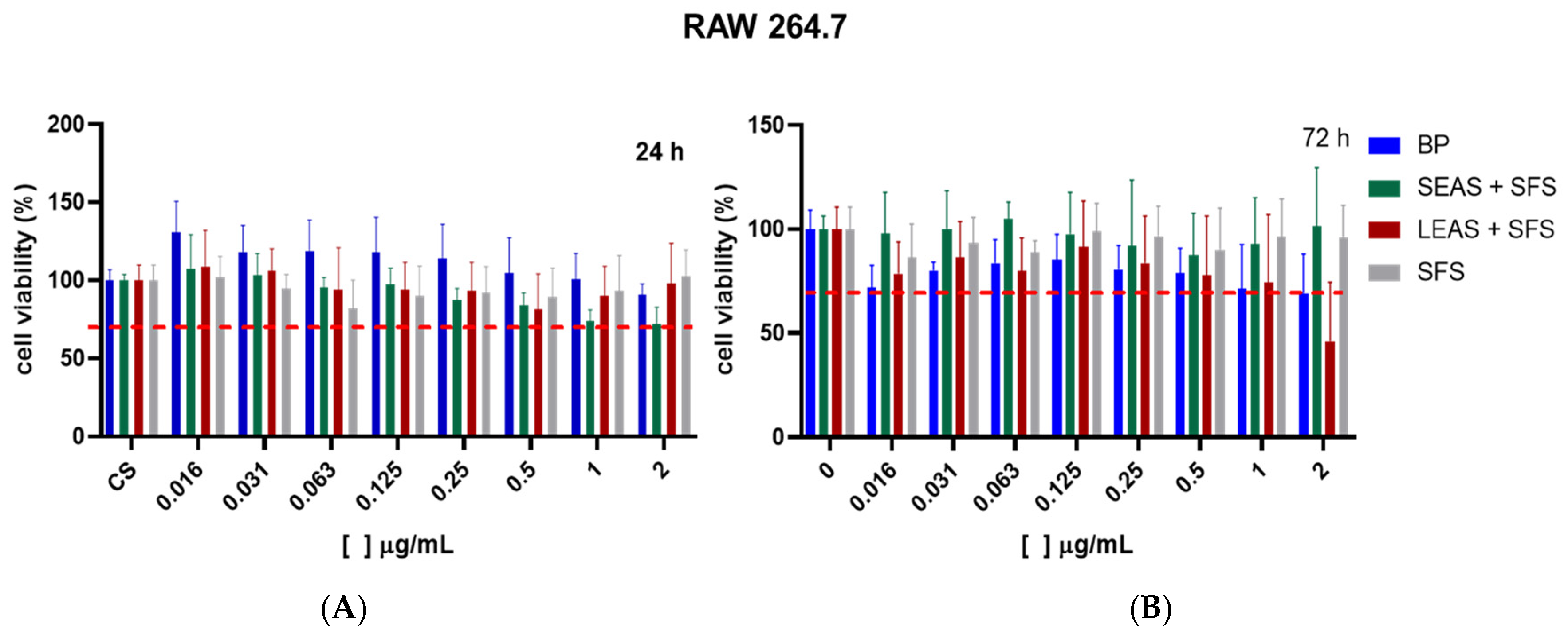
For RAW 264.7 cells, exposure to LEAS + SFS, SEAS + SFS, PB, and SFS for 24 and 72 h did not reduce cell viability below 70% at concentrations ranging from 0.016 to 1.0 µg/mL compared to the control group (p < 0.05) (Figure 8A,B). However, at a concentration of 2.0 µg/mL PB, and LEAS + SFS exhibited cytotoxic effects on macrophages, with viability decreasing to 69% and 46%, respectively, following prolonged exposure (72 h) (Figure 8B). These findings support previous evidence that bixin can reduce macrophage populations in inflammatory models [70,71].
Fibroblasts and macrophages are cells that play an important role in all stages of the healing process [72] and are widely used to evaluate the cell viability of bioactive compounds [41,59,67,73,74,75,76] and in screening the bioactivity of natural products [77,78,79,80].
Several bioactive materials and compounds have been investigated for their healing potential, with topical application being preferred in experimental trials due to its direct application to the damaged tissue [81]. Considering that dermal fibroblasts and macrophages represent cell populations abundantly distributed in the epidermal and dermal layers [82], these cell types constitute relevant biological models for evaluating the cutaneous response to topical exposure to bioactive substances. Furthermore, because they are susceptible to irritants, analyzing their viability and functionality can provide important parameters regarding the cytotoxic or immunomodulatory potential of the tested compounds [83].
Furthermore, studies addressing the use of these cell lines to evaluate the cellular viability of these materials are scarce in the literature. The results obtained (non-cytotoxic concentrations) may be used in subsequent studies to evaluate biological activity in vivo.

4. Conclusions

This study demonstrated the technical feasibility and sustainability of using silk fibroin solution (SFS) as a green solvent for the extraction of bixin from annatto seeds (Bixa orellana L.). Optimization using the Box-Behnken design identified the optimal extraction conditions, as 1.5% SFS, 60 °C, and 60 min, resulting in bixin concentrations of 10.87 mg/mL (LEAS + SFS) and 150.72 mg/g (SEAS + SFS). These values are comparable to those obtained with conventional organic solvents, without the environmental and toxicological risks associated with these methods.
Chemical characterization confirmed the presence of bixin in the extracts, and cell viability assays showed that SFS and bixin-rich extracts maintained ≥70% viability in HDFn fibroblasts and RAW 264.7 macrophages, indicating low cytotoxicity and good biocompatibility. These findings suggest the potential of extracts obtained with SFS for applications in the food, cosmetics, and biomedical sectors, contributing to clean-label and environmentally responsible production chains.
Limitations of this study include the focus on in vitro analyses with two cell lines and the lack of functional or long-term evaluations. Future work should include in vivo validation of therapeutic properties, assessment of physicochemical stability in formulations, and comparisons with other green solvents to more precisely define the role of SFS in sustainable extraction technologies.
Overall, the use of SFS as a biocompatible and biodegradable solvent is in line with the principles of green chemistry. This approach contributes to the advancement of sustainable extraction processes to obtain high-value-added natural compounds, reinforcing the viability of green solvents.

Supplementary Materials

The following supporting information can be downloaded at: https://www.mdpi.com/article/10.3390/su17167518/s1, Table S1: Analysis of variance (ANOVA) on the effect of different combinations of seed/SFS concentration ratio, temperature and extraction time on bixin content in LEAS + SFS; Table S2: Analysis of variance (ANOVA) on the effect of different combinations of seed/SFS concentration ratio, temperature and extraction time on the bixin content in SEAS + SFS.

Author Contributions

Conceptualization, S.F.B. and I.M.F.; methodology, S.F.B., F.H.e.H., K.C.D.M., S.d.S.d.C.O., D.E.Q.J., C.M.L., V.Z. and I.M.F.; software, F.H.e.H.; formal analysis, S.F.B., F.H.e.H., K.C.D.M., S.d.S.d.C.O., D.E.Q.J., C.M.L., V.Z. and I.M.F.; resources, I.M.F.; data curation, S.F.B., F.H.e.H., D.E.Q.J., C.M.L., V.Z. and I.M.F.; writing—original draft preparation, S.F.B., C.M.L., V.Z. and I.M.F.; writing—review and editing, S.F.B., C.M.L., V.Z. and I.M.F.; supervision, I.M.F.; project administration, I.M.F.; funding acquisition, I.M.F. All authors have read and agreed to the published version of the manuscript.

Funding

This research was funded by the National Council for Scientific and Technological Development (Process No. 444009/2024-8 and 445078/2024-3) and Borges, S.F. received a scholarship funded by the Coordination for the Improvement of Higher Education Personnel—Brazil (CAPES) No. 52001016062P2.

Institutional Review Board Statement

Not applicable.

Informed Consent Statement

Not applicable.

Data Availability Statement

The original contributions presented in this study are included in the article. Further inquiries can be directed to the corresponding author.

Acknowledgments

The authors would like to thank Valtencir Zucolotto, from the Nanomedicine and Nano-toxicology Group of the Physics Institute of São Carlos, University of São Paulo (São Carlos—SP), for kindly providing the HDFn and RAW 264.7 cell lines, as well as for providing the infrastructure necessary to perform the in vitro cytotoxicity assays.

Conflicts of Interest

The authors declare no conflicts of interest. The funders had no role in the design of the study; in the collection, analyses, or interpretation of data; in the writing of the manuscript; or in the decision to publish the results.

Abbreviations

The following abbreviations are used in this manuscript:
PBPurified bixin
SFSSilk fibroin solution
BBDBox-Behnken experimental design
FTIRFourier transform infrared
NMRNuclear magnetic resonance
MTT3-(4,5-dimethylthiazol-2-yl)-2,5-diphenyltetrazolium bromide
LEAS + SFSLiquid extract of annatto seeds from silk fibroin solution
SEAS + SFSSolid extract of annatto seeds from silk fibroin solution
LOXLipoxygenase
HDFnHuman neonatal dermal fibroblasts
RAW 264.7Murine macrophages
DMENDulbecco’s Modified Eagle’s Medium
DMSODimethyl sulfoxide or dimethyl sulfoxide
Na2CO3Sodium carbonate
H2O: EtOH: CaCl2Water: Ethanol: Calcium chloride
KDaKilo Dalton
CO2Carbon dioxide
GlyGlycine
AlaAlanine
SerSerine
MHzMegahertz
CDCl3Deuterated chloroform
CaCl2Calcium chloride

References

  1. De Oliveira Júnior, R.G.; Bonnet, A.; Braconnier, E.; Groult, H.; Prunier, G.; Beaugeard, L.; Grougnet, R.; da Silva Almeida, J.R.G.; Ferraz, C.A.A.; Picot, L. Bixin, an Apocarotenoid Isolated from Bixa orellana L., Sensitizes Human Melanoma Cells to Dacarbazine-Induced Apoptosis through ROS-Mediated Cytotoxicity. Food Chem. Toxicol. 2019, 125, 549–561. [Google Scholar] [CrossRef]
  2. Cruz Lima, R.J.; De Deus Moreno, A.J.; Loureiro De Castro, S.F.; Santos Gonçalves, J.D.R.; De Olivera, A.B.; Sasaki, J.M.; Cavalcante Freire, P.D.T. Taninos Hidrolisáveis Em Bixa orellana L. Quim. Nova 2006, 29, 507–509. [Google Scholar] [CrossRef]
  3. Medina-Flores, D.; Ulloa-Urizar, G.; Camere-Colarossi, R.; Caballero-García, S.; Mayta-Tovalino, F.; del Valle-Mendoza, J. Antibacterial Activity of Bixa orellana L. (Achiote) against Streptococcus mutans and Streptococcus sanguinis. Asian Pac. J. Trop. Biomed. 2016, 6, 400–403. [Google Scholar] [CrossRef]
  4. Keita, H.; dos Santos, C.B.R.; Ramos, M.M.; Padilha, E.C.; Serafim, R.B.; Castro, A.N.; Amado, J.R.R.; da Silva, G.M.; Ferreira, I.M.; Giuliatti, S.; et al. Assessment of the Hypoglycemic Effect of Bixin in Alloxan-Induced Diabetic Rats: In vivo and in silico Studies. J. Biomol. Struct. Dyn. 2021, 39, 1017–1028. [Google Scholar] [CrossRef] [PubMed]
  5. Nathan, V.K.; Rani, M.E.; Rathinasamy, G.; Narayanan Dhiraviam, K. Antioxidant and Antimicrobial Potential of Natural Colouring Pigment Derived from Bixa orellana L. Seed Aril. Proc. Natl. Acad. Sci. India Sect. B Biol. Sci. 2019, 89, 137–143. [Google Scholar] [CrossRef]
  6. Oliveira, S.D.S.D.C.; Araújo, R.D.C.; da Silva, G.A.; Leitão, J.H.; da Silva Sousa, S.A.B.; Fonseca, L.P.; Carvalho, J.C.T.; Cantuária, P.; Hage-Melim, L.I.D.S.; Ferreira, I.M. Bixa orellana L. from Northern Brazil: Morphological Analysis, Phenolic Content, Antioxidant and Antibacterial Activities. Braz. J. Bot. 2022, 45, 883–896. [Google Scholar] [CrossRef]
  7. Gatsou Djibersou, D.; Rosnay Tietcheu Galani, B.; Dieudonne Djamen Chuisseu, P.; Yanou Njintang, N. Anti-Oxidant and Anti-Inflammatory Potential of Aqueous Extracts of Leaves, Barks and Roots of Bixa orellana L. (Bixaceae) on Acetaminophen-Induced Liver Damage in Mice. Avicenna J. Phytomed. 2020, 10, 428–439. [Google Scholar]
  8. Pacheco, S.D.G.; Gasparin, A.T.; Jesus, C.H.A.; Sotomaior, B.B.; Ventura, A.C.S.S.B.; Redivo, D.D.B.; Cabrini, D.D.A.; Gaspari Dias, J.D.F.; Miguel, M.D.; Miguel, O.G.; et al. Antinociceptive and Anti-Inflammatory Effects of Bixin, a Carotenoid Extracted from the Seeds of Bixa orellana. Planta Med. 2019, 85, 1216–1224. [Google Scholar] [CrossRef]
  9. Rivera-Madrid, R.; Aguilar-Espinosa, M.; Cárdenas-Conejo, Y.; Garza-Caligaris, L.E. Carotenoid Derivates in Achiote (Bixa orellana) Seeds: Synthesis and Health Promoting Properties. Front. Plant Sci. 2016, 7, 1406. [Google Scholar] [CrossRef]
  10. Kapoor, L.; Ramamoorthy, S. Strategies to Meet the Global Demand for Natural Food Colorant Bixin: A Multidisciplinary Approach. J. Biotechnol. 2021, 338, 40–51. [Google Scholar] [CrossRef]
  11. Tao, S.; Park, S.L.; De La Vega, M.R.; Zhang, D.D.; Wondrak, G.T. Systemic Administration of the Apocarotenoid Bixin Protects Skin against Solar UV-Induced Damage through Activation of NRF2. Free Radic. Biol. Med. 2015, 89, 690–700. [Google Scholar] [CrossRef] [PubMed]
  12. Sudha, P.; Manoja, V.; Deepa, J.; Jayakumar, J.; Kishore, S.G.; Pandiselvam, R. Optimization of Microwave-Assisted Aqueous Extraction of Pigments from Annatto Seeds Using Box-Behnken Design. Biomass Convers. Biorefin 2023, 14, 18775–18788. [Google Scholar] [CrossRef]
  13. Jayakumar, J.; Sudha, P.; Rajkumar, P.; Pandiselvam, R.; Gurusamy, K.; Kumaran, K.; Subramanian, P. Comparative Study on the Effect of Solvents on Extraction of Bixin from Annatto Seed (Bixa orellana L.) and Optimization of Process Parameters Using Box–Behnken Design. Biomass Convers. Biorefin. 2023, 14, 19863–19874. [Google Scholar] [CrossRef]
  14. Alcázar-Alay, S.C.; Osorio-Tobón, J.F.; Forster-Carneiro, T.; Meireles, M.A.A. Obtaining Bixin from Semi-Defatted Annatto Seeds by a Mechanical Method and Solvent Extraction: Process Integration and Economic Evaluation. Food Res. Int. 2017, 99, 393–402. [Google Scholar] [CrossRef]
  15. David, S.L.J.; Julián, G.C.C.; Liliana, V.S.C.; Ayala-Aponte, A.A.; Liliana, S.C. Kinetics and Bixine Extraction Time from Achiote (Bixa orellana L.). J. Food Sci. Technol. 2022, 59, 1239–1246. [Google Scholar] [CrossRef]
  16. Hirko, B.; Getu, A. Bixa orellana (Annatto Bixa): A Review on Use, Structure, Extraction Methods and Analysis. J. Agron. Technol. Eng. Manag. 2022, 5, 687–696. [Google Scholar]
  17. Husa, N.N.; Hamzah, F.; Said, H.M. Characterization and Storage Stability Study of Bixin Extracted from Bixa orellana Using Organic Solvent. IOP Conf. Ser. Mater. Sci. Eng. 2018, 358, 012035. [Google Scholar] [CrossRef]
  18. Quiroz, J.Q.; Torres, A.C.; Ramirez, L.M.; Garcia, M.S.; Gomez, G.C.; Rojas, J. Optimization of the Microwave-Assisted Extraction Process of Bioactive Compounds from Annatto Seeds (Bixa orellana L.). Antioxidants 2019, 8, 37. [Google Scholar] [CrossRef]
  19. Taham, T.; Silva, D.O.; Barrozo, M.A.S. Improvement of Bixin Extraction from Annatto Seeds Using a Screen-Topped Spouted Bed. Sep. Purif. Technol. 2016, 158, 313–321. [Google Scholar] [CrossRef]
  20. Yolmeh, M.; Habibi Najafi, M.B.; Farhoosh, R. Optimisation of Ultrasound-Assisted Extraction of Natural Pigment from Annatto Seeds by Response Surface Methodology (RSM). Food Chem. 2014, 155, 319–324. [Google Scholar] [CrossRef]
  21. Handayani, I.; Septiana, A.T.; Sustriawan, B. Propriedades de Pigmentos Naturais e Antioxidantes Do Extrato de Urucum Em Vários PHs de Solventes de Água Destilada e Tempos de Extração. Food Res. 2024, 8, 489–494. [Google Scholar] [CrossRef]
  22. Bachtler, S.; Bart, H.J. Increase the Yield of Bioactive Compounds from Elder Bark and Annatto Seeds Using Ultrasound and Microwave Assisted Extraction Technologies. Food Bioprod. Process. 2021, 125, 1–13. [Google Scholar] [CrossRef]
  23. Zheng, H.; Zuo, B. Functional Silk Fibroin Hydrogels: Preparation, Properties and Applications. J. Mater. Chem. B 2021, 9, 1238–1258. [Google Scholar] [CrossRef]
  24. Kundu, B.; Rajkhowa, R.; Kundu, S.C.; Wang, X. Silk Fibroin Biomaterials for Tissue Regenerations. Adv. Drug Deliv. Rev. 2013, 65, 457–470. [Google Scholar] [CrossRef] [PubMed]
  25. Partlow, B.P.; Tabatabai, A.P.; Leisk, G.G.; Cebe, P.; Blair, D.L.; Kaplan, D.L. Silk Fibroin Degradation Related to Rheological and Mechanical Properties. Macromol. Biosci. 2016, 16, 666–675. [Google Scholar] [CrossRef] [PubMed]
  26. Floren, M.; Migliaresi, C.; Motta, A. Processing Techniques and Applications of Silk Hydrogels in Bioengineering. J. Funct. Biomater. 2016, 7, 26. [Google Scholar] [CrossRef]
  27. Ferreira, I.M.; Yoshioka, S.A.; Comasseto, J.V.; Porto, A.L.M. Immobilization of Amano Lipase from Pseudomonas Fluorescens on Silk Fibroin Spheres: An Alternative Protocol for the Enantioselective Synthesis of Halohydrins. RSC Adv. 2017, 7, 12650–12658. [Google Scholar] [CrossRef]
  28. Kasoju, N.; Bora, U. Silk Fibroin in Tissue Engineering. Adv. Healthc. Mater. 2012, 1, 393–412. [Google Scholar] [CrossRef]
  29. Debari, M.K.; King, C.I.; Altgold, T.A.; Abbott, R.D. Silk Fibroin as a Green Material. ACS Biomater. Sci. Eng. 2021, 7, 3530–3544. [Google Scholar] [CrossRef]
  30. Marinho, V.H.S.; Neves, F.B.; Jimenez, D.E.Q.; Oliveira, F.R.; Santos, A.V.T.L.T.; Ferreira, R.M.A.; Souto, R.N.P.; Carvalho, J.C.T.; Yoshioka, S.A.; Ferreira, I.M. Development of an Environmentally Friendly Formulation of Silk Fibroin Combined with Fatty Acid from Astrocaryum murumuru Mart. Effective against Aedes aegypti Larvae. J. Drug Deliv. Sci. Technol. 2022, 75, 103626. [Google Scholar] [CrossRef]
  31. Sarquis, I.R.; Sarquis, R.S.F.R.; Marinho, V.H.S.; Neves, F.B.; Araújo, I.F.; Damasceno, L.F.; Ferreira, R.M.A.; Souto, R.N.P.; Carvalho, J.C.T.; Ferreira, I.M. Carapa guianensis Aubl. (Meliaceae) Oil Associated with Silk Fibroin, as Alternative to Traditional Surfactants, and Active against Larvae of the Vector Aedes Aegypti. Ind. Crops Prod. 2020, 157, 112931. [Google Scholar] [CrossRef]
  32. Marinho, V.H.S.; Holanda, F.H.; Araújo, I.F.; Jimenez, D.E.Q.; Pereira, R.R.; Porto, A.L.M.; Ferreira, A.M.; Carvalho, J.C.T.; Albuquerque de Freitas, A.C.G.; Fernandes, C.P.; et al. Nanoparticles from Silk Fibroin and Amazon Oils: Potential Larvicidal Activity and Oviposition Deterrence against Aedes aegypti. Ind. Crops Prod. 2023, 203, 117133. [Google Scholar] [CrossRef]
  33. Holanda, F.H.; Pereira, R.R.; Marinho, V.H.S.; Jimenez, D.E.Q.; Costa Ferreira, L.M.M.; Ribeiro-Costa, R.M.; de Sousa, F.F.O.; Ferreira, I.M. Development of Nanostructured Formulation from Naringenin and Silk Fibroin and Application for Inhibition of Lipoxygenase (LOX). RSC Adv. 2023, 13, 23063–23075. [Google Scholar] [CrossRef]
  34. de Castro Cantuária, P.; Medeiros, T.D.S.; Cantuária, M.F.; Soares, A.C.S.; e Silva, B.M.D.S.; Krahl, A.H.; Costa-Campos, C.E.; e Silva, R.B.L. Você Conhece a Nomenclatura Biológica? Aprenda a Forma Correta de Escrever Os Nomes Dos Organismos. Res. Soc. Dev. 2022, 11, e21711326378. [Google Scholar] [CrossRef]
  35. Maciel Ferreira, I.; Coutinho Rocha, L.; Akinobo Yoshioka, S.; Nitschke, M.; Haroldo Jeller, A.; Pizzuti, L.; Seleghim, M.H.R.; Porto, A.L.M. Chemoselective Reduction of Chalcones by Whole Hyphae of Marine Fungus Penicillium citrinum CBMAI 1186, Free and Immobilized on Biopolymers. Biocatal. Agric. Biotechnol. 2014, 3, 358–364. [Google Scholar] [CrossRef]
  36. de Oliveira Penido, C.A.F.; Pacheco, M.T.T.; Novotny, E.H.; Lednev, I.K.; Silveira, L. Quantification of Cocaine in Ternary Mixtures Using Partial Least Squares Regression Applied to Raman and Fourier Transform Infrared Spectroscopy. J. Raman Spectrosc. 2017, 48, 1732–1743. [Google Scholar] [CrossRef]
  37. Leite, C.M.; Araujo-Neto, J.H.; Guedes, A.P.M.; Costa, A.R.; Demidoff, F.C.; Netto, C.D.; Castellano, E.E.; Nascimento, O.R.; Batista, A.A. Copper(I)/Triphenylphosphine Complexes Containing Naphthoquinone Ligands as Potential Anticancer Agents. Inorganics 2023, 11, 367. [Google Scholar] [CrossRef]
  38. Wang, H.; Cheng, H.; Wang, F.; Wei, D.; Wang, X. An Improved 3-(4,5-Dimethylthiazol-2-Yl)-2,5-Diphenyl Tetrazolium Bromide (MTT) Reduction Assay for Evaluating the Viability of Escherichia Coli Cells. J. Microbiol. Methods 2010, 82, 330–333. [Google Scholar] [CrossRef] [PubMed]
  39. de Toledo, C.D.; Teixeira, D.A.; da Vitória Archanjo, E.; Pereira, F.F.; Siqueira, F.C.; Mininel, F.J.; de Lima, J.S.; da Mota Silveira, K. Revisitação Dos Métodos de Extração de Pigmentos Do Urucum (Review of Urucum Pigment Extraction Methods). Ciências Exatas e Tecnologia 2021, 16, 24–29. [Google Scholar] [CrossRef]
  40. Taham, T.; Cabral, F.A.; Barrozo, M.A.S. The Journal of Supercritical Fluids Extraction of Bixin from Annatto Seeds Using Combined Technologies. J. Supercrit. Fluids 2015, 100, 175–183. [Google Scholar] [CrossRef]
  41. Puri, S.; Mandal, S.K.; Jain, H.; Sharma, P.K.; Deepa, P.R. Comparative bioactivity assessment of bixin pigment and associated phytochemicals extracted from annatto seeds using conventional and green solvents. J. Food Drug Anal. 2024, 32, 168–183. [Google Scholar] [CrossRef]
  42. Miguel, K.B.; Cardoso, V.L.; Reis, M.H.M. Concentration of Pigments and Health-Promoting Bioactive Compounds from Annatto Seeds by Ultrafiltration. Waste Biomass Valorization 2024, 15, 127–137. [Google Scholar] [CrossRef]
  43. Witono, J.R.; Ramadhany, P.; Santoso, H.; Putri, A. The Potency of Norbixin as an Active Compound of Natural Dye in Textile Industry. Mater. Today Proc. 2022, 63, S248–S254. [Google Scholar] [CrossRef]
  44. Rodrigues, L.M.; Alcázar-Alay, S.C.; Petenate, A.J.; Meireles, M.A.A. Bixin Extraction from Defatted Annatto Seeds. Comptes Rendus Chim. 2014, 17, 268–283. [Google Scholar] [CrossRef]
  45. Peters, A.T.; Freeman, H.S. Physico-Chemical Principles of Color Chemistry, 1st ed.; Springer: Dordrecht, The Netherlands, 1996; 299p, ISBN 978-0-7514-0210-0. [Google Scholar]
  46. Satyanarayana, A.; Prabhakara Rao, P.G.; Rao, D.G. Chemistry, Processing and Toxicology of Annatto (Bixa orellana L.). J. Food Sci. Technol. 2003, 40, 131–141. [Google Scholar]
  47. Erdawati, E.; Allanas, E.; Wesnina, W. Extraction of Bixin from Annatto Seeds with Microwave. J. Phys. Conf. Ser. 2021, 1869, 012016. [Google Scholar] [CrossRef]
  48. Jin, X.; Streett, D.A.; Dunlap, C.A.; Lyn, M.E. Application of Hydrophilic–Lipophilic Balance (HLB) Number to Optimize a Compatible Non-Ionic Surfactant for Dried Aerial Conidia of Beauveria bassiana. Biol. Control 2008, 46, 226–233. [Google Scholar] [CrossRef]
  49. Weuts, I.; Kempen, D.; Verreck, G.; Decorte, A.; Heymans, K.; Peeters, J.; Brewster, M.; Mooter, G. Van den Study of the Physicochemical Properties and Stability of Solid Dispersions of Loperamide and PEG6000 Prepared by Spray Drying. Eur. J. Pharm. Biopharm. 2005, 59, 119–126. [Google Scholar] [CrossRef]
  50. Marelli, B.; Brenckle, M.A.; Kaplan, D.L.; Omenetto, F.G. Silk Fibroin as Edible Coating for Perishable Food Preservation. Sci. Rep. 2016, 6, 25263. [Google Scholar] [CrossRef]
  51. Kua, Y.L.; Gan, S.; Morris, A.; Ng, H.K. Ethyl Lactate as a Potential Green Solvent to Extract Hydrophilic (Polar) and Lipophilic (Non-Polar) Phytonutrients Simultaneously from Fruit and Vegetable by-Products. Sustain. Chem. Pharm. 2016, 4, 21–31. [Google Scholar] [CrossRef]
  52. Bitencourt, A.P.R.; Duarte, J.L.; Oliveira, A.E.M.F.M.; Cruz, R.A.S.; Carvalho, J.C.T.; Gomes, A.T.A.; Ferreira, I.M.; Ribeiro-Costa, R.M.; Silva-Júnior, J.O.C.; Fernandes, C.P. Preparation of Aqueous Nanodispersions with Annatto (Bixa orellana L.) Extract Using an Organic Solvent-Free and Low Energy Method. Food Chem. 2018, 257, 196–205. [Google Scholar] [CrossRef]
  53. Patra, S.; Kar, S.; Bag, B.G. Isolation and Biological Activities of Naturally Occurring Apocarotenoid Bixin. Prayogik Rasayan 2021, 5, 112–119. [Google Scholar] [CrossRef]
  54. Mitra, I.; Mukherjee, S.; Reddy Venkata, P.B.; Dasgupta, S.; Jagadeesh Bose, C.K.; Mukherjee, S.; Linert, W.; Moi, S.C. Benzimidazole Based Pt(II) Complexes with Better Normal Cell Viability than Cisplatin: Synthesis, Substitution Behavior, Cytotoxicity, DNA Binding and DFT Study. RSC Adv. 2016, 6, 76600–76613. [Google Scholar] [CrossRef]
  55. Stockert, J.C.; Horobin, R.W.; Colombo, L.L.; Blázquez-Castro, A. Tetrazolium Salts and Formazan Products in Cell Biology: Viability Assessment, Fluorescence Imaging, and Labeling Perspectives. Acta Histochem. 2018, 120, 159–167. [Google Scholar] [CrossRef]
  56. Mazurek, Ł.; Szudzik, M.; Rybka, M.; Konop, M. Silk Fibroin Biomaterials and Their Beneficial Role in Skin Wound Healing. Biomolecules 2022, 12, 1852. [Google Scholar] [CrossRef]
  57. Chouhan, D.; Mandal, B.B. Silk Biomaterials in Wound Healing and Skin Regeneration Therapeutics: From Bench to bedside. Acta Biomater. 2020, 103, 24–51. [Google Scholar] [CrossRef]
  58. Guo, P.; Du, P.; Zhao, P.; Chen, X.; Liu, C.; Du, Y.; Li, J.; Tang, X.; Yang, F.; Lv, G. Regulating the Mechanics of Silk Fi Broin Scaffolds Promotes Wound Vascularization. Biochem. Biophys. Res. Commun. 2021, 574, 78–84. [Google Scholar] [CrossRef]
  59. Hodgkinson, T.; Yuan, X.; Bayat, A. Electrospun Silk Fibroin Fiber Diameter Influences In Vitro Dermal Fibroblast Behavior and Promotes Healing of Ex Vivo Wound Models. J. Tissue Eng. 2014, 5, 2041731414551661. [Google Scholar] [CrossRef] [PubMed]
  60. Millán-Rivero, J.E.; Martínez, C.M.; Romecín, P.A.; Aznar-cervantes, S.D.; Carpes-Ruiz, M.; Cenis, J.L.; Moraleda, J.M.; Atucha, N.M.; García-bernal, D. Silk Fibroin Scaffolds Seeded with Wharton’ s Jelly Mesenchymal Stem Cells Enhance Re-Epithelialization and Reduce Formation of Scar Tissue after Cutaneous Wound Healing. Stem Cell Res. Ther. 2019, 10, 126. [Google Scholar] [CrossRef]
  61. Woo, H.; Joo, O.; Min, J.; Mi, B.; Jung, H.; Ri, Y.; Chae, M.; Hyeon, S.; Ren, J.; Seok, C.; et al. Wound Healing Effect of Electrospun Silk Fibroin Nanomatrix In burn-model. Int. J. Biol. Macromol. 2016, 85, 29–39. [Google Scholar] [CrossRef]
  62. Zhang, W.; Chen, L.; Chen, J.; Wang, L.; Gui, X.; Ran, J.; Xu, G.; Zhao, H.; Zeng, M.; Ji, J.; et al. Silk Fibroin Biomaterial Shows Safe and Effective Wound Healing in Animal Models and a Randomized Controlled Clinical Trial. Adv. Healthc. Mater. 2017, 6, 1700121. [Google Scholar] [CrossRef]
  63. Zhang, J.; Liu, N.; Sun, C.; Wang, Y. Polysaccharides from Polygonatum Sibiricum Delar. Ex Redoute Induce an Immune Response in the RAW264.7 Cell Line via an NF-ΚB/MAPK Pathway. RSC Adv. 2019, 9, 17988–17994. [Google Scholar] [CrossRef] [PubMed]
  64. Sultan, M.T.; Lee, O.J.; Kim, S.H.; Ju, H.W.; Park, C.H. Silk Fibroin in Wound Healing Process. In Novel Biomaterials for Regenerative Medicine; Chun, H.J., Park, K., Kim, C.-H., Khang, G., Eds.; Springer: Singapore, 2018; pp. 115–126. ISBN 978-981-13-0947-2. [Google Scholar]
  65. Capella, S.O.; Tillmann, M.T.; Félix, A.O.C.; Fontoura, E.G.; Fernandes, C.G.; Freitag, R.A.; Santos, M.A.Z.; Félix, S.R.; Nobre, M.O. Potencial Cicatricial Da Bixa orellana L. Em Feridas Cutâneas: Estudo Em Modelo Experimental. Arq. Bras. Med. Vet. Zootec. 2016, 68, 104–112. [Google Scholar] [CrossRef]
  66. Franklin, V.A.; Bach Hi, E.M.; Wadt, N.S.Y.; Bach, E.E. Aqueous Extract from Urucum (Bixa orellana L.): Antimicrobial, Antioxidant, and Healing Activity. Porto Biomed. J. 2023, 8, e183. [Google Scholar] [CrossRef] [PubMed]
  67. Pizoón-Garcia, A.D.; Cassini-vieira, P.; Ribeiro, C.C.; Eduardo, C.; Jensen, D.M.; Barcelos, L.S.; Cortes, M.E.; Sinisterra, R.D. Efficient Cutaneous Wound Healing Using Bixin-Loaded PCL Nanofibers in Diabetic Mice. J. Biomed. Mat. Res. 2016, 105, 1938–1949. [Google Scholar] [CrossRef]
  68. Nikolaev, A.; Sokolov, N.N.; Kozlov, E.A.; Kutsman, M.E. Isolation and Properties of a Homogenous L Asparaginase Preparation from Pseudomonas Fluorescens AG. Biokhimiya 2009, 40, 984–989. (In Russian) [Google Scholar]
  69. ISO 10993-5:2009; Biological Evaluation of Medical Devices–Part 5: Tests for In Vitro Cytotoxicity. International Organization for Standardization: Geneva, Switzerland, 2009.
  70. Figueiredo-Junior, A.T.; Valença, S.S.; Finotelli, P.V.; Anjos, F.d.F.d.; de Brito-Gitirana, L.; Takiya, C.M.; Lanzetti, M. Treatment with Bixin-Loaded Polymeric Nanoparticles Prevents Cigarette Smoke-Induced Acute Lung Inflammation and Oxidative Stress in Mice. Antioxidants 2022, 11, 1293. [Google Scholar] [CrossRef]
  71. Somacal, S.; Schüler da Silva, L.C.; de Oliveira, J.; Emanuelli, T.; Fabro de Bem, A. Bixin, a New Atheroprotective Carotenoid Candidate, Prevents oxLDL-Induced Cytotoxicity and Mitochondrial Dysfunction in Macrophages: Involvement of the Nrf2 and NF-κB Pathways. Foods 2024, 13, 2002. [Google Scholar] [CrossRef]
  72. Krzyszczyk, P.; Schloss, R.; Palmer, A.; Berthiaume, F. The Role of Macrophages in Acute and Chronic Wound Healing and Interventions to Promote Pro-Wound Healing Phenotypes. Front. Physiol. 2018, 9, 419. [Google Scholar] [CrossRef] [PubMed]
  73. Ferreira, L.M.; Sobral, C.S.; Blanes, L.; Ipolito, M.Z.; Horibe, E.K. Proliferation of Fibroblasts Cultured on a Hemi-Cellulose Dressing. J. Plast. Reconstr. Aesthetic Surg. 2010, 63, 865–869. [Google Scholar] [CrossRef]
  74. Gragnani, A.; Müller, B.R.; Silva, I.D.C.G.D.; Noronha, S.M.R.D.; Ferreira, L.M. Keratinocyte Growth Factor, Tumor Necrosis Factor-Alpha and Interleukin-1 Beta Gene Expression in Cultured Fibroblasts and Keratinocytes from Burned Patients. Acta Cir. Bras. 2013, 28, 551–558. [Google Scholar] [CrossRef]
  75. Keira, S.M.; Ferreira, L.M.; Gragnani, A.; Duarte, I.D.S.; Santos, I.A.N.D. Experimental Model for Fibroblast Culture. Acta Cir. Bras. 2004, 19, 11–16. [Google Scholar] [CrossRef]
  76. Campaner, A.; Filho, A.; Morgan, J.; Ferreira, L. Animal Model of Keloids Using Genetically Modified Fibroblasts. Rev. Bras. Cir. Plást. 2007, 22, 137–142. [Google Scholar]
  77. Ahmad, W.; Jantan, I.; Kumolosasi, E.; Haque, A. Immunomodulatory effects of Tinospora crispa Extract and Its Major Compounds on the Immune Functions of RAW 264.7 Macrophages. Int. Immunopharmacol. 2018, 60, 141–151. [Google Scholar] [CrossRef]
  78. Kang, C.; Kim, J.; Kim, H.; Park, H.M.; Paek, N. Heat—Killed Lactic Acid Bacteria Inhibit Nitric Oxide Production via Inducible Nitric Oxide Synthase and Cyclooxygenase-2 in RAW 264.7 cells. Probiotics Antimicrob. Proteins 2021, 13, 1530–1538. [Google Scholar] [CrossRef]
  79. Rivera, D.E.; Ocampo, Y.C.; Castro, J.P.; Barrios, L.; Diaz, F.; Franco, L.A. A Screening of Plants Used in Colombian Traditional Medicine Revealed the Anti-Inflammatory Potential of Physalis Angulata Calyces. Saudi J. Biol. Sci. 2019, 26, 1758–1766. [Google Scholar] [CrossRef]
  80. Trinh, T.A.; Park, J.; Oh, J.H.; Park, J.S.; Lee, D.; Kim, C.E.; Choi, H.-S.; Kim, S.-B.; Hwang, G.S.; Koo, B.A.; et al. Effect of Herbal Formulation on Immune Response Enhancement in RAW 264.7 Macrophages. Biomolecules 2020, 10, 424. [Google Scholar] [CrossRef]
  81. Eriksson, E.; Griffith, G.L.; Nuutila, K. Topical Drug Delivery in the Treatment of Skin Wounds and Ocular Trauma Using the Platform Wound Device. Pharmaceutics 2023, 15, 1060. [Google Scholar] [CrossRef]
  82. Hofmann, E.; Schwarz, A.; Fink, J.; Kamolz, L.-P.; Kotzbeck, P. Modelling the Complexity of Human Skin In Vitro. Biomedicines 2023, 11, 794. [Google Scholar] [CrossRef]
  83. Gupta, R.; Polaka, S.; Rajpoot, K.; Tekade, M.; Sharma, M.C.; Tekade, R.K. Chapter 6—Importance of Toxicity Testing in Drug Discovery and Research. In Advances in Pharmaceutical Product Development and Research; Tekade, R.K., Ed.; Academic Press: Cambridge, MA, USA, 2022; Volume 2, pp. 117–144. ISBN 978-0-323-98367-9. [Google Scholar]
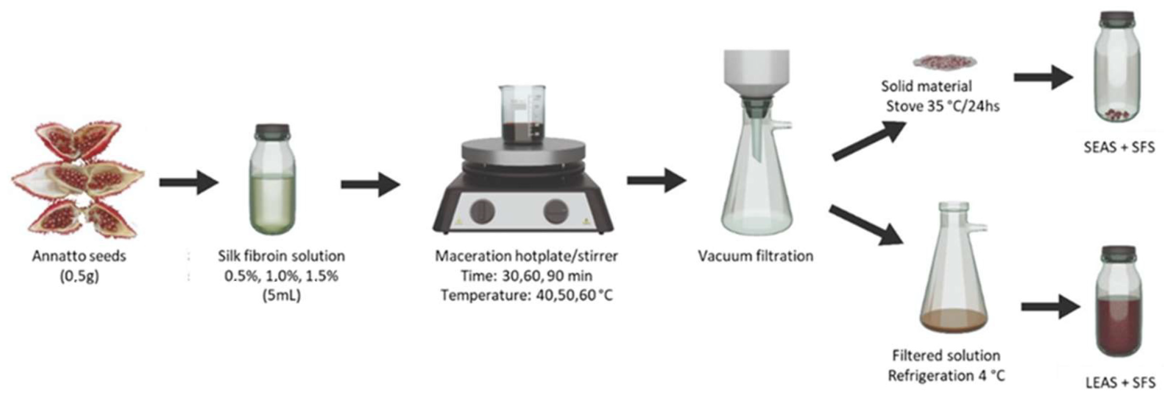
Figure 1. Summary process of bixin extraction with silk fibroin solution.
Sustainability 17 07518 g001
Figure 2. UV-Vis spectrum of samples SEAS + SFS (Run 2), LEAS + SFS (Run 2) and PB.
Sustainability 17 07518 g002
Figure 3. Bixin content response in LEAS + SFS. Pareto chart (A) and three-dimensional response surface plots showing the effects of (B) time and temperature, (C) SFS concentrations and temperature, and (D) SFS concentrations and time. The red dotted vertical line of the Pareto chart (subfigure (A)) represents the boundary between significant and non-significant effects with a 5% risk of error. The color gradient (subfigures (BD)) indicates the magnitude of the response: green regions indicate lower values, yellow regions intermediate values and orange/red regions the highest values.
Sustainability 17 07518 g003
Figure 4. Bixin content response in SEAS + SFS. Pareto chart (A) and three-dimensional response surface plots showing the effects of (B) time and temperature, (C) SFS concentrations and temperature, (D) SFS concentrations and time. The red dotted vertical line of the Pareto chart (subfigure (A)) represents the boundary between significant and non-significant effects with a 5% risk of error. The color gradient (subfigures (BD)) indicates the magnitude of the response: green regions indicate lower values, yellow regions intermediate values and orange/red regions the highest values.
Sustainability 17 07518 g004
Figure 5. FTIR spectra of purified Bixin (A); SFS (B); SEAS + SFS (C); LEAS + SFS (D).
Sustainability 17 07518 g005
Figure 6. 1H NMR (400 MHz, CDCl3) of purified bixin.
Sustainability 17 07518 g006
Figure 7. Cytotoxic activity of LEAS + SFS, SEAS + SFS, PB, and SFS at concentrations of 0.016 to 2.0 µg/mL on neonatal human dermal fibroblasts (HDFn cells) at 24 h (A) and 72 h (B). Data were expressed as a percentage of cell viability. The red dotted horizontal line represents the cutoff point for 70% viability.
Sustainability 17 07518 g007
Figure 8. Cytotoxic activity of LEAS + SFS, SEAS + SFS, PB and SFS at concentrations of 0.016 to 2.0 µg/mL on macrophages (RAW 264.7 cells) at 24 h (A) and 72 h (B). The data were expressed as a percentage of cell viability. The red dotted horizontal line represents the cutoff point for 70% viability.
Sustainability 17 07518 g008
Table 1. Input factors and their levels applied in the Box-Behnken (BBD) experimental design.
FactorVariablesLevels
−101
x1Temperature (°C) 40 50 60
x2Time (min) 30 60 90
x3SFS concentration (%)0.51.01.5
Table 2. Bixin content in each experimental combination of the Box-Behnken design (BBD).
Experiment
(Run)
Temperature (°C)Time
(min)
Concentration SFS (%)Bixin Content
(LEAS + SFS)
mg/mL
Bixin Content
(SEAS + SFS)
mg/g
140301. 06.4472.48
260601.510.87150.72
350300.52.89106.68
460301.03.54216.36
550601.04.38103.32
640901.05.1270.56
750601.03.4995.52
840600.52.16175.44
950900.54.94134.52
1050601.06.94136.56
1160901.05.5290.6
1250901.59.20130.56
1350301.56.73150.24
1440601.56.48137.4
1560600.52.9684.72
Table 3. Experimental data from 13C NMR spectroscopy (CDCl3, 100 MHz) for bixin.
Sustainability 17 07518 i001
Carbon Number *δ 13C (ppm)
Present Study[53]
1124.18130.73
2130.70130.73
3131.42131.46
4135.18135.22
5137.12137.14
6117.52123.38
7140.36140.39
8133.27133.57
9142.34142.38
10114.83117.53
11167.97168.01
1251.5851.61
1320.2620.56
1412.6212.63
15170.75171.67
16114.83115.0
17151.03151.08
18134.18134.21
19140.44140.50
20123.36124.21
21137.91137.95
22136.53136.56
23131.58131.60
2412.9913.01
2512.7612.77
Disclaimer/Publisher’s Note: The statements, opinions and data contained in all publications are solely those of the individual author(s) and contributor(s) and not of MDPI and/or the editor(s). MDPI and/or the editor(s) disclaim responsibility for any injury to people or property resulting from any ideas, methods, instructions or products referred to in the content.