Article

In Vitro Culture Initiation and Micropropagation Optimization of Plantago Halophytes: A Sustainable Approach to Exploring Valuable Plant Species

by
Aleksandra Koźmińska
1,*,
Dawid Kocot
2,* and
Karolina Kaleta
3
1
Department of Botany, Physiology and Plant Protection, Faculty of Biotechnology and Horticulture, University of Agriculture in Krakow, Mickiewicza 21 Ave., 31-120 Krakow, Poland
2
Department of Ornamental Plants and Garden Art, Faculty of Biotechnology and Horticulture, University of Agriculture in Krakow, Mickiewicza 21 Ave., 31-120 Krakow, Poland
3
Faculty of Biotechnology and Horticulture, University of Agriculture in Krakow, Mickiewicza 21 Ave., 31-120 Krakow, Poland
*
Authors to whom correspondence should be addressed.
Sustainability 2025, 17(16), 7471; https://doi.org/10.3390/su17167471
Submission received: 24 June 2025 / Revised: 10 August 2025 / Accepted: 13 August 2025 / Published: 18 August 2025

Abstract

Halophytes are salt-tolerant plants with ethnomedicinal value and growing potential in food and cosmetics; their adaptability to extreme conditions makes them promising candidates for sustainable agriculture and crop development in salt-affected areas. In vitro plant tissue culture further supports this by enabling resilient plant production in the face of climate and food security challenges. In this study, in vitro cultures of two medicinal halophytes from the genus Plantago (P. coronopus and P. crassifolia) were established to optimize their micropropagation protocol. Seed germination percentages, growth parameters, micropropagation rates, rooting efficiency, and physiological condition were evaluated. Growth media (modified MS medium) differed in the type of cytokinin. The seed germination efficiency was monitored at weekly intervals for 8 weeks, and other growth parameters were evaluated in 6- and 12-week cultures. Differences in both the rate and efficiency of in vitro germination between the two species were observed, with approximately 73% germination reached by P. coronopus and 47% by P. crassifolia after 4 weeks, and 80% and 53% after 8 weeks, respectively. The addition of 0.5 mg dm−3 kinetin plus 0.5 mg dm−3 IAA (indole acetic acid) proved to be effective in promoting growth in P. coronopus, resulting in longer plantlets and higher multiplication rates, while the addition of meta-topolin (mT) was a better stimulator of shoot and root growth in P. crassifolia. The highest multiplication coefficient, 6.22 for P. coronopus and 4.90 for P. crassifolia, was obtained on the P1 medium for both species. Importantly, medium with mT also had a stimulating effect on rooting in both species over the long term (12-week culture). The developed PTC enables efficient propagation and trait selection in halophytes, supporting sustainable large-scale production of the studied Plantago species, and facilitating future research on salt stress tolerance.

1. Introduction

The genus Plantago belongs to the Plantaginaceae family and includes approximately 260 species, primarily herbaceous (annual or perennial), with several shrubby types. These species are distributed worldwide, particularly in temperate and cold regions, especially in Europe and America. Many species (up to 200) are local or regional endemics, a result of geographical isolation showing a high degree of speciation. Only 29 species have a wide or discontinuous distribution [1,2]. The Plantago section Coronopus comprises seven species, with two focal species, Plantago crassifolia Forssk. and P. coronopus L., which are the subject of the present study. Both species have a distinct bihemispheric range: the Mediterranean and Middle Eastern regions in the northern hemisphere and South Africa in the southern hemisphere [3]. Within the genus Plantago, both halophyte species (P. crassifolia and P. coronopus) and glycophyte species (P. ovata and P. afra) are present [4].
Plants of the genus Plantago are rich in biologically active compounds such as glycosides, flavonoids, polysaccharides, and vitamins, which contribute to their diverse beneficial properties. These plants exhibit significant antioxidant, antimicrobial, and anti-inflammatory activities, supporting their use in traditional medicine and modern phytotherapy to treat various ailments, including wound healing and inhibition of apoptosis [5,6]. In addition to medicinal applications, Plantago species have notable nutritional value. Their leaves and seeds are used in various culinary traditions as food ingredients and dietary supplements, due to their rich phytochemical composition. Moreover, Plantago plants are used in cosmetic products because of their bioactive compounds, which promote skin health and possess anti-inflammatory effects. The ethnomedical importance of these species further motivates ongoing research on their potential as ingredients in food, cosmetics, and health supplements. Soil salinity, a critical environmental variable, greatly affects the natural distribution of plants and is one of the main threats to agriculture. Plants that show high tolerance to this environmental variable are halophytes. These plants can develop in extreme environmental conditions with high salinity but also intense light, drought, or floods. Their broad adaptability to a salty environment makes this group of plants valuable for sustainable agriculture, especially in saline soils and areas irrigated with salt water, the area of which is gradually increasing [7]. Plantago crassifolia and P. coronopus naturally occur in saline environments such as coastal salt marshes and saline sandy or clayey soils, which are characterized by high salt concentrations and variable moisture conditions. Plantago species have different levels of tolerance to salt stress. Some, such as P. crassifolia and P. coronopus, are examples of halophytes and exhibit significant salt tolerance, while others are more susceptible to salt stress [8,9]. Although the salinity tolerance of P. crassifolia and P. coronopus has already been studied from various perspectives, micropropagation protocols for these species are not found in the scientific literature. Scientific research conducted in vitro makes it possible to control cultivation conditions and design very different experiments without negatively impacting the natural environment. This research enables the identification and production of tolerant genotypes of Plantago species in the future [10,11]. By thriving in challenging environments, requiring fewer inputs and offering ecological and economic benefits, the study and cultivation of Plantago species align closely with the principles of sustainable agriculture, including resource efficiency, biodiversity support, soil conservation, and resilience to climate change.
Micropropagation is a vital technique for the mass production of medicinal plants [12], including various species of the genus Plantago, for which propagation protocols have been developed [13]. This method allows for the rapid multiplication of plants under controlled conditions that contribute to environmental sustainability by reducing the use of chemicals and pesticides, conserving water, minimizing land use, and allowing the development of resilient crops. Efficient propagation protocols have been developed under in vitro conditions on modified MS medium (Murashige and Skoog medium) [14], for Plantago major [15] using culturing shoot-tips, P. camtschatica showing adventitious shoot regeneration from hypocotyl explants [16], P. asiatica using shoot-tip multiplication [17] and P. maritima [18]. In vitro germination and seedling formation for P. tomentosa were tested [19] on different variants of MS medium, and in vitro germinating seeds of P. lanceolata served as a source of explants for the establishment of cultures [20]. In vitro techniques can be useful to ensure the preservation of genetic material and to promote the conservation of biodiversity. Micropropagation serves as a crucial tool for the conservation of endangered species. For two species, P. algarbiensis and P. almogravensis, endemic to Portugal, which are threatened with global extinction, micropropagation methods have been developed. During acclimatization, which is often a critical step, these species showed a high survival rate, effectively coping with oxidative stress. The transition from in vitro to ex vitro environments was successful [21]. Research on in vitro cultures of halophyte Plantago species has been steadily advancing in recent years with the development of several micropropagation protocols. In vitro techniques now enable efficient propagation, production of valuable metabolites, and investigation of salt tolerance mechanisms in various other halophyte groups [22,23]. Despite increasing attention to the ecological adaptability of Plantago, tissue culture and in vitro propagation of halophytes of the genus Plantago remain understudied.
Plantago crassifolia and P. coronopus are notable for their ability to tolerate high salinity environments, which makes them subjects of interest in studies on salt stress and plant adaptation mechanisms. Their adaptability to different environments and wide range of uses highlight their significance in various fields. The challenges of Plantago species hinder the increase in demand in the health and beauty sectors. As an alternative method, in vitro propagation is crucial for the mass production of Plantago, which is also a rapid method. In this study, the lack of efficient micropropagation protocols for P. crassifolia and P. coronopus was addressed by optimizing in vitro propagation conditions for these two halophytic medicinal species. Initiation and micropropagation were conducted with the aim of optimizing their micropropagation protocol. The use of PTC may help develop innovative production systems and obtain bioactive products derived from halophytes [24]. This constitutes a crucial way to address the challenges of modern agriculture, especially in the context of climate change and growing global food demand. In addition, the technique supports the industrial demand for medicinal plants by providing a reliable source of consistent quality plant material with the desired characteristics. These advances enable efficient propagation, metabolite production, and a deeper understanding of salt tolerance, positioning Plantago halophytes as valuable resources for sustainable agriculture and biotechnology.

2. Materials and Methods

2.1. Plant Material

The plant material used for the initiation of culture consisted of seeds of Plantago crassifolia Forssk. and P. coronopus L., collected from the Natural Park “La Albufera” (Province of Valencia, Spain; 39°28′ N, 0°23′ W). The seeds were harvested in August 2023, approximately 10 months before the start of the experiment and stored at 4 °C until use in the experimental procedures. The plants were chosen based on specific criteria to ensure the genetic diversity and representativeness of the natural population. These criteria included spatial distribution (maintaining a minimum distance between selected plants), visible health and maturity of the individuals, and the presence of fully developed seeds.

2.2. Culture Initiation

To initiate the cultures, properly developed seeds were used, which were cleaned and rinsed with tap water for 15 min prior to surface disinfection. The seeds were then disinfected in 70% ethyl alcohol for 30 s and transferred to a 2% sodium hypochlorite solution (NaOCl) with the addition of a few drops of Tween 20 (a surfactant) and agitated for 10–15 min. The explants were then thoroughly rinsed five times with sterile distilled water under aseptic conditions. All procedures requiring aseptic conditions were performed under a laminar flow chamber, ensuring sterile handling during the initiation and subculture of culture. Decontaminated seeds were placed on sowing medium, consisting of ½ MS mineral medium [14] with an addition of 20 g of sucrose, and 8.0 g dm−3 of agar (BTL, Poland), pH 5.6 before autoclaving. The seeds were placed in 100 cm3 Erlenmeyer flasks filled with 10 cm3 of medium. The single experiment for each studied species consisted of 10 flasks with 10 seeds per flask. Cultures were maintained in closed vessels, where the relative humidity remained near 100%, providing a stable microclimate for explant development. Germination was carried out in a growth chamber illuminated with white light during the day, using a 16/8 photoperiod and a temperature of 22/14 °C (day/night). Seed germination was monitored and the percentage of seedlings obtained (germination efficiency) was counted weekly for 8 weeks.

2.3. Multiplication—Culture Condition

The seedlings obtained in vitro were used to establish shoot cultures of the tested species, where all apical parts were collected and placed on three types of media. The explants were placed in 200 cm3 Erlenmeyer flasks on multiplication medium (abbreviated as P1–P3 medium). Three variants of P medium (P1, P2, P3) with different combinations of plant growth regulators were tested. The detailed composition of the three media variants tested is presented in Table 1. The pH was adjusted to 5.8 using 0.1 M HCl and 0.1 M NaOH, as appropriate, prior to autoclaving at 121 °C for 20 min. The Erlenmeyer flasks were kept under a 16/8 h day/night photoperiod in a growing room at a temperature of 24 ± 2 °C. Micropropagation rates, growth parameters, such as plant height, root height, root number and leaf number, and rooting efficiency were evaluated in 6- and 12-week cultures.

2.4. Physiological Conditions of Plants

2.4.1. Photosynthetic Pigments

The levels of chlorophyll and total carotenoids were evaluated following the method of Lichtenthaler and Wellburn [25], with some modifications. Fresh plant tissues were homogenized using a mortar and pestle, and pigments were extracted using 80% acetone that had been pre-chilled to −20 °C. The extracts were then thoroughly vortexed and centrifuged at 13,000 × g for 15 min at 4 °C. After centrifugation, the supernatant was carefully collected, and the absorbance was measured at wavelengths of 663 nm, 646 nm, and 470 nm.
Pigment concentrations were calculated using the following equations from Lichtenthaler and Wellburn [25]:
  • Chlorophyll a (abbreviated as Chl a) (μg cm−3) = 12.21 × A663 − 2.81 × A646
  • Chlorophyll b (abbreviated as Chl b) (μg cm−3) = 20.13 × A646 − 5.03 × A663
  • Total chlorophyll = Chl a + Chl b
  • Total carotenoids (μg cm−3) = (1000 × A470 − 3.27 × [Chl a] − 104 × [Chl b])/229
The final pigment concentrations were converted to milligrams per 100 grams of fresh weight (mg 100 g−1 FW) for standardized comparison.

2.4.2. Proline Determination

The concentration of proline (Pro) was measured in fresh leaf tissue following the ninhydrin-acetic acid method described by Bates [26]. Free proline was extracted using 3% aqueous sulfosalicylic acid and then reacted with an acid ninhydrin solution (the ninhydrin solution was prepared by dissolving 1.25 g of ninhydrin in a mixture of 30 cm3 glacial acetic acid and 20 cm3 of 6 M orthophosphoric acid). The mixture was incubated at 95 °C for 1 h, subsequently cooled on ice, and extracted with toluene. The absorbance of the resulting organic phase was measured at 520 nm, using toluene as a blank. The proline content was determined from a standard curve prepared with known proline concentrations and expressed as mg per 100 g of fresh weight (mg 100 g−1 FW).

2.4.3. Total Phenolic Compounds Determination

The concentration of total phenolic compounds (TPC) in leaf tissues was determined using the Folin–Ciocalteu colorimetric method, as outlined by Blainski [27]. The methanolic extracts were combined with the Folin–Ciocalteu reagent and the sodium bicarbonate solution. The reaction mixtures were incubated in the dark for 90 min to allow color development. The absorbance was then measured at 765 nm using a Hitachi U-2900 spectrophotometer (Hitachi High-Tech Science Corporation, Westford, MA, USA). The phenolic content was quantified using a standard curve generated with gallic acid and expressed as milligrams of equivalent gallic acid (GA) per 100 g of fresh weight (mg GA g−1 FW).

2.5. Statystical Analysis

Each treatment was tested in two independent experiments, each with eight replicates, where one replicate consisted of three plants (three plants per Erlenmeyer flask). Quantitative data for the content of biochemical compounds were obtained from three biological replicates. Data were statistically analyzed using STATISTICA 13.0 software (StatSoft, Tulsa, OK, USA). If the ANOVA assumptions were met, the differences among the three medium variants were evaluated using one-way analysis of variance (ANOVA) at a confidence level of 95% (p < 0.05), followed by post- hoc comparisons with the Tukey’s HSD test. Where applicable, the Student’s t-test was also used to assess significant differences between the selected treatment pairs.

3. Results

3.1. Germination Efficiency of Plantago Seeds

The average percentage of contaminated explants was 20% in the case of P. coronopus and 30% for P. crassifolia. Differences in both the rate and efficiency of in vitro germination were observed between tested species. P. coronopus seeds first started germination, reaching 53% after the first week of measurements. The maximum germination efficiency for P. coronopus was reached after 7 weeks and reached approximately 80%. The seeds of P. crassifolia started germinating after the second week, reaching a maximum of 53% germinated seeds after 6 weeks (Figure 1).

3.2. Growth Parameters of Plantago Plants

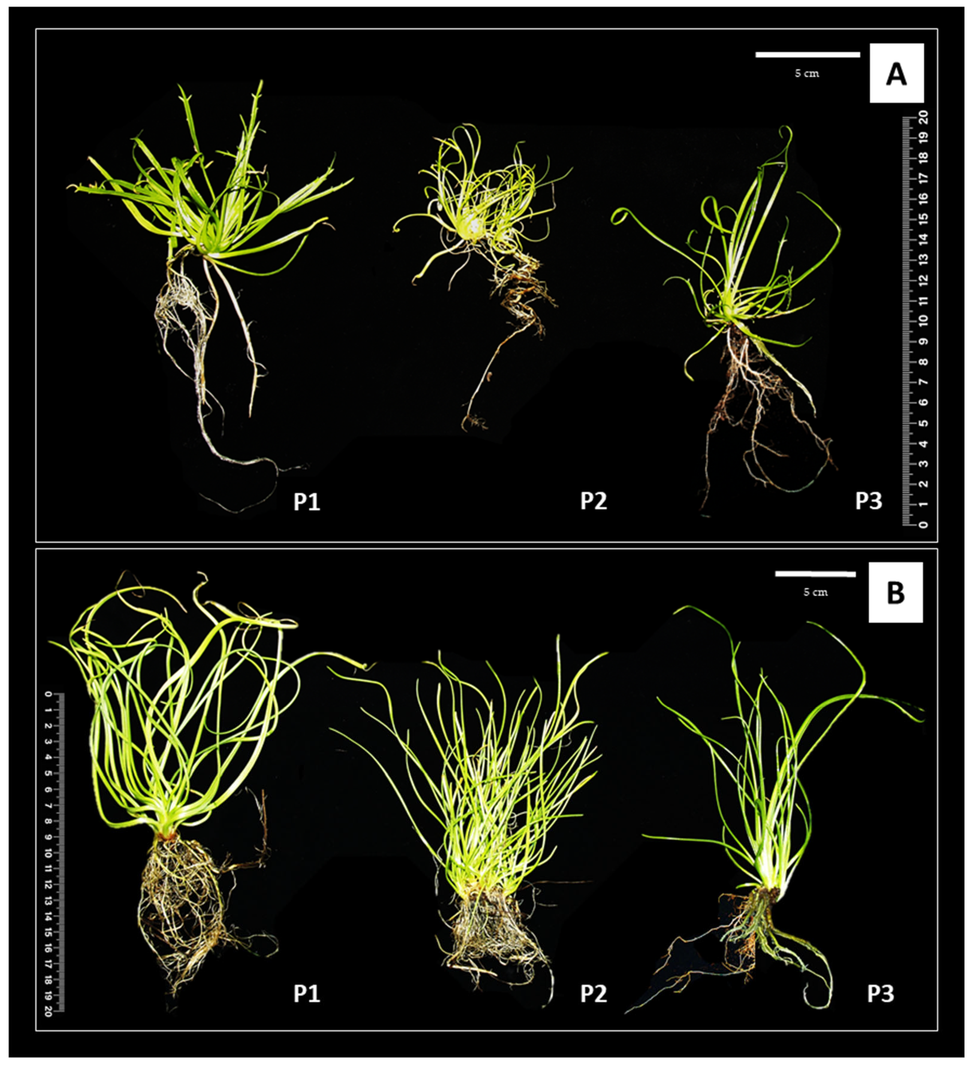
MS medium supplemented with 0.5 mg dm3 kinetin and 0.5 mg dm3 IAA (P1) proved to be effective in promoting shoot growth in P. coronopus, which reached 11.86 cm after 6 weeks of cultivation and 11.81 after 12 weeks. After 6 weeks, the plants grown on P1 medium were 22% higher than those on P2 and 16% higher than those on P3 (Figure 2).
After 12 weeks, they were approximately 35% higher than plants on P2 and 11% higher than those on P3 (Table 2). Plantago crassifolia microplants reached their maximum lengths on MS medium supplemented with 0.5 mg dm−3 meta-topoline and 0.5 mg dm−3 IAA (P3 medium), measuring 16.73 cm and 19.53 cm after 6 and 12 weeks, respectively. After 6 weeks, the plants grown on P3 medium were 34% higher than those on P1 and 90% higher than those on P2. After 12 weeks, they were approximately 40% higher than plants on P1 and 52% higher than those on P2.
The length of the roots of the explants placed in the medium supplemented with meta-topoline (P3) was higher than that from the medium supplemented with KIN and BAP in both studied species. For P. coronopus, the root length on P3 was 8% and 24% greater than on P1 and P2, respectively. After 12 weeks, it was 17% higher than on P1 and 60% higher than on P2. For P. crassifolia, the stimulating effect of meta-topoline on root growth was not evident at 6 weeks, as similar root lengths were recorded in the three media. Significant differences (p ≤ 0.05) appeared only after 12 weeks, when P. crassifolia plants grown on P3 medium exhibited markedly longer roots compared to those on P1 and P2 achieving, respectively: 19.14, 15.17, 13.57 centimeters (Table 2). Regarding the average number of leaves in plants cultivated on the three media variants, no clear trend was observed. After 6 weeks, P. coronopus had a similar number of leaves on media P1 and P3 (with differences statistically insignificant), and both were greater than the number of leaves observed on P2 medium. P. crassifolia had the highest number of leaves on P1 medium after 6 weeks of cultivation. After 12 weeks, the number of leaves was highest in P. coronopus on P1 medium (31 leaves) and in P. crassifolia on P3 medium (24 leaves) (Table 2). In 12-week-old cultures of P. coronopus, slight leaf yellowing was observed on P1 medium. The percentage of water content followed a similar pattern across all three MS media in both species (Table 2). The highest fresh weight was observed in P. coronopus plants cultivated on the P1 medium for 6 weeks, reaching 1.75 g (Supplementary Materials Table S1).
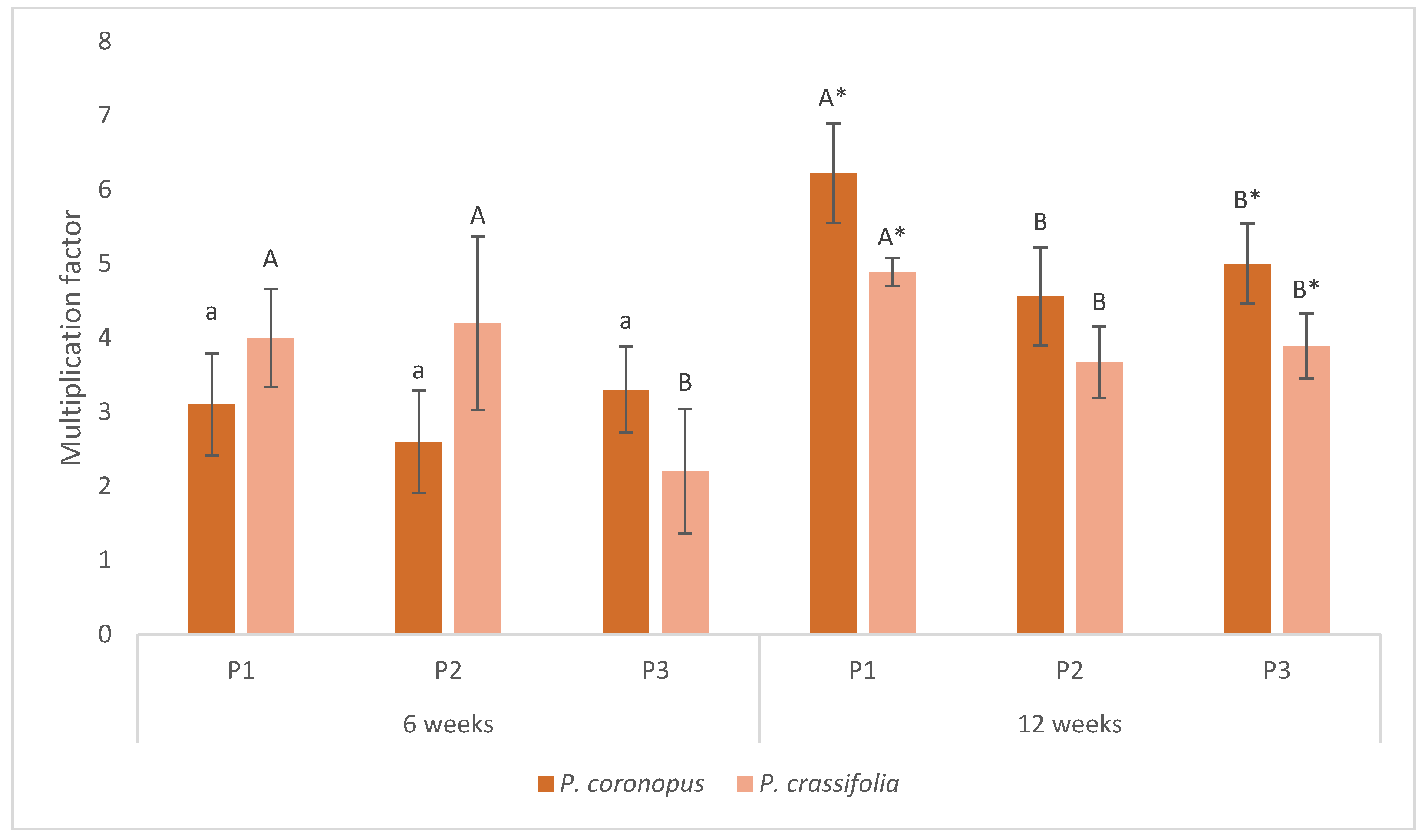
After 6 weeks of plant culture, no statistically significant differences were observed in the multiplication coefficient among the three media used for each species. However, after 12 weeks of in vitro propagation, both P. coronopus and P. crassifolia showed the highest multiplication factor when cultured on MS medium supplemented with 1 mg dm−3 kinetin and 0.5 mg dm−3 IAA (P1). The multiplication coefficient reached 6.22 for P. coronopus and 4.90 for P. crassifolia per explant, respectively (Figure 3).

3.3. Physiological Analysis of Plant Material

3.3.1. Total Chlorophylls Content

In P. coronopus, after 6 weeks of cultivation, no statistically significant differences were observed between the media types, with a total chlorophyll content that narrowly ranged between 70 and 75 mg 100 g−1 FW across P1, P2, and P3. However, after 12 weeks, statistically significant differences were found (p ≤ 0.05): medium P2 (BAP) led to the highest chlorophyll accumulation (95 mg 100 g−1 FW), significantly higher than both P1 and P3. In contrast, P. crassifolia showed a more consistent trend across time points. After 6 weeks, the P1 medium (kinetin) yielded significantly higher chlorophyll content (≈93 mg 100 g−1 FW) compared to P2 and P3. This trend persisted at 12 weeks, although the pattern changed: the highest chlorophyll content was observed on P3 (94 mg 100 g−1 FW), followed by P2 (83 mg 100 g−1 FW) and P1 (70 mg 100 g−1 FW), with all differences statistically significant (p ≤ 0.05) (Figure 4a).

3.3.2. Carotenoids Content

The accumulation of carotenoids varied significantly between species. In P. coronopus, carotenoid content ranged from approximately 16 to 24 mg 100 g−1 FW across all media. After 6 weeks, no statistically significant differences were observed between plants from different media. The highest accumulation was recorded after 12 weeks, particularly on medium P1 (≈24 mg 100 g−1 FW), which was significantly higher (p ≤ 0.05) than P2 and P3. In contrast, P. crassifolia consistently exhibited significantly higher carotenoid levels than P. coronopus, with values ranging from 20 to 27 mg 100 g−1 FW (p ≤ 0.05). At 6 weeks, a significantly higher accumulation of carotenoids was recorded on P1 (approximately 27 mg 100 g−1 FW), while P2 and P3 showed uniform levels (approximately 20 mg 100 g−1 FW). After 12 weeks, the carotenoid content increased further on P2 and P3 (approximately 25–27 mg 100 g−1 FW), with statistically significant differences observed between time points and media, indicating a time- and medium-dependent increase in carotenoid biosynthesis in P. crassifolia (Figure 4b).

3.3.3. Proline Concentration

In P. coronopus, a significant increase in proline content was observed after 12 weeks, with BAP (P2) inducing the highest levels (approximately 36 mg 100 g−1 FW; p ≤ 0.05) compared to KIN (P2) and MT (P3). In contrast, P. crassifolia showed medium-dependent differences at both time points: after 6 weeks, kinetin (P1) led to the highest accumulation of proline (p ≤ 0.05), while after 12 weeks, BAP (P2) and MT (P3) were the most effective (p ≤ 0.05) (Figure 4c).

3.3.4. Total Phenolic Compounds Content

The total phenolic compounds content was significantly higher (p ≤ 0.05) in P. crassifolia compared to P. coronopus, regardless of the growth medium, at both 6 and 12 weeks of culture. The highest levels of phenolic compounds were recorded in 12-week cultures of P. crassifolia, reaching 93.46 mg g−1, 96.88 mg g−1, and 112.08 mg g−1 on P1, P2, and P3 media, respectively (all differences statistically significant at p ≤ 0.05) (Figure 4d).
Data represent mean values of six replicates. Means ± SE within a column followed by different lowercase letters indicate statistically significant differences between media for P. coronopus (a,b,c) and P. crassifolia (x,y,z) after 6 weeks of culture. Means ± SE within a column followed by different uppercase letters indicate statistically significant differences between media for P. coronopus (A,B,C) and P. crassifolia (X,Y,Z) after 12 weeks of culture. An asterisk indicates significant differences between P. coronopus and P. crassifolia for the same medium and time point; (p ≤ 0.05) according to Tukey’s test.

4. Discussion

The present study aimed to establish an efficient micropropagation protocol for Plantago crassifolia and P. coronopus by in vitro optimization of the effect of different plant growth regulators (PGRs). Seeds were used to initiate cultures. Seed germination in Plantago species has been widely studied and generally shows high efficiency. For example, P. ovata reached germination of 80.71% [28], P. algarbiensis 80%, and P. almogravensis 58% [21]. In P. tomentosa, germination rates of 78–91% were recorded at various concentrations of MS medium [19]. Similarly, for P. lanceolata, cold pretreatment, light exposure, and reduced MS salt concentration (1/8×) enhanced germination, achieving rates of up to 78% [20]. Temperature variability, population origin, and seed age have also been shown to significantly influence germination, with reported ranges from 27% to 85% [29,30].
In our study, we observed somewhat lower germination efficiencies for the tested halophyte species compared to other Plantago species. P. coronopus reached a maximum germination rate of approximately 80% after 7 weeks, which is consistent with the results obtained for other species under optimized conditions. However, a delayed and reduced response was observed in P. crassifolia, with a maximum germination rate of 53% after 6 weeks. These differences may be attributed to species-specific seed dormancy mechanisms or different ecological adaptations. In particular, the contamination rate was also higher for P. crassifolia (30%) compared to P. coronopus (20%), which may have further impacted the germination results. Comparison of our results with previously reported data highlights the importance of tailoring germination protocols to specific species, especially in the case of halophytes, where environmental adaptations can affect in vitro responses.
The studied species P. coronopus produces mucilaginous seeds, which facilitate seed germination in dry environments. Previous studies have shown that seeds of this species germinate more effectively when soil crust is disturbed and that germination is more successful on filter paper or sand than in soil [31,32]. Temperatures favourable for P. coronopus germination range from 10 °C to 25 °C, with the optimal temperature reported as 25 °C [33,34]. Seed age, post-maturation temperature during dry storage, and environmental conditions during imbibition have also been shown to influence germination. Although both studied species are halophytes, previous research indicated that physiological saline solutions significantly inhibit the germination of P. coronopus and, to a lesser extent, P. crassifolia, with no germination occurring at 0.2 M NaCl [33]. Conversely, other in vitro studies reported very high germination percentages (>96%) for both species under control conditions; P. coronopus was more salt tolerant than P. crassifolia at 100–400 mM NaCl [8]. In our study, the germination efficiency was noticeably lower under the applied in vitro conditions: a maximum of 80% germination was observed for P. coronopus, while only 53% was achieved for P. crassifolia. Furthermore, differences in the onset of germination were evident: P. coronopus began to germinate in the first week, while P. crassifolia germinated from the third week onwards. One possible explanation for the lower germination rates could be the age of the seeds, as in our experiment, seeds were used approximately 10 months after harvest. Despite this, the germination levels obtained can be considered satisfactory and sufficient to establish viable in vitro cultures. These cultures formed the basis for further experimental stages, including the evaluation of plant growth dynamics and physiological responses under different cytokinin treatments. Research on the use of cytokinins in the in vitro propagation of Plantago species is relatively limited. A notable study by Makowczyńska and Andrzejewska-Golec [17] focused on the micropropagation of Plantago asiatica via shoot-tip culture. They reported that Murashige and Skoog (MS) medium supplemented with 0.1 mg dm−3 indole-3-acetic acid (IAA) and 1 mg dm−3 benzylaminopurine (BAP) was the most effective for shoot multiplication. Further investigations by the same authors demonstrated that combining BAP with 2,4-dichlorophenoxyacetic acid (2,4-D) or α-naphthaleneacetic acid (NAA) could induce both somatic organogenesis and embryogenesis in P. asiatica. These findings suggest that specific combinations of cytokinins and auxins can significantly influence the success of in vitro propagation protocols for Plantago species. Cytokinins (CKs) are plant growth regulators that play a key role in controlling the cell cycle and development in plants. When applied externally in plant tissue culture (PTC), they help regulate various morphogenic processes. The selection of the appropriate type or concentration of CK affects the mass propagation of plants, their growth parameters, and physiological condition. In our study, three different types of cytokinins (kinetin, BAP and m-topoline) were used in the propagation media at an appropriate ratio to the auxin IAA, in order to optimize the multiplication protocol and obtain Plantago crassifolia and P. coronopus plants with optimal growth parameters and physiological conditions.
Kinetin, a synthetic cytokinin, is widely used in plant tissue culture due to its capacity to regulate growth and development. In combination with auxins such as indole-3-acetic acid (IAA), it plays a crucial role in the influence of shoot multiplication and root development. Although direct studies on the in vitro response of Plantago coronopus and P. crassifolia to kinetin are lacking in the literature, comparisons with other Plantago species are useful to relate our findings. For instance, in P. ovata, kinetin combined with auxins (e.g., 2,4-D or NAA) has been used to induce callus formation, followed by shoot regeneration when transferred to BAP- and IAA-supplemented media [35]. However, in P. asiatica, the IAA with BAP combination was more effective for shoot multiplication than kinetin [17]. In our study, the use of medium P1, containing 0.5 mg dm−3 kinetin and 0.5 mg dm−3 IAA, resulted in the highest multiplication factor for both P. coronopus and P. crassifolia after 12 weeks of in vitro culture. Furthermore, in P. coronopus, a positive effect on plant height and number of leaves was observed, indicating the effectiveness of kinetin in promoting shoot development in these species. It is worth noting that although previous studies on P. ovata report callus induction under kinetin with 2,4-D conditions [36], in our experiments with P. coronopus and P. crassifolia, no callus formation was observed on any of the tested media. This difference may be due to genotypic variation or the specific combinations of cytokinin-auxin used.
To broaden the interpretation of the effect of kinetin, we also refer to studies on other plant species. For example, in Solanum tuberosum, kinetin and IAA have been shown to support root development [37,38], and in Hippophae rhamnoides, a combination of kinetin, IAA, and BAP stimulated efficient shoot formation [39]. However, in our research, the best results regarding root length and number were observed not on P1 but on media enriched with meta-topolin (P3), suggesting that for P. coronopus and P. crassifolia, mT may be more effective for rooting than kinetin.
The rooting of individual microshoots obtained through micropropagation is a crucial step in studies that aim to cultivate plants in greenhouses, open fields, or in their natural habitats. Although root induction can sometimes occur spontaneously in the basal or propagation medium, healthy shoots are typically excised and transferred to a dedicated rooting medium prior to acclimatization [23]. Our results clearly demonstrate that m-topolin (mT) promotes root formation in the two halophytic species studied. As shown in Figure 2 and Table 2, microshoots cultured on the P3 medium developed significantly longer roots and a higher number of roots compared to P1 (kinetin) and P2 (BAP) media. This effect may be associated with the cytokinin activity of mT, which has been reported to promote not only shoot proliferation but also root induction. Studies by Aremu et al. [40] and Ahmad and Anis [41] indicated that mT often improves rooting efficiency in micropropagation systems due to its favorable influence on hormonal balance and oxidative stress levels in regenerating tissues. Our findings support these observations that mT may act as a physiologically more suitable cytokinin for promoting balanced shoot and root growth in certain halophytes, possibly by better supporting stress resilience mechanisms and promoting efficient nutrient uptake under in vitro conditions.
M-topolin (mT) is a benzyladenine analog [N 6-(3-hydroxybenzylamino) purine] and its derivatives have been proposed as effective alternatives to commonly used CK, such as BAP and thidiazuron, in several plant species. These compounds have shown potential to address micropropagation challenges, including shoot abnormalities, reduced rooting capacity, and negative effects during ex vitro acclimatization [40,42,43]. Furthermore, m-topolins have been reported to delay senescence, improve in vitro photosynthetic pigment levels, and modulate antioxidant enzyme activity [43,44,45]. The effect of mT on the rooting of plants in in vitro cultures has been studied extensively. M-topolin, a cytokinin, has been shown to enhance rooting and overall plant development in various plant species when used in tissue culture media. In our study, the application of meta-topolin (mT) at 0.5 mg dm−3 (medium P3) significantly enhanced root formation in both P. coronopus and P. crassifolia, resulting in the longest roots and highest number of roots observed among all combinations of media tested (Figure 2, Table 2). These findings confirm the positive effect of mT on rhizogenesis, which has also been documented in other species. For example, mT was previously shown to improve rooting efficiency and plantlet quality in species such as Pterocarpus marsupium, Corylus colurna, and Vanilla planifolia [41,46]. In these studies, mT outperformed traditional cytokinins such as BAP in stimulating both shoot multiplication and root development. Moreover, Kulpa et al. [47] demonstrated that mT enhanced spontaneous rooting in the halophyte Honckenya peploides, suggesting its potential usefulness in plants adapted to saline environments. Our findings are consistent with these reports and extend the understanding of mT’s role to two new halophyte species. Since no previous data are available for P. coronopus and P. crassifolia, this comparison with related genera and functional plant groups (e.g., halophytes) provides a valuable context for interpreting the observed phenomena. This also supports the conclusion that mT may be particularly effective in promoting root system development in species that grow under saline or stressful conditions.
Depending on the species of plants, different types of cytokinins can stimulate rooting. For example, in Juncus roemerianus, regenerated shoots with BAP supplementation induced the formation of adventitious roots, while those supplemented with TDZ did not [48]. It should also be noted that when rooting does not occur spontaneously or after transfer to a PGR-free medium, the main factors that influence root induction are the type and concentration of auxins. Among these, IBA is the most commonly used, followed by NAA and IAA—either individually, in combination, or in association with cytokinins such as BAP and kinetin, or with gibberellins like GA3 [49]. In our study, only IAA was used at a constant concentration of 0.5 mg dm−3. Therefore, it is difficult to assess the specific effects of different auxins on rooting in Plantago species. However, the auxin-to-cytokinin ratio likely plays a significant role. The ratios used in our experiments were as follows: 1:1 for kinetin to IAA, 2:1 for BAP to IAA, and 1:1 for m-topolin to IAA.
The type of cytokinin in the growth medium affects not only plant growth (shoot and root length, leaf number), but also physiological, biochemical parameters, and stress response [50,51,52,53]. The specific type of cytokinin influences both the chlorophyll a and chlorophyll b content, as well as their ratio. In our study, no single cytokinin consistently stimulated chlorophyll or carotenoid production at both species and time points. Depending on the duration of the in vitro culture, different cytokinin treatments led to varied responses in the two studied species. After 6 weeks, P. coronopus showed the most effective shoot multiplication and root formation on medium P1 (containing 0.5 mg dm−3 kinetin and 0.5 mg dm−3 IAA), as reflected in the highest number of shoots and the longest shoots (Figure 2, Table 2). Conversely, at this same time point, P. crassifolia responded more favourably to medium P3 (with 0.5 mg dm−3 m-topolin), which promoted greater shoot height and more developed roots. After 12 weeks, both species showed increased growth and development, but with species-specific preferences. P. coronopus still performed best on P1, exhibiting the highest shoot multiplication rate and number of leaves, while P. crassifolia again responded more strongly to P3, with the longest roots and highest number of roots per plantlet (Figure 2, Table 2). These findings underscore the differential responses to cytokinins based on both the species and the culture duration, emphasizing the importance of tailoring hormonal treatments in micropropagation protocols for halophyte species. Previous studies confirm that cytokinin effects are species- and dose-dependent. For example, BAP increased the chlorophyll content in apple leaves, while m-topolin improved the chlorophyll a/b ratio [54]. In Annona glabra, both kinetin and BAP increased chlorophyll a and carotenoids, improving chloroplast development and acclimatization [55]. Previous studies have shown that cytokinins such as kinetin and BAP can influence photosynthetic pigment levels, although their effects depend on concentration and species [53,54]. Our results confirm this in Plantago species. In P. coronopus, chlorophyll content remained stable across treatments after 6 weeks, but increased significantly at week 12 on BAP-containing medium (P2), reaching 95 mg 100 g−1 FW. In P. crassifolia, kinetin (P1) was most effective at 6 weeks, while meta-topolin (P3) led to the highest chlorophyll content at 12 weeks. Carotenoid accumulation was generally higher in P. crassifolia than in P. coronopus. In both species, the highest levels were observed after 12 weeks, particularly on media with kinetin (P1) or m-topolin (P3). These results emphasize the importance of cytokinin selection and exposure time in optimizing pigment content and improving plant physiological quality in vitro.
With regard to phenolic compounds, our results clearly indicate that P. crassifolia accumulated significantly higher levels of total phenolics than P. coronopus, regardless of the growth medium or culture duration. The highest concentrations were observed in 12-week-old P. crassifolia cultures, reaching 93.46 mg g−1 (P1), 96.88 mg g−1 (P2), and 112.08 mg g−1 (P3). These findings suggest that the synthesis of phenolic compound synthesis in P. crassifolia may intensify over time, particularly under prolonged in vitro cultivation. Although the literature on the relationship between culture age and phenolic content remains limited and sometimes inconclusive, our data provide evidence of a time-dependent increase in phenolic accumulation in P. crassifolia. This supports the hypothesis that such accumulation is both species- and condition-specific. In contrast to our results, studies on Deschampsia antarctica reported no significant differences in phenolic content between one- and two-year-old cultures [56], while in Ageratina pichichensis, variations in phenolic levels were attributed more to culture type than to duration [57]. These discrepancies highlight the complexity of phenolic biosynthesis pathways and underscore the necessity of species-specific optimization in in vitro systems aimed at improving secondary metabolite production.
The application of plant tissue culture (PTC) techniques to halophytes such as Plantago coronopus and Plantago crassifolia enables their effective propagation and facilitates the evaluation of physiological responses under controlled conditions [24,58]. In our study, the optimized micropropagation protocol proved effective for both species, supporting high multiplication rates and allowing for detailed assessment of growth parameters and secondary metabolite accumulation. Beyond propagation, in vitro culture systems serve as platforms to study salt stress tolerance, conserving elite genotypes, and stimulate the synthesis of valuable secondary metabolites, including phenolic compounds and antioxidants. This study provides a foundational protocol for the micropropagation of P. crassifolia and P. coronopus. While only a limited number of factors were examined, the results offer a basis for future investigations that involve more complex experimental designs and additional variables that influence the response of tissue culture. Under in vitro conditions, changes in phytochemical constituents compared to those in vivo plants and the elicitation of desired compounds may occur. In the case of micropropagation of P. ovata, an increase in phenols and alkaloids was observed, while others, such as carbohydrates and tannins, decreased [22]. This approach opens new avenues for the use of halophytes as alternative crops in saline or marginal environments, thus reducing dependence on conventional agriculture and contributing to the protection of wild plant populations. Integrating biotechnological advances with ecological considerations, the in vitro culture of halophytes promotes the preservation of biodiversity, sustainable agriculture, and the development of resilient food systems. The development of micropropagation protocols for halophytic species not only facilitates their conservation and cultivation, but also supports sustainable agricultural practices on saline soils, offering a potential strategy to address ecological challenges linked to climate change and land degradation. Ultimately, these methods support long-term environmental sustainability and global food security.

5. Conclusions

In the present study, P. coronopus and P. crassifolia were successfully propagated in vitro from seeds, indicating that seeds of these species are a suitable source of initial explants for culture establishment due to their high germination rates and effective surface disinfection. Efficient micropropagation protocols were developed by initiating in vitro cultures from explants excised from seedlings or native adult plants. Culture of P. coronopus is recommended in MS medium supplemented with 0.5 mg dm−3 kinetin and 0.5 mg dm−3 IAA for shoot growth and multiplication. For both studied species (P. coronopus and P. crassifolia), MS medium containing 0.5 mg dm−3 meta-topolin and 0.5 mg dm−3 IAA is recommended for effective rhizogenesis and enhanced synthesis of total phenolic compounds. The results confirm that phytohormonal requirements can vary even among closely related species, highlighting the importance of an individual approach. Even related species within the same ecological category may require different types and concentrations of growth regulators to achieve optimal stimulation. This underscores the importance of tailoring growth regulator treatments to the specific physiological needs of each species.

Supplementary Materials

The following supporting information can be downloaded at: https://www.mdpi.com/article/10.3390/su17167471/s1. Table S1: Effect of kinetin (P1), BAP (P2) and meta-topolin (P3) on fresh weight and dry weight of P. coronopus and P. crassifolia after six and twelve weeks of cultivation.

Author Contributions

Conceptualization, A.K.; methodology, A.K., D.K. and K.K.; validation, A.K. and D.K.; formal analysis, A.K. and D.K.; investigation, A.K. and K.K.; resources, A.K.; data curation, A.K. and D.K.; writing—original draft preparation, A.K. and D.K.; writing—review and editing, A.K. and D.K.; visualization, D.K. and K.K.; supervision, A.K. and D.K.; funding acquisition, A.K. All authors have read and agreed to the published version of the manuscript.

Funding

This research was funded by the Ministry of Science and Higher Education of Poland—statutory funding of the research activity held at the University of Agriculture in Kraków, Poland (050012-D011).

Institutional Review Board Statement

Not applicable.

Informed Consent Statement

Not applicable.

Data Availability Statement

The original contributions presented in this study are included in the article/Supplementary Materials. Further inquiries can be directed to the corresponding author.

Conflicts of Interest

The authors declare that they have no conflicts of interest.

References

  1. Henderson, L. Floral anatomy of several species of Plantago. Am. J. Bot. 1926, 13, 397–405. [Google Scholar] [CrossRef]
  2. Brown, M. A synopsis of the genus Plantago L. in Tasmania Pap. Proc. R. Soc. Tasman. 1991, 124, 65–74. [Google Scholar] [CrossRef]
  3. Höpke, J.; Mucina, L.; Albach, D. Phylogenetic and morphometric analysis of Plantago section Coronopus (Plantaginaceae). Taxon 2019, 68, 315–339. [Google Scholar] [CrossRef]
  4. Ltaeif, H.; Sakhraoui, A.; González-Orenga, S.; Faz, A.; Boscaiu, M.; Vicente, Ó.; Rouz, S. Responses to Salinity in Four Plantago Species from Tunisia. Plants 2021, 10, 1392. [Google Scholar] [CrossRef]
  5. Weryszko-Chmielewska, E.; Matysik-Woźniak, A.; Sulborska, A.; Rejdak, R. Commercially important properties of plants of the genus Plantago. Acta Agrobot. 2012, 65, 11–20. [Google Scholar] [CrossRef]
  6. Gonçalves, S.; Romano, A. The medicinal potential of plants from the genus Plantago (Plantaginaceae). Ind. Crops Prod. 2016, 83, 213–226. [Google Scholar] [CrossRef]
  7. Boscaiu, M.; Ballesteros, G.; Naranjo, M.A.; Vicente, O.; Boira, H. Responses of halophytes to salt stress. Bul. USAMV-CN 2007, 64, 13–18. [Google Scholar]
  8. Al Hassan, M.; Pacurar, A.; Gaspar, A.; Vicente, O.; Boscaiu, M. Growth and Reproductive Success under Saline Conditions of Three Plantago Species with Different Levels of Stress Tolerance. Not. Bot. Horti Agrobot. Cluj-Napoca 2014, 42, 180–186. [Google Scholar] [CrossRef]
  9. Al Hassan, M.; Pacurar, A.; López-Gresa, M.P.; Donat-Torres, M.P.; Llinares, J.V.; Boscaiu, M.; Vicente, O. Effects of Salt Stress on Three Ecologically Distinct Plantago Species. PLoS ONE 2016, 11, e0160236. [Google Scholar] [CrossRef] [PubMed]
  10. Safarnejad, A.; Shoorvarzi, M.; Dalir, M. In vitro selection of Plantago ovata for NaCl tolerance. Iran. J. Rangel. For. Plant Breed. Genet. Res. 2017, 25, 275–287. [Google Scholar]
  11. Safarnejad, A.; Shoorvarzi, M.; Dalir, M. In vitro selection of Plantago psyllium L. for salt tolerance and Changes of Sodium, Calcium and Potassium levels at callus stage. Iran. J. Rangel. For. Plant Breed. Genet. Res. 2017, 24, 221–231. [Google Scholar]
  12. Norouzi, O.; Hesami, M.; Pepe, M.; Dutta, A.; Jones, A.M.P. In vitro plant tissue culture as the fifth generation of bioenergy. Sci. Rep. 2022, 12, 5038. [Google Scholar] [CrossRef]
  13. Fons, F.; Gargadennec, A.; Rapior, S. Culture of Plantago species as bioactive components resources: A 20-year review and recent applications. Acta Bot. Gall. 2008, 155, 277–300. [Google Scholar] [CrossRef]
  14. Murashige, T.; Skoog, F. A Revised Medium for Rapid Growth and Bio Assays with Tobacco Tissue Cultures. Plant Physiol. 1962, 15, 473–497. [Google Scholar] [CrossRef]
  15. Mederos, S.; Martin, C.; Navarro, E.; Ayuso, M. Micropropagation of a medicinal plant, Plantago major L. Biol. Plant. 1997, 39, 465–468. [Google Scholar] [CrossRef]
  16. Andrzejewska-Golec, E.; Makowczyńska, J. Micropropagation of Plantago camtschatica Link. Acta Soc. Bot. Pol. 2008, 77, 269–273. [Google Scholar] [CrossRef]
  17. Makowczyńska, J.; Andrzejewska-Golec, E. Micropropagation of Plantago asiatica L. through culture of shoot-tips. Acta Soc. Bot. Pol. 2003, 72, 191–194. [Google Scholar] [CrossRef]
  18. Makowczyńska, J.; Andrzejewska-Golec, E. Micropropagation of Plantago maritima L.—A vanishing species in Poland. Acta Soc. Bot. Pol. 2009, 78, 13–18. [Google Scholar] [CrossRef]
  19. Gastmann, J.; Klaus, M.V.V.; Winhelmann, M.C.; de Campos, S.S.; Hoehne, L.; de Freitas, E.M. In vitro germination and seedling formation of Plantago tomentosa Lam. (Plantaginaceae): Influence of concentrations of the MS medium. Ciência Nat. 2024, 46, e71895. [Google Scholar] [CrossRef]
  20. Rahamooz-Haghighi, S.; Bagheri, K.; Danafar, H.; Sharafi, A. Tissue culture, in vitro organogenesis and regeneration of Plantago lanceolata. J. Appl. Biotechnol. Rep. 2020, 7, 258–265. [Google Scholar] [CrossRef]
  21. Gonçalves, S.; Martins, N.; Romano, A. Micropropagation and conservation of endangered species Plantago algarbiensis and P. almogravensis. Biol. Plant 2009, 53, 774–778. [Google Scholar] [CrossRef]
  22. Sharma, M.; Kumari, A.; Mahant, E. Micropropogation and analysis of phytochemical profile of tissue culture grown Plantago ovata Forsk. Asian J. Pharm. Clin. Res. 2017, 10, 202–206. [Google Scholar] [CrossRef]
  23. Custódio, L.; Charles, G.; Magné, C.; Barba-Espín, G.; Piqueras, A.; Hernández, J.; Hamed, K.; Castañeda-Loaiza, V.; Fernandes, E.; Rodrigues, M. Application of In Vitro Plant Tissue Culture Techniques to Halophyte Species: A Review. Plants 2022, 12, 126. [Google Scholar] [CrossRef]
  24. Pieracci, Y.; Vento, M.; Pistelli, L.; Lombardi, T.; Pistelli, L. Halophyte Artemisia caerulescens L.: Metabolites from In Vitro Shoots and Wild Plants. Plants 2022, 11, 1081. [Google Scholar] [CrossRef]
  25. Lichtenthaler, H.K.; Wellburn, A.R. Determinations of total carotenoids and chlorophylls a and b of leaf extracts in different solvents. Biochem. Soc. Trans. 1983, 11, 591–592. [Google Scholar] [CrossRef]
  26. Bates, L.S.; Waldren, R.P.; Teare, I.D. Rapid determination of free proline for water stress studies. Plant Soil 1973, 39, 205–207. [Google Scholar] [CrossRef]
  27. Blainski, A.; Lopes, G.C.; Palazzo de Mello, J.C. Application and analysis of the Folin Ciocalteu method for the determination of the total phenolic content from Limonium brasiliense L. Molecules 2013, 18, 6852–6865. [Google Scholar] [CrossRef] [PubMed]
  28. Mahato, S.; Meht, A. Optimization of in-vitro propagation protocol for Plantago ovata Forsk. J. Indian Bot. Soc. 2015, 94, 245–251. [Google Scholar]
  29. Mira, S.; Veiga-Barbosa, L.; González-Benito, M.; Pérez-García, F. Inter-population variation in germination characteristics of “Plantago lanceolata” seeds: Effects of temperature, osmotic stress and salinity. Appl. Bot. 2018, 39, 89–96. [Google Scholar] [CrossRef]
  30. Sarihan, E.; Ipek, A.; Mahmood, K.; Atak, M.; Gürbüz, B. Role of GA3 and KNO3 in improving the frequency of seed germination in Plantago lanceolata L. Pak. J. Bot. 2005, 37, 883–887. [Google Scholar]
  31. Waite, S.; Hutchings, M.J. The effects of sowing density, salinity and substrate upon the germination of seeds of Plantago coronopus L. New Phytol. 1978, 81, 341–348. [Google Scholar] [CrossRef]
  32. Zaady, E.; Gutterman, Y.; Boeken, B. The germination of mucilaginous seeds of Plantago coronopus, Reboudia pinnata, and Carrichtera annua on cyanobacterial soil crust from the Negev Desert. Plant Soil 1997, 190, 247–252. [Google Scholar] [CrossRef]
  33. Luciani, F.; Cristaudo, A.; Aricò, D. Germination ecology of three Plantago L. (Plantaginaceae) species living in a saline environment. Plant Biosyst. 2001, 135, 213–221. [Google Scholar] [CrossRef]
  34. Gutterman, Y.; Shem-Tov, S.; Gozlan, S. The effect of post-maturation temperatures and duration on seed germinability of Plantago coronopus occurring in natural populations in the Negev Desert highlands, Israel. J. Arid Environ. 1998, 38, 451–463. [Google Scholar] [CrossRef]
  35. Pramanik, S.; Raychaudhuri, S.; Chakraborty, S. Changes in esterase and superoxide dismutase isozymes during in vitro morphogenesis in Plantago ovata Forssk. Plant Cell Tissue Organ Cult. 1996, 44, 123–127. [Google Scholar] [CrossRef]
  36. Budzianowska, A.; Kikowska, M.; Budzianowski, J. Phenylethanoid glycosides accumulation and antiradical activity of fractionated extracts of Plantago ovata Forssk. callus cultures lines. Plant Cell Tissue Organ Cult. (PCTOC) 2024, 156, 54. [Google Scholar] [CrossRef]
  37. Gaspar, T.; Xhaufflaire, A. Effect of kinetin on growth, auxin catabolism, peroxidase and catalase activities. Planta 1966, 72, 252–257. [Google Scholar] [CrossRef]
  38. Hartmans, K.; Es, A. The influence of growth regulators GA3, ABA, kinetin and IAA on sprout and root growth and plant development using excised potato buds. Potato Res. 1979, 22, 319–332. [Google Scholar] [CrossRef]
  39. Lee, S.; Cho, W.; Jang, H.; Chandra, R.; Lee, S.; Kang, H. Effect of Plant Growth Regulators in In Vitro Culture of Hippophae rhamnoides. J. For. Environ. Sci. 2021, 37, 148–153. [Google Scholar] [CrossRef]
  40. Aremu, A.O.; Bairu, M.W.; Doležal, K.; Finnie, J.F.; Van Staden, J. Topolins: A panacea to plant tissue culture challenges? Plant Cell Tissue Organ Cult. 2012, 108, 1–16. [Google Scholar] [CrossRef]
  41. Ahmad, A.; Anis, M. Meta-topolin Improves In Vitro Morphogenesis, Rhizogen-esis and Biochemical Analysis in Pterocarpus marsupium Roxb.: A Potential Drug-Yielding Tree. J. Plant Growth Regul. 2019, 38, 1007–1016. [Google Scholar] [CrossRef]
  42. Amoo, S.O.; Van Staden, J. Influence of plant growth regulators on shoot proliferation and secondary metabolite production in micropropagated Huernia hystrix. Plant Cell Tissue Organ Cult. 2013, 112, 249–256. [Google Scholar] [CrossRef]
  43. Amoo, S.O.; Aremu, A.O.; Moyo, M.; Sunmonu, T.O.; Plíhalová, L.; Doležal, K.; Van Staden, J. Physiological and biochemical effects of a tetrahydropyranyl-substituted meta-topolin in micropropagated Merwilla plumbea. Plant Cell Tissue Organ Cult. 2015, 121, 579–590. [Google Scholar] [CrossRef]
  44. Aremu, A.O.; Bairu, M.W.; Novák, O.; Plačková, L.; Zatloukal, M.; Doležal, K.; Finnie, J.F.; Strnad, M.; Van Staden, J. Physiological responses and endogenous cytokinin profiles of tissue-cultured ‘Williams’ bananas in relation to roscovitine and an inhibitor of cytokinin oxidase/dehydrogenase (INCYDE) treatments. Planta 2012, 236, 1775–1790. [Google Scholar] [CrossRef]
  45. Mutui, T.M.; Mibus, H.; Serek, M. The influence of plant growth regulators and storage on root induction and growth in Pelargonium zonale cuttings. Plant Growth Regul. 2010, 61, 185–193. [Google Scholar] [CrossRef]
  46. Gentile, A.; Frattarelli, A.; Nota, P.; Condello, E.; Caboni, E. The aromatic cytokinin meta-topolin promotes in vitro propagation, shoot quality and micrografting in Corylus colurna L. Plant Cell Tissue Organ Cult. 2017, 128, 693–703. [Google Scholar] [CrossRef]
  47. Kulpa, D.; Wrobel, M.; Bednarek, M. Type of Explant Affects In Vitro Development and Multiplication Success of the Rare Halophyte Plant Honckenya peploides L. Ehrh. Plants 2020, 9, 1526. [Google Scholar] [CrossRef] [PubMed]
  48. Wang, J.; Seliskar, D.M.; Gallagher, J.L. Tissue Culture and Plant Regeneration of the Salt Marsh Monocots Juncus roemerianus and Juncus gerardi. Vitr. Cell. Dev. Biol.-Plant 2005, 41, 274–280. [Google Scholar] [CrossRef]
  49. Adem, M.; Sharma, L.; Shekhawat, G.; Šafranek, M.; Jásik, J. Auxin Signaling Transportation and Regulation during Adventitious Root Formation. Curr. Plant Biol. 2024, 40, 100385. [Google Scholar] [CrossRef]
  50. Baldwin, I.; Meza-Canales, I.; Schäfer, M.; Vaňková, R.; Grosskinsky, D.; Brütting, C.; Meldau, S. The role of cis-zeatin-type cytokinins in plant growth regulation and mediating responses to environmental interactions. J. Exp. Bot. 2015, 66, 4873–4884. [Google Scholar] [CrossRef]
  51. Liu, Y.; Wang, B.; Meng, Z.; Chen, M.; Zhang, M. Research Progress on the Roles of Cytokinin in Plant Response to Stress. Int. J. Mol. Sci. 2020, 21, 6574. [Google Scholar] [CrossRef]
  52. Al-Qudah, T.S.; Shibli, R.A.; Zatimeh, A.; Tahtamouni, R.W.; Al-Zyoud, F. A Sustainable Approach to In Vitro Propagation and Conservation of Salvia dominica L.: A Wild Medicinal Plant from Jordan. Sustainability 2023, 15, 14218. [Google Scholar] [CrossRef]
  53. Martini, A.N.; Papafotiou, M. In Vitro Seed and Clonal Propagation of the Mediterranean Bee Friendly Plant Anthyllis hermanniae L. Sustainability 2023, 15, 4025. [Google Scholar] [CrossRef]
  54. Dobránszki, J.; Mendler-Drienyovszki, N. Cytokinin-induced changes in the chlorophyll content and fluorescence of in vitro apple leaves. J. Plant Physiol. 2014, 171, 1472–1478. [Google Scholar] [CrossRef] [PubMed]
  55. Alves, E.; Paiva, R.; Nogueira, R.; Pereira, F.; De Oliveira, L.; Santana, J. Effect of cytokinins on in vitro development of autotrophism and acclimatization of Annona glabra L. Vitr. Cell. Dev. Biol. Plant 2008, 44, 128–135. [Google Scholar] [CrossRef]
  56. Andreev, I.; Lystvan, K.; Konvalyuk, I.; Kunakh, V.; Twardovska, M. The content of phenolic compounds and flavonoids in in vitro plants and tissue culture of Deschampsia antarctica E. Desv. Fakt. Eksperimental Noi Evol. Org. 2020, 26, 276–281. [Google Scholar] [CrossRef]
  57. Cruz-Sosa, F.; Franco-Vásquez, A.; Arreguín-Espinosa, R.; Nieto-Camacho, A.; Rodríguez-Monroy, M.; Román-Guerrero, A.; Motolinía-Alcántara, E. Phenolic Compounds from Wild Plant and In Vitro Cultures of Ageratina pichichensis and Evaluation of Their Antioxidant Activity. Plants 2023, 12, 1107. [Google Scholar] [CrossRef]
  58. Jurado-Mañogil, C.; Díaz-Vivancos, P.; Hernández, J.; Piqueras, A.; Barba-Espín, G. Efficient In Vitro Platform for Multiplication, Acclimatization, and Deliver of High-NaCl-Tolerant Clones of the Halophyte Arthrocaulon macrostachyum. J. Plant Growth Regul. 2024, 43, 1631–1641. [Google Scholar] [CrossRef]
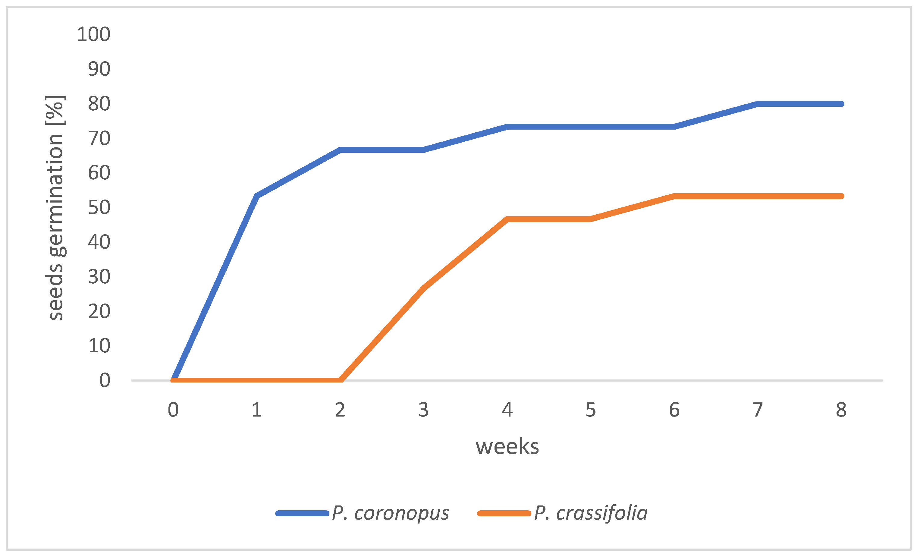
Figure 1. Plantago (P. coronopus and P. crassifolia) seeds germination on the medium for 8 weeks cultivation.
Sustainability 17 07471 g001
Figure 2. Plantago (P. coronopus—(A) and P. crassifolia—(B)) plants growth on different culture medium variants (P1–P3) after 6 weeks of cultivation, bar: 5 cm.
Sustainability 17 07471 g002
Figure 3. Effect of kinetin (P1), BAP (P2), and meta-topolin (P3) on the multiplication factor of P. coronopus and P. crassifolia growth after two cultivation terms. Data represent mean values of eight replicates ± SE (n = 8). Different lowercase letters indicate statistics significant differences between media for P. coronopus (analyzed separately for 6 and 12 weeks); while different uppercase letters indicate differences for P. crassifolia. Asterisks denote significant differences between species for the same medium and time point (p ≤ 0.05). One-way analysis of variance (ANOVA), followed by Tukey’s HSD test, where applicable, was used.
Sustainability 17 07471 g003
Figure 4. Selected physiological parameters of Plantago (P. coronopus and P. crassifolia) plants after 6 and 12 weeks of cultivation on different culture media; (a) total chlorophylls; (b) carotenoids; (c) proline; (d) total phenolic compounds. Different lowercase letters indicate significant differences between media for P. coronopus and P. crassifolia after 6 weeks; uppercase letters indicate differences between media for species after 12 weeks. Asterisks denote significant differences between species for the same medium and time point (p ≤ 0.05). One-way analysis of variance (ANOVA), followed by Tukey’s HSD test, where applicable, was used.
Sustainability 17 07471 g004
Table 1. Composition of the multiplication media (modified MS medium: P1, P2, and P3).
Medium 1 (P1)Medium 2 (P2)Medium 3 (P3)
MS macronutrients and micronutrients
20 cm3 dm−3 iron (Fe)
2 mg dm−3 glycine
0.1 mg dm−3 thiamine
0.5 mg dm−3 pyridoxine
0.5 mg dm−3 nicotinic acid
0.5 mg dm−3 kinetin
0.5 mg dm−3 IAA
1 mg dm−3 BAP
0.5 mg dm−3 IAA
0.5 mg dm−3 meta-topolin
0.5 mg dm−3 IAA
0.5 g dm−3 mesoinositol
0.65 g dm−3 calcium gluconate
20 g dm−3 saccharose
0.6 g dm−3 activated carbon
pH 5.8
7.5 g dm−3 agar
Table 2. Effect of kinetin (P1), BAP (P2), and meta-topolin (P3) on selected indices of growth of P. coronopus and P. crassifolia after two terms of cultivation.
Plant SpeciesTime [Weeks]Culture MediumShoots Height [cm]Roots Length [cm]Number of RootsNumber of LeavesWater Content (WC) [%]
P. coronopus6 P111.86 ± 0.87 a9.10 ± 1.08 b13.22 ± 0.95 b22.64 ± 1.99 a93.70 ± 0.94 a
P29.72 ± 0.90 b10.42 ± 0.85 b13.14 ± 1.02 b17.67 ± 1.47 b95.78 ± 1.02 a
P310.23 ± 0.31 b11.27 ± 0.48 a15.98 ± 0.82 a21.5 ± 1.67 a95.48 ± 1.34 a
12 P111.81 ± 0.81 A7.67 ± 0.60 C9.36 ± 0.56 B31.00 ± 2.08 A*93.91 ± 0.75 A
P28.78 ± 0.77 B10.5 ± 0.67 B11.28 ± 1.24 B24.20 ± 3.27 B*94.70 ± 0.98 A
P310.64 ± 1.37 A12.24 ± 0.94 A17.44 ± 1.42 A22.14 ± 2.38 B93.65 ± 1.14 A
P. crassifolia6 P112.50 ± 1.70 b11.43 ± 1.20 a*15.46 ± 2.06 b24.40 ± 3.29 a*95.26 ± 1.52 a
P28.79 ± 1.23 c10.92 ± 0.57 a20.08 ± 1.94 a*16.14 ± 0.83 b96.04 ± 1.68 a
P316.73 ± 0.44 a*11.21 ± 1.04 a17.14 ± 1.78 ab20.42 ± 2.10 a93.68 ± 0.98 a
12 P113.93 ± 0.89 B*15.17 ± 1.22 B*16.32 ± 1.14 B*18.00 ± 2.30 BA93.55 ± 1.18 A
P212.81 ± 1.09 B*13.57 ± 2.04 B*24.5 ± 2.22 A*16.11 ± 1.84 B93.82 ± 2.02 A
P319.53 ± 2.92 A*19.14 ± 1.54 A*21.62 ± 2.04 A*23.75 ± 1.79 A92.26 ± 1.44 A
Data represent mean values of eight replicates ± SE (n = 8). Different lowercase letters indicate statistically significant differences between media for P. coronopus (analyzed separately for 6 and 12 weeks), while different uppercase letters indicate significant differences for P. crassifolia. Asterisks denote significant differences between P. coronopus and P. crassifolia for the same medium and time point (6 or 12 weeks; p ≤ 0.05). Statistical analyses were performed using one-way ANOVA followed, where applicable, by Tukey’s HSD test.
Disclaimer/Publisher’s Note: The statements, opinions and data contained in all publications are solely those of the individual author(s) and contributor(s) and not of MDPI and/or the editor(s). MDPI and/or the editor(s) disclaim responsibility for any injury to people or property resulting from any ideas, methods, instructions or products referred to in the content.