Review

Insect-Derived Chitin and Chitosan: A Still Unexploited Resource for the Edible Insect Sector

1
DIL—Deutsches Institut für Lebensmitteltechnik e.V. (German Institute of Food Technologies), 49610 Quackenbruck, Germany
2
Department of Microbiology, Faculty of Veterinary and Animal Sciences, The Islamia University of Bahawalpur, Bahawalpur 63100, Pakistan
3
State Key Laboratory of Agricultural Microbiology, National Engineering Research Center of Microbial Pesticides, College of Life Science and Technology, Huazhong Agricultural University, Wuhan 430070, China
4
Livestock and Dairy Development Department, Poultry Research Institute, Rawalpindi 43600, Pakistan
5
Department of Energy and Chemical Engineering, Incheon National University, Incheon 22012, Republic of Korea
6
Department of Environmental Sciences, Kohsar University, Murree 47150, Pakistan
7
Laboratory of Entomology and Agricultural Zoology, University of Thessaly, 38221 Volos, Greece
*
Authors to whom correspondence should be addressed.
Sustainability 2023, 15(6), 4864; https://doi.org/10.3390/su15064864
Submission received: 19 January 2023 / Revised: 3 March 2023 / Accepted: 6 March 2023 / Published: 9 March 2023
(This article belongs to the Collection Sustainable Insect Farming: Feed the Future)

Abstract

:
Chitin and chitosan are biopolymers that are frequently found in nature and have a broad range of applications in the food, biomedical and industrial sectors, due to their high biological activity. The primary source of chitin and chitosan is shellfish, however, shortages in the supply chain, seasonality issues in their availability, as well as ecological degradation are only a few of the problems with the main chitin resources. Due to the broad spectrum of applications for which chitin can be used, the demand for chitin and its derivatives is increasing. Therefore, the market is looking for widely available, greener alternatives to the main commercial chitin sources. Insects appear as a suitable candidate to fill this gap. During insect rearing and processing, a number of side streams are generated, e.g., exuviae of larvae and pupae, dead adults, etc. which are currently mostly discarded as waste. However, these side streams could constitute a novel and long-term supply of chitin for industrial applications. Recent research has demonstrated the suitability of several edible insect species for the production of chitin and chitosan, wherein the exoskeleton of the black soldier fly and field cricket are rich in chitin, making them a good source for chitin and chitosan extraction and purification among other farmed insect candidates. Moreover, several potential uses have been identified for insect-derived chitin and chitosan. Thus, this review aims to present recent advances in the production of chitin and chitosan from edible insects, specifically on their extraction and purification, as well as on their applications for agriculture, food and nutrition, biomedicine and bioplastic production.

1. Introduction

1.1. Chitin

Chitin is a polysaccharide made up of two N-acetyl-D-glucosamine units joined together by covalent-1,4 bonds, first reported in 1811 by a French chemist, Henri Braconnot [1,2,3,4]. It is predominantly composed of amino acids and deoxy-D-glucose residues, whereas its name comes from the Greek word “chiton”, which means “covering”. The structure of chitin is given in Figure 1. Chitin is found in animals, the cell walls of fungi, green algae, and yeast, as well as in crustacean shells and insect cuticles in various amounts [5,6]. In fungi, chitin is present as a chitin–glucan complex, which is quite hard to deconstruct into its component polysaccharides and can therefore only be extracted via chemical means [7]. There are three allomorphs of chitin namely α-, β-, and γ-chitin. The chains of α-chitin are aligned in a parallel manner, and the majority of arthropods and crustaceans have this form of chitin, whereas β-chitin, which can be found in sea diatoms, has an antiparallel chain structure. In comparison, the structure of the γ-chitin chain is slightly more complex than that of other types of chitin and the existence of γ-chitin is a matter of controversy [8,9,10]. The most stable form of chitin is γ-chitin, while β-chitin can easily be changed to γ-chitin by treating it with lithium thiocyanate or letting it settle out with formic acid [11,12].
The only chemical or inorganic solvents that can dissolve chitin are very concentrated mineral acids. This is because of the strong intramolecular and intermolecular hydrogen bonds that are produced by chitin [13,14]. Lithium chloride/dimethylacetamide, lithium chloride/N-methyl-2-pyrolidone, and calcium chloride dihydrate/methanol are well-studied polar solvent systems for chitin application [15]. Ionic liquids [16,17], deep eutectic solvents [18], and alkali/urea aqueous solutions [19] are used to dissolve chitin. These solvents are environmentally friendly and have been shown to be effective. Several ionic liquids, including 1-allyl-3-methylimidazolium bromide, 1-butyl-3-methylimidazolium acetates, and tetrabutylphosphonium amino acid salts, are compatible with chitin [20]. Since the intra- and intermolecular hydrogen bonds that ordinarily keep chitin together are broken during the alkaline freeze–thawing process, it has been demonstrated that this approach may improve chitin solubility and make it more receptive to subsequent modification [21].

1.2. Chitosan

Chitin is often converted into its analogue chitosan by the addition of an amino group in place of the acetyl group during the transformation process [1,2,3,4] (Figure 1). Chitosan is an aminated polysaccharide that is found in high concentrations in nature and is produced through the chemical, or less frequently, the enzymatic, deacetylation of chitin [4]. By deacetylation with alkali, chitosan is commercially made from chitin. Chitosan has a unique set of useful properties that come from its chemical characteristics [13,22]. Some of chitosan’s most distinguishing features are its molecular weight and its level of acetylation. Chitosan’s solubility, its ability to form materials, its biodegradability, and a wide range of bioactive characteristics are all influenced by these two factors. Chitosan has long been known for its wide range of uses, as its biological characteristics make it ideal for agricultural, food, and environmental engineering applications [23,24]. For instance, as a result of its polycationic character, it has found usage in the chemical and textile industries, in membranes, as well as in wastewater treatment [25,26,27]. It also has significant uses in the industry of cosmetics, papermaking, and tissue engineering [28,29]. More recently, chitosan-based nanoparticles and hydrogels have been produced with a variety of uses in the biomedical, pharmacological, and agricultural fields [30,31,32,33].
Both the degree of deacetylation (DD) and the molecular weight of chitosan have a substantial influence on its physicochemical properties, such as its solubility, hydrophilicity, and crystallinity, as well as on the biological properties of chitosan. For example, the DD of chitosan is proportional to its molecular weight. Analytical techniques, such as infrared spectroscopy, ultraviolet–visible spectrophotometry, and nuclear magnetic resonance spectroscopy, are among those that are used in the process of determining the DD [27,34]. In general, people think of chitosan as a polymer that dissolves in weak organic acids, which would indicate a deacetylation level of around 40–45%. The worldwide market for chitin and chitosan derivatives had a value of $3.8 billion in 2020, and it is predicted to reach $12.3 billion by 2027, growing at a CAGR of 18.4% over the period 2020–2027 [35].

2. Chitin and Chitosan Sources

The main source of chitin and chitosan for commercial use are the waste streams of the marine food industry [36]. This waste mostly consists of the exoskeletons of crustaceans. In 2016, eight million tons of crustaceans were gathered for human food worldwide, and 40% of that amount consisted of discarded exoskeletons with a chitin percentage of 15–40% [37]. However, fisheries waste is very seasonal, since commercial crab fishing begins in spring following the spawning season [6,38]. After crustaceans, fungi are the second most common source of chitin [5]. Fungi have recently gained a lot of interest as an alternative and vegan source of chitin and chitosan, which has led to a number of research efforts focusing on fungi and their potential economic worth [39]. Chitin constitutes 1 to 15% of the bulk of fungal cell walls, and its structure is similar to that of crustacean cell walls [40]. Chitin is found in a broad variety of fungal phyla, including Basidiomycota, Ascomycota, and Zygomycota, despite the fact that it is not present in every kind of fungal cell [41]. In most cases, procedures as stringent as those needed for the production of crustacean chitin are also necessary to produce chitosan from fungal chitin [42]. Chitosan, on the other hand, may be easily extracted from the cell wall of some fungal species without the need for the cleavage of the acetyl groups that are present in the cell wall [43]. Absidia spp. (Zygomycetes), Aspergilus niger (Ascomycetes), Mucor rouxii (Zygomycetes), Rhizophus oryzae (Zygomycetes), and Lentinus edodes (Basidiomycetes) are some of the species that have been extensively studied for their potential to directly produce chitosan [44]. Despite this, neither the production of fungal chitin nor chitosan have reached the level of industrialization. A variety of arthropods, including centipedes and woodlice, have been studied for their potential to serve as chitin suppliers [45]. Notably, giant centipedes’ body segments may be used to make chitin rings in a three-dimensional format. Chitin has also been derived from the guano of insectivorous bats, as well as from poriferans, bryozoans, and tardigrades [38].

3. Methods for Chitin and Chitosan Extraction

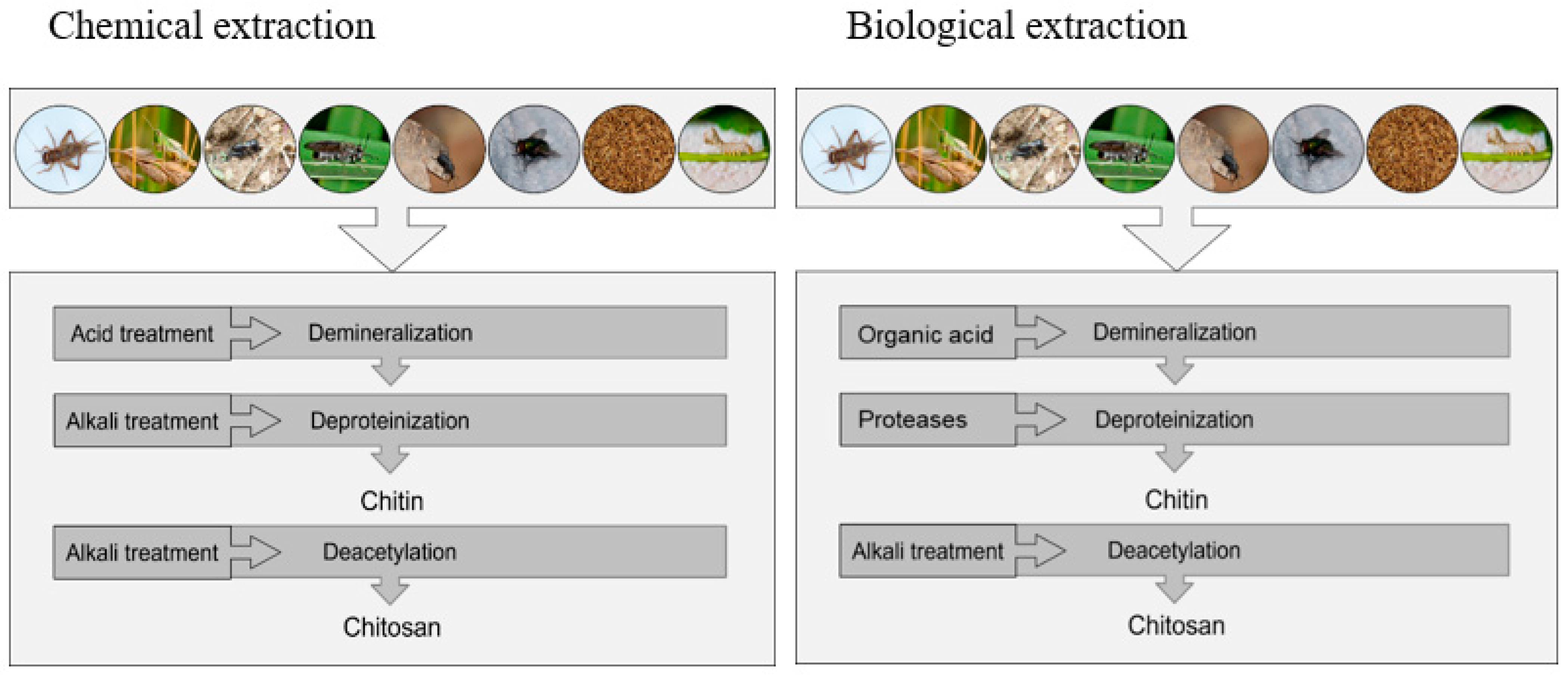
Chitin and chitosan are extracted through two kinds of extraction processes: chemical or biological [46]. Calcium carbonate and proteins can only be dissolved using powerful acids and bases, respectively, in order for the chemical processes to work [46,47]. Chemical procedures remain the most popular treatment at industrial scale due to the speed with which they can extract the desired material [47]. Extracting chitin and chitosan using biological methods might reduce the need for acidic and alkaline processes that are not so environmentally friendly [48]. Both lactic acid bacteria and protease-producing bacteria have been used in the demineralization and deproteinization processes [49]. Enzymatic techniques are used to carry out chitin deacetylation, and the enzyme chitin deacetylase is responsible for this process. The differences between chemical and biological extraction of chitin and chitosan are shown in Table 1.
Extensive studies on the use of these approaches to extract chitin and chitosan from a variety of materials have been published. The demineralization and deproteinization steps in the chitin synthesis process are used to remove inorganic calcium carbonate and proteins, trace amounts of colors and lipids [51,52]. Acid treatment is used to remove minerals, such as calcium carbonate (CaCO3), from chitin. Acids such as hydrochloric acid (HCl), nitric acid (HNO3), sulfuric acid (H2SO4), or acetic acid (CH3COOH) are often used in the process, with the diluted hydrochloric acid being used in the conventional demineralization procedure [53,54]. Different extraction times, temperatures, particle sizes, acid contents, and solute-to-solvent ratios have been proposed to obtain the desired demineralization results. Since it takes two HCl molecules to convert one molecule of calcium carbonate into calcium chloride, the quantity of calcium chloride generated is directly related to the concentration of the acid. The stoichiometric concentration of the minerals must be larger than or equal to an acid concentration for the reaction to be successful [55,56]. Since it is challenging to extract all the minerals, a larger volume of acid solution or a more concentrated acid solution is needed (due to the heterogeneity of the material). In most cases, demineralization with HCl takes 2–3 h and involves stirring [55]; however, reaction times may yet vary widely amongst methods, ranging from 15 min to 48 h [56]. A minor drop in ash content and polymer breakdown are both caused by a longer demineralization period, even up to several days [57,58]. To promote solvent penetration into the chitinous matrix, heat can be used to facilitate the demineralization process. For this reason, demineralization is most effective at higher temperatures [58].
During the process of deproteinization, the chemical connections that hold chitin and proteins together are broken. Heterogeneous application of chemicals that may depolymerize the biopolymer is employed here. The NaOH, Na2CO3, NaHCO3, KOH, K2CO3, Ca (OH)2, Na2SO3, NaHSO3, CaHSO3, Na3PO4, and Na2S are among the compounds employed as reagents for chemical deproteinization [38]. In each study, the reaction circumstances differ significantly. The most common use calls for NaOH at concentrations ranging from 0.125 to 5.0 M, temperatures ranging from 0 to 160 °C, and periods that can cover a broad spectrum (from several minutes to several days) [59]. The NaOH not only deproteinizes, but also partially deacetylates chitin, hydrolyzes the biopolymer, and so decreases its molecular weight. Enzymatic deproteinization was tested as a replacement for chemical deproteinization, however the findings did not prove that all proteins were effectively eliminated [60]. Rarely, further bleaching is needed to eliminate any leftover color. Pigments must be removed during decolorization to get a clear product. Chitin sources of any composition undergo these reactions. Protein and pigment residues are removed for medicinal and culinary uses. Several decolorizing chemicals (acetone or organic solvent) remove color from crustacean shell and insect exoskeleton chitin [61,62].
Reactive amino groups are substituted for acetyl groups on chitin during the deacetylation process. The amount of deacetylation determines the proportion of free amino groups in a structure, which may be utilized to distinguish between chitin and chitosan. The DD of chitosan must be taken into account since it affects both its physical and biological properties, including its acid–base ratio, electrostatic properties, biodegradability, and its capacity to bind metal ions [63,64]. Chemical technologies make it possible to produce chitosan from chitin on a large scale in an industrial setting [65,66]. Use of alkali–NaOH [67] or acids to deacetylate chitin for crustacean shell debris and insects is the preferred method of chemical deacetylation [63,64]. Alkali is suggested as a superior chemical alternative due to glycosidic linkages’ vulnerability to acid [38]. There are several variables that might affect the final product’s qualities when it is being deacetylated. Temperature and the amount of time spent processing the material were the most crucial factors to consider in relation to DDA and molecular weight [63,64].
Alterations in the physicochemical properties of the extracted chitin, the use of expensive chemicals in the purification process, and the discharge of toxic effluent wastewater into the environment are some of the potential drawbacks associated with modifying the conventional method for chitin chemical extraction. These issues wreak havoc on the ecosystem [68,69] and diminish the number of vital proteins available for animal feed [70]. Green extraction technologies have been gaining popularity because of their cleaner and more environmentally friendly approaches [71,72].
Microorganisms such as Lactobacillus [73,74], Pseudomonas aeruginosa K-187 [49], and Bacillus subtilis [75] can be used in the biological extraction process to reduce chitin degradation and impurities to acceptable levels for specific applications. For example, Tan et al. [73] and Gopal et al. [75] compared different chemical and microbiological techniques for isolating chitin and chitosan from shrimp waste and concluded that the use of microbes was a more effective method than the use of chemicals. The microbial technique needed much less time, had a straightforward process, produced chitin mostly from shrimp waste, and made use of enzymatic [76,77], microwave-assisted [77,78], and ultrasonic-assisted [79] reactions, as well as phytoextraction [80].
Ionic liquids have the potential to be used as a volatile organic solvent for the production of chitin; however, many ionic liquids have limitations that make them unsuitable for use in biological applications [81]. These downsides include high cost and toxicity [81]. Therefore, deep eutectic solvents (DES) are an environmentally friendly substitute for conventional methods of chitin synthesis [82,83]. When compared with older technologies, DES stands out for its many advantages, including its low or non-toxicity, cheap cost, easy production, and biodegradability [84]. Extraction using DES has been used to create chitin in shrimp [85] and lobster [86], in addition to the black soldier fly, Hermetia illucens (L.) (Diptera: Stratiomyidae) [85]. Brigode et al. [87] recently discovered that acid detergent fiber and acid detergent lignin (ADF–ADL) production processes have been used in order to create chitin from H. illucens. More research, however, is needed to understand the development of carbon-footprint-reducing, environmentally friendly processes for extracting chitin and chitosan from insects. The diagrammatic representation of the extraction of insect chitin and chitosan through chemical and biological processes is shown in Figure 2.

4. Insects as an Alternative Chitin and Chitosan Source

Humans have long been aware of the economic benefits associated with beneficial insects [88,89]. The silkworm, Bombyx mori (L.) (Lepidoptera: Bombycidae), was first farmed commercially in China for the production of silk at some time around the Neolithic era [90]. Over time, humans have determined how to rear many insect species and use them in various ways [90,91]. After the middle of the 20th century, several insect species were mass-produced as biological control agents. For instance, the large production of the screw worm, Cochliomyia hominivorax (Coquerel) (Diptera: Calliphoridae) for biocontrol began in Florida in the late 1950s [92,93]. Large-scale, commercial insect farming for human consumption and as animal feed is a relatively new development; the first industrial plant was opened in 2015 [93]. Since then, there has been a rapid expansion of the insect breeding industry [90]. For instance, in terms of value, the edible insect market is expected to record a CAGR of 28.3% from 2022 to 2030 to reach $9.60 billion by 2030 [92]. According to various reports, there are hundreds of startups and established companies globally that are actively involved in the production and sale of insect-based food products, including whole insects, insect-based flour, and insect-based protein powder [94]. In Europe alone, around 6 kt of protein meal of H. illucens, the yellow mealworm, Tenebrio molitor L. (Coleoptera: Tenebrionidae) and the lesser mealworm, Alphitobius diaperinus (Panzer) (Coleoptera: Tenebrionidae), were produced in 2019–2020 as animal feed [93,94].
Apart from being a valuable nutrient source, insects are a promising and sustainable source of chitin and chitosan, though they have not received a lot of attention in the past in this regard [44]. When compared with crustaceans, insects have a number of advantages, including the facts that they are not affected by seasonality and that they can be easily mass reared as a result of their short life cycle and their high reproductive rate [95]. In addition to this, industrial insect rearing facilities are currently being established all over the world [95]. Moreover, insects can be utilized as bioconverters, i.e., they can be raised on organic side streams and wastes [89,96,97,98]. In that context, insects could also be used as a viable alternative to crustaceans for the production of chitin and chitosan, resulting in greater ecological and economic sustainability [99,100]. The exoskeleton is a protective outer layer that covers the body of many types of insects, crustaceans, and arthropods. The insect exoskeleton is the hard, protective shell that covers the inner soft body tissues [93,100] and can serve a variety of purposes, including acting as a protective covering and assisting in metamorphosis [101]. Indicatively, chitin constitutes up to 25% of the exoskeletons of H. illucens, rendering it one of the principal chemicals involved in the processes of the industrial scale production of insects for potential extraction through the insect farming sector [100,101]. Based on the above, this chitin-rich byproduct of insect farming could provide a novel and long-term supply of chitin for industrial applications [95,100,102]. Given the sustainable profile of insect farming and the predicted expansion of the insect sector in the near future, leftovers from insect farming could constitute a highly attractive future supply of chitin [94]. Chitin is naturally organized into tiny threads that are enmeshed in a protein matrix. Generally speaking, whole insects include 30–60% protein, 10–25% fat, 15–25% chitin, and 2–10% minerals such as calcium, phosphorus, potassium, and magnesium salts [100,103]. In the context of farmed insects, the chitin content can vary depending on the species, the conditions under which they are raised, and the stage of their life cycle. Some species of insects, such as the black soldier fly (BSF) have been reported to range from 10–20% chitin, however, some studies have reported higher chitin content, up to 25%, house flies have been reported to range from 10–20%, yellow mealworms have been reported to range from 16–17%, superworm 3–14%, house cricket 4–7%, field cricket 23%, and silk worm has been reported to range from 3–20% (Table 2). Similarly, the chitosan extraction from the chitin of insect species proposed for food and feed was up to 53% in BSF, 70% in house fly, 50% in yellow mealworm, 83% in superworm, 5.8% in house cricket, 94% in field cricket, 55% in desert locust and 97% in silkworm (Table 3). Proteins, lipids, minerals, and pigments that are present in the insect cuticle are removed during the purification process in order to extract the chitin [100,104,105]. Extraction techniques and the physicochemical qualities of insect chitin have received little attention until recently. The up-to-date available information on the production and purification of chitin and chitosan from the major edible insect species is presented in the following sections.

4.1. Black Soldier Fly

Hermetia illucens is a ubiquitous insect species that can be found all over the globe, especially in Asia, Europe, and the southern United States [130,131]. Is it not considered to be a pest species due to the fact that adults do not need to feed, do not bite or sting, and have not been described as vector of any specific diseases [89,132,133]. Various agricultural byproducts of either animal or plant origin are among the substrates that H. illucens larvae can break down from animal manure to food waste [96,134,135]. This fact opens up new possibilities for a novel waste bioconversion technique based on insects [93,136]. Moreover, H. illucens larvae are a good source of protein that may also be exploited for the production of livestock animal feed and fat that can be utilized either as feed or as biodiesel [134,137,138]. Production of protein and fat from H. illucens was shown to be promising for poultry, aquaculture, and other animal feed [89,98,139]. Chitin may also be extracted from H. illucens, the physicochemical structure of which has been well described [100,140,141]. Pure chitin and chitosan can be extracted from the H. illucens only during the early stages of development, up to the fifth instar larval stage. Later on (prepupae, pupae, and adults), a pigment (melanin) is produced and is chemically bound to the insect’s chitin exoskeleton [142,143].
Several methods have been established for chitin extraction, most of which are listed and presented in Table 2. All methods rely on the successive removal of impurities from a raw material source [100]. In one of the proposed methods, the raw materials (pupal exuviae, corpses, etc.) were first cleaned and dried until they achieved a consistent weight, and then pulverized in a laboratory grinder [140]. After demineralizing with 1 M HCl for 1 h at room temperature, the water was drained and re-used. All trace minerals were washed out of the demineralized powder by cleaning it with distilled water. Deproteinization was carried out at 80 °C for 24 h using 1 M NaOH. Using 1% KMnO4, the extract was filtered and decolored. A solution containing 4% oxalic acid was used to remove the surplus KMnO4. The finished product was filtered, washed with distilled water, and dried to a white-gray color. In another method for chitin isolation from H. illucens, petroleum ether was used to defat the insect biomass in two stages [140]. The defatted material was subsequently demineralized using a 2 N HCl and a 1 M NaOH solution. The precipitate was then rinsed in water and dried after centrifugation. Depending on the degree of acetylation, the chitin output with this method ranged from 11.7 to 14.6% [107], however, no information was provided regarding the chitin purity.
Khayrova et al. [60] used a novel technique for extracting amorphous chitin from H. illucens larvae, including an additional deacetylation step. In that study, direct extraction with high phosphoric acid yielded amorphous chitin. Physicochemical procedures were then used to evaluate the purity of the resulting chitin (elemental analysis, IR spectroscopy, measurement of primary amines, amino acid analysis). Afterwards, the usual process, which includes demineralization, deproteinization, and deacetylation stages, was used to create crystalline chitin and high molecular weight chitosan. Treatment with hexane for 6 h has been proposed to defat H. illucens larvae prior to chitin extraction [109]. When Purkayastha et al. [112] extracted chitin from H. illucens pupal exuviae and adults, chitin contents of 9 and 23% were reported, respectively. In both cases, chitin was identified as α-chitin. The dried pupal exuviae and adult samples were first placed in liquid nitrogen, then ground to a fine powder and kept at 20 °C in an airtight container until chitin extraction. In order to remove minerals, the powdered pupal exuviae and adult samples were demineralized and heated to 100 °C for 30 min. Following demineralization, the samples were washed repeatedly with distilled water until the sample solution’s pH was neutral. Using 250 mL of 1 M NaOH solution, the samples were deproteinized in a water bath at 80 °C for 24 h. Afterwards, the sample solution was neutralized by repeating the deproteinization process with distilled water. As a last step, the precipitate was washed thoroughly with deionized distilled water and treated with a 1% potassium permanganate solution for an hour. The final product was a brownish precipitate of chitin that was dried for two days at 50 °C. Caligiani et al. [141] reported a method for chitosan production from larval exoskeletons and compared the characteristics of the final product with crab shell chitosan. Following this method, the demineralization efficiency of using formic acid (5 mol/kg of larval exoskeletons) was 89%, whereas there were 8.8% minerals. For the deproteinization of small volumes, 1.9 M NaOH was used at 81 °C for 2.8 h; for larger volumes (10 L), 2 M alkali was used at 80 °C for 2 h. Using a linear regression model, the influence of deproteinization parameters on the chitin content was evaluated and it was found that the chitin content of the deproteinized material ranged from 83% on the lab scale to 87% on the industrial scale. Two types of deacetylation reactions were carried out: at 120 °C, heterogeneous deacetylation resulted in a 72% DD and a maximum chitosan yield of 43%. At 4 °C, homogeneous deacetylation resulted in a DD of 34% and a low chitosan yield of 13%. When chitosan was dissolved in acetic acid, it had good film-forming characteristics and a high level of viscosity. The study’s findings indicate that insect chitosan has quantitative characteristics that are equivalent to crustacean chitosan, which is highly reliant on the conditions under which it is produced.
Wang et al. [100] studied the physicochemical properties of the chitin matrix throughout the developmental stages of H. illucens (larva, prepupa, puparium, and adult) and reported chitin contents of 3.6, 3.1, 14.1, and 2.9%, respectively. Fourier transform infrared spectroscopy, thermogravimetric analysis, and X-ray diffraction research revealed that H. illucens chitin was all chitin with equal thermal stability. From larva to adult, the chitin crystalline index increased progressively reaching 33.09, 35.14, 68.44, and 87.92%, respectively. The materials were first demineralized for 1 h at 55 °C in a 2 M HCl solution. Deproteinization occurred in a 2 M NaOH solution at 200 rpm and 50 °C for 18 h. The material was filtered, then soaked in 3.6% HCl for 30 min, and then mixed with NaClO that was diluted by a factor of 10. After drying the samples, these were heated in an oil bath at 80 °C for 4 h to remove the pigments.

4.2. Housefly

The housefly, Musca domestica L. (Diptera: Muscidae), is a synanthropic species with global distribution [144]. The body of housefly larvae, pupae, and adults contains around 16 unique amino acids, along with a high level of protein and an extremely pure form of chitin, which can be used in a variety of industrial applications in the food, agricultural and the medical sector [145]. Several methods have been proposed for chitin and chitosan production from M. domestica [114,116]. For chitosan production, Ai et al. [116] treated powdered larvae with a 1 mol/L aqueous sodium hydroxide solution for 6 h at 95 °C to remove the protein. Deionized water was then used to wash the mixture to remove any impurities. The precipitate was neutral and crude. It took 4 h to decolorize the crude chitin with a potassium permanganate solution (10 mg/mL) and 3 h to treat it with an oxalic acid solution (10 mg/mL) to get the final color of the crude chitin. Deionized water was used to neutralize the chitin, which was subsequently freeze-dried. A sodium hydroxide solution at 400 mg/mL was used to N-deacetylate the crude chitin for 8 h at 70 °C. The chitosan was obtained and kept at 20 °C after filtering, neutral washing with deionized water, and freeze-drying.
Zhang et al. [115] proposed a method for the extraction of food-grade chitin and chitosan by macerating 4-d old M. domestica larvae for 10–15 min in a home juicer. The crude cuticle granules that were formed were filtered through a mesh screen, thoroughly washed, and then freeze-dried. By heating the dry cuticle in a 1 mol/l NaOH solution at 100 °C for 3 h, protein and fat were removed. Meanwhile, to extract crude chitin, the mixture was rinsed with water until it reached a neutral pH, then filtered through a mesh sieve to remove any remaining water before being freeze-dried. To extract chitin and chitosan from M. domestica pupa shells, the shells were treated with hydrochloric acid and sodium hydroxide [114]. Decalcification and deproteinization were performed using solutions of 2 N HCl and 1.25 N NaOH, respectively. In order to accomplish deacetylation during chitosan extraction, 50% NaOH solution was utilized. With this method, the chitin and chitosan content extracted from M. domestica pupa were 8.02 and 5.87%, respectively. The first alkali treatment at 105 °C with a 50% NaOH (w/w) solution deacetylated chitosan (made from chitin C1 and C2) by 89.76 and 92.39% after 3 and 5 h, respectively. The viscosities of the chitosan produced from chitin C1 and C2, were, 33.6 and 19.2 centiPoise (cP), respectively [114].

4.3. Yellow Mealworm

Tenebrio molitor has recently attracted a lot of scientific and commercial interest as a source of protein for food and feed all around the world [146]. Briefly, it has a high feed conversion ratio (which describes the efficiency in converting feed into body weight) in comparison with traditional livestock animals, it has high growth rate and reproductive output, and its edible portion is almost 100%, in contrast with chickens and pigs (55%) or cattle (40%) [147]. Mealworm larvae have 47–49% protein, 38–43% fat, 7–8% fiber, 0.2–3% nitrogen-free extract, and 2–3% ash (based on their dry matter (DM) composition) [148,149]. Regarding chitin and chitosan, Song et al. [117] studied the extraction of chitin and chitosan from the exuviae and whole bodies of T. molitor larvae. Demineralization and deproteinization were achieved by chemically removing chitin from the exuviae and the whole bodies with acid and alkaline solutions, respectively. The demineralization and deproteinization percentages were 32.56 and 73.16% from larval exuviae, respectively, while from the whole body the respective figures were 41.68 and 91.53% of dry mass. To produce chitosan, chitin particles were extracted from the exuviae and the larval bodies and cooked at various temperatures in varied concentrations of NaOH. The average chitin yields from the exuviae and whole bodies were 18.01 and 4.92% of DW, respectively. The chitosan production from the whole bodies resulted in an average of 3.65% of dry mass. Over 50% of the chitosan that was produced from the whole bodies had been deacetylated, while the overall chitosan viscosity in the body ranged from 48.0 to 54.0 cP. When compared with dry weight, the amount of chitin that was present in the dry byproducts of the entire body were 13.07%. The chitosan concentration of the dry byproducts was 14.48. These results suggest that the exuviae, as well as the whole bodies of T. molitor larvae, could be successfully used as a chitin and chitosan source.
Similarly, Luo et al. [131] developed a number of procedures for extracting chitin and chitosan from T. molitor. Larvae were first decontaminated by being soaked in ethyl alcohol for 1 h at room temperature, then were washed with deionized water, and finally dried in an oven overnight at 60 °C until a constant weight was obtained. After that, the particles were crushed to a size that would fit through a screen with a 100-mesh sieve. Samples were demineralized in a water bath at 30 °C with a solution of 1 mg per liter of hydrochloric acid for 2 h, so that calcium carbonate and other calcium salts could be removed (1:15 solid-to-solution ratio). Before the samples were considered to be pH-balanced, they were filtered and washed many times with deionized water. In order to remove protein and fat from the samples, they were first placed in a solution containing sodium hydroxide with a concentration of 1 M (w/v 15:1), and were then refluxed for 2 h at 90 °C. The insoluble substance was neutralized in pH after filtering and washing with deionized water. The samples were decolored by first oxidizing them in an aqueous solution of potassium permanganate (2%), and then reducing them in an aqueous solution of oxalic acid (2%), both for 2 h. After filtering out and neutralizing the chitin in the samples, these were placed in a preheated oven for drying overnight at 60 ℃. Chitin in its purest form was obtained, and it had a solid appearance that was somewhat yellow. The deacetylation of the separated chitin was performed by placing an aqueous solution containing 60 wt% NaOH (w/v:15) in a coil bath heated at 100 °C for 8 h, while continuously shaking the mixture at 200 rpm. The samples were separated using a filter and were then washed with distilled water until a pH of 7 was reached.

4.4. Superworm

The superworm, Zophobas morio (F.) (Coleoptera:Tenebrionidae), is an insect species which has not attracted considerable interest as a nutrient source, being overlooked by both researchers and insect growers [150]. Late instar larvae can grow up to 50–60 mm long, whereas they contain significant amounts of lipids (40–41% DM) and crude protein (44–47% DM), as well as antimicrobial peptides and essential amino acids [151,152,153]. Additionally, Z. morio larvae are rich in iron and zinc, as well as in vitamins [154,155,156]. Due to its large size, as well as its good nutritional composition, Z. morio is considered to have great potential as food and feed source [150].
The chitin content of Z. morio larvae is between 4.60 and 8.40% [120,121,157,158]. Soon et al. [121] provided a full physicochemical characterization of chitin and chitosan isolated from Z. morio larvae. Briefly, after larvae had been cleaned under running water to eliminate any leftover contaminants, they were dried in a convection oven at 70 °C for 12 h. After being heated in a water bath containing a 1 M HCl solution at 35 °C for 30 min, the samples were filtered and then washed with distilled water to neutralize the pH. Next, the demineralization process was completed by heating the samples. Then, to observe what would happen, the deionized samples were mixed with 0.5, 1, and 2 M NaOH solutions. The 20 h of deproteinization were carried out in an 80 °C water bath. The samples were filtered and given a thorough washing with distilled water in order to bring their pH levels back to normal. Chitin samples were treated with ice-cold acetone and then subjected to a 30 min treatment at room temperature in order to remove organic pigment. Chitin was air-dried for some time after being extracted from the acetone and was then further dried in a convection oven at 70 °C. The resulting chitin was deacetylated in a water bath at 90 °C with 50% NaOH for 30 h. The samples were filtered, then twice washed with distilled water to bring back their neutral pH. The samples of chitosan were then dried overnight at 70 °C in an oven. The overall production was 5.43%, with the least concentrated alkali producing the greatest yield. The findings show that the yields of chitosan extracted from chitin generated in 0.5, 1, and 2 M alkali were 78%, and 83%, respectively. The degree of chitin acetylation was 82.4, 92.8, and 99.4% in the presence of 0.5, 1, and 2 M NaOH, whereas the degree of chitosan deacetylation was 81.1, 64.8, and 74.1% in the same solutions.
Shin et al. [120] used a modification of previously described techniques [159,160] to extract chitin and chitosan from larvae and adults of Z. morio. The protein, fat, and color were all removed from the dried insect powder (20 g) by subjecting it to an alkaline treatment with a 10% (w/v) sodium hydroxide solution at 80 °C for 24 h. The samples were then washed in distilled water until the pH value was neutralized and then subjected to a second acid treatment with a 7% (v/v) HCl solution at 25 °C for 24 h to remove minerals. After being dried in an air oven at 60 °C for 24 h, the chitin took on a color palette that went from white to light brown. Acetyl groups were removed from the samples by treating them with a 55% (w/v) NaOH solution at 90 °C for 9 h, followed by rinsing with distilled water until the pH value reverted to neutral. After drying in an air oven at 60 °C for 24 h, the weight of the acquired chitosan was measured.

4.5. House Cricket

The house cricket, Acheta domesticus (L.) (Orthoptera: Gryllidae), was first discovered in southwestern Asia. However, being one of the favorable insect species for pet food, its global distribution increased dramatically between the years 1950 and 2000 [161]. It has a considerable amount of protein (20–25 g/100 g dry weight) and fat (4–7 g/100 g dry weight), comparable to those of more common meats, like beef or chicken [162]. In addition to being a good protein source, house cricket fat contains 29–31% polyunsaturated fatty acids [163]. Crickets are often used as adults, whereas for flies and beetles larvae are typically the ones harvested and processed as food and feed. To obtain chitin from house crickets, it is suggested to treat dried crickets with 200 mL of a 1 M NaOH solution for 6 h at 95 °C to achieve deproteinization, providing a sample-to-NaOH ratio of 1:20 [122]. The substance must then be filtered through a 100-mesh screen after being rinsed with distilled water until the pH of the distilled water has been adjusted. The sample is then dried. The deproteinized sample is demineralized using the same ratio of 200 mL of 1 g 100 mL−1 oxalic acid at room temperature for 3 h with gentle stirring, then filtered through a 100-mesh sieve and washed with distilled water until the pH of the distilled water becomes neutral. The sample is decolored by combining 200 mL of 1% sodium hypochlorite solution (1%, w/v) with the sample and allowing it to rest at room temperature for three hours while stirring gently. After neutralizing the pH of the distilled water, the sample is filtered through a 100-mesh sieve and washed with distilled water. Following an overnight drying at 60 °C, the dry weight of the sample is determined. The chitosan can then be purified by deacetylating the chitin sample following a process previously described [164].
An alternative technique for chitin separation from A. domesticus has also been proposed [165]. The relative degrees of demineralization for chemical demineralization, citric acid treatment, Lactococcus lactis fermentation, and microwave therapy were 91.1, 70.5, 97.3, and 85.8%, respectively. Fermentation using Bacillus subtilis, and enzymatic digestion produced materials with a chitin content that was less than half of that produced by alkaline deproteinization. Chemical treatment, or an alternative method that combines the fermentation of L. lactis with the deproteinization of bromelain, are both used to obtain deacetylated chitinous material for use in large-scale chitosan production. Chitosan content (81.9 and 88.0%, respectively), antioxidant activity (59 and 49%, respectively), and degree of deacetylation (66.6 and 62.9%, respectively) were all similarly achieved using the chemical and the alternative methods.

4.6. Field Cricket

The field cricket, Gryllus bimaculatus De Geer (Orthoptera: Gryllidae) is rich in protein (60–70% DM), including all of the essential amino acids, as well as in lipids (10–23% DM) and minerals such as phosphorus, sodium, and calcium, whereas it has a good ratio of omega-3 to omega-6 fatty acids. Regarding chitin, Kim et al. [105] used chemical treatment with an acid and an alkali to extract chitin from G. bimaculatus specimens. To demineralize and deproteinize the chitin, they utilized 2 N HCl and 1.25 N NaOH solutions. Deacetylation of chitosan was achieved using 50% NaOH (w/v) solutions. On a dry weight basis, the percentage of chitosan extracted from G. bimaculatus exoskeletons treated with 50% NaOH (w/v) at 95 °C for 3 h was 79.1–94.2%, whereas the chitin yield was 20.9–23.3%.

4.7. Desert Locust

The desert locust, Schistocerca gregaria Forskål (Orthoptera: Acrididae), is a short-horned grasshopper that is commonly found in the deserts and arid regions of northern and eastern Africa, Arabia, and southwest Asia [166,167]. As for all hemimetobolous species, there are three life stages in the lifecycle of the desert locust, i.e., the egg, the nymph stage that is referred to as a hopper, and the adult stage that has wings [166]. Marei et al. [125] extracted chitin from S. gregaria following the industry standard practices. The demineralization process was completed by treating the samples with a 1 M HCl solution at room temperature with a solution-to-solid ratio of 15 mL/gm for 15 min. The solid fraction obtained after this process was rinsed with distilled water until a neutral pH was reached. Deproteinization was accomplished using an alkaline treatment with 1.0 M sodium hydroxide at 100 °C for 8 h. The resulting chitin was then rinsed with distilled water to bring it back to its neutral state. As a last step before usage, it was boiled in acetone to remove any lingering pollutants after being cleaned with hot ethanol. The cleaned chitin was dried in a vacuum oven at 50 °C until it reached a constant weight. The chitin content of the raw materials was determined by comparing the relative weight changes of the raw materials and the chitin generated after the acid and alkaline treatments. In a follow-up study of the same research group, the insect exoskeletons were immersed and cooked in 1 N NaOH until they yielded a clear solution [124]. Finally, the solid fraction was demineralized, being treated with 36.5% HCl solution at a solution-to-solid ratio of 15 mL/g. The cleaned chitin was dried in a vacuum oven at 50 °C until it reached a constant weight. The next step was an 8-h soak in 50% NaOH at 100 °C, transforming the chitin into chitosan.

4.8. Silkworm

The silkworm, B. mori, is usually reared for silk production. Once the silk is produced, the pupae (chrysalides) are discarded since they are of limited use [168]. To make one kilogram of silk, it is necessary to produce two kilograms of dry pupae. Since the world’s silk output is around 1.6 million tons per year, at least 3 million tons of pupae are accessible each season. Carbohydrates, proteins, and fats are abundant in silkworm pupae. Components in the pupae may be isolated and utilized in particular applications if they are not fed to the animals. For example, oil derived from pupae is utilized for biodiesel production, whereas the proteins found in pupae have the potential to be utilized in food and pharmaceutical applications [169]. As a result, following defatting and protein extraction, carbohydrate-dense pupae are ready for consumption. However, there are differences in the amount of chitin present in silkworms depending on the race and the sex [129]. The chitin content of multivoltine silkworms was greater than that of univoltine silkworms, and male insects had higher chitin content than female insects [129,168,169].
There have been a number of works studying the process of removing chitin from silkworm pupae and processing it into chitosan. Chitin was extracted from silkworm pupa exuviae by treating them with 1 N HCl and then 1 N NaOH, and the resulting chitosan was further treated with 40% NaOH. Chitinase was more effective in breaking down silkworm chitin than beetle chitin, converting around 55% of the N-acetyl groups [127,170]. Pupal chitin outputs were from 2.6 to 2.4%, while chitosan conversion rates ranged from 73 to 97% [128].
There are several methods described for the extraction of chitin from silkworms (Table 2). Pupae fat was extracted using acetone or alcohol in a Soxhlet equipment for 3–4 h. At a ratio of 1:5 and a temperature of 95–100 °C, the deproteinization (DP) process used 5–7% NaOH, whereas the demineralization reaction used 2% HCl. The chitin was dried and bleached with 10% H2O2 after the washing process. Overall, the extraction process was successful in removing 80% of the fat and wax. After the DP phase, the yield dropped to 5.4%. An estimated 4% of the yield was obtained when demineralization was completed. Chitin bleaching was the last process, and the output dropped to 3.6%. Original chitin samples were crystallized between 76 and 86%, based on the yield of each sample [171].
Silkworm pupae cuticles were processed to obtain chitin with 1 N HCl at 100 °C for 20 min, 1 N NaOH at 80 °C for 36 or 24 h and were refluxed with 0.4 % Na2CO3 [170]. Finally, the residue was vacuum dried on P2O5 after being rinsed with distilled water. The output of chitin varied between 15 and 20%. The chitin sample was heated for 4 h at 110 °C in a solution of 40% NaOH containing 0.1% NaBH4. The product was dissolved in water with 2% acetic acid as a solvent, then filtered, and the pH of the resultant supernatant solution was reduced to 8–9. The 70–80% of chitosan was extracted from the precipitate after carefully washing and drying it.
Paulino et al. [128] tested two techniques for comparing the quantity and quality of chitin that may be obtained from silkworm larvae. Chitin from pupae was extracted in a sealed Teflon reactor, which was then cooked in an oven within a stainless-steel reactor. The second extraction technique used a heated plate in an open system (beaker). At first, the dried pupae were subjected to 1 M HCl at 100 °C in a closed reactor for 20 min. The pupae were demineralized at a quantity of 10 mL HCl/g dry weight. Deionized water was filtered and used for several washings and the residue was finally neutralized. At 80 °C, the residue was treated for 24 h with 1 M NaOH (the same concentration as the acid) in the same way. To achieve a pH of 7, the heated solution was filtered off and the precipitate was washed multiple times with deionized water. The chitin was cleaned with 0.4% Na2CO3 and dried at 80 °C in an oven a number of times. During the alkali treatment, the temperature ranged from 65 to 100 °C and lasted from 1 to 24 h; between 2.6 and 4.2% of chitin was produced with this method. Chitin was deacetylated using a NaOH and NaBH4 solution. There was a 73–97% chitosan production after one to six h of alkali treatment at 100 °C.
Finally, Battampara et al. [129] used a multi-step process to isolate and purify chitin and chitosan from silkworm pupae and eggshell. Chitin and chitosan were separated using hydrochloric acid (HCl) and sodium hydroxide (NaOH). The waste materials from the production process, such as pupae and shells, were ground up and treated with hexane to extract the oil. A 1:10 (material: alkali solution) mixture of the defatted material and 10% (w/w) NaOH was heated at 80 °C with constant stirring at 250 rpm for 24 h. After treatment, the dissolved components were filtered out and the solid residue (chitin) was neutralized by washing with distilled water. When the pupae and shells were treated with alkali, the proteins were eliminated, and the shells were decolored. To remove any minerals, the alkali-treated materials were diluted 1:10 with HCl solution containing 7% (v/v) HCl and heated to 25 °C for 24 h. After filtering, the solids were washed with distilled water and then dried for 24 h at 60 °C in a vacuum oven. Deacetylation was used to transform chitin into chitosan. A 55% (w/v) NaOH solution was used to treat the chitin powder for 9 h at 90 °C in a water bath at 150 rpm. Alkali solution was eliminated after treatment, with residue being rinsed in distilled water to achieve a pH balance.

5. Applications of Chitin and Chitosan

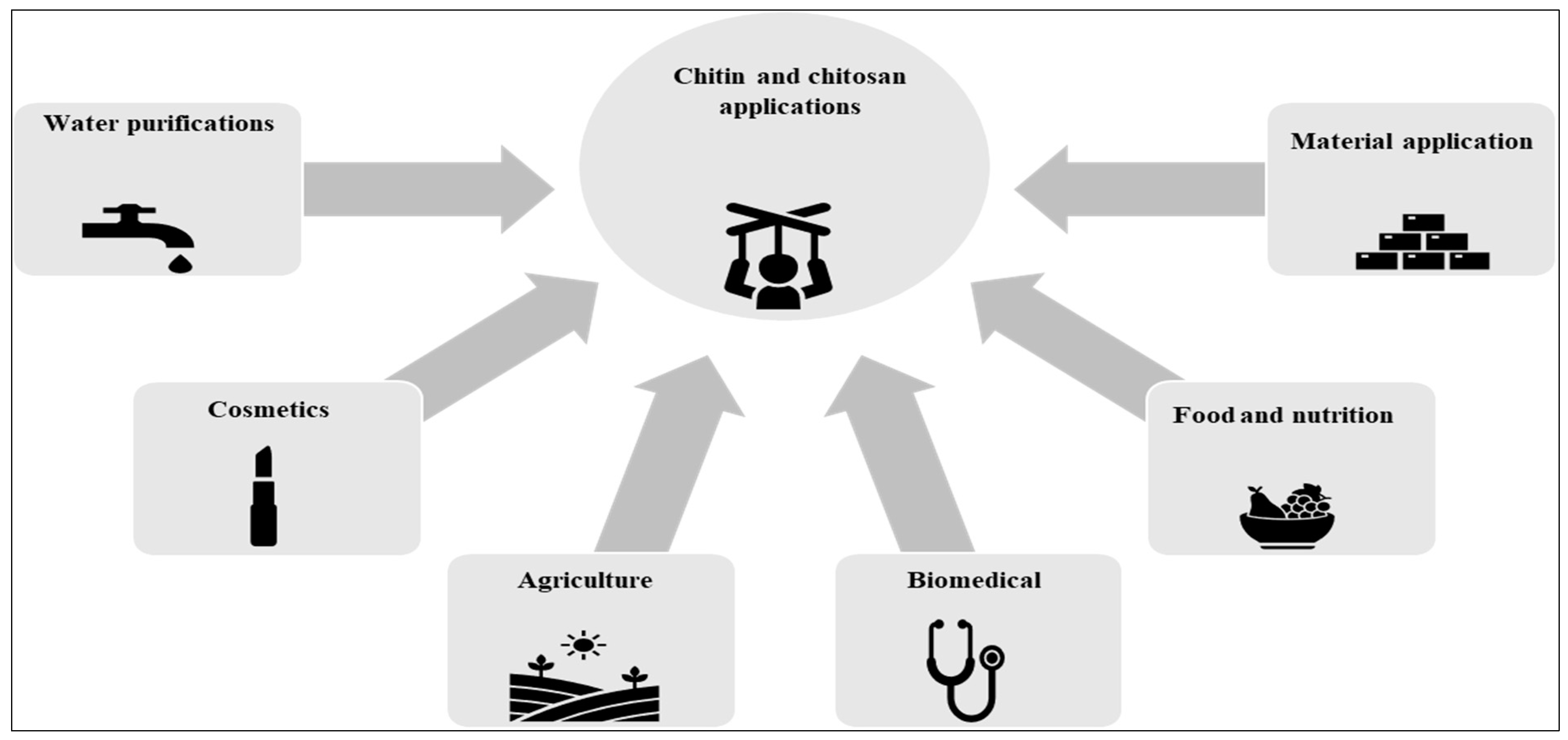
Recent studies have shown that chitin and chitosan offer a variety of useful qualities (Figure 3). In this section, we will mostly discuss the uses of chitin and chitosan in biomedicine, agriculture, materials, and water purification.

5.1. Chitin and Chitosan Applications as Biomaterials

Producers of antimicrobial films for the food sector, such as those used to store perishable items such as fresh produce and meat, have long made use of chitosan as a plastic [172]. There have also been a number of initiatives to create new bifunctional materials, such as the grafting of poly (ethylene glycol) ester derivatives or phosphitylation. The use of chitosan as a catalyst support is one of the more recent innovative uses to garner more attention. Indeed, this application utilizes a number of cutting-edge methods, including freeze drying and the use of supercritical CO2 to boost surface exchange capacities and/or ionic liquid use. Using renewable raw materials (the world’s second most prevalent biopolymer) and reducing the quantity of product needed (through catalytic rather than stoichiometric proportions) aids in the adoption of green chemistry concepts [172].

5.2. Chitin and Chitosan Applications in Food and Nutrition

Chitin and its derivatives have several uses in the food industry, such as color stabilizers, thickening agents, emulsifying agents, natural taste enhancers, and more. Chitin has several potential uses in the food and beverage industry, including the regulation of moisture, heat, respiration, antioxidant release, and enzymatic browning in fruits and vegetables. Chitin oligomers need to be made in such a manner. Researchers have shown that the degree of polymerization (DP) affects the physiological activities and functional features of chitin oligomers. Functionality is enhanced in high DP oligomers compared with low DP oligomers. Preparation of chito-oligosaccharide is carried out with little intervention, using a crude enzyme [173]. Medical supplies, biological insecticides, and nutritional supplements are just some of the areas where these materials show great promise for use [174].
Food and drinks include melanoidins, complex biopolymers of amino carbonyl molecules. With a rise in temperature, chitin nanofibers absorb more melanoidins (higher than other chitin-derived adsorbents) [175]. As another example, chitin nanofibers have been depolymerized into their component monomers through acid hydrolysis, which, due to their fineness are dispersed readily in a melanoidin solution. Additionally, NH3+ groups boost melanoidin adsorption [176]. Since chitin nanofibers are effective in removing unwanted colors, such as melanoidins, from sugar syrup, they find use in the sugar industry [177].
As a result of the association between obesity and calorie consumption, which is greatest for lipids, several studies have demonstrated that one method for treating obesity is to reduce a person’s capacity to absorb lipids. This treatment method has been proved to be both successful and verified. Triglycerides are the most common form in which lipids are taken in via meals. Triglycerides are subsequently broken down by the pancreatic secretions, and the pancreatic lipase enzyme is responsible for the hydrolysis of triglycerides into glycerol and fatty acids, which are then absorbed by enterocytes [178,179]. Therefore, preventing this metabolic response is one approach that may be used to manage obesity.
By monitoring lipid accumulation, the anti-obesity activity of carboxymethyl chitin (CM-chitin) has been examined. Triglyceride content and quantity of released glycerol were measured in order to study the inhibitory impact of CM-chitin on lipid accumulation [180,181]. The collected findings demonstrate that CM-chitin modulates phosphorylated adenosine monophosphate-activated protein kinase and aquaporin-7 to decrease triglyceride content and boost glycerol release [178,179]. Overall, it is clear that CM-chitin reduces lipid accumulation and has an anti-adipogenic impact.
Researchers are looking at the possibility of using chitin, chitosan, and acylated derivatives as thickeners of vegetable oils rather than metallic soaps or polyurea derivatives. Once these biopolymers have been described chemically and thermally, oleo gels may be made from them. The findings demonstrate that, when acylated chitosan or soya bean oil are utilized in the formulation of oleo gels, the linear viscoelasticity functions rise by biopolymer concentration, whereas those for other biopolymers decrease [182]. Oleo gels made from chitin and chitosan have greater thermostability than those made using acylated chitosan. Although these oleo gels tend to have good thermostability, they are generally not considered to be very mechanically stable when used in rolling components [183].

5.3. Chitin and Chitosan Applications in Biomedicine

Biocompatibility, biodegradability, and non-toxicity are just a few of the notable qualities that make chitin stand out as a material with potential biomedical uses [45]. A large number of chitin derivatives with biological uses have been created in the past fifty years. Gels, membranes, beads, scaffolds, nanofibers and microfibers, and nanoparticles are all forms of chitin that have potential uses in biomedicine, including tissue engineering, wound dressing, drug delivery, and cancer identification [1,184]. When applied to wounds, these nanoparticles reduce fibroplasia and boost the development of engineered tissues. While other absorbable sutures dissolve and cause complications in media including bile, urine, and pancreatic juice, chitin sutures can withstand these substances without deteriorating [45].
Chitosan’s major use in medicine is to aid in the recovery from wounds. In this context, chitosan’s antibacterial activity and biocompatibility are brought together [185]. The first goods of this kind appeared on store shelves in the early 1990s, and they quickly gained popularity throughout Asia and North America. Businesses, including BioSyntech (Canada), Hemcon (USA), Medovent (Germany), Marine Polymers Technologies (USA), and Cytosial (France), market and sell these items to consumers. Bone replacements, blood interactions, drug vectorization, implants, and medications for treating inflammation, hypertension, and cancer are only some of the additional biological uses of chitosan that have been investigated [184]. However, few of these uses have yet to make it to market. Finally, chitosan was shown to be effective against goat cryptosporidiosis, with reduced Cryptosporidum parvum oocyst discharge and improved weight growth in young animals.

5.4. Chitin and Chitosan Applications in Agriculture

Chitin and its derivatives are excellent prospects for agricultural uses due to their bactericidal, fungicidal, and other similar qualities [100,186,187]. They are also used to gauge how much agricultural items are contaminated with mold [187]. It has been said that seeds treated with chitin, such as wheat, develop more quickly because the addition of chitinous materials to the soil and plant combination clearly reduces the penetration of insects and harmful fungus [188,189]. By hydrolyzing chitinase/chitosanase, the oligosaccharides of chitin/chitosan have been created [190]. These may be employed as biofertilizers, antioxidants, and anti-microbial compounds in the agricultural sector [187]. It has been shown that the presence of microorganisms such as fungus may cause the manufacture of stilbenoids in several plants, including peanuts (trans-resveratrol and trans-piceatannol) [187]. Gram-negative microorganisms have been shown to be ineffective elicitors. Regarding the chitin composition of the cell wall, many treatments involving fungus and Gram-negative bacteria have been conducted [191]. The findings show that trans-resveratrol and trans-piceatannol may be produced in controlled peanut culture using sterilized fungus and chitin as an efficient and quick elicitor that poses no threat from live bacteria. In plant cells, chitin oligosaccharides may be employed to generate defensive responses [191]. In a broad variety of plant species, chitin and its fragments, chitin oligosaccharides or N-acetylchitooligosaccharides, cause the fungal microbe-associated molecular pattern, which is the first line of protection in plants’ multi-layer defensive system [192,193].

5.5. Chitin and Chitosan Applications in Cosmetics

Chitin and one of its most significant derivatives, chitosan, have special qualities that make them suitable for cosmetic applications [99,100]. These qualities include being fungistatic, fungicidal, and the capacity to dissolve in organic acids [194]. Chitin and its derivatives are used cosmetically for skin, mouth, and hair care. Its primary benefits include the preservation of skin tone and suppleness, lowering static electricity in hair, treating acne, and retaining skin hydration [194]. It may thus be used in a variety of products, including creams, lotions, and permanent waving lotions, as well as toothpastes, chewing gums, nail polish, foundation, eye makeup, lipstick, and cleaning and bath products [195]. A few of its derivatives are also used to make nail lacquers. In order to create clean false teeth, they may also be utilized as dental fillers to absorb the fungus thicans, which is similar to candida and attaches to teeth [196] and in so doing stopping plaque buildup and tooth decay [195].

5.6. Chitin and Chitosan Applications in Water Purification

The improvement of technology that does not harm the environment is a priority for the relevant sectors today as environmental protection is becoming a global issue [197]. The water-soluble derivatives of hydroxymethyl chitin and other compounds show promise for the treatment of anionic wastewater [198]. According to reports, chitin may help industrial effluent that contains heavy metals to be chelated. Chitin may bind to typical water contaminants and is cheap and non-toxic. Mercury, copper, iron, nickel, chromium, lead, zinc, cadmium, silver, and cobalt are all components of this binding. The findings demonstrate that mercury and cobalt had the greatest binding and the lowest binding, respectively. The sol-gel method has recently been used to create hybrids of chitin hydrogel and SiO2 for use as biosorbents. Four dyes were examined for possible adsorption on them (Remazol Black B, Erythrosine B, Neutral Red and Gentian Violet) [199]. The findings demonstrate that a spontaneous charge-related interaction was the primary mechanism of dye adsorption, with the exception of EB adsorption. Biocomposites combining n-HAp and chitin have been developed for the first time in order to remove Fe (III) from water. Both n-HAp and n-Hap/chitin were tested for their ability to adsorb Fe (III) (4238 and 5800 mg/kg, respectively) [200,201,202]. Water engineering could make use of the sorption process since it was spontaneous, endothermic, and obeyed the Langmuir isotherm. The study was performed using aqueous solutions containing copper and chromium. The data reveal that the sorbents exhibited metal ion selectivity in the following order: Fe (III) > Cu (II) > Cr (VI) [203,204].
Water affected by mining operations often has a low pH and high concentration of hazardous metals. Chitorem SC-20 (raw crushed crab shell containing 40% (w/w), CaCO3 (30% protein, 20% chitin, 7% moisture, and 3% ash), and chitorem SC-80 (the chitin polymer composed of 88% chitin and 12% moisture) have both been employed in recent studies to remove heavy metals from municipal wastewater [205]. The findings show that SC-20 neutralized the high acidity of mining influenced water and was successful in removing all traces of iron, lead, and zinc, as well as a significant amount of copper, cobalt, and manganese. Ultimately, physical adsorption helped precipitation remove the metal. Additionally, SC-80 was somewhat effective in the absence of precipitation for the removal of cobalt, manganese, lead, and cadmium. Specifically, it was discovered that pH has a significant role in the total quantity of metal removed. Overall, the CaCO3 in chitin products effectively eliminates metals from mining influenced water by neutralizing and precipitating them [205]. Microporous chitin/cellulose composite membranes have been developed by Tang et al. [206]. Their composite membranes’ microporous structure allowed them to efficiently remove heavy metal ions including mercury, copper, and lead. In light of their microporous structure, huge surface area, and propensity for absorbing the dyes, chitin hydrogels may find value in wastewater treatment [207].

5.7. Chitin and Chitosan-Based Food Packaging Bioplastics Production

In a world producing more than 380 million tons of plastic on a yearly basis and with the projections for plastic production to double within the next 20 years, plastic is a material which has become a significant source of environmental pollution [180,208]. The transition toward a “circular plastic economy” is driven by growing concerns about the negative effects of plastic waste on the environment and the release of greenhouse gases (GHGs) from the production and disposal of plastic products [181,208]. In a circular economy, the use of nonrenewable resources and the generation of waste are both kept to a minimum. Instead, reuse and recycling play a significant role in the material life cycle. However, most commercial plastics still originate from fossil fuels and, due to its inability to decompose, it is almost impossible to deal with waste from packaging made from petro-based materials [209,210]. Therefore, alternatives to petroleum-based polymers that produce less emissions and can be degraded or processed without hazardous wastes must be evaluated and considered. The European Union has recently approved the “European Strategy for Plastics in a Circular Economy”, which highlights the environmental burden that is caused by the usage of plastics [211]. In accordance with the plan, by 2030, all plastic packaging shall be recyclable.
‘Bioplastics’ is the name given to a new generation of plastic materials that have been produced as part of an effort to find a sustainable alternative to plastics made from petroleum [212]. Biopolymers or bioplastics represent a sustainable alternative in terms of food packaging since they are bio-based and biodegradable. In addition, the use of these biobased alternatives reduces fossil resources and GHG emissions and is thus associated with a lower carbon footprint [213,214]. Bioplastics are produced from renewable biomass sources, such as vegetable oil, cellulose, starch, and chitin [215]. The most superior differentiation of biodegradable films depending on their resources is the distinction between polysaccharides, proteins, lipids and their derivatives [197,216]. Lipid films are derived by e.g., beeswax or carnauba wax, and are mainly used to lower water transmission when material is packed. An advantage of plastics or films composed of proteins such as gelatins, collagen, whey protein, etc. (animal-derived products) or soy, pea, wheat etc. (plant-derived products) is their mechanical stability. Polysaccharides in biodegradable films (e.g., starch or galactomannans) can control the transmission of oxygen and other gases promoting a potential antioxidant effect of the packaging. Pectins and alginate are particularly widely used as biodegradable packaging material for food products [217].
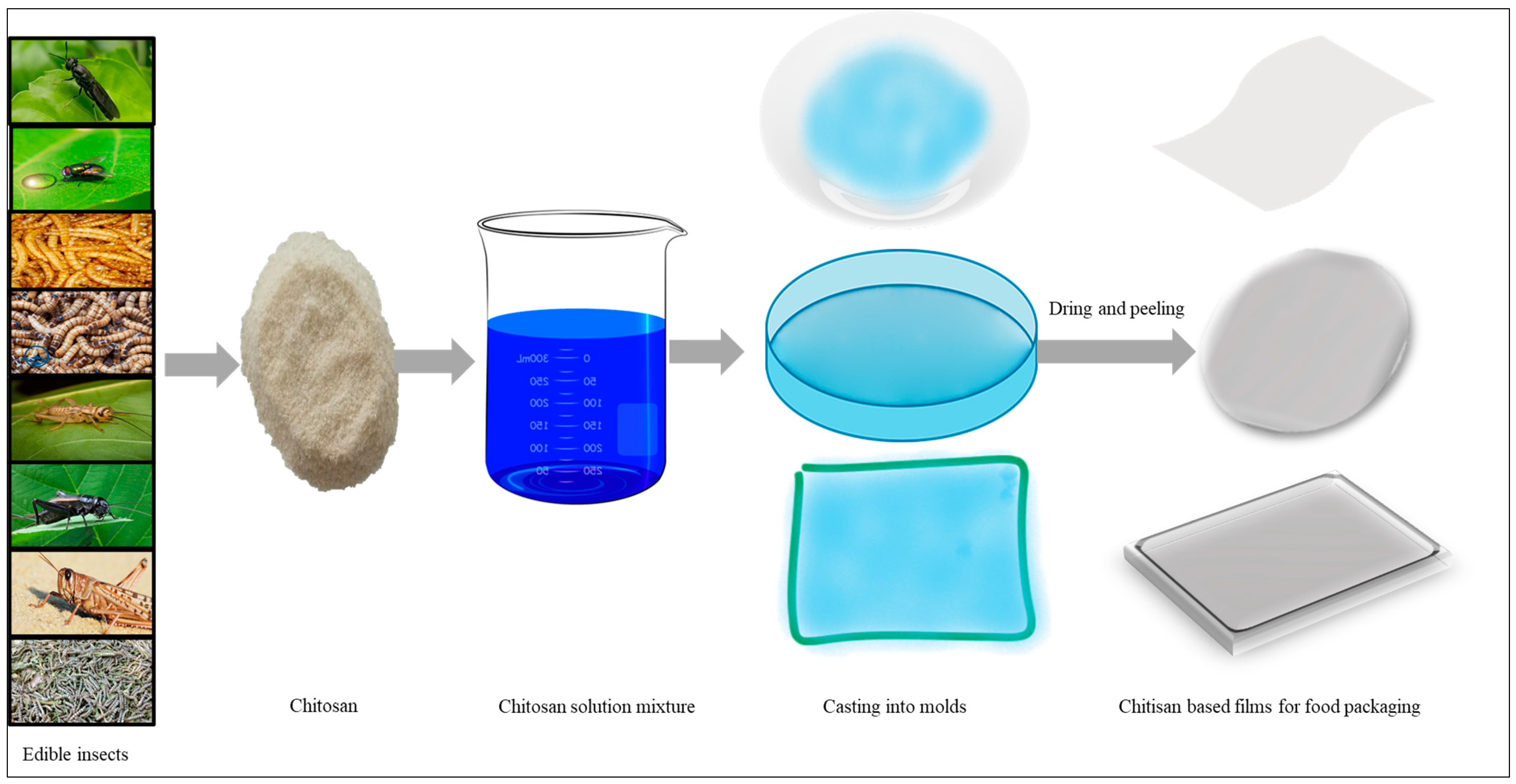
Another prominent polysaccharide material for biobased films is chitosan since it is biodegradable and simultaneously chemically stable during its processing to food packaging. Moreover, its antioxidant and antimicrobial effect as well as non-toxic characteristics may constitute a benefit for food shelf-life stabilization. However, this benefit is adversely affected by the chitosan’s sensitivity to humidity as well as the way that its lower stability is influenced by thermal and mechanical impact. An effective way to avoid and mitigate these disadvantages is to blend chitosan with other, ideally biodegradable polymer/plastics [217]. Natural chitosan blends with other macromolecules are widely found. Polysaccharides, such as starch, cellulose or pectins, are usually selected for blends with chitosan [218]. Chitosan blends with e.g., gliadin proteins isolated from wheat gluten, have resulted in the formation of films with a higher water resistance and, importantly, with anti-microbial activity against S. aureus which, interestingly, was enhanced when chitosan concentrations were increased [219]. Water capacity was influenced when blending chitosan with pectin: an increase in pectin concentration from orange peel enhances moisture of the films and did not influence the water vapor permeability [220]. Film blends of chitosan with purple yam starch which were applied to fruits surfaces as packaging material delayed the natural ripening process of apples, resulting in lower weight loss and, thus, the ability to stabilize the quality and shelf life of the fruit products [221]. Chitosan blends with animal or plant-derived protein molecules proved to be beneficial compared with single-sourced films for use in packaging material. Different studies have shown that the film blends had better functional and gas barrier properties. Blending chitosan with porcine plasma protein lowers water solubility and water vapor permeability. Moreover, these blends were more stable to thermal exposure and transparency was decreased, protecting the quality of food products when packed in these films [222]. Blended films with synthetic polymers, such as polyvinyl alcohol (PVA), polyvinyl pyrrolidone (PVP) or polylactic acid (PLA), are preferably used for packaging material due to their mechanical and biological properties. A big benefit of synthetic polymers is low production costs as well as their preferable functionality in chemical resistance. Moreover, its mechanical flexibility allows usage in diverse food packaging forms. Blending chitosan with synthetic polymers can therefore be advantageous in terms of food packaging production. The hydroxyl and amine groups of chitosan allow the formation of intermolecular hydrogen bonds between hydroxyl groups of some synthetic polymers [24]. A schematic diagram showing the preparation of chitosan-based bioplastic is shown in Figure 4.
There are several studies analyzing the properties of chitosan–synthetic polymer blends. Blending chitosan with PVA affected the elasticity of films and so affected its capability for use as a packaging material. Interestingly, the blend was more thermally stable than films with PVA alone. Moreover, light transmittance was reduced, and the blend exhibited an antimicrobial activity against Pseudomonas aeruginosa, a primary biofilm producer [223]. Another study showed similar results and, beyond that, demonstrated that increasing the chitosan concentration enhanced the antimicrobial potential against other bacteria as Staphylococcus aureus, Bacillus cereus and Escherichia coli [224,225].
However, a recent development in the utilization of insect chitin without blending with other polymers in the bioplastic food packaging is the formation of chitin hydrogels and chitin films that have good mechanical properties. According to the finding of Zhou et al. [226] chitin KOH/urea solution was used to create a high-performance biodegradable plastic. To make chitin bioplastic, the solution was first converted to hydrogel by crosslinking with epichlorohydrin and ethanol immersion, and then pressed under high heat. When the concentration of the solution is increased, the solution becomes more viscous, crystallizes, and attains a smoother texture. Barrier qualities, flame retardancy, high temperature resistance, mechanical capabilities (tensile strength up to 107.1 MPa), and soil degradation properties are all shown by a 4% chitin bioplastic. In about seven weeks, bacteria may break down chitin bioplastic entirely. Furthermore, chitin has been shown to be harmless to both cells and plants in biosafety studies (wheat and mung beans). Water resistance and transparency of the chitin bioplastic were found to be equivalent to those of commercial polypropylene plastic, and it was used in a variety of other applications including containers, straws, and photo protection. It has been concluded that chitin and chitosan have potential for use in bioplastic manufacturing due to their great performance, safety, and sustainability, making them a promising replacement for traditional plastic made from fossil fuels.
There is presently only a small amount of literature on the effectiveness of insect-derived chitosan biofilms. Although chitosan films have been effectively manufactured from G. bimaculatus [227], the relevant authors did not evaluate the films’ mechanical and barrier capabilities for food packaging applications in favor of focusing on antibacterial and color features. Pine weevil chitosan has also been used to make solution cast films that were then plasticized with glycerol [102]. Films were reported to have antioxidant and antibacterial properties, whereas their mechanical qualities were also examined. However, there was no comparison of these films with those made from crustacean chitosan. Similarly, the larvae of H. illucens were used to produce both heterogeneous and homogeneous chitosan, which was then used in solution casting with glycerol as a plasticizer to create films [103]. In this work on chitosan films made from H. illucens larvae, quantitative data on film thickness and qualitative remarks on film color and transparency were provided. In contrast, there was no quantitative analysis of the films’ functional qualities. Films made using chitosan from two different species of crickets have outstanding film forming characteristics, comparable to films made with commercially available chitosan from shrimp [228]. Some biobased food packaging may benefit from the added complexity that cricket chitosan may have because of the inherent differences amongst insects [228]. The water resistance, transparency, and vapor barrier characteristics of films made from cricket chitosan were significantly improved over those of shrimp chitosan [228].

5.8. Antibacterial Activities of Chitin and Chitosan

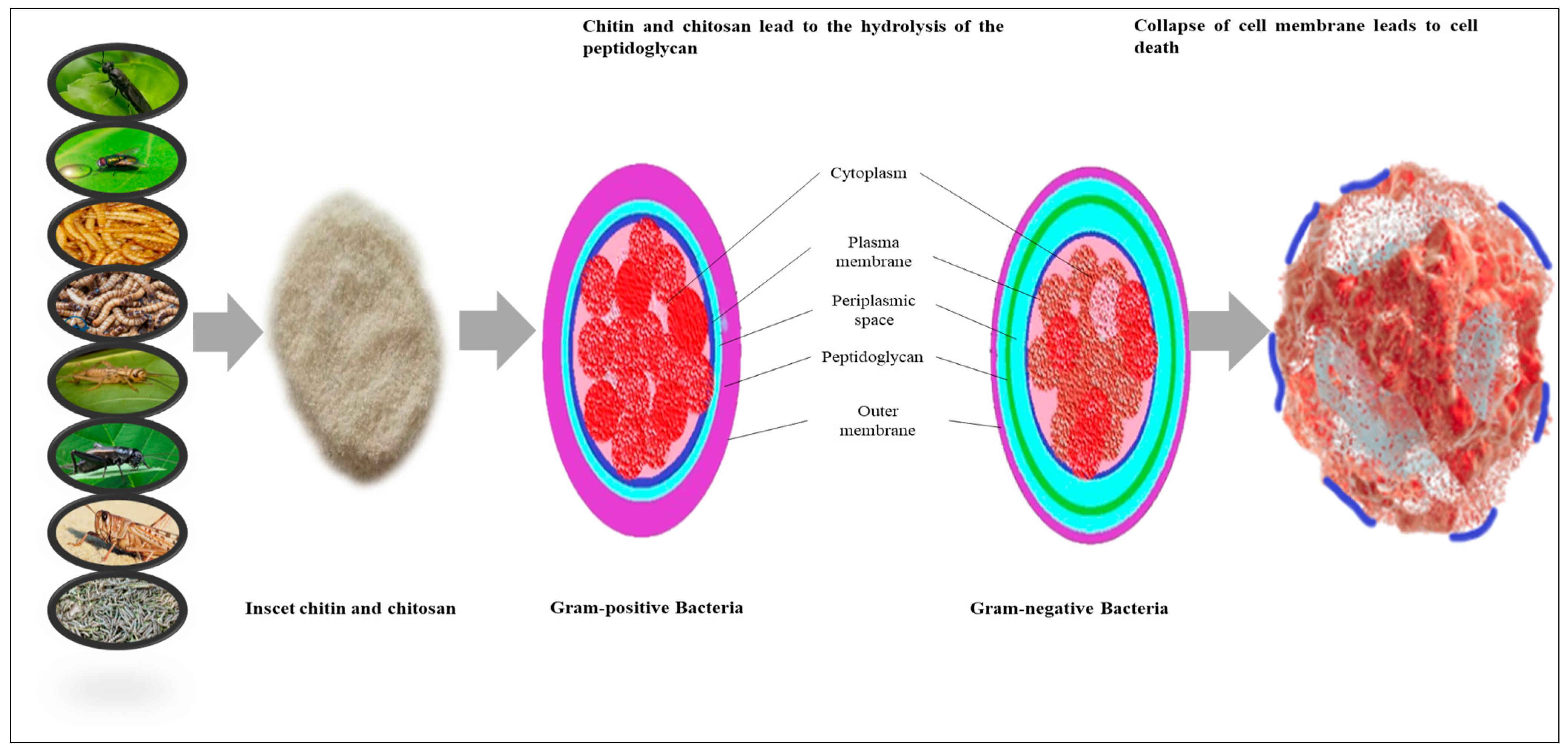
Chitosan has been shown to exhibit antimicrobial activity against a wide range of microorganisms, including Gram-positive and Gram-negative bacteria (Escherichia coli, Vibrio cholerae, Shigella dysenteriae, and Bacteroides fragilis), filamentous fungus, and yeast, however the exact mechanism is still unclear [229]. Electrostatic interactions between the negatively charged membranes of microorganisms and the positively charged NH3+ sites of chitosan have been proposed as a possible mode of action [31]. Because of these interactions, modifications are made to the permeability of the bacterial cell which result in the release of substances from inside the cell [31,230]. It has been shown that the cellular structure of E. coli and S. aureus is disrupted by chitosan’s binding to microbial enzymes and nucleotides [231,232]. It has also been shown that chitosan may change the concentration of calcium on cell walls [233]. To exercise its antibacterial effect, chitosan here modifies the osmotic balance of the membrane wall and hence influences the stability of peptidoglycan (Figure 5) [233,234,235]. The membrane energy stability may potentially be compromised by chitosan due to its interference with the oxygen reduction and electron transport chain activities [235,236]. Chitosan’s ability to bind metal ions is another proposed mechanism, one in which the chitosan’s propensity to bind metal ions is detrimental to the stability of bacterial membranes through the sequestering of bivalent cations such as iron, zinc, copper, cadmium, and magnesium [237,238]. Chitosan that is positively charged may also function to prevent the creation of RNA and proteins, which inhibits the growth of bacteria [239,240]. However, for this process to operate, chitosan must first undergo a size reduction for it to be able to penetrate the cellular system [31]. There is evidence that the polycationic form of chitosan is bactericidal against both Gram-positive and Gram-negative bacteria, however the mechanism by which it does so differs with the lipid composition of the bacterium’s cell membranes. Gram-positive bacteria’s outer cell wall is a direct target of chitosan’s interactions, which is made up of negatively charged peptidoglycan and teichoic acids. The polymer binds to the surface of Gram-negative bacteria where it reacts with anionic compounds such as proteins and lipopolysaccharides [241,242]. Some studies and authors have reported a more potent effect against Gram-negative bacteria, while others have reported a larger effect against Gram-positive bacteria [219,243]. The antibacterial effectiveness of chitosan increases with its molecular weight. This is because high-molecular-weight chitosan may condense into an impermeable polymeric coating on the surface of the microbial cell, altering the cell’s permeability and, in the end, blocking the nutrition supply. Conversely, low-molecular-weight chitosan may penetrate the cytosol of the cells and interact with the DNA of the cells, preventing the cells from creating the mRNA and proteins they need to live [241,244]. Polycationic chitosan, the most well-known mechanism for cell death, is formed when the positively charged amino groups of the chitosan polymer interact with negatively charged components on microbial cell membranes under acidic circumstances (pH > 6.3) [245]. Further evidence for this theory can be found in the study of Lou et al. [246], which tested chitosan’s antibacterial efficacy against Burkholderia seminalis, the bacterium responsible for serious apricot fruit deterioration. They hypothesized that the bacterial cell membranes leaked and ruptured because the positively charged chitosan attracted to the negatively charged microbial cell membrane (carbonyl and phosphoryl groups of the phospholipid components) [237,246]. The antibacterial activity of chitosan is influenced by a number of physicochemical variables, including its molecular weight and degree of deacetylation [172,247].
The bactericidal properties of H. illucens-derived chitosan were tested using a variety of methods, including agar diffusion and microdilution assays, qualitative studies, and quantitative analyses [234]. These findings provided more evidence for H. illucens chitosan’s antibacterial action, opening up new avenues for study in the biological sciences [234].
Jing et al. [248] instigated the chitosan of M. domestica larvae for antimicrobial activities. Six microorganisms were used as test subjects for the antibacterial properties of various molecular weights of chitosan. The antibacterial mechanisms of chitosan were analyzed by measuring membrane permeability and observing bacterial cell health. Results indicate that, as molecular weight increased, chitosan’s antibacterial effectiveness dropped. Ca2+ and Mg2+ may significantly reduce the antibacterial activity of chitosan, which demonstrates the increased antibacterial activity at low pH. The minimum inhibitory concentrations of chitosans varied depending on the kind of bacteria and molecular weight of chitosan and ranged from 0.03% to 0.25%. If the bacterial cell wall is compromised by the chitosan, the contents of the cell might be released.
The antibacterial activity of chitosan of T. molitor larvae was examined and confirmed by Mohan et al. [34] and Shin et al. [120], whose studies found that 4% of the chitosan from T. molitor larvae had no effect on Listeria monocytogenes, S. aureus, B. cereus, and E. coli, however, increasing the chitosan content to 8% inhibited development by 1–2 mm. Furthermore, chitosan isolated from the Z. morio demonstrated a 1–2 mm inhabitation zone against Gram-negative (E. coli and S. aureus) and Gram-positive (B. cereus and L. monocytogenes) bacteria [109].
Chitosan produced from A. domesticus has the potential to serve as a natural antibacterial agent that can inhibit the growth of foodborne infections. The effects of chitosan with varied degrees of deacetylation, including 72%, 76%, and 80%, on the development of Listeria innocua and E. coli after incubation at 37 °C for 24 h have been confirmed [248]. Moreover, chitosan from G. bimaculatus inhibited the growth B. cereus, creating an inhibition zone of 280 ± 69 mm2, whereas it did not have any effect against E. coli and L. monocytogenes [249].
Ali et al. [249] studied the S. gregaria chitosan and demonstrated the antimicrobial activity of that biopolymer. Using agar well diffusion methods, chitosan was examined for its ability to inhibit the growth of three different microbes, i.e., two Gram-positive bacterial species (E. coli and Salmonella typhi), and one Gram-negative bacterial species (S. aureus). According to the findings, the chitosan was effective in preventing the proliferation of bacteria in vitro.
The antibacterial activity of chitosan extracted from B. mori pupae was comparable to that of chitosan obtained from commercial sources and was superior to that of chitosan extracted from eggshells. Excellent inhibition was reported for E. coli among the common bacteria that were investigated, while a much lower level of activity was reported for B. cereus [129].
As a result, we can see that chitin and chitosan produced from insects have antimicrobial characteristics. These characteristics make them excellent for use in the food business, where they might enhance food safety, shelf life, and quality control [34]. The antibacterial mechanism of chitin and chitosan obtained from insects is shown in Figure 5.

6. Conclusions

To conclude, chitin and chitosan have a wide range of useful applications in various fields and sectors. Due to the increasing demand for chitin and its derivatives, it is questionable as to whether the traditional industrial chitin supply, i.e., crustacean waste from the fishing industry, can adequately provide the necessary quantities. Based on the evidence provided to date, we can see that, in addition to their remarkable nutrient content, insects appear to be a promising source of chitin. The byproducts of insect rearing and processing, e.g., exuviae of larvae and pupae, dead adults etc., which currently are discarded as waste, can be purified and used for the production of chitin and chitosan, which can then be functionalized in several applications, providing an additional revenue to the sector. However, there is still a dearth of sufficient information on the production and applications of insect-derived chitin and chitosan, as there are still several questions that need to be answered before these materials can be used to their full potential. Optimizing and scaling up the purification process of chitin from insect production byproducts and developing suitable procedures to improve its chemical, physical and biological characterization are two of the biggest hurdles the scientific community has to face with regarding to insect chitin and its derivatives. Further research in this direction will unfold the potential of insects as sources of chitin and chitosan.

Funding

This research received no external funding.

Institutional Review Board Statement

Not applicable.

Informal Consent of Statement

Not applicable.

Data Availability Statement

Not applicable.

Acknowledgments

The authors would like to sincerely thank the Department of Advance technologies, German Institute of Food technologies, Quakenbruck, Germany and the Department of Microbiology, Faculty of Veterinary and Animal Sciences, The Islamia University of Bahawalpur, Punjab, Pakistan for their support and for the provision of essential facilities needed for this work.

Conflicts of Interest

The authors declare no conflict of interest.

References

  1. Islam, S.; Bhuiyan, M.A.R.; Islam, M.N. Chitin and Chitosan: Structure, Properties and Applications in Biomedical Engineering. J. Polym. Environ. 2017, 25, 854–866. [Google Scholar] [CrossRef]
  2. Crini, G. Historical Landmarks in the Discovery of Chitin. In Sustainable Agriculture Reviews; Springer: Berlin, Germany, 2019; pp. 1–47. [Google Scholar]
  3. Peter, S.; Lyczko, N.; Gopakumar, D.; Maria, H.J.; Nzihou, A.; Thomas, S. Chitin and Chitosan Based Composites for Energy and Environmental Applications: A Review. Waste Biomass Valorization 2021, 12, 4777–4804. [Google Scholar] [CrossRef]
  4. Elieh-Ali-Komi, D.; Hamblin, M.R. Chitin and Chitosan: Production and Application of Versatile Biomedical Nanomaterials. Int. J. Adv. Res 2016, 4, 411–427. [Google Scholar]
  5. Bastiaens, L.; Soetemans, L.; D’Hondt, E.; Elst, K. Sources of Chitin and Chitosan and Their Isolation. In Chitin and Chitosan: Properties and Applications; Wiley: Hoboken, NJ, USA, 2019; pp. 1–34. [Google Scholar]
  6. Terkula Iber, B.; Azman Kasan, N.; Torsabo, D.; Wese Omuwa, J. A Review of Various Sources of Chitin and Chitosan in Nature. J. Renew Mater. 2022, 10, 1097–1123. [Google Scholar] [CrossRef]
  7. Hong, Y.; Ying, T. Characterization of a Chitin-Glucan Complex from the Fruiting Body of Termitomyces albuminosus (Berk.) Heim. Int. J. Biol. Macromol. 2019, 134, 131–138. [Google Scholar] [CrossRef] [PubMed]
  8. Kumirska, J.; Czerwicka, M.; Kaczyński, Z.; Bychowska, A.; Brzozowski, K.; Thöming, J.; Stepnowski, P. Application of Spectroscopic Methods for Structural Analysis of Chitin and Chitosan. Mar. Drugs 2010, 8, 1567–1636. [Google Scholar] [CrossRef] [Green Version]
  9. Abdou, E.S.; Nagy, K.S.A.; Elsabee, M.Z. Extraction and Characterization of Chitin and Chitosan from Local Sources. Bioresour. Technol. 2008, 99, 1359–1367. [Google Scholar] [CrossRef]
  10. John Kasongo, K.; Tubadi, D.J.; Bampole, L.D.; Kaniki, T.A.; Kanda, N.J.M.; Lukumu, M.E. Extraction and Characterization of Chitin and Chitosan from Termitomyces titanicus. SN Appl. Sci. 2020, 2, 406. [Google Scholar] [CrossRef] [Green Version]
  11. Wang, J.; Chen, C. Chitosan-Based Biosorbents: Modification and Application for Biosorption of Heavy Metals and Radionuclides. Bioresour. Technol. 2014, 160, 129–141. [Google Scholar] [CrossRef]
  12. Zemskova, L.; Egorin, A.; Tokar, E.; Ivanov, V. Chitosan-Based Biosorbents: Immobilization of Metal Hexacyanoferrates and Application for Removal of Cesium radionuclide from Aqueous Solutions. J. Solgel. Sci. Technol. 2019, 92, 459–466. [Google Scholar] [CrossRef]
  13. El Knidri, H.; Belaabed, R.; Addaou, A.; Laajeb, A.; Lahsini, A. Extraction, Chemical Modification and Characterization of Chitin and Chitosan. Int. J. Biol. Macromol. 2018, 120, 1181–1189. [Google Scholar] [CrossRef] [PubMed]
  14. Jantzen da Silva Lucas, A.; Quadro Oreste, E.; Leão Gouveia Costa, H.; Martín López, H.; Dias Medeiros Saad, C.; Prentice, C. Extraction, Physicochemical Characterization, and Morphological Properties of Chitin and Chitosan from Cuticles of Edible Insects. Food Chem. 2021, 343, 128550. [Google Scholar] [CrossRef] [PubMed]
  15. Liao, J.; Huang, H. A Fungal Chitin Derived from Hericium erinaceus Residue: Dissolution, Gelation and Characterization. Int. J. Biol. Macromol. 2020, 152, 456–464. [Google Scholar] [CrossRef] [PubMed]
  16. Prasad, K.; Murakami, M.A.; Kaneko, Y.; Takada, A.; Nakamura, Y.; Kadokawa, J. ichi Weak Gel of Chitin with Ionic Liquid, 1-Allyl-3-Methylimidazolium Bromide. Int. J. Biol. Macromol. 2009, 45, 221–225. [Google Scholar] [CrossRef]
  17. Takada, A.; Kadokawa, J.-i. Preparation of Cellulosic Soft and Composite Materials Using Ionic Liquid Media and Ion Gels. Cellulose 2021, 29, 2745–2754. [Google Scholar] [CrossRef]
  18. Mukesh, C.; Mondal, D.; Sharma, M.; Prasad, K. Choline Chloride-Thiourea, a Deep Eutectic Solvent for the Production of Chitin Nanofibers. Carbohydr. Polym. 2014, 103, 466–471. [Google Scholar] [CrossRef]
  19. Hu, X.; Tang, Y.; Wang, Q.; Li, Y.; Yang, J.; Du, Y.; Kennedy, J.F. Rheological Behaviour of Chitin in NaOH/Urea Aqueous Solution. Carbohydr. Polym. 2011, 83, 1128–1133. [Google Scholar] [CrossRef]
  20. Kadokawa, J.-i. Dissolution, Derivatization, and Functionalization of Chitin in Ionic Liquid. Int. J. Biol. Macromol. 2019, 123, 732–737. [Google Scholar] [CrossRef]
  21. Huang, L.; Bi, S.; Pang, J.; Sun, M.; Feng, C.; Chen, X. Preparation and Characterization of Chitosan from Crab Shell (Portunus trituberculatus) by NaOH/Urea Solution Freeze-Thaw Pretreatment Procedure. Int. J. Biol. Macromol. 2020, 147, 931–936. [Google Scholar] [CrossRef]
  22. Silvestre, J.; Delattre, C.; Michaud, P.; de Baynast, H. Optimization of Chitosan Properties with the Aim of a Water Resistant Adhesive Development. Polymers 2021, 13, 4031. [Google Scholar] [CrossRef]
  23. Abhinaya, M.; Parthiban, R.; Kumar, P.S.; Vo, D.V.N. A Review on Cleaner Strategies for Extraction of Chitosan and Its Application in Toxic Pollutant Removal. Environ. Res. 2021, 196, 110996. [Google Scholar] [CrossRef] [PubMed]
  24. Bonilla, J.; Fortunati, E.; Atarés, L.; Chiralt, A.; Kenny, J.M. Physical, Structural and Antimicrobial Properties of Poly Vinyl Alcohol-Chitosan Biodegradable Films. Food Hydrocoll. 2014, 35, 463–470. [Google Scholar] [CrossRef]
  25. Niederhofer, A.; Müller, B.W. A Method for Direct Preparation of Chitosan with Low Molecular Weight from Fungi. Eur. J. Pharm. Biopharm. 2004, 57, 101–105. [Google Scholar] [CrossRef] [PubMed]
  26. Wang, Y.; Wang, E.L.; Wu, Z.M.; Li, H.; Zhu, Z.; Zhu, X.S.; Dong, Y. Synthesis of Chitosan Molecularly Imprinted Polymers for Solid-Phase Extraction of Methandrostenolone. Carbohydr. Polym. 2014, 101, 517–523. [Google Scholar] [CrossRef]
  27. Ravi Kumar, M.N.V. A Review of Chitin and Chitosan Applications. React. Funct. Polym. 2000, 46, 1–27. [Google Scholar] [CrossRef]
  28. Morin-Crini, N.; Lichtfouse, E.; Torri, G.; Crini, G. Applications of Chitosan in Food, Pharmaceuticals, Medicine, Cosmetics, Agriculture, Textiles, Pulp and Paper, Biotechnology, and Environmental Chemistry. Environ. Chem. Lett. 2019, 17, 1667–1692. [Google Scholar] [CrossRef] [Green Version]
  29. Bano, I.; Arshad, M.; Yasin, T.; Ghauri, M.A.; Younus, M. Chitosan: A Potential Biopolymer for Wound Management. Int. J. Biol. Macromol. 2017, 102, 380–383. [Google Scholar] [CrossRef]
  30. Naskar, S.; Sharma, S.; Kuotsu, K. Chitosan-Based Nanoparticles: An Overview of Biomedical Applications and Its Preparation. J. Drug Deliv Sci. Technol. 2019, 49, 66–81. [Google Scholar] [CrossRef]
  31. Rashki, S.; Asgarpour, K.; Tarrahimofrad, H.; Hashemipour, M.; Ebrahimi, M.S.; Fathizadeh, H.; Khorshidi, A.; Khan, H.; Marzhoseyni, Z.; Salavati-Niasari, M.; et al. Chitosan-Based Nanoparticles against Bacterial Infections. Carbohydr. Polym. 2021, 251, 117108. [Google Scholar] [CrossRef]
  32. Mohammadzadeh Pakdel, P.; Peighambardoust, S.J. Review on Recent Progress in Chitosan-Based Hydrogels for Wastewater Treatment Application. Carbohydr. Polym. 2018, 201, 264–279. [Google Scholar] [CrossRef]
  33. Shariatinia, Z.; Jalali, A.M. Chitosan-Based Hydrogels: Preparation, Properties and Applications. Int. J. Biol. Macromol. 2018, 115, 194–220. [Google Scholar] [CrossRef] [PubMed]
  34. Mohan, K.; Ganesan, A.R.; Muralisankar, T.; Jayakumar, R.; Sathishkumar, P.; Uthayakumar, V.; Chandirasekar, R.; Revathi, N. Recent Insights into the Extraction, Characterization, and Bioactivities of Chitin and Chitosan from Insects. Trends Food Sci. Technol. 2020, 105, 17–42. [Google Scholar] [CrossRef] [PubMed]
  35. Chitin and Chitosan Derivatives: Global Strategic Business Report. Available online: https://www.researchandmarkets.com/reports/338576/chitin_and_chitosan_derivatives_global_strategic (accessed on 17 January 2023).
  36. Arbia, W.; Arbia, L.; Adour, L.; Amrane, A. Chitin Extraction from Crustacean Shells Using Biological Methods—A Review. Food Technol. Biotechnol. 2013, 51, 12–25. [Google Scholar]
  37. Tacon, A.G.J. Global Trends in Aquaculture and Compound Aquafeed Production; World Aquaculture Society: Stavanger, Norway, 2018. [Google Scholar]
  38. Joseph, S.M.; Krishnamoorthy, S.; Paranthaman, R.; Moses, J.A.; Anandharamakrishnan, C. A Review on Source-Specific Chemistry, Functionality, and Applications of Chitin and Chitosan. Carbohydr. Polym. Technol. Appl. 2021, 2, 100036. [Google Scholar] [CrossRef]
  39. Rana, K.L.; Kour, D.; Sheikh, I.; Dhiman, A.; Yadav, N.; Yadav, A.N.; Rastegari, A.A.; Singh, K.; Saxena, A.K. Endophytic Fungi: Biodiversity, Ecological Significance, and Potential Industrial Applications. In Recent Advancement in White Biotechnology through Fungi, Volume 1: Diversity and Enzymes Perspectives; Springer: Berlin, Germany, 2019. [Google Scholar]
  40. Fernando, L.D.; Dickwella Widanage, M.C.; Penfield, J.; Lipton, A.S.; Washton, N.; Latgé, J.P.; Wang, P.; Zhang, L.; Wang, T. Structural Polymorphism of Chitin and Chitosan in Fungal Cell Walls from Solid-State NMR and Principal Component Analysis. Front. Mol. Biosci. 2021, 8, 814. [Google Scholar] [CrossRef]
  41. Naranjo-Ortiz, M.A.; Gabaldón, T. Fungal Evolution: Diversity, Taxonomy and Phylogeny of the Fungi. Biol. Rev. 2019, 94, 2101–2137. [Google Scholar] [CrossRef]
  42. Huq, T.; Khan, A.; Brown, D.; Dhayagude, N.; He, Z.; Ni, Y. Sources, Production and Commercial Applications of Fungal Chitosan:: A Review. J. Bioresour. Bioprod. 2022, 7, 85–98. [Google Scholar] [CrossRef]
  43. Hazmi, A.T.; Ahmad, F.B.; Maziati Akmal, M.H.; Md Ralib, A.A.; Binti Ali, F. Fungal Chitosan for Potential Application in Piezoelectric Energy Harvesting: Review on Experimental Procedure of Chitosan Extraction. Alex. Eng. J. 2022, 67, 105–116. [Google Scholar] [CrossRef]
  44. Crognale, S.; Russo, C.; Petruccioli, M.; D’annibale, A. Chitosan Production by Fungi: Current State of Knowledge, Future Opportunities and Constraints. Fermentation 2022, 8, 76. [Google Scholar] [CrossRef]
  45. Shahbaz, U. Chitin, Characteristic, Sources, and Biomedical Application. Curr. Pharm. Biotechnol. 2020, 21, 1433–1443. [Google Scholar] [CrossRef]
  46. el Knidri, H.; Dahmani, J.; Addaou, A.; Laajeb, A.; Lahsini, A. Rapid and Efficient Extraction of Chitin and Chitosan for Scale-up Production: Effect of Process Parameters on Deacetylation Degree and Molecular Weight. Int. J. Biol. Macromol. 2019, 139, 1092–1102. [Google Scholar] [CrossRef] [PubMed]
  47. Yeul, V.S.; Rayalu, S.S. Unprecedented Chitin and Chitosan: A Chemical Overview. J. Polym. Environ. 2013, 21, 606–614. [Google Scholar] [CrossRef]
  48. Mathew, G.M.; Sukumaran, R.K.; Sindhu, R.; Binod, P.; Pandey, A. Green Remediation of the Potential Hazardous Shellfish Wastes Generated from the Processing Industries and Their Bioprospecting. Environ. Technol. Innov 2021, 24, 101979. [Google Scholar] [CrossRef]
  49. Doan, C.T.; Tran, T.N.; Nguyen, V.B.; Vo, T.P.K.; Nguyen, A.D.; Wang, S.L. Chitin Extraction from Shrimp Waste by Liquid Fermentation Using an Alkaline Protease-Producing Strain, Brevibacillus parabrevis. Int. J. Biol. Macromol. 2019, 131, 706–715. [Google Scholar] [CrossRef] [PubMed]
  50. Hamed, I.; Özogul, F.; Regenstein, J.M. Industrial Applications of Crustacean By-Products (Chitin, Chitosan, and Chitooligosaccharides): A Review. Trends Food Sci. Technol. 2016, 48, 40–50. [Google Scholar] [CrossRef]
  51. Gonil, P.; Sajomsang, W. Applications of Magnetic Resonance Spectroscopy to Chitin from Insect Cuticles. Int. J. Biol. Macromol. 2012, 51, 514–522. [Google Scholar] [CrossRef]
  52. Younes, I.; Rinaudo, M. Chitin and Chitosan Preparation from Marine Sources. Structure, Properties and Applications. Mar. Drugs 2015, 13, 1133–1174. [Google Scholar] [CrossRef] [Green Version]
  53. No, H.K.; Hur, E.Y. Control of Foam Formation by Antifoam during Demineralization of Crustacean Shell in Preparation of Chitin. J. Agric. Food Chem. 1998, 46, 3844–3846. [Google Scholar] [CrossRef]
  54. Bradić, B.; Novak, U.; Likozar, B. Crustacean Shell Bio-Refining to Chitin by Natural Deep Eutectic Solvents. Green Process Synth. 2020, 9, 13–25. [Google Scholar] [CrossRef]
  55. Santos, V.P.; Marques, N.S.S.; Maia, P.C.S.V.; de Lima, M.A.B.; de Franco, L.O.; de Campos-Takaki, G.M. Seafood Waste as Attractive Source of Chitin and Chitosan Production and Their Applications. Int. J. Mol. Sci. 2020, 21, 4290. [Google Scholar] [CrossRef]
  56. Varun, T.K.; Senani, S.; Jayapal, N.; Chikkerur, J.; Roy, S.; Tekulapally, V.B.; Gautam, M.; Kumar, N. Extraction of Chitosan and Its Oligomers from Shrimp Shell Waste, Their Characterization and Antimicrobial Effect. Vet. World 2017, 10, 170–175. [Google Scholar] [CrossRef] [PubMed] [Green Version]
  57. Roberts, G.A.F. Preparation of Chitin and Chitosan. In Chitin Chemistry; Palgrave: London, UK, 1992; pp. 54–84. [Google Scholar]
  58. Kostag, M.; el Seoud, O.A. Sustainable Biomaterials Based on Cellulose, Chitin and Chitosan Composites—A Review. Carbohydr. Polym. Technol. Appl. 2021, 2, 100079. [Google Scholar] [CrossRef]
  59. Kou, S. Peters, L.M.; Mucalo, M.R. Chitosan: A Review of Sources and Preparation Methods. Int. J. Biol. Macromol. 2021, 169, 85–94. [Google Scholar] [CrossRef] [PubMed]
  60. Khayrova, A.; Lopatin, S.; Varlamov, V. Obtaining Chitin, Chitosan and Their Melanin Complexes from Insects. Int. J. Biol. Macromol. 2021, 167, 1319–1328. [Google Scholar] [CrossRef]
  61. Rinaudo, M. Chitin and Chitosan: Properties and Applications. Prog. Polym. Sci. 2006, 31, 603–632. [Google Scholar] [CrossRef]
  62. Van den Broek, L.A.M.; Boeriu, C.G. Chitin and Chitosan: Properties and Applications; Wiley: Hoboken, NJ, USA, 2019; ISBN 9781119450467. [Google Scholar]
  63. Pakizeh, M.; Moradi, A.; Ghassemi, T. Chemical Extraction and Modification of Chitin and Chitosan from Shrimp Shells. Eur. Polym. J. 2021, 159, 110709. [Google Scholar] [CrossRef]
  64. Hussain, R.; Maji, T.K.; Maji, T.K. Determination of Degree of Deacetylation of Chitosan and Their Effect on the Release Behavior of Essential Oil from Chitosan and Chitosan-Gelatin Complex Microcapsules. Int. J. Adv. Eng. Appl. 2013, 2, 4–12. [Google Scholar]
  65. Mukarram, M.; Naeem, M.; Aftab, T.; Khan, M.M.A. Chitin, Chitosan, and Chitooligosaccharides: Recent Advances and Future Perspectives. In Radiation-Processed Polysaccharides; Academic Press: Cambridge, MA, USA, 2022; pp. 339–353. [Google Scholar] [CrossRef]
  66. Philibert, T.; Lee, B.H.; Fabien, N. Current Status and New Perspectives on Chitin and Chitosan as Functional Biopolymers. Appl. Biochem. Biotechnol. 2017, 181, 1314–1337. [Google Scholar] [CrossRef]
  67. Anand, M.; Kalaivani, R.; Maruthupandy, M.; Kumaraguru, A.K.; Suresh, S. Extraction and Characterization of Chitosan from Marine Crab and Squilla Collected from the Gulf of Mannar Region, South India. J. Chitin Chitosan Sci. 2014, 2, 280–287. [Google Scholar] [CrossRef]
  68. Gadkari, R.R.; Suwalka, S.; Yogi, M.R.; Ali, W.; Das, A.; Alagirusamy, R. Green Synthesis of Chitosan-Cinnamaldehyde Cross-Linked Nanoparticles: Characterization and Antibacterial Activity. Carbohydr. Polym. 2019, 226, 115298. [Google Scholar] [CrossRef]
  69. Sivanesan, I.; Gopal, J.; Muthu, M.; Shin, J.; Mari, S.; Oh, J. Green Synthesized Chitosan/Chitosan Nanoforms/Nanocomposites for Drug Delivery Applications. Polymers 2021, 13, 2256. [Google Scholar] [CrossRef] [PubMed]
  70. Shirai, K.; Guerrero, I.; Huerta, S.; Saucedo, G.; Castillo, A.; Obdulia Gonzalez, R.; Hall, G.M. Effect of Initial Glucose Concentration and Inoculation Level of Lactic Acid Bacteria in Shrimp Waste Ensilation. Enzym. Microb. Technol. 2001, 28, 446–452. [Google Scholar] [CrossRef] [PubMed]
  71. De Holanda, H.D.; Netto, F.M. Recovery of Components from Shrimp (Xiphopenaeus kroyeri) Processing Waste by Enzymatic Hydrolysis. J. Food Sci. 2006, 71, C298–C303. [Google Scholar] [CrossRef]
  72. Van Nguyen, N.; Hai, P.D.; My My, V.T.; Men, D.T.; Trung, L.D.; Bavor, H.J. Improving Product Added-Value from Shrimp (Litopenaeus vannamei) Waste by Using Enzymatic Hydrolysis and Response Surface Methodology. J. Aquat. Food Prod. Technol. 2021, 30, 880–892. [Google Scholar] [CrossRef]
  73. Tan, Y.N.; Lee, P.P.; Chen, W.N. Microbial Extraction of Chitin from Seafood Waste Using Sugars Derived from Fruit Waste-Stream. AMB Express 2020, 10, 17. [Google Scholar] [CrossRef]
  74. Rao, M.S.; Muñoz, J.; Stevens, W.F. Critical Factors in Chitin Production by Fermentation of Shrimp Biowaste. Appl. Microbiol. Biotechnol. 2000, 54, 808–813. [Google Scholar] [CrossRef]
  75. Yang, J.K.; Shih, I.L.; Tzeng, Y.M.; Wang, S.L. Production and Purification of Protease from a Bacillus Subtilis That Can Deproteinize Crustacean Wastes. Enzym. Microb. Technol. 2000, 26, 406–413. [Google Scholar] [CrossRef]
  76. Gartner, C.; Peláez, C.A.; López, B.L. Characterization of Chitin and Chitosan Extracted from Shrimp Shells by Two Methods. E-Polymers 2010, 10, 69. [Google Scholar] [CrossRef]
  77. Trung, T.S.; Tram, L.H.; van Tan, N.; van Hoa, N.; Minh, N.C.; Loc, P.T.; Stevens, W.F. Improved Method for Production of Chitin and Chitosan from Shrimp Shells. Carbohydr. Res 2020, 489, 107913. [Google Scholar] [CrossRef]
  78. Hongkulsup, C.; Khutoryanskiy, V.-v.; Niranjan, K. Enzyme Assisted Extraction of Chitin from Shrimp Shells (Litopenaeus Vannamei). J. Chem. Technol. Biotechnol. 2016, 91, 1250–1256. [Google Scholar] [CrossRef]
  79. Valdez-Peña, A.U.; Espinoza-Perez, J.D.; Sandoval-Fabian, G.C.; Balagurusamy, N.; Hernandez-Rivera, A.; De-la-Garza-Rodriguez, I.M.; Contreras-Esquivel, J.C. Screening of Industrial Enzymes for Deproteinization of Shrimp Head for Chitin Recovery. Food Sci. Biotechnol. 2010, 19, 553–557. [Google Scholar] [CrossRef]
  80. Gopal, J.; Muthu, M.; Dhakshanamurthy, T.; Kim, K.J.; Hasan, N.; Kwon, S.J.; Chun, S. Sustainable Ecofriendly Phytoextract Mediated One Pot Green Recovery of Chitosan. Sci. Rep. 2019, 9, 1–12. [Google Scholar] [CrossRef] [Green Version]
  81. Singh, S.K. Solubility of Lignin and Chitin in Ionic Liquids and Their Biomedical Applications. Int. J. Biol. Macromol. 2019, 132, 265–277. [Google Scholar] [CrossRef] [PubMed]
  82. Morais, E.S.; da Costa Lopes, A.M.; Freire, M.G.; Freire, C.S.R.; Coutinho, J.A.P.; Silvestre, A.J.D. Use of Ionic Liquids and Deep Eutectic Solvents in Polysaccharides Dissolution and Extraction Processes towards Sustainable Biomass Valorization. Molecules 2020, 25, 3652. [Google Scholar] [CrossRef] [PubMed]
  83. Idenoue, S.; Yamamoto, K.; Kadokawa, J.I. Dissolution of Chitin in Deep Eutectic Solvents Composed of Imidazolium Ionic Liquids and Thiourea. Chem. Eng. 2019, 3, 90. [Google Scholar] [CrossRef] [Green Version]
  84. Ramón, D.J.; Guillena, G. Deep Eutectic Solvents: Synthesis, Properties, and Applications; Wiley: Hoboken, NJ, USA, 2019; ISBN 9783527818471. [Google Scholar]
  85. Huang, W.C.; Zhao, D.; Guo, N.; Xue, C.; Mao, X. Green and Facile Production of Chitin from Crustacean Shells Using a Natural Deep Eutectic Solvent. J. Agric. Food Chem. 2018, 66, 11897–11901. [Google Scholar] [CrossRef]
  86. Hong, S.; Yuan, Y.; Yang, Q.; Zhu, P.; Lian, H. Versatile Acid Base Sustainable Solvent for Fast Extraction of Various Molecular Weight Chitin from Lobster Shell. Carbohydr. Polym. 2018, 201, 211–217. [Google Scholar] [CrossRef] [PubMed]
  87. Brigode, C.; Hobbi, P.; Jafari, H.; Verwilghen, F.; Baeten, E.; Shavandi, A. Isolation and Physicochemical Properties of Chitin Polymer from Insect Farm Side Stream as a New Source of Renewable Biopolymer. J. Clean. Prod. 2020, 275, 122924. [Google Scholar] [CrossRef]
  88. Ji-bin, Z.; Jia, Z.; Jia-hui, L.I.; Tomerlin, J.K.; Xiao-peng, X.; Rehman, K. Black Soldier Fly: A New Vista for Livestock and Poultry Manure Management. J. Integr. Agric. 2020, 19, 2–14. [Google Scholar] [CrossRef]
  89. Rehman, K.U.; Hollah, C.; Wiesotzki, K.; Rehman, R.U.; Rehman, A.U.; Zhang, J.; Zheng, L.; Nienaber, T.; Heinz, V.; Aganovic, K. Black Soldier Fly, Hermetia illucens as a Potential Innovative and Environmentally Friendly Tool for Organic Waste Management: A Mini-Review. Waste Manag. Res. J. A Sustain. Circ. Econ. 2023, 41, 734242X2211054. [Google Scholar] [CrossRef]
  90. Lourenço, F.; Calado, R.; Medina, I.; Ameixa, O.M.C.C. The Potential Impacts by the Invasion of Insects Reared to Feed and Pet Animals in Europe and Other Regions: A Critical Review. Sustainability 2022, 14, 6361. [Google Scholar] [CrossRef]
  91. Liu, Y.; Li, Y.; Li, X.; Qin, L. The Origin and Dispersal of the Domesticated Chinese Oak Silkworm, Antheraea pernyi, in China: A Reconstruction Based on Ancient Texts. J. Insect Sci. 2010, 10, 180. [Google Scholar] [CrossRef] [PubMed] [Green Version]
  92. Edible Insects Market Worth $9.60 Billion by 2030. Available online: https://www.meticulousresearch.com/pressrelease/184/edible-insects-market-2030 (accessed on 18 January 2023).
  93. Delgado, L.; Garino, C.; Moreno, F.J.; Zagon, J.; Broll, H. Sustainable Food Systems: EU Regulatory Framework and Contribution of Insects to the Farm-To-Fork Strategy. Food Rev. Int. 2022, 8, 1–22. [Google Scholar] [CrossRef]
  94. Petrescu, C.; Malina Petrescu-Mag, R.; Rizov, M.; Burny, P.; Markham Kim, H.; Joo, K.; Hwang, J. Are Customers Willing to Pay More for Eco-Friendly Edible Insect Restaurants? Focusing on the Internal Environmental Locus of Control. Sustainability 2022, 14, 10075. [Google Scholar] [CrossRef]
  95. Triunfo, M.; Tafi, E.; Guarnieri, A.; Salvia, R.; Scieuzo, C.; Hahn, T.; Zibek, S.; Gagliardini, A.; Panariello, L.; Coltelli, M.B.; et al. Characterization of Chitin and Chitosan Derived from Hermetia illucens, a Further Step in a Circular Economy Process. Sci. Rep. 2022, 12, 1–17. [Google Scholar] [CrossRef]
  96. Wang, H.; Kashif ur Rehman; Liu, X.; Yang, Q.; Zheng, L.; Li, W.; Cai, M.; Li, Q.; Zhang, J.; Yu, Z. Insect Biorefinery: A Green Approach for Conversion of Crop Residues into Biodiesel and Protein. Biotechnol. Biofuels 2017, 10, 304. [Google Scholar] [CrossRef] [Green Version]
  97. Rehman, K.U.; Cai, M.; Xiao, X.; Zheng, L.; Wang, H.; Soomro, A.A.; Zhou, Y.; Li, W.; Yu, Z.; Zhang, J. Cellulose Decomposition and Larval Biomass Production from the Co-Digestion of Dairy Manure and Chicken Manure by Mini-Livestock (Hermetia illucens L.). J. Environ. Manag. 2017, 196, 458–465. [Google Scholar] [CrossRef]
  98. Somroo, A.A.; ur Rehman, K.; Zheng, L.; Cai, M.; Xiao, X.; Hu, S.; Mathys, A.; Gold, M.; Yu, Z.; Zhang, J. Influence of Lactobacillus Buchneri on Soybean Curd Residue Co-Conversion by Black Soldier Fly Larvae (Hermetia illucens) for Food and Feedstock Production. Waste Manag. 2019, 86, 114–122. [Google Scholar] [CrossRef]
  99. Triunfo, M.; Tafi, E.; Guarnieri, A.; Scieuzo, C.; Hahn, T.; Zibek, S.; Salvia, R.; Falabella, P. Insect Chitin-Based Nanomaterials for Innovative Cosmetics and Cosmeceuticals. Cosmetics 2021, 8, 40. [Google Scholar] [CrossRef]
  100. Wang, H.; Rehman, K.U.; Feng, W.; Yang, D.; Rehman, R.U.; Cai, M.; Zhang, J.; Yu, Z.; Zheng, L. Physicochemical Structure of Chitin in the Developing Stages of Black Soldier Fly. Int. J. Biol. Macromol. 2020, 149, 901–907. [Google Scholar] [CrossRef]
  101. Valdés, F.; Villanueva, V.; Durán, E.; Campos, F.; Avendaño, C.; Sánchez, M.; Domingoz-araujo, C.; Valenzuela, C. Insects as Feed for Companion and Exotic Pets: A Current Trend. Animals 2022, 12, 1450. [Google Scholar] [CrossRef]
  102. Greven, H.; Kaya, M.; Sargin, I.; Baran, T.; Møbjerg Kristensen, R.; Vinther Sørensen, M. Characterisation of Chitin in the Cuticle of a Velvet Worm (Onychophora). Turk. J. Zool. 2019, 43, 416–424. [Google Scholar] [CrossRef]
  103. Hahn, T.; Tafi, E.; Paul, A.; Salvia, R.; Falabella, P.; Zibek, S. Current State of Chitin Purification and Chitosan Production from Insects. J. Chem. Technol. Biotechnol. 2020, 95, 2775–2795. [Google Scholar] [CrossRef]
  104. Hahn, T.; Roth, A.; Ji, R.; Schmitt, E.; Zibek, S. Chitosan Production with Larval Exoskeletons Derived from the Insect Protein Production. J. Biotechnol. 2020, 310, 62–67. [Google Scholar] [CrossRef]
  105. Kim, M.W.; Song, Y.S.; Han, Y.S.; Jo, Y.H.; Choi, M.H.; Park, Y.K.; Kang, S.H.; Kim, S.A.; Choi, C.; Jung, W.J. Production of Chitin and Chitosan from the Exoskeleton of Adult Two-Spotted Field Crickets (Gryllus bimaculatus). Entomol. Res. 2017, 47, 279–285. [Google Scholar] [CrossRef]
  106. Soetemans, L.; Uyttebroek, M.; Bastiaens, L. Characteristics of Chitin Extracted from Black Soldier Fly in Different Life Stages. Int. J. Biol. Macromol. 2020, 165, 3205–3214. [Google Scholar] [CrossRef]
  107. Caligiani, A.; Marseglia, A.; Leni, G.; Baldassarre, S.; Maistrello, L.; Dossena, A.; Sforza, S. Composition of Black Soldier Fly Prepupae and Systematic Approaches for Extraction and Fractionation of Proteins, Lipids and Chitin. Food Res. Int. 2018, 105, 812–820. [Google Scholar] [CrossRef]
  108. D’Hondt, E.; Soetemans, L.; Bastiaens, L.; Maesen, M.; Jespers, V.; Van den Bosch, B.; Voorspoels, S.; Elst, K. Simplified Determination of the Content and Average Degree of Acetylation of Chitin in Crude Black Soldier Fly Larvae Samples. Carbohydr. Res. 2020, 488, 107899. [Google Scholar] [CrossRef]
  109. Nafisah, A.; Nahrowi; Mutia, R.; Jayanegara, A. Chemical Composition, Chitin and Cell Wall Nitrogen Content of Black Soldier Fly (Hermetia illucens) Larvae after Physical and Biological Treatment. In Proceedings of the IOP Conference Series: Materials Science and Engineering, Wuhan, China, 1 June 2019; IOP Publishing: Bristol, UK, 2019; Volume 546, p. 42028. [Google Scholar]
  110. Khayrova, A.A.; Lopatin, S.A.; Sinitsyna, O.A.; Sinitsyn, A.P.; Varlamov, V.P. Obtaining chitin from the black soldier fly Hermetia illucens by direct extraction. Proc. RAS Ufa Sci. Cent. 2018, 3, 84–88. [Google Scholar] [CrossRef]
  111. Khayrova, A.; Lopatin, S.; Varlamov, V. Black Soldier Fly Hermetia illucens as a Novel Source of Chitin and Chitosan. Int. J. Sci. 2019, 8, 81–86. [Google Scholar] [CrossRef] [Green Version]
  112. Purkayastha, D.; Sarkar, S. Physicochemical Structure Analysis of Chitin Extracted from Pupa Exuviae and Dead Imago of Wild Black Soldier Fly (Hermetia illucens). J. Polym Environ. 2020, 28, 445–457. [Google Scholar] [CrossRef]
  113. Khayrova, A.; Lopatin, S.; Varlamov, V.; Antonov, A.; Ivanov, G.; Pastukhova, N.; Bovykina, G. Production of Chitin from Dead Hermetia illucens. IOP Conf. Ser. Earth Environ. Sci. 2019, 315, 042003. [Google Scholar] [CrossRef]
  114. Kim, M.W.; Han, Y.S.; Jo, Y.H.; Choi, M.H.; Kang, S.H.; Kim, S.A.; Jung, W.J. Extraction of Chitin and Chitosan from Housefly, Musca domestica, Pupa Shells. Entomol. Res. 2016, 46, 324–328. [Google Scholar] [CrossRef]
  115. Zhang, A.J.; Qin, Q.L.; Zhang, H.; Wang, H.T.; Li, X.; Miao, L.; Wu, Y.J. Preparation and Characterisation of Food-Grade Chitosan from Housefly Larvae. Czech J. Food Sci. 2011, 29, 616–623. [Google Scholar] [CrossRef] [Green Version]
  116. Ai, H.; Wang, F.; Yang, Q.; Zhu, F.; Lei, C. Preparation and Biological Activities of Chitosan from the Larvae of Housefly, Musca Domestica. Carbohydr. Polym. 2008, 72, 419–423. [Google Scholar] [CrossRef]
  117. Song, Y.S.; Kim, M.W.; Moon, C.; Seo, D.J.; Han, Y.S.; Jo, Y.H.; Noh, M.Y.; Park, Y.K.; Kim, S.A.; Kim, Y.W.; et al. Extraction of Chitin and Chitosan from Larval Exuvium and Whole Body of Edible Mealworm, Tenebrio Molitor. Entomol. Res. 2018, 48, 227–233. [Google Scholar] [CrossRef]
  118. Son, Y.J.; Hwang, I.K.; Nho, C.W.; Kim, S.M.; Kim, S.H. Determination of Carbohydrate Composition in Mealworm (Tenebrio molitor L.) Larvae and Characterization of Mealworm Chitin and Chitosan. Foods 2021, 10, 640. [Google Scholar] [CrossRef]
  119. Song, Y.S.; Jo, Y.H.; Han, Y.S.; Jung, W.J. Production of Chitin- and Chitosan-Oligosaccharide Using the Edible Insect, Tenebrio molitor. Entomol. Res. 2022, 52, 207–213. [Google Scholar] [CrossRef]
  120. Shin, C.S.; Kim, D.Y.; Shin, W.S. Characterization of Chitosan Extracted from Mealworm Beetle (Tenebrio molitor, Zophobas morio) and Rhinoceros Beetle (Allomyrina dichotoma) and Their Antibacterial Activities. Int. J. Biol. Macromol. 2019, 125, 72–77. [Google Scholar] [CrossRef]
  121. Soon, C.Y.; Tee, Y.B.; Tan, C.H.; Rosnita, A.T.; Khalina, A. Extraction and Physicochemical Characterization of Chitin and Chitosan from Zophobas morio Larvae in Varying Sodium Hydroxide Concentration. Int. J. Biol. Macromol. 2018, 108, 135–142. [Google Scholar] [CrossRef]
  122. Ibitoye, E.B.; Lokman, I.H.; Hezmee, M.N.M.; Goh, Y.M.; Zuki, A.B.Z.; Jimoh, A.A. Extraction and Physicochemical Characterization of Chitin and Chitosan Isolated from House Cricket. Biomed. Mater. 2018, 13, 025009. [Google Scholar] [CrossRef] [Green Version]
  123. Psarianos, M.; Dimopoulos, G.; Ojha, S.; Cavini, A.C.M.; Bußler, S.; Taoukis, P.; Schlüter, O.K. Effect of Pulsed Electric Fields on Cricket (Acheta Domesticus) Flour: Extraction Yield (Protein, Fat and Chitin) and Techno-Functional Properties. Innov. Food Sci. Emerg. Technol. 2022, 76, 102908. [Google Scholar] [CrossRef]
  124. Marei, N.; Elwahy, A.H.M.; Salah, T.A.; el Sherif, Y.; El-Samie, E.A. Enhanced Antibacterial Activity of Egyptian Local Insects’ Chitosan-Based Nanoparticles Loaded with Ciprofloxacin-HCl. Int. J. Biol. Macromol. 2019, 126, 262–272. [Google Scholar] [CrossRef]
  125. Marei, N.H.; El-Samie, E.A.; Salah, T.; Saad, G.R.; Elwahy, A.H.M. Isolation and Characterization of Chitosan from Different Local Insects in Egypt. Int. J. Biol. Macromol. 2016, 82, 871–877. [Google Scholar] [CrossRef]
  126. Milusheva, R.Y.; Rashidova, S.S. Bombyx Mori Chitin and Chitosan: Synthesis, Properties, and Use, Tashkent; FAN: Tashkent, Uzbekistan, 2009; pp. 220–246. [Google Scholar]
  127. Zhang, M.; Haga, A.; Sekiguchi, H.; Hirano, S. Structure of Insect Chitin Isolated from Beetle Larva Cuticle and Silkworm (Bombyx mori) Pupa Exuvia. Int. J. Biol. Macromol. 2000, 27, 99–105. [Google Scholar] [CrossRef]
  128. Paulino, A.T.; Simionato, J.I.; Garcia, J.C.; Nozaki, J. Characterization of Chitosan and Chitin Produced from Silkworm Crysalides. Carbohydr. Polym. 2006, 64, 98–103. [Google Scholar] [CrossRef]
  129. Battampara, P.; Nimisha Sathish, T.; Reddy, R.; Guna, V.; Nagananda, G.S.; Reddy, N.; Ramesha, B.S.; Maharaddi, V.H.; Rao, A.P.; Ravikumar, H.N.; et al. Properties of Chitin and Chitosan Extracted from Silkworm Pupae and Egg Shells. Int. J. Biol. Macromol. 2020, 161, 1296–1304. [Google Scholar] [CrossRef]
  130. Pazmiño, M.F.; Del Hierro, A.G.; Flores, F.J. Genetic Diversity and Organic Waste Degrading Capacity of Hermetia illucens from the Evergreen Forest of the Equatorial Choco Lowland. PeerJ 2023, 11, e14798. [Google Scholar] [CrossRef]
  131. Salam, M.; Alam, F.; Dezhi, S.; Nabi, G.; Shahzadi, A.; Hassan, S.U.; Ali, M.; Saeed, M.A.; Hassan, J.; Ali, N.; et al. Exploring the Role of Black Soldier Fly Larva Technology for Sustainable Management of Municipal Solid Waste in Developing Countries. Environ. Technol. Innov 2021, 24, 101934. [Google Scholar] [CrossRef]
  132. Luo, Q.; Wang, Y.; Han, Q.; Ji, L.; Zhang, H.; Fei, Z.; Wang, Y. Comparison of the Physicochemical, Rheological, and Morphologic Properties of Chitosan from Four Insects. Carbohydr. Polym. 2019, 209, 266–275. [Google Scholar] [CrossRef]
  133. Wang, Y.-S.; Shelomi, M. Review of Black Soldier Fly (Hermetia illucens) as Animal Feed and Human Food. Foods 2017, 6, 91. [Google Scholar] [CrossRef] [PubMed] [Green Version]
  134. Liu, X.; Chen, X.; Wang, H.; Yang, Q.; Ur Rehman, K.; Li, W.; Cai, M.; Li, Q.; Mazza, L.; Zhang, J.; et al. Dynamic Changes of Nutrient Composition throughout the Entire Life Cycle of Black Soldier Fly. PLoS ONE 2017, 12, e0182601. [Google Scholar] [CrossRef] [PubMed] [Green Version]
  135. Rehman, K.U.; Abdul; Cai, M.; Zheng, L.; Xiao, X.; Somroo, A.A.; Wang, H.; Li, W.; Yu, Z.; Zhang, J. Conversion of Mixtures of Dairy Manure and Soybean Curd Residue by Black Soldier Fly Larvae (Hermetia illucens L.). J. Clean. Prod. 2017, 154, 366–373. [Google Scholar] [CrossRef]
  136. Mazza, L.; Xiao, X.; ur Rehman, K.; Cai, M.; Zhang, D.; Fasulo, S.; Tomberlin, J.K.; Zheng, L.; Soomro, A.A.; Yu, Z.; et al. Management of Chicken Manure Using Black Soldier Fly (Diptera: Stratiomyidae) Larvae Assisted by Companion Bacteria. Waste Manag. 2020, 102, 312–318. [Google Scholar] [CrossRef]
  137. Zhu, Z.; Rehman, K.U.; Yu, Y.; Liu, X.; Wang, H.; Tomberlin, J.K.; Sze, S.H.; Cai, M.; Zhang, J.; Yu, Z.; et al. De Novo Transcriptome Sequencing and Analysis Revealed the Molecular Basis of Rapid Fat Accumulation by Black Soldier Fly (Hermetia illucens, L.) for Development of Insectival Biodiesel. Biotechnol. Biofuels 2019, 12, 194. [Google Scholar] [CrossRef] [Green Version]
  138. Rehman, K.U.; Liu, X.; Wang, H.; Zheng, L.; Rehman, R.U.; Cheng, X.; Li, Q.; Li, W.; Cai, M.; Zhang, J.; et al. Effects of Black Soldier Fly Biodiesel Blended with Diesel Fuel on Combustion, Performance and Emission Characteristics of Diesel Engine. Energy Convers. Manag. 2018, 173, 489–498. [Google Scholar] [CrossRef]
  139. Kashif, R.U.; Clemens, H.; Heinz, V. Insects: Alternative Source of Protein and Fat in Livestock, Pets and Aquaculture Feed FEEDPLANET. 2021; pp. 28–35. Available online: https://feedplanetmagazine.com/blog/insects-alternative-source-of-protein-and-fat-in-livestock-pets-and-aquaculture-feed-1415 (accessed on 9 January 2023).
  140. Waśko, A.; Bulak, P.; Polak-Berecka, M.; Nowak, K.; Polakowski, C.; Bieganowski, A. The First Report of the Physicochemical Structure of Chitin Isolated from Hermetia illucens. Int. J. Biol. Macromol. 2016, 92, 316–320. [Google Scholar] [CrossRef]
  141. Smets, R.; Verbinnen, B.; Van De Voorde, I.; Aerts, G.; Claes, J.; Van Der Borght, M. Sequential Extraction and Characterisation of Lipids, Proteins, and Chitin from Black Soldier Fly (Hermetia illucens) Larvae, Prepupae, and Pupae. Waste Biomass Valorization 2020, 11, 6455–6466. [Google Scholar] [CrossRef]
  142. Khayrova, A.; Lopatin, S.; Varlamov, V. Obtaining Chitin/Chitosan-Melanin Complexes from Black Soldier Fly Hermetia illucens. In Proceedings of the IOP Conference Series: Materials Science and Engineering, Ufa, Russia, 5–9 October 2020; Volume 809. [Google Scholar]
  143. Kurchenko, V.P.; Kukulyanskaya, T.A.; Azarko, I.I.; Zueva, O.Y.; Khizmatullin, R.G.; Varlamov, V.P. Physicochemical Properties of Chitin-Melanin and Melanoprotein Complexes from Bee Corpses. Appl. Biochem. Microbiol. 2006, 42, 331–334. [Google Scholar] [CrossRef]
  144. Scott, J.G. Evolution of Resistance to Pyrethroid Insecticides in Musca Domestica. Pest. Manag. Sci. 2017, 73, 716–722. [Google Scholar] [CrossRef]
  145. Cheng, Z.; Yu, L.; Li, H.; Xu, X.; Yang, Z. Use of Housefly (Musca domestica L.) Larvae to Bioconversion Food Waste for Animal Nutrition and Organic Fertilizer. Environ. Sci. Pollut. Res. 2021, 28, 48921–48928. [Google Scholar] [CrossRef]
  146. Rumbos, C.I.; Karapanagiotidis, I.T.; Mente, E.; Psofakis, P.; Athanassiou, C.G. Evaluation of Various Commodities for the Development of the Yellow Mealworm, Tenebrio Molitor. Sci. Rep. 2020, 10, 11224. [Google Scholar] [CrossRef] [PubMed]
  147. Errico, S.; Spagnoletta, A.; Verardi, A.; Moliterni, S.; Dimatteo, S.; Sangiorgio, P. Tenebrio molitor as a Source of Interesting Natural Compounds, Their Recovery Processes, Biological Effects, and Safety Aspects. Compr. Rev. Food Sci. Food Saf. 2022, 21, 148–197. [Google Scholar] [CrossRef] [PubMed]
  148. Gkinali, A.-A.; Matsakidou, A.; Vasileiou, E.; Paraskevopoulou, A. Potentiality of Tenebrio molitor Larva-Based Ingredients for the Food Industry: A Review. Trends Food Sci. Technol. 2022, 119, 495–507. [Google Scholar] [CrossRef]
  149. Nascimento Filho, M.A.; Pereira, R.T.; Oliveira, A.B.S.; Suckeveris, D.; Burin Junior, A.M.; Soares, C.A.P.; Menten, J.F.M. Nutritional Value of Tenebrio Molitor Larvae Meal for Broiler Chickens: Metabolizable Energy and Standardized Ileal Amino Acid Digestibility. J. Appl. Poult. Res. 2021, 30, 100102. [Google Scholar] [CrossRef]
  150. Rumbos, C.I.; Athanassiou, C.G. The Superworm, Zophobas Morio (Coleoptera:Tenebrionidae): A ‘Sleeping Giant’ in Nutrient Sources. J. Insect Sci. 2021, 21, 13. [Google Scholar] [CrossRef] [PubMed]
  151. Dragojlović, D.; Ðuragić, O.; Pezo, L.; Popović, L.; Rakita, S.; Tomičić, Z.; Spasevski, N. Comparison of Nutritional Profiles of Super Worm (Zophobas Morio) and Yellow Mealworm (Tenebrio Molitor) as Alternative Feeds Used in Animal Husbandry: Is Super Worm Superior? Animals 2022, 12, 1277. [Google Scholar] [CrossRef]
  152. Benzertiha, A.; Kierończyk, B.; Kołodziejski, P.; Pruszyńska–Oszmałek, E.; Rawski, M.; Józefiak, D.; Józefiak, A. Tenebrio Molitor and Zophobas Morio Full-Fat Meals as Functional Feed Additives Affect Broiler Chickens’ Growth Performance and Immune System Traits. Poult. Sci. 2020, 99, 196–206. [Google Scholar] [CrossRef]
  153. Abd, M.D.; Jabir, R.; Razak, S.A.; Vikineswary, S. Nutritive Potential and Utilization of Super Worm (Zophobas Morio) Meal in the Diet of Nile Tilapia (Oreochromis Niloticus) Juvenile. Afr. J. Biotechnol. 2014, 11, 6592–6598. [Google Scholar] [CrossRef]
  154. Sogari, G.; Amato, M.; Biasato, I.; Chiesa, S.; Gasco, L. The Potential Role of Insects as Feed: A Multi-Perspective Review. Animals 2019, 9, 119. [Google Scholar] [CrossRef] [Green Version]
  155. Soares Araújo, R.R.; dos Santos Benfica, T.A.R.; Ferraz, V.P.; Moreira Santos, E. Nutritional Composition of Insects Gryllus assimilis and Zophobas morio: Potential Foods Harvested in Brazil. J. Food Compos. Anal. 2019, 76, 22–26. [Google Scholar] [CrossRef]
  156. Finke, M. Complete Nutrient Composition of Commercially Raised Invertebrates Used as Food for Insectivores. Zoo Biol. 2002, 21, 269–285. [Google Scholar] [CrossRef]
  157. Adámková, A.; Mlček, J.; Kouřimská, L.; Borkovcová, M.; Bušina, T.; Adámek, M.; Bednářová, M.; Krajsa, J. Nutritional Potential of Selected Insect Species Reared on the Island of Sumatra. Int. J. Environ. Res Public Health 2017, 14, 521. [Google Scholar] [CrossRef] [PubMed] [Green Version]
  158. Kulma, M.; Kouřimská, L.; Homolková, D.; Božik, M.; Plachý, V.; Vrabec, V. Effect of Developmental Stage on the Nutritional Value of Edible Insects. A Case Study with Blaberus craniifer and Zophobas morio. J. Food Compos. Anal. 2020, 92, 103570. [Google Scholar] [CrossRef]
  159. Kaya, M.; Erdogan, S.; Mol, A.; Baran, T. Comparison of Chitin Structures Isolated from Seven Orthoptera Species. Int. J. Biol. Macromol. 2015, 72, 797–805. [Google Scholar] [CrossRef] [PubMed]
  160. Kaya, M.; Seyyar, O.; Baran, T.; Turkes, T. Bat Guano as New and Attractive Chitin and Chitosan Source. Front. Zool. 2014, 11, 59. [Google Scholar] [CrossRef]
  161. Osimani, A.; Milanović, V.; Cardinali, F.; Roncolini, A.; Garofalo, C.; Clementi, F.; Pasquini, M.; Mozzon, M.; Foligni, R.; Raffaelli, N.; et al. Bread Enriched with Cricket Powder (Acheta domesticus): A Technological, Microbiological and Nutritional Evaluation. Innov. Food Sci. Emerg. Technol. 2018, 48, 150–163. [Google Scholar] [CrossRef]
  162. Kulma, M.; Kouřimská, L.; Plachý, V.; Božik, M.; Adámková, A.; Vrabec, V. Effect of Sex on the Nutritional Value of House Cricket, Acheta domestica L. Food Chem. 2019, 272, 267–272. [Google Scholar] [CrossRef]
  163. Rumpold, B.A.; Schlüter, O.K. Nutritional Composition and Safety Aspects of Edible Insects. Mol. Nutr. Food Res. 2013, 57, 802–823. [Google Scholar] [CrossRef]
  164. Ploydee, E.; Chaiyanan, S. Production of High Viscosity Chitosan from Biologically Purified Chitin Isolated by Microbial Fermentation and Deproteinization. Int. J. Polym. Sci. 2014, 2014, 1–8. [Google Scholar] [CrossRef] [Green Version]
  165. Psarianos, M.; Ojha, S.; Schneider, R.; Schlüter, O.K. Chitin Isolation and Chitosan Production from House Crickets (Acheta Domesticus) by Environmentally Friendly Methods. Molecules 2022, 27, 5005. [Google Scholar] [CrossRef]
  166. Taylor, G.K.; Thomas, A.L.R. Dynamic Flight Stability in the Desert Locust Schistocerca gregaria. J. Exp. Biol. 2003, 206, 2803–2829. [Google Scholar] [CrossRef] [PubMed] [Green Version]
  167. Dillon, R.; Charnley, K. Mutualism between the Desert Locust Schistocerca gregaria and Its Gut Microbiota. Res. Microbiol. 2002, 153, 503–509. [Google Scholar] [CrossRef]
  168. Suresh, H.N.; Mahalingam, C.A.; Pallavi. Amount of Chitin, Chitosan and Chitosan Based on Chitin Weight in Pure Races of Multivoltine and Bivoltine Silkworm Pupae Bombyx mori L. Int. J. Sci. Nat. 2012, 3, 214–216. [Google Scholar]
  169. Mahesh, D.; Subbarayappa, C.T.; Shruthi, R. A Review—Bionutritional Science of Silkworm Pupal Residue to Mine New Ways for Utilization. Int. J. Adv. Res. Biol. Sci. 2015, 2, 135–140. [Google Scholar]
  170. Sánchez-Pérez, L.dC.; Barranco-Florido, J.E.; Rodríguez-Navarro, S.; Cervantes-Mayagoitia, J.F.; Ramos-López, M.Á. Enzymes of Entomopathogenic Fungi, Advances and Insights. Adv. Enzyme Res. 2014, 2, 65–76. [Google Scholar] [CrossRef] [Green Version]
  171. Milusheva, R.Y.; Rashidova, S.S. Bombyx Mori Chitosan Nanoparticles: Synthesis and Properties. Open J. Org. Polym. Mater. 2019, 9, 63–73. [Google Scholar] [CrossRef] [Green Version]
  172. Kumar, S.; Mukherjee, A.; Dutta, J. Chitosan Based Nanocomposite Films and Coatings: Emerging Antimicrobial Food Packaging Alternatives. Trends Food Sci. Technol. 2020, 97, 196–209. [Google Scholar] [CrossRef]
  173. Giraldo, J.D.; Garrido-Miranda, K.A.; Schoebitz, M. Chitin and Its Derivatives: Functional Biopolymers for Developing Bioproducts for Sustainable Agriculture—A Reality? Carbohydr. Polym. 2023, 299, 120196. [Google Scholar] [CrossRef]
  174. Schmitz, C.; Auza, L.G.; Koberidze, D.; Rasche, S.; Fischer, R.; Bortesi, L. Conversion of Chitin to Defined Chitosan Oligomers: Current Status and Future Prospects. Mar. Drugs 2019, 17, 452. [Google Scholar] [CrossRef] [Green Version]
  175. Ali, G.; Sharma, M.; Salama, E.S.; Ling, Z.; Li, X. Applications of Chitin and Chitosan as Natural Biopolymer: Potential Sources, Pretreatments, and Degradation Pathways. Biomass Convers. Biorefin. 2022 2022, 1, 1–15. [Google Scholar] [CrossRef]
  176. Rizvi, S.; Goswami, L.; Gupta, S.K. A Holistic Approach for Melanoidin Removal via Fe-Impregnated Activated Carbon Prepared from Mangifera indica Leaves Biomass. Bioresour. Technol. Rep. 2020, 12, 100591. [Google Scholar] [CrossRef]
  177. Ghourbanpour, J.; Sabzi, M.; Shafagh, N. Effective Dye Adsorption Behavior of Poly (Vinyl alcohol)/ Chitin Nanofiber/Fe(III) Complex. Int. J. Biol. Macromol. 2019, 137, 296–306. [Google Scholar] [CrossRef] [PubMed]
  178. Lv, J.; Lv, X.; Ma, M.; Oh, D.-H.; Jiang, Z.; Fu, X. Chitin and Chitin-Based Biomaterials: A Review of Advances in Processing and Food Applications. Carbohydr. Polym. 2023, 299, 120142. [Google Scholar] [CrossRef]
  179. Huang, J.; Wu, Q.; Lin, Z.; Liu, S.; Su, Q.; Pan, Y. Therapeutic effects of chitin from Pleurotus eryngii on high-fat diet induced obesity in rats. Acta Sci. Pol. Technol. Aliment. 2020, 19, 279–289. [Google Scholar] [CrossRef] [PubMed]
  180. Konti, A.; Mamma, D.; Scarlat, N.; Damigos, D. The Determinants of the Growth of the European Bioplastics Sector—A Fuzzy Cognitive Maps Approach. Sustainability 2022, 14, 6035. [Google Scholar] [CrossRef]
  181. Nachod, B.; Keller, E.; Hassanein, A.; Lansing, S. Assessment of Petroleum-Based Plastic and Bioplastics Degradation Using Anaerobic Digestion. Sustainability 2021, 13, 13295. [Google Scholar] [CrossRef]
  182. Negi, H.; Verma, P.; Singh, R.K. A Comprehensive Review on the Applications of Functionalized Chitosan in Petroleum Industry. Carbohydr. Polym. 2021, 266, 118125. [Google Scholar] [CrossRef]
  183. Davidovich-Pinhas, M. Oil Structuring Using Polysaccharides. Curr. Opin. Food Sci. 2019, 27, 29–35. [Google Scholar] [CrossRef]
  184. Ahmad, S.I.; Ahmad, R.; Khan, M.S.; Kant, R.; Shahid, S.; Gautam, L.; Hasan, G.M.; Hassan, M.I. Chitin and Its Derivatives: Structural Properties and Biomedical Applications. Int. J. Biol. Macromol. 2020, 164, 526–539. [Google Scholar] [CrossRef]
  185. Baharlouei, P.; Rahman, A. Chitin and Chitosan: Prospective Biomedical Applications in Drug Delivery, Cancer Treatment, and Wound Healing. Mar. Drugs 2022, 20, 460. [Google Scholar] [CrossRef] [PubMed]
  186. Synowiecki, J.; Al-Khateeb, N.A. Production, Properties, and Some New Applications of Chitin and Its Derivatives. Crit. Rev. Food Sci. Nutr. 2010, 43, 145–171. [Google Scholar] [CrossRef] [PubMed]
  187. el Hadrami, A.; Adam, L.R.; el Hadrami, I.; Daayf, F. Chitosan in Plant Protection. Mar. Drugs 2010, 8, 968–987. [Google Scholar] [CrossRef] [PubMed]
  188. Hadwiger, L.A. Multiple Effects of Chitosan on Plant Systems: Solid Science or Hype. Plant Sci. 2013, 208, 42–49. [Google Scholar] [CrossRef]
  189. Katiyar, D.; Hemantaranjan, A.; Singh, B. Chitosan as a Promising Natural Compound to Enhance Potential Physiological Responses in Plant: A Review. Ind. J. Plant Physiol. 2015, 20, 1–9. [Google Scholar] [CrossRef]
  190. Malerba, M.; Cerana, R. Recent Applications of Chitin- and Chitosan-Based Polymers in Plants. Polymers 2019, 11, 839. [Google Scholar] [CrossRef] [PubMed] [Green Version]
  191. Veliz, E.A.; Martínez-Hidalgo, P.; Hirsch, A.M. Chitinase-Producing Bacteria and Their Role in Biocontrol. AIMS Microbiol. 2017, 3, 689. [Google Scholar] [CrossRef]
  192. Li, K.; Xing, R.; Liu, S.; Li, P. Chitin and Chitosan Fragments Responsible for Plant Elicitor and Growth Stimulator. J. Agric. Food Chem. 2020, 68, 12203–12211. [Google Scholar] [CrossRef]
  193. Katiyar, D.; Hemantaranjan, A.; Singh, B.; Nishant Bhanu, A. A Future Perspective in Crop Protection: Chitosan and Its Oligosaccharides Pigeonpea View Project Scaling City Institutions for India (SCI-FI): Sanitation, Centre for Policy Research, New Delhi View Project; MedCrave: Edmond, OK, USA, 2014. [Google Scholar] [CrossRef] [Green Version]
  194. Hasan, S.; Boddu, V.M.; Viswanath, D.S.; Ghosh, T.K. Chitosan Uses in Cosmetics; Springer: Cham, Switzerland, 2022; pp. 377–404. [Google Scholar] [CrossRef]
  195. Casadidio, C.; Peregrina, D.V.; Gigliobianco, M.R.; Deng, S.; Censi, R.; di Martino, P. Chitin and Chitosans: Characteristics, Eco-Friendly Processes, and Applications in Cosmetic Science. Mar. Drugs 2019, 17, 369. [Google Scholar] [CrossRef] [Green Version]
  196. Aranaz, I.; Acosta, N.; Civera, C.; Elorza, B.; Mingo, J.; Castro, C.; de los Gandía, M.L.; Caballero, A.H. Cosmetics and Cosmeceutical Applications of Chitin, Chitosan and Their Derivatives. Polymers 2018, 10, 213. [Google Scholar] [CrossRef] [Green Version]
  197. Caner, C. The Effect of Edible Eggshell Coatings on Egg Quality and Consumer Perception. J. Sci. Food Agric. 2005, 85, 1897–1902. [Google Scholar] [CrossRef]
  198. Bhatnagar, A.; Sillanpää, M. Applications of Chitin- and Chitosan-Derivatives for the Detoxification of Water and Wastewater—A Short Review. Adv. Colloid Int. Sci. 2009, 152, 26–38. [Google Scholar] [CrossRef] [PubMed]
  199. Nasrollahzadeh, M.; Sajjadi, M.; Iravani, S.; Varma, R.S. Starch, Cellulose, Pectin, Gum, Alginate, Chitin and Chitosan Derived (Nano)Materials for Sustainable Water Treatment: A Review. Carbohydr. Polym. 2021, 251, 116986. [Google Scholar] [CrossRef] [PubMed]
  200. Kumari, S.; Kishor, R. Chitin and Chitosan: Origin, Properties, and Applications. In Handbook of Chitin and Chitosan: Volume 1: Preparation and Properties; Elsevier: Amsterdam, The Netherlands, 2020; pp. 1–33. ISBN 9780128179703. [Google Scholar] [CrossRef]
  201. Li, Q.; Dunn, E.T.; Grandmaison, E.W.; Goosen, M.F.A. Applications and Properties of Chitosan. Appl. Chitin Chitosan 2020, 7, 3–29. [Google Scholar] [CrossRef]
  202. Amar Cheba, B.; Khan, F.I.; Ali, S. Chitin and Chitosan: Marine Biopolymers with Unique Properties and Versatile Applications. Glob. J. Biotechnol. Biochem. 2011, 6, 149–153. [Google Scholar]
  203. Rajiv Gandhi, M.; Kousalya, G.N.; Meenakshi, S. Removal of Copper(II) Using Chitin/Chitosan Nano-Hydroxyapatite Composite. Int. J. Biol. Macromol. 2011, 48, 119–124. [Google Scholar] [CrossRef]
  204. Silvianti, F.; Siswanta, D.; Aprilita, N.H.; Kiswandono, A.A. Adsorption Characteristic of Iron onto Poly[Eugenol-Co-(Divinyl benzene)] from Aqueous Solution. J. Nat. 2017, 17, 108. [Google Scholar] [CrossRef]
  205. Pinto, P.X.; Al-Abed, S.R.; Reisman, D.J. Biosorption of Heavy Metals from Mining Influenced Water onto Chitin Products. Chem. Eng. J. 2011, 166, 1002–1009. [Google Scholar] [CrossRef]
  206. Tang, H.; Chang, C.; Zhang, L. Efficient Adsorption of Hg2+ Ions on Chitin/Cellulose Composite Membranes Prepared via Environmentally Friendly Pathway. Chem. Eng. J. 2011, 173, 689–697. [Google Scholar] [CrossRef]
  207. Tang, H.; Zhou, W.; Zhang, L. Adsorption Isotherms and Kinetics Studies of Malachite Green on Chitin Hydrogels. J. Hazard. Mater. 2012, 209–210, 218–225. [Google Scholar] [CrossRef]
  208. Rosenboom, J.G.; Langer, R.; Traverso, G. Bioplastics for a Circular Economy. Nat. Rev. Mater. 2022, 7, 117–137. [Google Scholar] [CrossRef] [PubMed]
  209. Lawal, U.; Valapa, R.B. Bioplastics: An Introduction to the Role of Eco-Friendly Alternative Plastics in Sustainable Packaging. In Bio-Based Packaging; Wiley: Hoboken, NJ, USA, 2021. [Google Scholar]
  210. Jankowska, E.; Gorman, M.R.; Frischmann, C.J. Transforming the Plastic Production System Presents Opportunities to Tackle the Climate Crisis. Sustainability 2022, 14, 6539. [Google Scholar] [CrossRef]
  211. The New Industrial Strategy for Europe. Intereconomics 2021, 56, 132. [CrossRef]
  212. Shlush, E.; Davidovich-Pinhas, M. Bioplastics for Food Packaging. Trends Food Sci. Technol. 2022, 125, 66–80. [Google Scholar] [CrossRef]
  213. Ezgi, B.A.; Havva, D.O. A Review: Investigation of Bioplastics. J. Civ. Eng. Archit. 2015, 9, 188–192. [Google Scholar] [CrossRef]
  214. Kumar, S.; Thakur, K. Bioplastics—Classification, Production and Their Potential Food Applications. J. Hill Agric. 2017, 8, 118–129. [Google Scholar] [CrossRef]
  215. Faizan, M.; Nadeem, H.; Arif, A.; Zaheer, W. Bioplastics from Biopolymers: An Eco-Friendly and Sustainable Solution of Plastic Pollution. Polym. Sci.–Ser. C 2021, 63, 47–63. [Google Scholar] [CrossRef]
  216. Ramos, M.; Valdés, A.; Beltrán, A.; Garrigós, M. Gelatin-Based Films and Coatings for Food Packaging Applications. Coatings 2016, 6, 41. [Google Scholar] [CrossRef] [Green Version]
  217. Nur Hanani, Z.A.; Roos, Y.H.; Kerry, J.P. Use and Application of Gelatin as Potential Biodegradable Packaging Materials for Food Products. Int. J. Biol. Macromol. 2014, 71, 94–102. [Google Scholar] [CrossRef]
  218. Younis, H.G.R.; Zhao, G. Physicochemical Properties of the Edible Films from the Blends of High Methoxyl Apple Pectin and Chitosan. Int. J. Biol. Macromol. 2019, 131, 1057–1066. [Google Scholar] [CrossRef]
  219. Fernandez-Saiz, P.; Lagarón, J.M.; Ocio, M.J. Optimization of the Film-Forming and Storage Conditions of Chitosan as an Antimicrobial Agent. J. Agric. Food Chem. 2009, 57, 3298–3307. [Google Scholar] [CrossRef] [PubMed]
  220. Baron, R.D.; Pérez, L.L.; Salcedo, J.M.; Córdoba, L.P.; Sobral, P.J.d.A. Production and Characterization of Films Based on Blends of Chitosan from Blue Crab (Callinectes sapidus) Waste and Pectin from Orange (Citrus sinensis Osbeck) Peel. Int. J. Biol. Macromol. 2017, 98, 676–683. [Google Scholar] [CrossRef] [PubMed]
  221. Martins da Costa, J.C.; Lima Miki, K.S.; da Silva Ramos, A.; Teixeira-Costa, B.E. Development of Biodegradable Films Based on Purple Yam Starch/Chitosan for Food Application. Heliyon 2020, 6, e03718. [Google Scholar] [CrossRef] [PubMed]
  222. Samsalee, N.; Sothornvit, R. Development and Characterization of Porcine Plasma Protein-Chitosan Blended Films. Food Packag. Shelf Life 2019, 22, 100406. [Google Scholar] [CrossRef]
  223. Wu, Y.; Ying, Y.; Liu, Y.; Zhang, H.; Huang, J. Preparation of Chitosan/Poly Vinyl Alcohol Films and Their Inhibition of Biofilm Formation against Pseudomonas Aeruginosa PAO1. Int. J. Biol. Macromol. 2018, 118, 2131–2137. [Google Scholar] [CrossRef]
  224. Hu, Y.; Du, Y.; Yang, J.; Kennedy, J.F.; Wang, X.; Wang, L. Synthesis, Characterization and Antibacterial Activity of Guanidinylated Chitosan. Carbohydr. Polym. 2007, 67, 66–72. [Google Scholar] [CrossRef]
  225. Hamdi, M.; Hammami, A.; Hajji, S.; Jridi, M.; Nasri, M.; Nasri, R. Chitin Extraction from Blue Crab (Portunus segnis) and Shrimp (Penaeus kerathurus) Shells Using Digestive Alkaline Proteases from P. Segnis Viscera. Int. J. Biol. Macromol. 2017, 101, 455–463. [Google Scholar] [CrossRef]
  226. Zhou, Y.; He, Y.; Lin, X.; Yue, F.; Liu, M. Sustainable, High-Performance, and Biodegradable Plastic Made from Chitin. Res. Sq. 2022. preprint. [Google Scholar] [CrossRef]
  227. Jarolimkova, V. Preparation and Characterization of Antimicrobial Packaging Films from Cricket Chitosan Enriched with Schisandra Chinensis Extract; Lunds University: Lund, Sweden, 2015. [Google Scholar]
  228. Liceaga, A.A.; San Martin, F.; Jones, O.; Garcia Bravo, J.M.; Kaplan, I.; Bhunia, A. Purification and Characterization of Acheta Domesticus and Gryllodes Sigillatus Cricket Chitin and Chitosan for Bioactive and Biodegradable Food Packaging Applications. Ph.D. Thesis, Purdue University, West Lafayette, IN, USA, 2021. [Google Scholar] [CrossRef]
  229. Malm, M.; Liceaga, A.M. Physicochemical Properties of Chitosan from Two Commonly Reared Edible Cricket Species, and Its Application as a Hypolipidemic and Antimicrobial Agent. Polysaccharides 2021, 2, 339–353. [Google Scholar] [CrossRef]
  230. Wei, X.Y.; Xia, W.; Zhou, T. Antibacterial Activity and Action Mechanism of a Novel Chitosan Oligosaccharide Derivative against Dominant Spoilage Bacteria Isolated from Shrimp Penaeus Vannamei. Lett. Appl. Microbiol. 2022, 74, 268–276. [Google Scholar] [CrossRef]
  231. Chung, Y.C.; Chen, C.Y. Antibacterial Characteristics and Activity of Acid-Soluble Chitosan. Bioresour. Technol. 2008, 99, 2806–2814. [Google Scholar] [CrossRef] [PubMed]
  232. Chung, Y.C.; Yeh, J.Y.; Tsai, C.F. Antibacterial Characteristics and Activity of Water-Soluble Chitosan Derivatives Prepared by the Maillard Reaction. Molecules 2011, 16, 8504–8514. [Google Scholar] [CrossRef] [PubMed] [Green Version]
  233. Abd El-Hack, M.E.; El-Saadony, M.T.; Shafi, M.E.; Zabermawi, N.M.; Arif, M.; Batiha, G.E.; Khafaga, A.F.; Abd El-Hakim, Y.M.; Al-Sagheer, A.A. Antimicrobial and Antioxidant Properties of Chitosan and Its Derivatives and Their Applications: A Review. Int. J. Biol. Macromol. 2020, 164, 2726–2744. [Google Scholar] [CrossRef]
  234. Guarnieri, A.; Triunfo, M.; Scieuzo, C.; Ianniciello, D.; Tafi, E.; Hahn, T.; Zibek, S.; Salvia, R.; De Bonis, A.; Falabella, P. Antimicrobial Properties of Chitosan from Different Developmental Stages of the Bioconverter Insect Hermetia illucens. Sci. Rep. 2022, 12, 1–12. [Google Scholar] [CrossRef]
  235. Wang, Z.; Sun, Q.; Zhang, H.; Wang, J.; Fu, Q.; Qiao, H.; Wang, Q. Insight into Antibacterial Mechanism of Polysaccharides: A Review. LWT 2021, 150, 111929. [Google Scholar] [CrossRef]
  236. Abd El-Monaem, E.M.; Eltaweil, A.S.; Elshishini, H.M.; Hosny, M.; Abou Alsoaud, M.M.; Attia, N.F.; El-Subruiti, G.M.; Omer, A.M. Sustainable Adsorptive Removal of Antibiotic Residues by Chitosan Composites: An Insight into Current Developments and Future Recommendations. Arab. J. Chem. 2022, 15, 103743. [Google Scholar] [CrossRef]
  237. Ardean, C.; Davidescu, C.M.; Nemeş, N.S.; Negrea, A.; Ciopec, M.; Duteanu, N.; Negrea, P.; Duda-seiman, D.; Musta, V. Factors Influencing the Antibacterial Activity of Chitosan and Chitosan Modified by Functionalization. Int. J. Mol. Sci. 2021, 22, 7449. [Google Scholar] [CrossRef]
  238. Rezazadeh, N.; Kianvash, A. Preparation, Characterization, and Antibacterial Activity of Chitosan/Silicone Rubber Filled Zeolite, Silver, and Copper Nanocomposites against Pseudomonas aeruginosa and Methicillin-Resistant Staphylococcus aureus. J. Appl. Polym. Sci. 2021, 138, 50552. [Google Scholar] [CrossRef]
  239. Ke, C.L.; Deng, F.S.; Chuang, C.Y.; Lin, C.H. Antimicrobial Actions and Applications of Chitosan. Polymers 2021, 13, 904. [Google Scholar] [CrossRef]
  240. Liu, X.F.; Guan, Y.L.; Yang, D.Z.; Li, Z.; De Yao, K. Antibacterial Action of Chitosan and Carboxymethylated Chitosan. J. Appl. Polym. Sci. 2001, 79, 1324–1335. [Google Scholar] [CrossRef]
  241. Yilmaz Atay, H. Antibacterial Activity of Chitosan-Based Systems. In Functional Chitosan: Drug Delivery and Biomedical Applications; Springer: Singapore, 2020; pp. 457–489. ISBN 9789811502637. [Google Scholar]
  242. Chandrasekaran, M.; Kim, K.D.; Chun, S.C. Antibacterial Activity of Chitosan Nanoparticles: A Review. Processes 2020, 8, 1173. [Google Scholar] [CrossRef]
  243. Chandrika, K.S.V.P.; Prasad, R.D.; Godbole, V. Development of Chitosan-PEG Blended Films Using Trichoderma: Enhancement of Antimicrobial Activity and Seed Quality. Int. J. Biol. Macromol. 2019, 126, 282–290. [Google Scholar] [CrossRef]
  244. Hosseinnejad, M.; Jafari, S.M. Evaluation of Different Factors Affecting Antimicrobial Properties of Chitosan. Int. J. Biol. Macromol. 2016, 85, 467–475. [Google Scholar] [CrossRef] [PubMed]
  245. Li, X.-F.; Feng, X.-Q.; Yang, S.; Fu, G.-Q.; Wang, T.-P.; Su, Z.-X. Chitosan Kills Escherichia coli through Damage to Be of Cell Membrane Mechanism. Carbohydr. Polym. 2010, 79, 493–499. [Google Scholar] [CrossRef]
  246. Lou, M.M.; Zhu, B.; Muhammad, I.; Li, B.; Xie, G.L.; Wang, Y.L.; Li, H.Y.; Sun, G.C. Antibacterial Activity and Mechanism of Action of Chitosan Solutions against Apricot Fruit Rot Pathogen Burkholderia seminalis. Carbohydr. Res. 2011, 346, 1294–1301. [Google Scholar] [CrossRef]
  247. Kumar, S.; Ye, F.; Dobretsov, S.; Dutta, J. Chitosan Nanocomposite Coatings for Food, Paints, and Water Treatment Applications. Appl. Sci. 2019, 9, 2409. [Google Scholar] [CrossRef] [Green Version]
  248. Jing, Y.J.; Hao, Y.J.; Qu, H.; Shan, Y.; Li, D.S.; Du, R.Q. Studies on the Antibacterial Activities and Mechanisms of Chitosan Obtained from Cuticles of Housefly Larvae. Acta Biol. Hung. 2007, 58, 75–86. [Google Scholar] [CrossRef]
  249. Ali, U.; Baffa, M.U.; Shamsuddeen, Y. Physicochemical and Functional Characterization of Chitosan Prepared from Schistocerca gregaria (Desert Grasshopper) and the Investigation of Its Antimicrobial Activity. Bayero J. Pure Appl. Sci. 2021, 12, 104–110. [Google Scholar] [CrossRef]
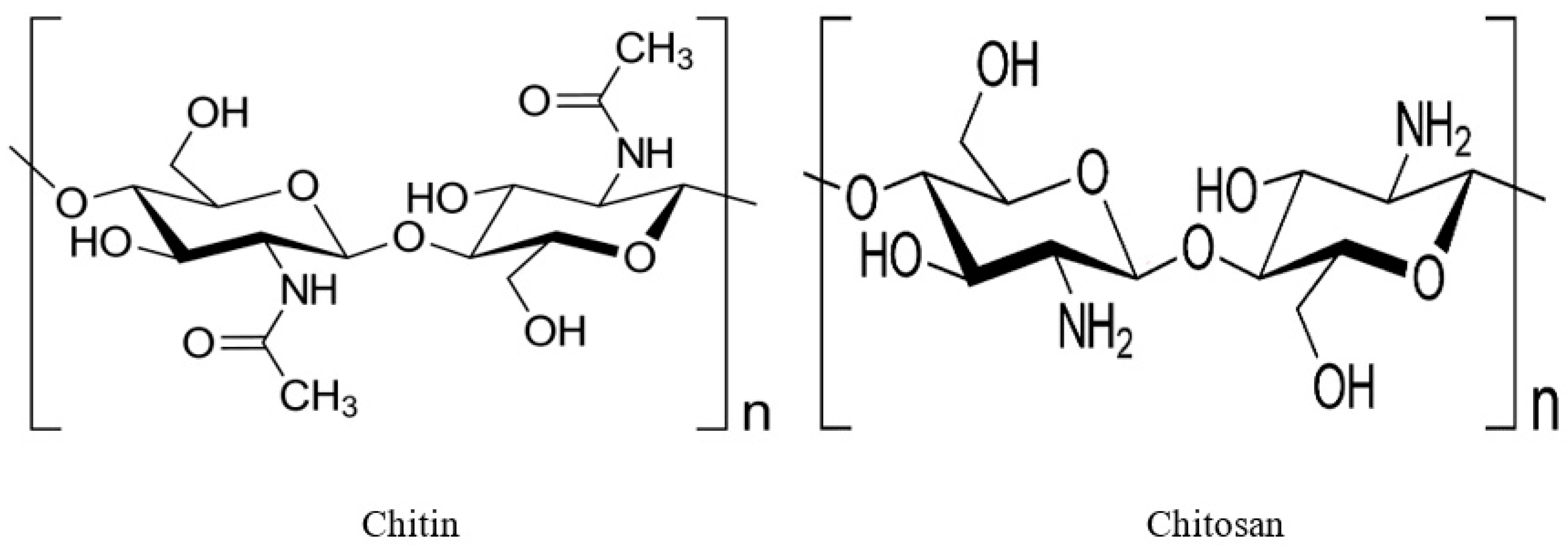
Figure 1. Structures of chitin, and chitosan.
Sustainability 15 04864 g001
Figure 2. Diagrammatic representation of the extraction of insect chitin and chitosan through chemical and biological processes.
Sustainability 15 04864 g002
Figure 3. Application of insect chitin and chitosan.
Sustainability 15 04864 g003
Figure 4. Schematic diagram shows the preparation of chitosan-based bioplastic.
Sustainability 15 04864 g004
Figure 5. Antibacterial mechanism of chitin and chitosan obtained from insects.
Sustainability 15 04864 g005
Table 1. Comparison of Chitin and chitosan extraction methods that use chemical vs biological methods [50,51].
#MethodMethods of TreatmentAdvantages and Disadvantages
1Chemical extractionDemineralization: by acidic treatment using HCl, HNO3, H2SO4.Deproteinization: by alkaline treatment using NaOH or KOH.Decoloration: acetone or organic solvent.Deacetylation: by alkaline treatment using a strong NaOH or KOH
  • Reduced processing time; High-end product DD%; applied on a large scale; organic salts are completely removed.
  • Unfriendly to the environment; minerals and proteins that have been dissolved in a solvent are not biodegradable and cannot be fed to humans or animals.
2Biological extractionDemineralization: Bacteria-produced lactic acid is used in demineralization.Deproteinization: Fermentation media proteases are released into the medium to deproteinize the cultureDecoloration: Acetone or organic solvents are effective decolorizers.Deacetylation: Bacteria produce chitin deacetylase, which deacetifies chitin.
  • High-quality product.
  • Non-harmful to the environment;minerals and proteins that have been removed from the food supply may be fed to humans and livestock.
  • Processing takes a long time (a few days) and is only suitable for laboratory size experiments.
Table 2. Various insect species have been identified as sources of chitin.
#SourceStageSteps Involved in ExtractionChitin YieldReferences
1Hermetia illucensLarvaeDemineralization (1:10, m/v with HCl 1 M at room temperature for 1 h), deproteinization (1 M NaOH treatment 1:25, m/v, 1 h at 80 °C), filtration (pore size 25 μm, 49 PA, 25/14, Solana), and washing with demineralized water until neutral pH and dried at 105 °C for 48 h9.5%[106]
Prepupae9.1%
Pupae10.3%
Shedding31.1%
Cocoon23.8%
Flies5.6%
PrepupaeDefatting (with petroleum ether), deproteinization (1 M NaOH)11.7–14.6%[107]
LarvaeAcidic hydrolysis (1 M NaOH), deproteinization (1 M NaOH) 8.50%[108]
Demineralization (HCOOH), deproteinization (1.9–2 M NaOH)8.3–8.7%[104]
LarvaeDepigmentation (3.6% HCl, NaClO), demineralization (2 M HCl), deproteinization (2 M NaOH)3.6%[100]
Prepupae3.1%
Pupae14.1%
Adult2.9%
LarvaeDefatting (C6H14), demineralization (1 M HCl), deproteinization (1 M NaOH)5.4%[109]
Defatting (CHCl3, CH3OH), demineralization (2% HCl), deproteinization (5% NaOH) 4.6%[110]
Defatting (CHCl3:CH3OH, 7:3), demineralization (2% HCl), deproteinization (5%, w/w NaOH)7%[111]
Pupae exuviaeDeproteinization (1 M NaOH), demineralization (1 M HCl solution)9%[112]
Imago23%
Dead fliesDemineralisation (HCl at 2 h), deproteinization (NaOH at 90 °C for 3 h) 21.3%[113]
2Musca domesticaPupaeDemineralisation (3 h in 500 mL of 2 N HCl solution at room), deproteinization (500 mL of 1.25 N NaOH at 95 °C for 3 h)8.02%[114]
LarvaeDeproteinization (1 mol/l NaOH solution at 100 °C for 3 h) 9.1%[115]
Deproteinization (100 mL of 1 mol/L NaOH at 95 °C for 6 h), decoloration (10 mg/mL KMnO4 for 4 h) Not determined (ND)[116]
3Tenebrio molitorLarvae exuviaeDecalcified (2 N HCl at 20 °C, the exuviae were decalcified for 3 h), deproteinization (500 mL of 5% NaOH at 95 °C)18.01%[117]
Whole bodyDecalcification (3 h in 500 mL of 2 N HCl at 20 °C), deproteinization (3 h at 95 °C in 500 mL of 1.25 N NaOH)4.92%
LarvaeDeproteinization (400 mL of 1.25 M NaOH solution at 80 °C for 24 h), demineralization (1.5 M HCl solution, 1:10 w/v in a shaker 120 rpm, 6 h, at 20 °C)4.72%[118]
Demineralization (2 N HCl, room temp., 3 h), deproteinization (5% NaOH, w/w for 3.5 h at 70 °C), decolorization (3% H2O2 for 1.5 h at 80 °C)6.82%[119]
4Zophobas morioLarvaeDeproteinization (10%, w/v NaOH at80 °C for 24 h), demineralization (7%, v/v HCl at 25 °C for 24 h)4.60%[120]
Adult8.40%
LarvaeDemineralization (1.0 M of HCl in 35 °C), deproteinization (0.5 M, 1.0 M and 2.0 M NaOH in 80 °C for 20 h), decoloration (glacial acetone for 30 min)5.43%[121]
5Acheta domesticusAdultDeproteinization (1 M NaOH at 95 °C for 6 h), demineralization (Oxalic acid for 3 h at room temperature), decoloration (1% sodium hypochlorite for 3 h)4.3–7.1%[122]
Deproteinization (NaOH, 1 M, s/l ratio = 1:50), demineralization (HCl, 1 M, s/l ratio 1:30)7.34[123]
6Gryllus bimaculatusAdultDeproteinization (1.25 M NaOH), demineralization (2 N HCl).20.9–23.3%[105]
7Schistocerca gregariaAdultDeproteinization (1 M NaOH),demineralization (1 N HCl) 22.5%[124]
Deproteinization (1.0 M NaOH at 100 °C for 8 h), demineralization (1 M HCl)12.2%[125]
8Bombyx moriPupae exuviaeDefatting (acetone / alcohol), deproteinization (5–7% NaOH), demineralization (2% HCl), depigmentation (H2O2)3.6%[60,126]
PupaeDemineralization (1 N HCl), deproteinization (1 N NaOH)15–20%[127]
ChrysalidesDemineralization (1 M HCl), deproteinization (1 M NaOH)2.6–4.2%[128]
Egg ShellDemineralization (7%, v/v HCl), deproteinization (10%, w/w NaOH)6%[129]
Pupae18%
Table 3. Various insect species have been identified as sources of chitosan.
#SourceSteps Involved in ExtractionChitin to Chitosan YieldReferences
1Hermetia illucensDemineralization (1:10, m/v with HCl 1 M at room temperature for 1 h), deproteinization (1 M NaOH treatment 1:25, m/v, 1 h at 80 °C), filtration (pore size 25 μm, 49 PA, 25/14, Solana), and washing with demineralized water (until neutral pH and dried at 105 °C for 48 h), deacetylation(1:30 m:v sample in 50 m% NaOH, 90 °C, 1 or 3 h)ND, whereas the degree of deacetylation of chitin was determined (89%)[106]
Demineralization (HCOOH), deproteinisation (1.9–2 M NaOH), deacetylation (10–12 M NaOH)13–43%[104]
Defatting (C6H14), demineralisation (1 M HCl), deproteinisation (1 M NaOH), deacetylation (NaOH, NaBH4)ND, whereas the degree of deacetylation of chitin was determined (66.11%)[109]
Deacetylation (30% NaOH), chitin precipitation (85% H3PO4)32%[111]
Demineralization (2% HCl), deproteinisation (5% NaOH), deacetylation (50% NaOH), defatting (CHCl3, CH3OH)53%[110]
Deproteinisation (30% NaOH), defatting ((C2H5)2O), demineralisation (1% HCl), deacetylation (50% NaOH)18–29%[60]
2Musca domesticaDeproteinization (1 mol/L NaOH solution at 100 °C for 3 h), deacetylation (NaOH, 50% w/v at 125 °C for 4 h)60–70%[115]
Decalcified (3 h in 500 mL of 2 N HCl solution at room temperature), deproteinization (500 mL of 1.25 N NaOH at 95 °C for 3 h), deacetylation (50% NaOH at 105 °C for 3 h)5.9%[114]
Deproteinization (100 mL of 1 mol/L NaOH at 95 °C for 6 h), decoloration (10 mg/mL KMnO4 for 4 h), deacetylation (400 mg/mL NaOH at 70 °C for 8 h)ND, whereas the degree of deacetylation of chitin was determined (90.3%)[116]
3Tenebrio molitorDeproteinization (500 mL 5% NaOH at 95 °C for 3) demineralization (3 h in 1500 mL 2 N HCl at 20 C), deacetylation (500 mL of NaOH at 95 or 105 °C for 3 h or 5 h)13.07–14.48%[117]
Demineralization (2 N HCl, room temp., 3 h), deproteinization (5% NaOH, w/w for 3.5 h at 70 °C), decolorization (3% H2O2 for 1.5 h at 80 °C), deacetylation (50% NaOH, w/w for 5 h at 105 °C)50%[119]
Deproteinization (enzymatic hydrolysis started by adding the alcalase enzyme in a proportion of 2%, w/w; enzyme/substrate), deacetylation (NaOH 40% w/v solution at 90 °C under 500 rpm, 8 h), demineralization (suspension was neutralized to pH 7.0 with HCl (1 M) and filtered again to separate the supernatant)31.9%[14]
Deproteinization (400 mL of 1.25 M NaOH solution and maintained at 80 °C for 24 h), demineralization (71.5 M HCl solution, 1:10, w/v, and shaken at 20 °C in a shaker, 120 rpm, 6 h)), deacetylation (50% NaOH at 80 °C for 4 h)ND, whereas the degree of deacetylation of chitin was determined (95.02%)[118]
4Zophobas morioDeproteinization (0.5 M, 1.0 M and 2.0 M NaOH in °C for 20 h), demineralization (1.0 M of HCl in 35 °C), decoloration (glacial acetone for 30 min), deacetylation (50 wt % NaOH in 90 °C for 30 h)65.84%[121]
Demineralization (1 M HCl); deproteinization (0.5–2 M NaOH); depigmentation ((CH3)2CO), deacetylation (50% NaOH)78–83%[120]
5Acheta domesticusDeproteinization (1 M NaOH at 95 °C for 6 h), demineralization (Oxalic acid for 3 h at room temperature), decoloration (1% sodium hypochlorite for 3 h), deacetylation (50% (w/v) NaOH in 121 °C for 5 h)2.4–5.8%[122]
Deproteinization (1.0 M NaOH at 100 °C for 8 h), demineralization (1 M HC), deacetylation (50% NaOH (15 mL/g) at 100 °C for 8 h)ND, whereas the degree of deacetylation of chitin was determined (98%)[124]
6Gryllus bimaculatusDeproteinization (1.25 M NaOH), demineralization(2 N HCl), deacetylation (50% NaOH (w/v))79.1–94.2%[105]
7Schistocerca gregariaDeproteinization (1.0 M NaOH at 100 °C for 8 h), demineralization (1 M HC), deacetylation (50% NaOH (15 mL/g) at 100 °C for 8 h)55%[125]
8Bombyx moriDemineralization (1 M HCl), deproteinization (1 M NaOH), deacetylation (NaOH, NaBH4)73–97%[128]
Demineralization (7% (v/v) HCl), deproteinization (10% (w/w) NaOH), deacetylation (55% (w/v) NaOH)4.4–16%[129]
Demineralization (1 N HCl), deproteinization (1 N NaOH), deacetylation (40% NaOH, NaBH4)70–80%[127]
Disclaimer/Publisher’s Note: The statements, opinions and data contained in all publications are solely those of the individual author(s) and contributor(s) and not of MDPI and/or the editor(s). MDPI and/or the editor(s) disclaim responsibility for any injury to people or property resulting from any ideas, methods, instructions or products referred to in the content.