Article

Role of Biotransformation of Acacia nilotica Metabolites by Aspergillus subolivaceus in Boosting Lupinus termis Yield: A Promising Approach to Sustainable Agriculture

by
Rasha M. E. Gamel
1,
Samia A. Haroun
1,
Omar Abdullah Alkhateeb
2,
Eman A. Soliman
1,
Arafat B. Tanash
1,
Abdel-Dayem A. Sherief
1,
Mamdoh Abdel-Mogib
3,
Ahmed Hassan Abdou
4,5,*,
Howayda Said Ahmed Mohamed Ali
6,
Nadi Awad Al-Harbi
7,
Khaled Abdelaal
8,* and
Amany M. Kazamel
1
1
Botany Department, Faculty of Science, Mansoura University, Mansoura 35516, Egypt
2
Department of Agribusiness and Consumer Sciences, College of Agriculture & Food Sciences, King Faisal University, Al-Ahsa 31982, Saudi Arabia
3
Chemistry Department, Faculty of Science, Mansoura University, Mansoura 35516, Egypt
4
Social Studies Department, College of Arts, King Faisal University, Al-Ahsa 31982, Saudi Arabia
5
Hotel Studies Department, Faculty of Tourism and Hotels, Mansoura University, Mansoura 35516, Egypt
6
Computer Science Department, Applied College, King Faisal University, Al-Ahsa 31982, Saudi Arabia
7
Biology Department, University College of Tayma, University of Tabuk, Tabuk 45527, Saudi Arabia
8
EPCRS Excellence Center, Plant Pathology and Biotechnology Lab, Faculty of Agriculture, Kafrelsheikh University, Kafr El Sheikh 33516, Egypt
*
Authors to whom correspondence should be addressed.
Sustainability 2023, 15(12), 9509; https://doi.org/10.3390/su15129509
Submission received: 5 May 2023 / Revised: 5 June 2023 / Accepted: 12 June 2023 / Published: 13 June 2023

Abstract

:
Biotransformation plays a significant role in sustainable agriculture. This process involves utilizing microorganisms, such as bacteria and fungi, to transform organic compounds and metabolites into bioactive compounds which have beneficial effects on plant growth, yield, and soil characters. Accordingly, the present study aims to explore the role of biotransformation of Acacia nilotica metabolites by Aspergillus subolivaceus in boosting L. termis yield, as an important strategy in agricultural sustainability. A pilot experiment was performed on five fungal strains (Fusarium oxysporium A. aculeatus, Aspergillus. subolivaceus, Rhizopus oryzae and Trichoderma viride) which were grown on different parts of plants (A. nilotica leaves; green tea leaves, green pepper fruits and pomegranate fruits), and the results indicated that the most active metabolite for the growth of L. termis seeds was the fungal metabolite of A. subolivaceus growing on A. nilotica. More specifically, we assess how metabolites produced by Aspergillus subolivaceus using A. nilotica leaves affect the biochemical properties and chemical composition of L. termis seeds. A. subolivaceus was grown on leaves from A. nilotica to obtain metabolites and fractionated into four extracts. Two concentrations of each extract were examined by pretreating the seeds of L. termis. The study found that all four extracts contributed to an increase in yield and some biochemical properties of the yielded seeds. The best results were obtained by treating the L. termis seeds with an extract obtained from diethyl ether, which led to a significant increase in total nitrogen, amino nitrogen, glucose and protein contents of the seeds. According to 1H NMR guided GC/MS analysis, our results showed an increase in phytochemicals such as terpenes, fatty materials, and flavonoids including 3′,4′,7-trimethoxyquercetin and 4-methyl-p-menth-8-en-3-one, which have not been stated before from A. nilotica suggesting that biotransformation may have occurred due to the presence of A. subolivaceus.

1. Introduction

The significance of biotransformation in sustainable agriculture cannot be ignored or disputed. This process, which includes the use of microorganisms to convert organic compounds into beneficial substances for plant growth and soil characteristics, has a crucial role in promoting more sustainable agricultural practices. By harnessing the power of soil microorganisms, such as plant-growth-promoting bacteria and fungi, to enhance plant nutrient acquisition, we can improve crop yields and reduce our reliance on chemical fertilizers [1]. Microbial biotransformation is an important approach, and commonly used to produce metabolites in large quantities with high efficiency [2]. This process entails several chemical reactions, such as hydrolysis, oxidation, condensation, reduction, isomerization, creating new C–C bonds, generating chiral molecules, and reversing hydrolytic reactions. The more frequent reactions are those involving oxidation, reduction, isomerization, and hydrolysis [3,4]. Recently, microbes have been used to bio-transform terpene. In order to create novel fragrances, several bacterial and fungal strains have been used, and new strains are continually chosen based on their capacity to bio-transform terpenes [5]. Aspergillus, Cunninghamella, and Penicilium strains are frequently used to bio-transform flavonoids, and they can virtually always complete all reactions with excellent yields, according to Cao et al. [6]. One of the microorganisms that is used the most frequently in the biotransformation of flavonoids is A. niger.
The fascinating organisms known as fungi are renowned for their capacity to produce a vast variety of secondary metabolites, which are bioactive compounds. These compounds, however, serve as virulence enhancers and are not necessary for the formation or expansion of the generating organism [7]. Other studies showed that the A. nilotica extract possesses antibacterial, antimalarial, antifungal, antibiotic, molluscoid, anti-hypertensive, anti-diarrheal, anti-denaturation, anthelmintic, anticancer, and antioxidant properties [8,9]. L. albus is sometimes referred to as Egyptian lupin (L. termis), European white lupin, bitter white lupin, broad-leaved lupin, broad-leaf lupin, and Mediterranean white lupin [10]. The entire lupin seed includes antioxidants, including vitamin E and vitamin C, as well as carbohydrates, lipids, protein, fatty acids, amino acids, fiber, ash, phosphorus, calcium, iron, zinc, copper, and manganese [11]. Additionally, lupine seeds are used as a protein source for both human and animal nutrition in many different parts of the world. This is due to both their nutritional content (rich in protein, lipids, and dietary fiber) and their ability to grow in a variety of climates [12].
Approximately 135 species of trees of the Fabaceae family, which includes the genus Acacia, are found across the dry and semi-arid tropics [13]. According to earlier research, some trees and bushes in Asia and Africa have a high nutritional value as animal feed [14]. Secondary metabolic substances present in acacia species include amines, cyanogenic glycosides, alkaloids, fatty acids, seed oils, cyclitols, fluoroacetate, nonprotein amino acids, gums, terpenes (including essential oils, diterpenes, phytosterols, and triterpenegenins and saponins), flavonoids, hydrolyzable and condensed tannins [15]. Cysteine, threonine, methionine, tryptophan, lysine, potassium, magnesium, phosphorus, manganese, and iron are all abundant in the plant [16]. Diester, pentacosanedioic acid dihexadecyl ester, and heptacosane 1, 2, and 3-triol are examples of A. nilotica chemical compounds found in plants [17]. The leaf has 8% digestible protein (12.4% crude protein), rutin, 6- and 8-bis-D-glucoside, and apigenin. Deseeded pods (5.4%), pods (50%), leaves (7.6%), twigs (15.8%), and bark (13.5%) are the plant parts that have the relatively highest tannin amounts [18]. Bark: It has terpenoids, glycosides, and a tannin content of 12–20%. Phlobetannin, gallic acid, protocatechuic acid, pyrocatechol, (+)-catechin, (−)-epigallocatechin-5, and 7-digallate [19] are examples of phenols. Between 9.2 and 16.5 g/100 gm of total phenols are included in its extract (see Table 1) [20].
The current study aims to explore the effect of metabolites produced by A. subolivaceus on A. nilotica leaves, on seed yield characteristics and biochemical features of L. termis-produced seeds, as well as on the chemical composition of the seeds. Additionally, the main goal is to detect a biologically safe way for the improving L. termis productivity as well as enhancement the production of active medicinal components which is in the pharmaceutical industry.

2. Materials and Methods

2.1. Microorganisms

The fungal strain was subordinated in order to perform complete identification using one of the most advanced sophisticated installations; an imaging analysis system using soft-imaging software (AnalysisPro ver.3.0) at the Regional Center for Mycology and Biotechnology (RCMB), Al-Azhar University, Egypt. Solid state fermentation medium (SSF): SSF medium was prepared [24]. Fungal strains were grown on PDA slants for 7 days at a temperature of 30 °C, the spore suspension was prepared by adding 10 mL of sterile basal medium. 50 mL of the sterile substrate in a 250 mL conical flask was inoculated by 2 mL (2 × 10⁷ spores) spore suspension.
The fungal strains (A. aculeatus, Trichoderma viride, A. subolivaceus, Rhizopus oryzae and Fusarium oxysporium) were grown on different plant parts (A. nilotica leaves; green tea leaves, pomegranate fruits and green pepper fruits). The fungal metabolites attained were tested by treating L. termis seeds. Fungal metabolites gained from the growth of A. subolivaceus on A. nilotica showed the finest results. So, this fungal metabolite was fractionated by diethyl ether and ethyl acetate into four fragments which were used for treating the Lupinus seeds.

2.2. Preparation of Metabolites

The purity of metabolic extract was confirmed by centrifugation at 15,000 rpm for 20 min, to ensure full removal of fungal culture.
(a) Water extract: Was carried out according to [25]. (b) Total diethyl ether extract was obtained according to [25,26]. Additionally, the aqueous extract is obtained by resuspension, shaking, and filtration of the residual substrate after the diethyl ether extract in distilled water. (c) Total diethyl ether extract separation according to [27]. Using a separation funnel, the diethyl ether extract was separated into two layers, the aqueous layer was re-extracted by ethyl acetate. However, the final diethyl ether extracts and ethyl acetate were used for Lupinus seeds treatments.

2.3. Experimental Design

L. termis seeds were surface sterilized by being submerged in a 0.01% HgCl2 solution for three minutes. After that, seeds were split into 100-seed bunches that were each equally sized. Each group was pre-soaked for 10 h in two different concentrations (50–100%) of one of the obtained fungal metabolite extracts (water, diethyl ether, ethyl acetate, and aqueous extract) before seeding. Additionally, one group served as control by soaking for the same amount of time in regular water. All seedlings were grown in pots measuring 30 cm in diameter, which contained uniformly distributed soil in a ratio of sand to clay of 1:2 (vol/vol). The plants were exposed to 11 h of light and 13 h of darkness (normal day-night conditions) and temperatures was 32 °C ± 2 and 20 °C ± 2 for day and night, respectively. The relative humidity was 58% during the growth period. The yield was recorded on the harvest day at 106 days from sowing and the analysis was done on the dried seeds that had been handled differently and not at all.

2.4. Statistical Analysis

The data were subjected to analysis utilizing L.S.D. test at the probability threshold of 0.05. ANOVA analysis was performed using IBM statistics software version 20 [28].

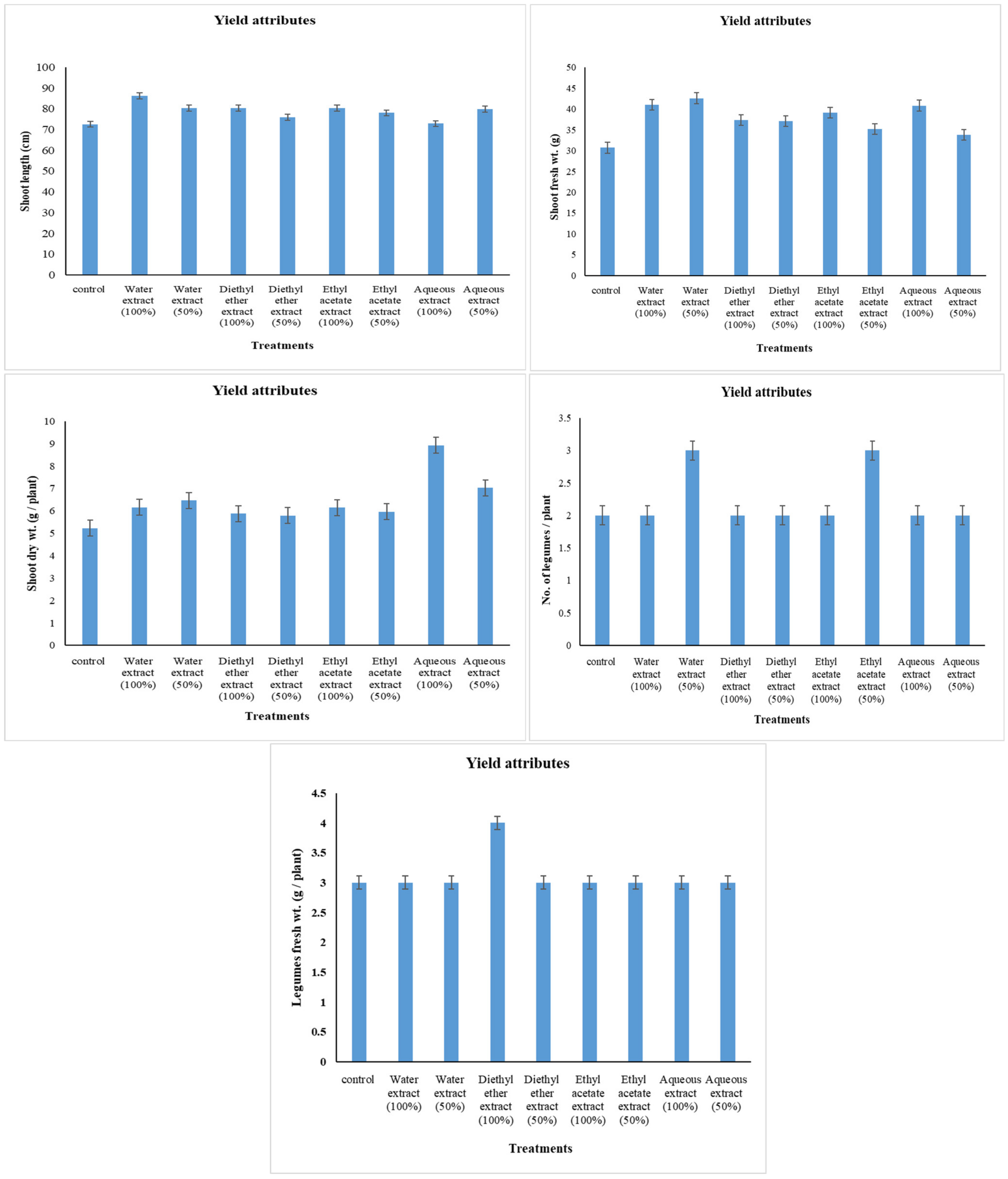
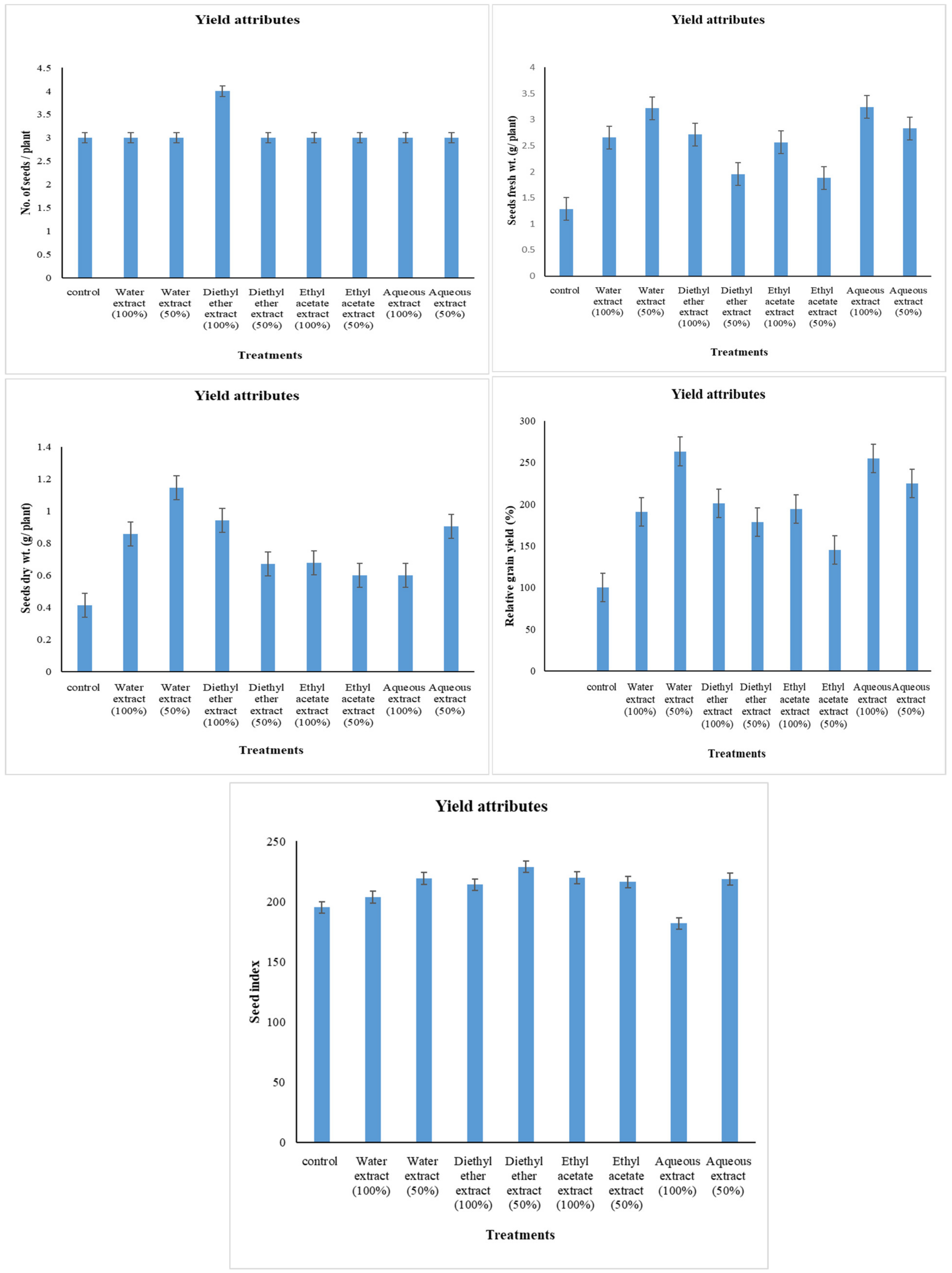
2.5. Yield Attributes

Seed index = the weight of 1000 seeds [29].
Relative grain yield = yield of treated plant/yield of untreated (normal) plant × 100 [30].

2.6. Physio-Biochemical Aspects

2.6.1. Estimation of Carbohydrates

The extraction techniques used for the various carbohydrate fractions put to the test were primarily those of Yemm and Willis [31] and Handel [32]. The o-toluidine method of Fetris [33], which was modified by Riazi et al. [30], was used to assess the glucose contents. A modified version of Handel’s procedures was used to determine the amount of sucrose in the sample [32]. The amount of total soluble sugar was calculated using a modified version of Yemm and Willis’ procedures [34]. Polysaccharides was estimated by the methods of Sadasivam and Manickam method [35].

2.6.2. Estimation of Nitrogenous Constituents

The Yemm and Willis method [36] was used as a basis to estimate the nitrogenous constituents, with some modifications. To estimate ammonia-N, the method described by Delory [37] and modified by Naguib [38] was utilized, with spectrophotometry and Nessler’s reagent employed. Naguib [38] suggested the technique used to estimate amide-N. By Muting and Kaiser [39], a method for estimating amino-N was developed. The traditional semi-micropropagation of the Kjeldahl method, as described by El-Shahaby [40] and Pirie [41], was used to calculate the total soluble nitrogen. The traditional semi-micropropagation Kjeldahl method developed by Rees and Williams [42] and described by Haroun [43], was utilized to determine the total nitrogen. The amount of total soluble nitrogen was subtracted to determine the amount of insoluble protein.

2.6.3. Estimation of Total Protein

The content of protein was measured in accordance with Bradford [44] using spectrophotometry.

2.7. Chemical Constituents of Diethyl Ether Extract

Instrumentation

1H NMR: The NMR spectra were recorded on a Varian Mercury VX-300 nmr spectrometer (Varian, Inc., Palo Alto, CA, USA). 1H-NMR spectra were run at 300 MHz in deuterated chloroform (CDCl3) or dimethyl sulphoxide (DMSO-d6). Chemical shifts are quoted in δ relative to that of TMS.
GC/MS: The Finnigan SSQ 7000 Mass Selective Detector (MSD) interfaced with an Avarian GC was used to determine the MS identification of the GC components through the ICIS V2.0 data system. The DB-5 column (J & W Scientific, Folsom, CA, USA) utilized for this purpose was a 30-meter-long fused silica capillary with an internal diameter of 0.25 mm and a polydimethylsiloxane coating at a film thickness of 0.5 μm. The oven temperature was initially set to 50 °C and maintained isothermally for 3 min before being heated at a rate of 7 °C per minute to a final temperature of 250 °C, at which it was maintained isothermally for 10 min. The injector temperature was set to 200 °C and the volume injected was 0.5 μL. The transition line and ion source temperature were 250 °C and 150 °C, respectively. The mass spectrometer scanned the range of m/z 50 to m/z 300 with an ionization energy of 70 ev and a delay of 3 min to avoid the solvent pea (National Research Center (NRC), Dokki, Cairo, Egypt).
For column chromatography (CC), a silica gel was used. TLC was performed on silica gel (Kieselgel 60, GF 254) of 0.25 mm thickness; Petroleum ether (60–80 °C), diethyl ether, hexane, methylene chloride, ethyl acetate, acetone, and methanol were obtained from Elgomhoria Company (Cairo, Egypt).
Processing of total diethyl ether extract (E) of A. subolivaceous/A. nilotica metabolite: The GC/MS technique was utilized to identify and characterize the components present in the total diethyl ether (E) extract sample.
After the diethyl ether extract had been analyzed by GC/MS to 4-methyl-p-menth-8-en-3-one (Rt 24.82 min, 4.29%), citronellyl butyrate (24.88, 6.31%), eicosane (28.72, 1.90%), 5-hydroxymethyl-1,3,3-trimethyl-2-(3-methyl-buta-1,3-dienyl)-cyclopentanol (33.62, 0.90%), 7-methyl-Z-tetradecen-1-ol acetate (34.57, 0.96%), tetradecane, 2,6,10-trimethyl- (35.93, 1.40%), 9-octadecenoic acid (Z)-(oleic acid) (39.66, 0.83%), docosane (40.20, 1.47%), pentadecanoic acid, 14-methyl-, methyl ester (40.44, 7.94%), hexadecanoic acid, ethyl ester (42.04, 3.58%), 5,8,11,14-eicosatetraenoic acid, (44.51, 5.30%), linoleic acid ethyl ester (45.82, 2.64%) and 3′,4′,7-trimethoxyquercetin(56.37, 0.89%), it was fractionated by CC.
Silica gel was employed as an adsorbent for the purpose of conducting column chromatography on a diethyl ether extract sample weighing 0.231 g. The column was subjected to elution through CC, wherein a sequence of progressively polar hexane/ethyl acetate eluent combinations was employed. The resulting effluents were then grouped into separate fractions based on their corresponding TLC patterns and 1H-NMR spectra. The final fractions were analyzed by GC/MS to 2-hydroxybicyclo [2,2,1] hept-5-ene-2-carboxylic acid (Rt 5.88, 18,40), 5-hydroxynicotinic acid (5.96, 1.39%), l-pipecolic acid (6.0, 0.45%), β-oplopenone (27.30, 1.85%), daucol (27.59, 2.53%), aromadendrene oxide-(1) (28.74, 27.83%), 1-eicosanol (41.42, 7.94%), heneicosane, 2-methyl (41.44, 0.64%), (Z,Z)-9,12-octadecadienoic acid, methyl ester (43.87, 2.91%), octadecanoic acid methyl ester (methyl stearate) (44.58, 12.28%), 3-ethyl-5-(2-ethylbutyl)-octadecane(45.38, 3.22%), ethyl oleate (45.43, 0.40%), 1-heneicosyl formate (45.85, 12.43%), octadecanoic acid, ethyl ester (46.01, 4.36%) and 1,3,5-trimethyl-2-(4-methyl-p-menth-8-en-3-one) cyclohexane (48.22, 1.02%).

3. Results

3.1. Changes in Yield Attributes

The data shown in Figure 1 and Figure 2 demonstrated that plants under drought stress treated with water and aqueous extracts showed a significant increase in shoot length by 10.78% and 10.06%, respectively; shoot fresh weight by 38.81% and 10.28%, respectively; shoot dry weight by 23.59% and 34.30%, respectively; legumes fresh weight by 132.75%, and 72.34%, respectively; seeds fresh weight by 150.70% and 120.41%, respectively; seeds dry weight by 176.63% and 118.55%, respectively; relative grain yield by 163.51% and 124.72%, respectively; and seed index by 12.41% and 11.98%, respectively, compared to control plants. Whereas drought-stressed plants treated with diethyl ether extract and ethyl acetate extract showed a significant increase in shoot length by 4.69% and 7.52%, respectively; shoot fresh weight by 20.93% and 14.64%, respectively; legumes fresh weight by 47.07% and 32.87%; respectively; seeds fresh weight by 52.18% and 46.34%, respectively; seeds dry weight by 61.69% and 45.06%, respectively; relative grain yield by 78.50% and 45.11%, respectively; and seed index by 17.26% and 10.81%, respectively, compared to control plants.

3.2. Changes in Biochemical Aspects in L. termis L. yielded Seeds

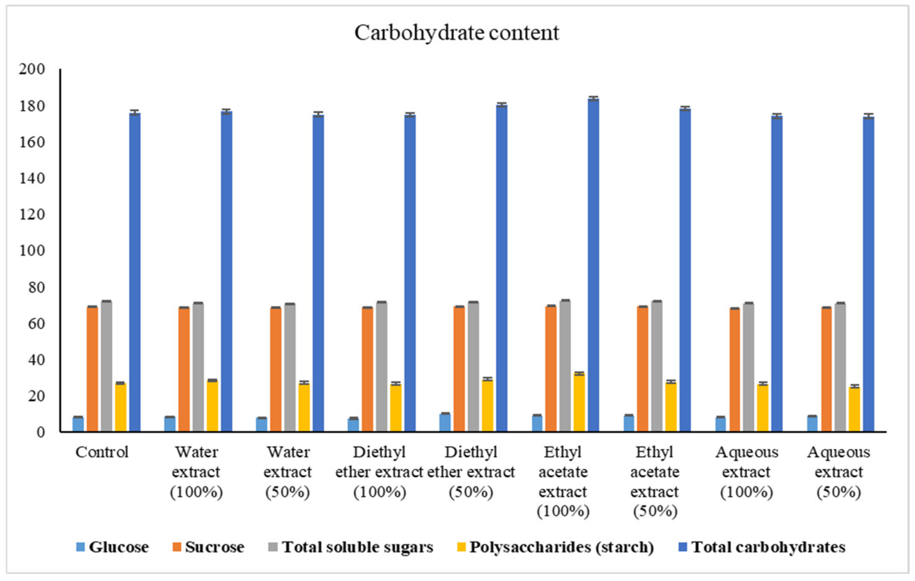
3.2.1. Changes in Carbohydrate Content

Figure 3 showed that A. subolivaceus water extracts (100% and 50%) led to a generally significant decrease in sucrose content of white lupine yielded seeds by 0.47% and 0.73%, respectively, and total soluble sugars content by 1.29% and 1.49%, respectively, while polysaccharides increased insignificantly by 5.60% and 0.61%, respectively. In this connection, water extract 50% decreased glucose and total carbohydrate content insignificantly by 2.47% and 0.56%, respectively, water extract 100% increased them insignificantly by 4.44% and 0.35%, respectively. Furthermore, diethyl ether extract 100% decreased all carbohydrate fractions of lupine yielded seeds insignificantly. On the other hand, the only insignificant decrease with diethyl ether 50% treatment was recorded for total soluble sugar content (0.28%). However, ethyl acetate extracts (100% and 50%) increased carbohydrate fractions significantly, in general. With the exception of glucose content that increased, the other carbohydrate fractions’ content showed a generally insignificant decrease with aqueous extracts (100% and 50%). On the whole, the diethyl ether extract 50% and ethyl acetate extracts (100% and 50%) caused the maximum carbohydrate fractions content.

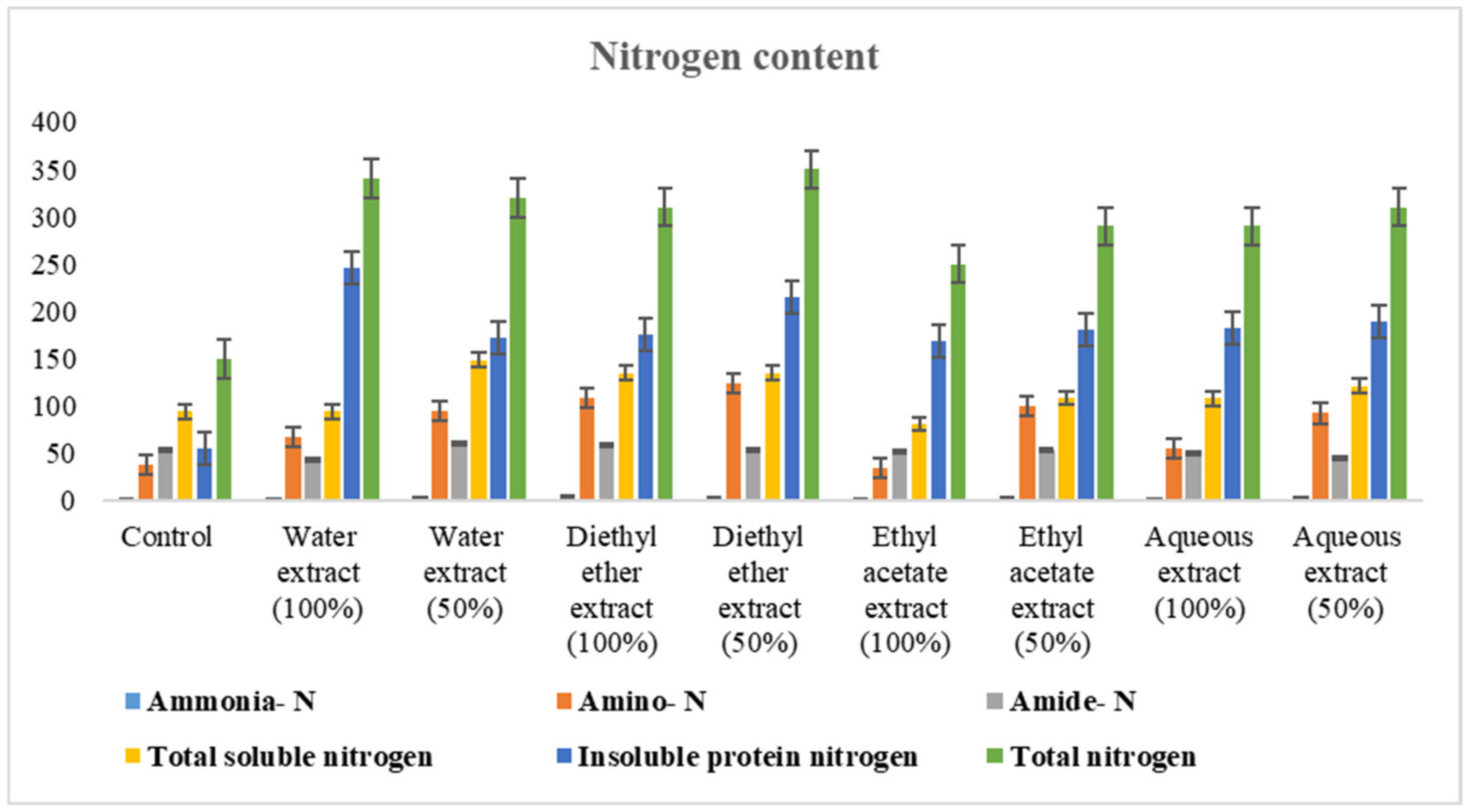
3.2.2. Changes in Nitrogen Content

Data presented in Figure 4 showed that the pretreatment with water and diethyl ether extracts (100% and 50%) caused a generally significant increase in different nitrogen fractions except water extract 100%, which caused a decrease in ammonia and amide content of the yielded seed by 1.96% and 19.56%, respectively. In this connection, the general significant increments in different nitrogen fractions were recorded for ethyl acetate 50% extract and aqueous extracts (100% and 50%), with the exception of a general significant decrease that was reported with those mentioned extracts for amide content, and with aqueous extract 100% for ammonia of lupine yielded seeds. On the other hand, ethyl acetate extract 100% caused an insignificant decrease in ammonia and amino nitrogen contents by 5.18% and 9.41%, respectively, a significant decrease in amide and total soluble nitrogen contents by 3.96% and 14.29%, respectively, and a significant increase in nitrogen, protein and total nitrogen of white lupine yielded seeds by 204.50% and 66.67%, respectively. However, the treatments that led to the maximum nitrogen content were recorded for water and diethyl ether extracts.

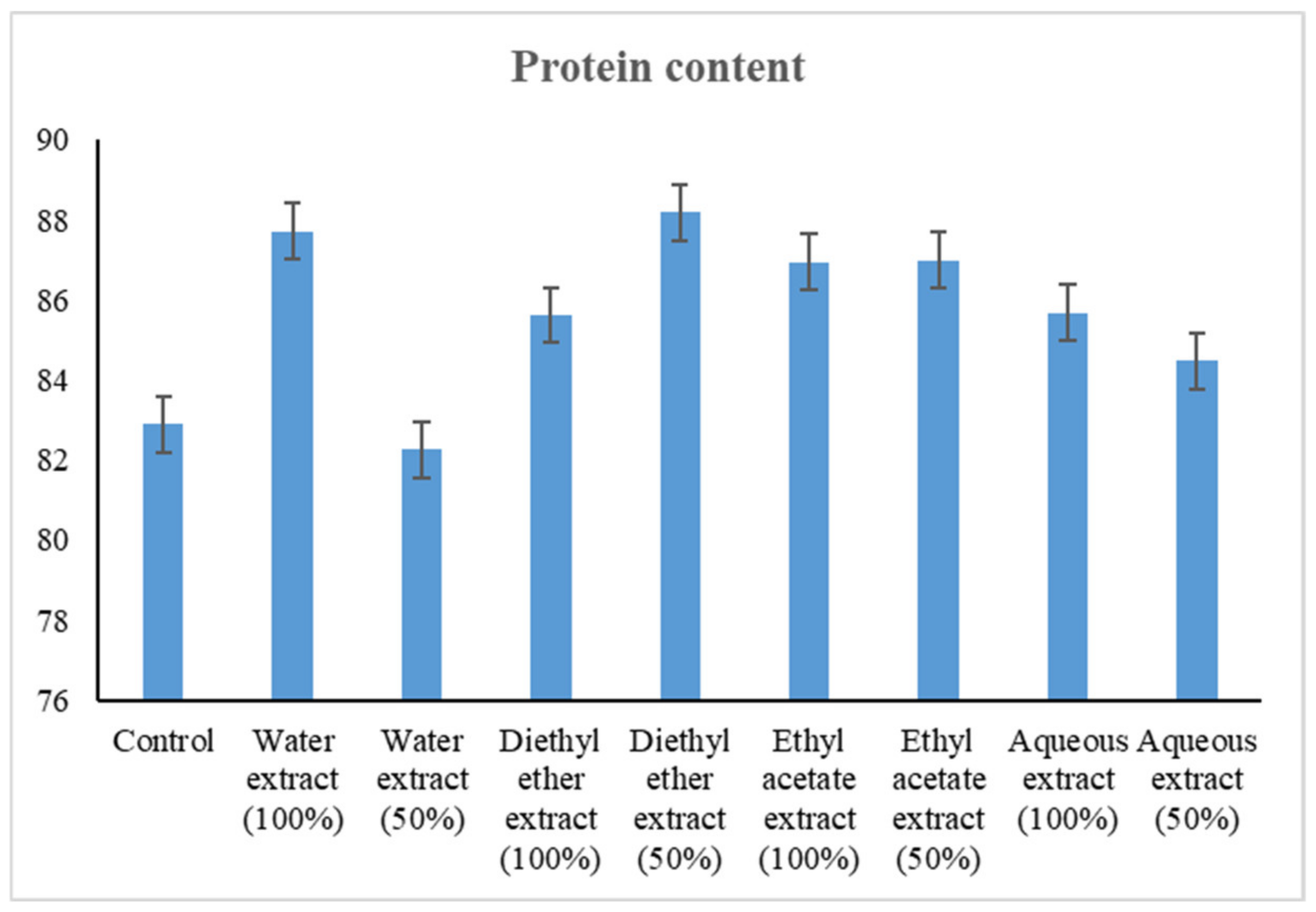
3.2.3. Changes in Protein Content

The obtained results showed that all treatments increased the protein content of the yielded Lupinus seeds significantly, except water extract 50%, which decreased it insignificantly by 0.76% (Figure 5). Moreover, the pretreatment of seeds with diethyl ether extract 50% caused the maximum protein content above than control by 6.40%.

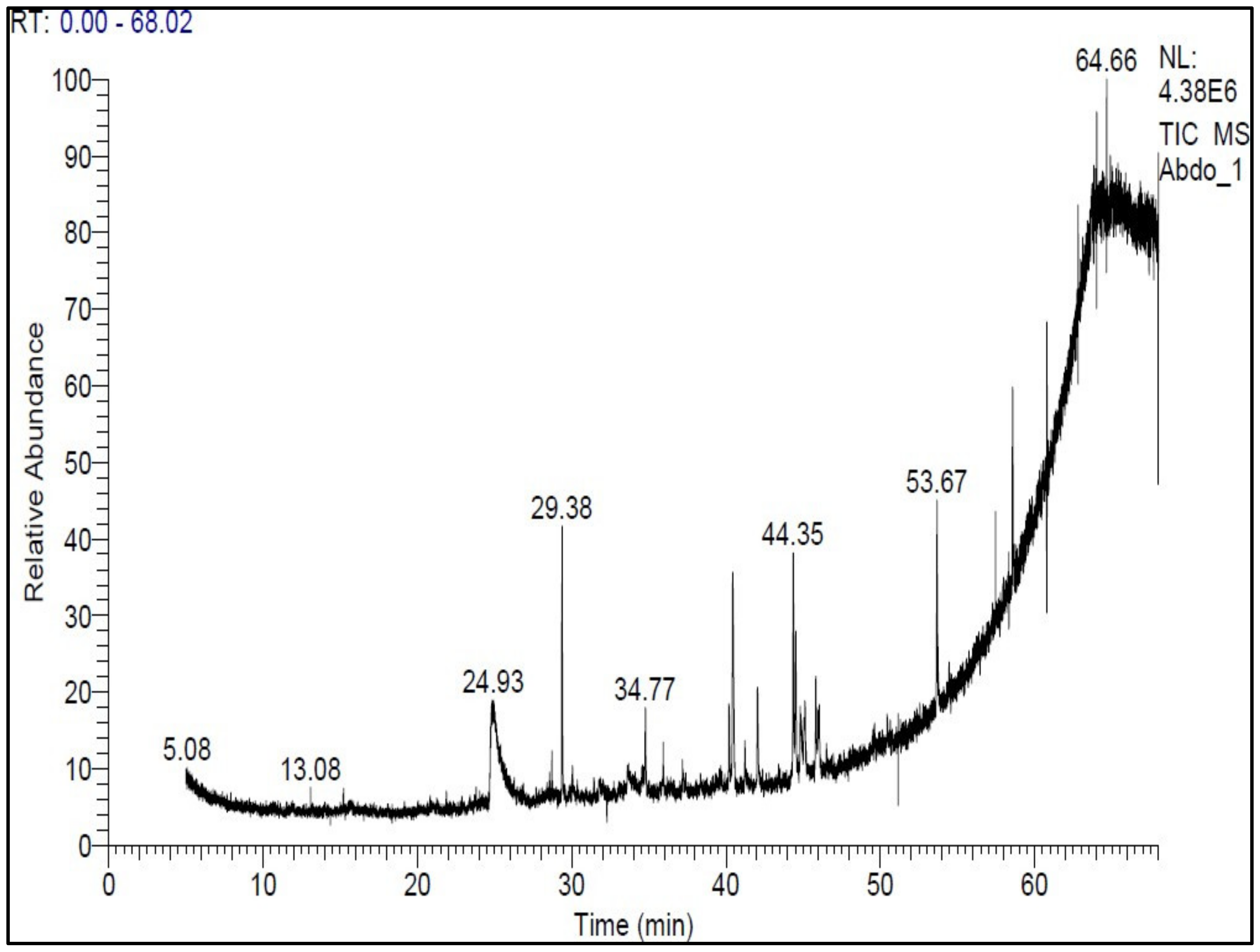
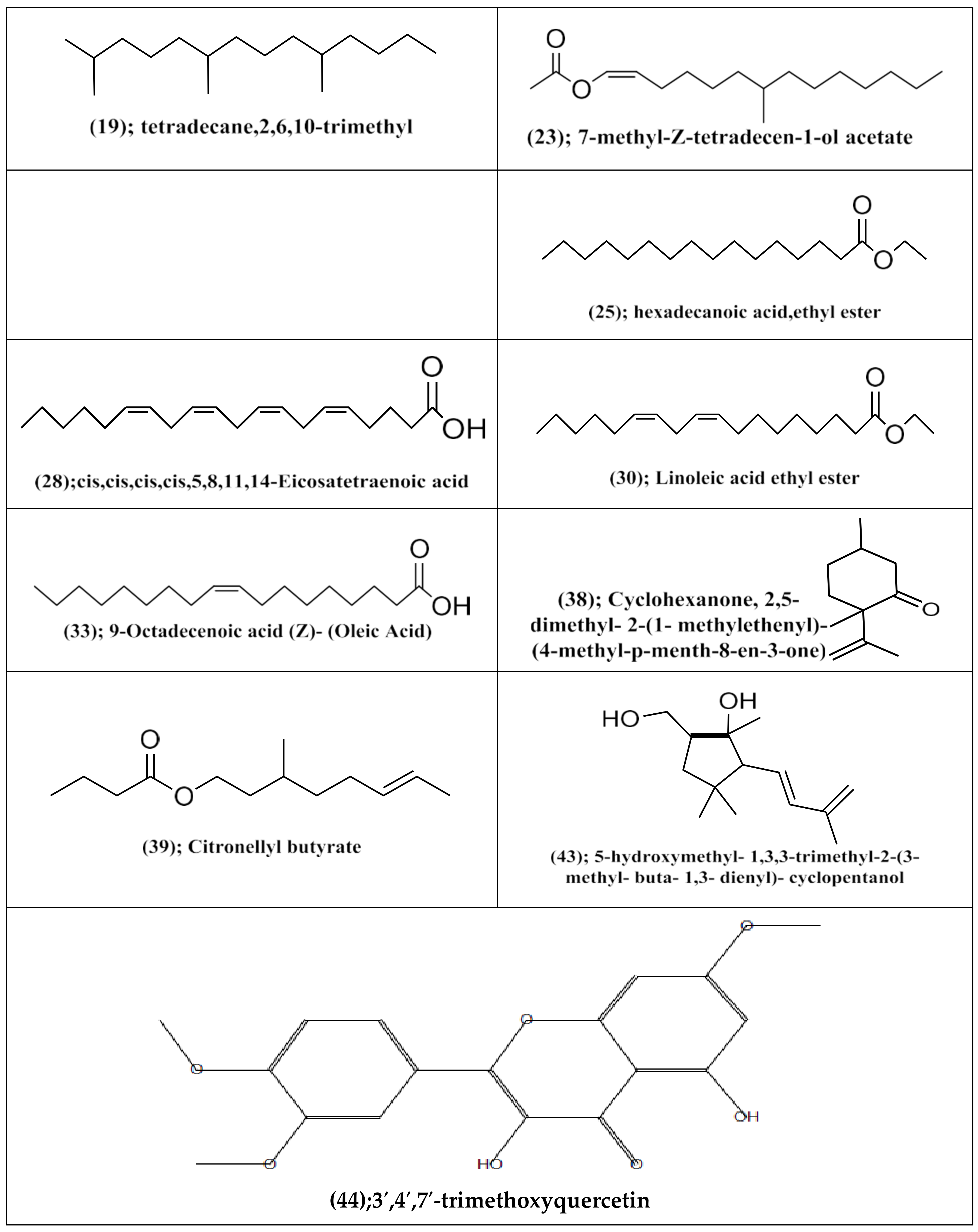
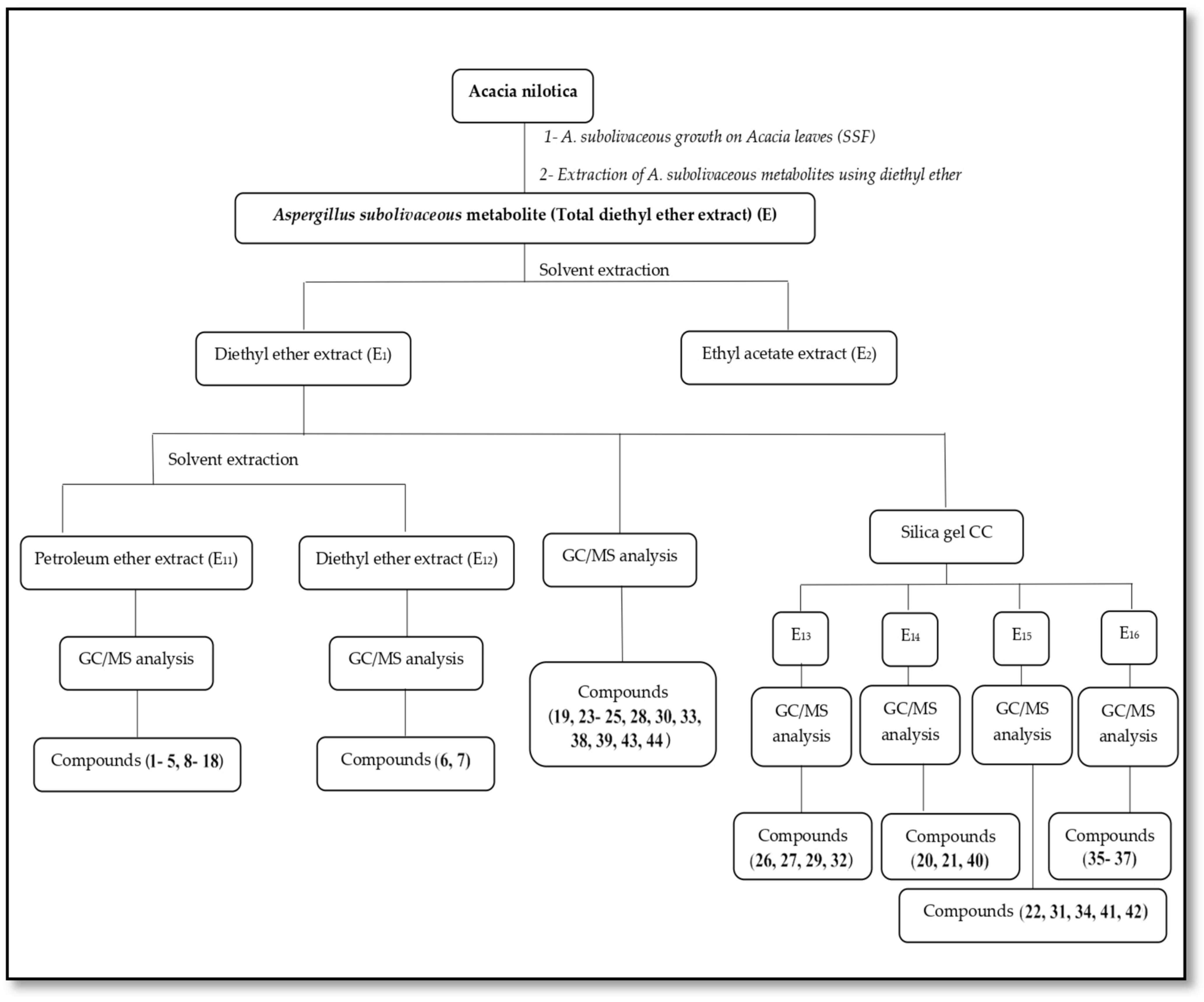
3.3. Processing of Total Diethyl Ether Extract (E) of A. subolivaceous/A. nilotica Metabolite

In order to explore the components of A. subolivaceous metabolites, the fungal metabolites produced by A. subolivaceous growth on powdered A. nilotica leaves were processed in a variety of ways (see Scheme 1). The extract’s constituents were then identified using Wiley 9, Wiley 7, NIST, and main lib libraries, or by comparing literature data [45] to mass spectral databases after fractionation of the A. subolivaceous metabolite extract using various solvents, separation of its components, and analysis using 1H-NMR spectra, and GC/MS.
The pretreatment of L. termis seeds with diethyl ether extract showed the best results as caused the maximum increase in (glucose, amino nitrogen, insoluble protein nitrogen, total nitrogen, and protein) contents in the yielded seeds, so the diethyl ether extract was processed in order to study its components. It was found that A. subolivaceous metabolite extract is rich in phytochemicals such as fatty materials, terpenes, flavonoids, and other minor phytochemicals as shown in Table 2 and Figure 6 and Figure 7.
In this investigation, the GC/MS analysis of diethyl ether extract from A. subolivaceous metabolite showed the presence of 4-methyl-p-menth-8-en-3-one and 3,4,7-trimethoxyquercetinas, shown in Table 3 and Figure 7, which have not reported previously from A. nilotica leaves extract as shown in Table 1, indicating the probable biotransformation as a result of the presence of A. subolivaceous. However, by a single or multiple step O-methylation process, A. subolivaceous can convert quercetin into 3′,4′,7′-trimethoxyquercetin. Additionally, A. subolivaceous has the ability to convert citronellal by one or more one-step C-methylation processes into 4-methyl-p-menth-8-en-3-one.

4. Discussion

4.1. Changes in Yield Attributes

Concerning this, Khalil and Ismael [46] reported that the yeast application in the L. termis L. yielded seeds enhanced yield attributes (number of pods/plants, number of seeds/plants, and 100 seeds weight). Anwer and Khan [47], who showed that the nursery application of A. niger isolates considerably affected yield and tomato plants highest with maximal growth, corroborated the observed results. A. niger also typically increases crop production, nitrogen uptake, and root growth and development [48].
These findings are also in line with those of El-Shahawy et al. [49], who revealed that the use of yeast as a fungal application increased the yield attributes of Linum usitatissimum L. plants, including total plant length, straw yield per plant, number of fruiting branches per plant, number of capsules per plant, the weight of 1000 seeds, biological yield per plant, and seed yield per plant. Connectively, the use of biofertilizer boosted the fennel (Foeniculum vulgare Mill.) plants’ yield features (plant fresh weight, plant dry weight, plant height, branches number, and fruit yield/plant) [50].

4.2. Changes in Biochemical Aspects in L. termis L. yielded Seeds

4.2.1. Changes in Carbohydrate Content

Application of A. subolivaceus extract resulted in a rise in the produced seeds’ carbohydrate content, according to research on the influence of A. subolivaceus extracts on that fraction’s content. However, the ethyl acetate (100% and 50%) and diethyl ether (50%) extracts produced the seeds with the highest carbohydrate fraction content. These findings are consistent with those of Khalil and Ismail [46], who observed an increase in the percentage of carbohydrates as a result of the use of yeast and attributed this increase to either an increase in chlorophyll a and b or to yeast’s enhanced role in cell division and cell elongation, which resulted in more leaf area [51]. Mahfouz and Sharaf-Eldin [51] investigated the biochemical features of fennel seeds and found that application of biofertilizers increased total carbs content. The availability of inorganic nitrogen in particular may have an impact on how proteins, soluble solids, and secondary metabolites are synthesized [52]. A considerable amount of variance was seen in the level of plant metabolites in seeds among the six examined mung bean types, and they also reported a larger buildup of amino acid content in leaves.

4.2.2. Changes in Nitrogen Content

The recorded increase in nitrogen content was found to be consistent with Gomaa and Abou-Aly’s findings [53], who investigated how yeast foliar treatment affected L. termis L. seeds and concluded that the yeast-produced growth hormones were considered to be the cause of the rise in nitrogen and protein content. El-Sayed et al. [54] further hypothesized that the rise in total nitrogen in the anise plants treated with bio-fertilizers may be the result of inoculation with non-symbiotic nitrogen-fixing organisms. Growth, which is impacted by several internal and external factors, in addition to its genetic make-up, is thus a valuable tool for assessing agricultural production across numerous crops. As a result, among other things, several factors, including water conductivity, osmotic potential, and threshold turgor, are essential components for plant growth and must all be present in order for growth to occur [55]. Nitrogen is an essential element of several components, such as proteins, enzymes, alkaloids, hormones, and vitamins [56]. It plays a significant role in photosynthesis process and the transfer of nutrients to the seed.

4.2.3. Changes in Protein Content

The observed increase in protein content was consistent with the results of Nafie [57], who reported that treatment with Streptomyces chibaensis increased protein content and biochemical components in L. termis L. seeds. Nonetheless, the same author also observed a decrease in protein content in L. termis L. seeds following treatment with Fusarium oxysporum, leading some to speculate that the plant’s relatively low protein content was due to a high concentration of soluble sugars. The protein content is closely correlated with the nitrogen content [58], the tuber’s protein content is crucial in terms of quality in potato tubers. The 100% manure × (Azospirillum + Pseudomonas) mixture had the highest tuber protein content (7.05%). By reactivating minerals found in soil and manure, the use of biofertilizers alongside manure to potatoes promotes the uptake of minerals from the soil and organic sources and decreases the demand for chemical fertilizers [59]. Application of both biofertilizer and other nitrogen fertilizers gave the high protein content of potato tuber [59,60].

4.3. Processing of Total Diethyl Ether Extract (E) of A. subolivaceous/A. nilotica Metabolite

The extract of the metabolite was discovered to be highly phytochemical. According to Kim et al., [61], A. subolivaceous, has various components, including sugars such as glucose, glucopyranose, galactose, turanose, and arabinofuranose, as well as sugar alcohols such as glycerol and inositol, organic acids such as citric acid and pyruvic acid, and fatty acids such as linoleic acid and palmitic acid. The current findings agreed with those of Senthilkumar et al. [62] and Devi and Prabakaran [63] who claimed that the metabolites generated by endophytic fungi Phomopsis sp. contained alkaloids, terpenoids, phenolics, and minor secondary compounds.
Devi and Prabakaran also studied the GC chromatogram and spectral analysis, as well as the name, molecular weight, and chemical structure of the compounds present in the ethyl acetate extract for Penicillium sp. These compounds include 4-hydroxybenzeneethanol, 2-tert-butyl-4-isopropyl-1-5-methyl phenol, and benzoic acid. 4-hydroxypropyl ester, p-hydroxyphenylacetamide, N-[2-methyl-1-prenylpropyl] formamide, cyclo (L-leucyl-L-propyl), 3-(3-azidopropyl)-1H-indene, and dihydroergotamine are some examples of these compounds. However, Waqas et al. [64] investigated the potential of two endophytic fungi to release the phytohormones gibberellins (GAs) and indoleacetic acid (IAA). They claimed that the study of these endophytic fungi’s pure cultures revealed varying levels of biologically active GAs (GA1, GA3, GA4, and GA7). The volatile compounds found in the culture samples of Trichoderma harzianum are members of the compound classes of alkanes, ketones, alcohols, pyrones (lactones), monoterpenes, furanes and sesquiterpenes [65]. A potent method for changing the structural makeup of bioactive natural and manmade materials is microbial transformation. The spectral analysis of the current study indicated biotransformation to a flavonoid (quercetin 7,3,4 trimethoxy) and a monoterpene (4-methyl P-menth-8-en-3-one), suggesting that biotransformation may be effective. The microorganism’s O-methylation of the flavonols quercetin and fisetin, which takes place at the C-3′ and C-4′ hydroxyls, supports this conclusion [6]. In this regard, quercetin was oxidized enzymatically by A. flavus, and quercetin was then broken down by quercetinase to produce carbon monoxide and 2-protocatechuoylphloroglucinol carboxylic acid [3]. O-methylated quercetin was found in Streptomyces griseus (ATCC 13273) [66]. Quercetin’s OH at C-3′ can be methylated by Beauveria bassiana ATCC 7159 to produce 3′-O-methylquercetin, and these bacteria can also produce derivatives of 3′-O-methylquercetin-7-glucuronide by glycosylating the hydroxyl at C-7 of quercetin [67]. Additionally, quercetin was transformed into a variety of metabolites through the processes of hydroxylation, glucosylation, and methylation by species of Bacillus, Aspergillus, Streptomyces, and Penicillium. Similar to this, Rueda et al. [5] investigated the biotransformation of (S)-citronellal using Penicillium and Fusarium sp. to produce a number of compounds. This outcome is also consistent with the monoterpene (tetra hydrogeraniol) to hydroxycitronellol conversion described for Glomerella cingulate [68]. In this regard, Rhodotorulaminuta free and immobilized cells were used in the biotransformation of (L)-citronellol to (L)-citronellol by Velankar and Heble [69]. Additionally, Pulicaria undulata plants were regarded by Alshammari et al. [70] as a useful source of physiologically active secondary metabolites.

5. Conclusions

Lupinus seed has an important place in the pharmaceutical industry, and has been used for the production of protein isolates with good functional and nutritional properties that are considered a protein source for human nutrition and animal feeding all over the world. According to this study’s findings, the fungal metabolite produced by A. subolivaceus growing on A. nilotica, is considered as an important strategy in agricultural sustainability. Hence, we must lessen our reliance on artificial fertilizers, which have detrimental effects on both the environment and human health, by using these naturally occurring substances to promote plant growth. This may lead to the development of a more ecologically conscious and economically viable farming system. It became clear that the bioactive natural and manufactured materials can have their structures changed effectively by microbial transformation as water and ether extracts improved the plant’s growth and yield and biochemical characteristics of the produced Lupine seeds. The spectral analysis of the present study revealed biotransformation to a flavonoid (3′,4′,7′-trimethoxyquercetin) and a monoterpene (4-methyl P-menth-8-en-3-one), as well as maximization of the active compound production that is used as a source of medicinal materials in the pharmaceutical industry.

Author Contributions

Conceptualization, R.M.E.G., S.A.H., E.A.S., A.B.T., A.-D.A.S., M.A.-M. and A.M.K.; methodology, R.M.E.G., S.A.H., E.A.S., A.B.T., A.-D.A.S., M.A.-M. and A.M.K.; software, R.M.E.G., S.A.H., E.A.S., A.B.T., A.-D.A.S., M.A.-M. and A.M.K.; validation, R.M.E.G., S.A.H., E.A.S., A.B.T., A.-D.A.S., M.A.-M. and A.M.K.; investigation, R.M.E.G., S.A.H., E.A.S., A.B.T., A.-D.A.S., M.A.-M. and A.M.K.; data curation, R.M.E.G., S.A.H., E.A.S., A.B.T., A.-D.A.S., M.A.-M. and A.M.K.; writing—original draft preparation, R.M.E.G., S.A.H., E.A.S., A.B.T., A.-D.A.S., M.A.-M. and A.M.K.; writing—review and editing, R.M.E.G., S.A.H., E.A.S., A.B.T., A.-D.A.S., M.A.-M. and A.M.K.; supervision, R.M.E.G., S.A.H., E.A.S., A.B.T., A.-D.A.S., M.A.-M. and A.M.K.; funding acquisition O.A.A., A.H.A., H.S.A.M.A., N.A.A.-H. and K.A. All authors have read and agreed to the published version of the manuscript.

Funding

This work was supported by the Deanship of Scientific Research, Vice Presidency for Graduate Studies and Scientific Research, King Faisal University, Al-Ahsa 31982, Saudi Arabia (Grant No. 3637).

Institutional Review Board Statement

The study was conducted in accordance with the Declaration of Helsinki and approved by the deanship of the scientific research ethical committee, King Faisal University (Project number: GRANT3637, date of approval: 1 May 2023).

Informed Consent Statement

Not applicable.

Data Availability Statement

The data presented in this study are available on request from the corresponding authors.

Conflicts of Interest

The authors declare no conflict of interest.

References

  1. Grandini, A.; Summa, D.; Costa, S.; Buzzi, R.; Tamburini, E.; Sacchetti, G.; Guerrini, A. Biotransformation of Waste Bile Acids: A New Possible Sustainable Approach to Anti-Fungal Molecules for Crop Plant Bioprotection? Int. J. Mol. Sci. 2022, 23, 4152. [Google Scholar] [CrossRef] [PubMed]
  2. Bianchini, L.F.; Arruda, M.F.C.; Vieira, S.R.; Campelo, P.M.S.; Gregio, A.M.T.; Rosa, E.A.R. Microbial biotransformation to obtain new antifungals. Front. Microbiol. 2015, 6, 1433. [Google Scholar] [CrossRef] [PubMed] [Green Version]
  3. Das, S.; Rosazza, J.P.N. Microbial and enzymatic transformations of flavonoids. J. Nat. Prod. 2006, 69, 499–508. [Google Scholar] [CrossRef] [PubMed]
  4. Smitha, M.S.; Singh, V.; Singh, R. Microbial biotransformation: A process for chemical alterations. J. Bacteriol. Mycol. 2017, 4, 85. [Google Scholar]
  5. Rueda, M.G.M.; Guerrinia, A.; Giovanninib, P.P.; Medicib, A.; Grandinia, A.; Sacchettia, G.; Pedrini, P. Biotransformations of terpenes by fungi from Amazonian citrus plants. Chem. Biodivers. 2013, 10, 1909–1919. [Google Scholar] [CrossRef]
  6. Cao, H.; Chen, X.; Jassbi, A.R.; Xiao, J. Microbial biotransformation of bioactive flavonoids. Biotechnol. Adv. 2015, 33, 214–223. [Google Scholar] [CrossRef]
  7. Liu, J.; Liu, G. Analysis of secondary metabolites from plant endophytic fungi. Methods Mol. Biol. 2018, 1848, 25–38. [Google Scholar]
  8. Hemamalini, G.; Jithesh, P.; Nirmala, P. In silico screening of phytochemicals identified from Acacia nilotica by GC/MS method for its anticancer activity. Int. J. Pharm. Sci. Res. 2014, 5, 2374–2381. [Google Scholar]
  9. Sadiq, M.B.; Tarning, J.; Aye, T.Z.; Anal, A.K. Antibacterial activities and possible modes of action of Acacia nilotica (L.) Del. against multidrug-resistant Escherichia coli and Salmonella. Molecules 2017, 22, 47. [Google Scholar] [CrossRef] [Green Version]
  10. Lim, T.K. Edible medicinal and non- medicinal plants. Springer Neth. 2012, 2, 763–769. [Google Scholar]
  11. Dubois, O.; Allanic, C.; Charvet, C.L.; Guégnard, F.; Février, H.; Théry-Koné, I.; Cortet, J.; Koch, C.; Bouvier, F.; Fassier, T.; et al. Lupin (Lupinus spp.) seeds exert anthelmintic activity associated with their alkaloid content. Sci. Rep. 2019, 9, 9070. [Google Scholar] [CrossRef] [PubMed] [Green Version]
  12. Sedláková, K.; Straková, E.; Suchý, P.; Krejcarová, J.; Herzig, I. Lupin as a perspective protein plant for animal and human nutrition. Areview. Acta Vet. Brno. 2016, 85, 165–175. [Google Scholar] [CrossRef] [Green Version]
  13. Zarei, M.; Asgarpanah, J.; Ziarati, P. Chemical composition profile of wild Acacia oerfota (Forssk) Schweinf seed growing in the south of Iran. Orient. J. Chem. 2015, 31, 2311–2318. [Google Scholar] [CrossRef]
  14. Zia-Ul-Haq, M.; Ćavar, S.; Qayum, M.; Khan, I.; Ahmad, S. Chemical composition and antioxidant potential of Acacia leucophloea Roxb. Acta Bot. Croat. 2013, 72, 133–144. [Google Scholar] [CrossRef]
  15. Abduljawad, E.A. Review of some evidenced medicinal activities of Acacia nilotica. Arch. Pharm. Pract. 2020, 11, 20–25. [Google Scholar]
  16. Rajbir, S.; Bikram, S.; Sukhpreeet, S. Anti-free radical activities of kaempferol isolated from Acacia nilotica (L.) Willd. Ex. Del. Toxicol. Vitro 2008, 22, 1965–1970. [Google Scholar]
  17. Banso, A. Phytochemical and antibacterial investigation of bark extracts of Acacia nilotica. J. Med. Plant Res. 2009, 3, 82–85. [Google Scholar]
  18. Wassel, G.M.; Abd-El-Wahab, S.M.; Aboutabl, E.A.; Ammar, N.M.; Afifi, M.S. Study of phenolic constituents and tannins isolated from Acacia nilotica L. Willd and Acacia farnesiana L. Willd growing in Egypt. Herba Hungarica 1990, 29, 43–50. [Google Scholar]
  19. Chaubal, R.; Tambe, A. Isolation of new straight chain compound from Acacia nilotica. Indian J. Chem. 2006, 45, 1231–1233. [Google Scholar] [CrossRef]
  20. Yadav, A.; Yadav, M.; Kumar, S.; Sharma, D.; Yadav, J.P. In vitro antioxidant activities and GC-MS analysis of different solvent extracts of Acacia nilotica leaves. Indian J. Pharm. Sci. 2018, 80, 892–902. [Google Scholar] [CrossRef]
  21. Bai, S.; Seasotiya, L.; Malik, A.; Bharti, P.; Dalal, S. GC-MS analysis of chloroform extract of Acacia nilotica L. leaves. J. Pharmacogn. Phytochem. 2014, 2, 79–82. [Google Scholar]
  22. Malviya, S.; Rawat, S.; Kharia, A.; Verma, M. Medicinal attributes of Acacia nilotica Linn. A comprehensive review on ethnopharmacological claims. Int. J. Pharm. Life Sci. 2011, 2, 830–837. [Google Scholar]
  23. Hemamalini, G.; Jithesh, P.; Nirmala, P. Phytochemical analysis of leafextract of plant Acacia nilotica by GCMS method. Adv. Biol. Res. 2013, 7, 141–144. [Google Scholar]
  24. Trevino-Cueto, B.; Luis, M.; Contreras-Esquivel, J.C.; Rodriguez, R.; Aguilera, A.; Aguilar, C.N. Gallic acid and tannase accumulation during fungal solid-state culture of a tannin-rich desert plant (Larrea tridentate Cov.). Bioresour. Technol. 2007, 98, 721–724. [Google Scholar] [CrossRef] [PubMed]
  25. Mukherjee, G.; Banerjee, R. Biosynthesis of tannase and gallic acid from tannin rich substrates by Rhizopus oryzae and Aspergillus foetidus. J. Basic Microbiol. 2004, 44, 42–48. [Google Scholar] [CrossRef] [PubMed]
  26. Samuel, P.; Prince, L.; Prabakaran, P. Antibacterial Activity of Marine derived Fungi Collected from South East Coast of Tamilnadu, India. J. Microbiol. Biotechnol. Res. 2011, 1, 86–94. [Google Scholar]
  27. Mohrig, J.R.; Hammond, C.N.; Schatz, P.F. Techniques in Organic Chemistry, 3rd ed.; Library of Congress: Washington, DC, USA, 2010; p. 200993463. [Google Scholar]
  28. Snedecor, G.W.; Cochran, W.G. Statistical Methods, 7th ed.; Iowa State University: Ames, IA, USA, 1980. [Google Scholar]
  29. Gregory, P.J. Crop growth and development. In Ressell”s Soil Conditions and Plant Growth; Wild, A., Ed.; Longman Scientific Technical: London, UK, 1988; pp. 31–68. [Google Scholar]
  30. Beadle, C.L. Growth analysis. In Photosynthesis and Production in a Changing Environment. A Field and Laboratory Manual; Hall, D.C., Scurlock, J.M.O., Bolhar-Nordenkampf, H.R., Leegood, R.C., Long, S.P., Eds.; Chapman and Hall: London, UK, 1993; pp. 36–46. [Google Scholar]
  31. Yemm, E.W.; Willis, A.J. The estimation of carbohydrates by anthrone. Biochem. J. 1954, 57, 508–514. [Google Scholar] [CrossRef] [PubMed] [Green Version]
  32. Handel, E.V. Direct micro determination of sucrose. Anal. Biochem. 1968, 22, 280–283. [Google Scholar] [CrossRef]
  33. Fetris, A.W. A serum glucose method without protein precipitation. Am. J. Med. Technol. 1965, 31, 17–21. [Google Scholar]
  34. Riazi, A.; Matsuda, K.; Arslan, A. Water stress induced changes in concentrations of proline and other solutes in growing regions of young barley leaves. J. Exp. Bot. 1985, 36, 1716–1725. [Google Scholar] [CrossRef]
  35. Sadasivam, S.; Manickam, A. Biochemical Methods, 2nd ed.; New Age Int. (P) Ltd.: Delhi, India, 1996. [Google Scholar]
  36. Yemm, E.W.; Willis, A.J. The respiration of barely plants. IX. The metabolism of roots during the assimilation of nitrogen. New Phytol. 1956, 55, 229–252. [Google Scholar] [CrossRef]
  37. Delory, M. Colourimetric Estimation of Ammonia. In Inorganic Chemistry; Vogel, H.J., Ed.; Longman: London, UK, 1949; pp. 126–132. [Google Scholar]
  38. Naguib, M.I. Effect of sevin on carbohydrates and nitrogen metabolism during the germination of cotton seeds. Indian J. Exp. Biol. 1964, 2, 149–155. [Google Scholar]
  39. Muting, D.; Kaiser, E. Spectrophotometric method of determination of α-amino-N in biological material by means of the ninhydrin reaction. Hopper Seyler’s J. Physiol. Chem. 1963, 332, 276–289. [Google Scholar]
  40. El-Shahaby, O.A. Studies on Growth and Metabolism of Certain Plants. Ph.D. Thesis, Mansoura University, Mansoura, Egypt, 1981. [Google Scholar]
  41. Pirie, N.W. Proteins. In Modern Methods of Plant Analysis IV; Paech, K., Tracey, M.B., Eds.; Springer: Berlin, Germany, 1955; pp. 23–68. [Google Scholar]
  42. Rees, M.W.; Williams, E.F. The total nitrogen content of egg albumin and other proteins. J. Biochem. 1943, 37, 354–359. [Google Scholar]
  43. Haroun, S.A. Studies on Adaptation of Plants to Water Stress. Ph.D. Thesis, Mansoura University, Mansoura, Egypt, 1985. [Google Scholar]
  44. Bradford, M.M. A rapid and sensitive method for the quantitative of microorgan quantities of protein utilizing the principle of protein dye binding. Anal. Biochem. 1976, 72, 248–251. [Google Scholar] [CrossRef]
  45. Adam, R.P. Identification of Essential Oil Components by Gas Chromatography/Mass Spectroscopy; Allured Publishing Corporation: Carol Stream, IL, USA, 1995; ISBN 0-931710-42-1. [Google Scholar]
  46. Khalil, S.E.; Ismael, E.G. Growth, yield and seed quality of Lupinus termis as affected by different soil moisture levels and different ways of yeast application. Am. J. Sci. 2010, 6, 141–153. [Google Scholar]
  47. Anwer, M.D.A.; Khan, M.R. Aspergillus niger as tomato fruit (Lycopersicum esculentum Mill.) quality enhancer and plant health promoter. J. Postharvest Technol. 2013, 1, 36–51. [Google Scholar]
  48. Sen, B. Biocontrol: A success story. Indian Phytopathol. 2000, 53, 243–249. [Google Scholar]
  49. El-Shahawy, T.A.; El-Rokiek, K.G.; Balbaa, L.K.; Abbas, S.M. Micronutrients, B-vitamins and yeast in relation to increasing flax (Linum usitatissimum L.) growth, yield productivity and controlling associated weeds. Asian Res. J. Agric. 2008, 2, 1–14. [Google Scholar] [CrossRef]
  50. Mahfouz, S.A.; Sharaf-Eldin, M.A. Effect of mineral vs. biofertilizer on growth, yield, and essential oil content of fennel (Foeniculum vulgare Mill.). Int. Agrophys. 2007, 21, 361–366. [Google Scholar]
  51. Hayat, A.E.H. Physiological Studies on Hibiscus sabdariffa L. Production in New Reclamated Soils. Master’s Thesis, Zagazig University, Zagazig, Egypt, 2007. [Google Scholar]
  52. Mondal, M.M.A.; Puteh, A.B.; Ismail, M.R.; Rafii, M.Y. Optimizing plant spacing for modern rice varieties. Int. J. Agric. Biol. 2013, 15, 175–178. [Google Scholar]
  53. Gomaa, A.O.; Abou-Aly, H.E. Efficiency of bioferitlization in the presence of both inorganic and organic fertilizers on growth, yield and chemical constituents of anise plant Pimpinellaanisum L. In Proceedings of the 5th Arabian Horticulture Conference, Ismailia, Egypt, 24–28 March 2001. [Google Scholar]
  54. El-Sayed, A.A.; El-Leithy, A.S.; Bazraa, W.M.; Abdel-Latef, M.S. Effect of growing media, bio and organic fertilization on the flowering and chemical constituents of Calendula officinalis L. plants. Biosci. Res. 2018, 15, 2029–2040. [Google Scholar]
  55. Poschenrieder, F.J.C. Plant water relations as affected by heavy metal stress: A review. J. Plant Nutr. 1990, 13, 1–37. [Google Scholar]
  56. Marschner, H. Mineral Nutrition of Higher Plants; Academic Press: San Diego, CA, USA, 1995; p. 889. [Google Scholar]
  57. Nafie, E.M. The possible induction of resistance in Lupinus termis L. against Fusarium oxysporum by Streptomyces chibaensis and its mode of action: I. changes in certain morphological criteria and biochemical composition related to induced resistance. Int. J. Agric. Biol. 2003, 5, 463–472. [Google Scholar]
  58. Naghdi, A.A.; Piri, S.; Khaligi, A.; Moradi, P. Enhancing the qualitative and quantitative traits of potato by biological, organic and chemical fertilizers. J. Saudi Soc. Agric. Sci. 2022, 21, 87–92. [Google Scholar] [CrossRef]
  59. Wichrowska, D.; Szczepanek, M. Possibility of limiting mineral fertilization in potato cultivation by using bio-fertilizer and its influence on protein content in potato tubers. Agriculture 2020, 10, 442. [Google Scholar] [CrossRef]
  60. Abdeldaym, E.A.; El-Sawy, M.; El-Helaly, M. Combined application of different sources of nitrogen fertilizers for improvement of potato yield and quality. Plant Arch. 2019, 19, 2513–2521. [Google Scholar]
  61. Kim, A.J.; Choi, J.N.; Kim, J.; Park, S.B.; Yeo, S.H.; Choi, J.H.; Lee, H. GC-MS Based Metabolite Profiling of Rice Koji Fermentation by Various Fungi. Biosci. Biotechnol. Biochem. 2010, 74, 2267–2272. [Google Scholar] [CrossRef] [Green Version]
  62. Senthilkumar, N.; Murugesan, S.; Babu, D.S.; Rajeshkannan, C. GC-MS analysis of the extract of endophytic fungus, Phomopsis sp. isolated from tropical tree species of India, Tectonagrandis L. Int. J. Innov. Res. Sci. Eng. Technol. 2014, 3, 10176–10179. [Google Scholar]
  63. Devi, N.N.; Prabakaran, J.J. Bioactive metabolites from an endophytic fungus Penicillium sp. isolated from Centellaasiatica. Environ. Appl. Mycol. 2014, 4, 34–43. [Google Scholar]
  64. Waqas, M.; Khan, A.L.; Kamran, M.; Hamayun, M.; Kang, S.; Kim, Y.; Lee, I. Endophytic Fungi Produce Gibberellins and Indoleacetic Acid and Promotes Host-Plant Growth during Stress. Molecules 2012, 17, 10754–10773. [Google Scholar] [CrossRef] [PubMed] [Green Version]
  65. Siddiquee, S.; Cheong, B.E.; Taslima, K.; Kausar, H.; Hasan, M.M. Separation and Identification of Volatile Compounds from Liquid Cultures of Trichodermaharzianum by GC-MS using Three Different Capillary Columns. J. Chromatogr. Sci. 2011, 2, 358–367. [Google Scholar]
  66. Hosny, M.; Dhar, K.; Rosazza, J.P.N. Hydroxylations and methylations of quercetin, fisetin, and628 catechin by Streptomyces griseus. J. Nat. Prod. 2011, 64, 462–465. [Google Scholar] [CrossRef] [PubMed]
  67. Costa, E.M.D.; Pimenta, F.C.; Luz, W.C.; Oliveira, V.D. Selection of filamentous fungi of the Beauveria genus able to metabolize quercetin like mammalian cells. Braz. J. Microbiol. 2008, 39, 405–408. [Google Scholar] [CrossRef] [Green Version]
  68. Hegazy, M.F.; Mohamed, T.A.; ElShamy, A.I.; Mohamed, A.H.; Mahalel, U.A.; Reda, E.H.; Shaheen, A.M.; Tawfik, W.A.; Shahat, A.A.K.; Shams, A.; et al. Microbial biotransformation as a tool for drug development based on natural products from mevalonic acid pathway. A review. J. Adv. Res. 2015, 6, 17–33. [Google Scholar] [CrossRef] [Green Version]
  69. Velankar, H.R.; Heble, M.R. Biotransformation of (L)-citronellal to (L)-citronellol by free and immobilized Rhodotorula minuta. Electron. J. Biotechnol. 2003, 6, 90–102. [Google Scholar] [CrossRef] [Green Version]
  70. Alshammari, S.O.; Abd El Aty, A.A.; Hegazy, M.E.F. New sesquiterpene lactone via fungal transformation of Rhizopus oryzaeKX685359: Antimicrobial In-Vitro and In-Silico Study. Catal. Lett. 2022. [Google Scholar] [CrossRef]
Figure 1. Effect of different extracts of A. subolivaceus metabolite on shoot length (cm), shoot fresh and dry weight (g), No. of legumes/plant, and legumes fresh weight (g/plant) of lupines termis plants.
Sustainability 15 09509 g001
Figure 2. Effect of different extracts of A. subolivaceus metabolite on No. of seeds/plant, seeds fresh weight (g/plant), seeds dry weight (g/plant), relative grain yield (%) and seed index of lupines termis plants.
Sustainability 15 09509 g002
Figure 3. Effect of different metabolite extracts on carbohydrate content (mg/g dry weight) of L. termis L. seeds.
Sustainability 15 09509 g003
Figure 4. Effect of different metabolite extracts on nitrogen content (mg/g dry weight) of L. termis L. seeds.
Sustainability 15 09509 g004
Figure 5. Effect of different metabolite extracts on protein content (mg/g dry weight) of L. termis L. seeds.
Sustainability 15 09509 g005
Scheme 1. Processing of A. subolivaceous metabolite.
Sustainability 15 09509 sch001
Figure 6. GC chromatogram of total diethyl ether extract (E).
Sustainability 15 09509 g006
Figure 7. Structural formulas of the constituents identified in total diethyl ether extract (E) of A. subolivaceous metabolite by GC/MS technique.
Sustainability 15 09509 g007
Table 1. GC/MS study of Acacia nilotica and a review of related literature.
CompoundReferenceCompoundReference
AcetylbetacarbolineHemamalini et al. [8]MyristicacidMyristicacid
ArachidonicacidBai et al. [21]NeophytadieneNeophytadiene
ApigeninMalviya et al. [22]Nonane,5-(2-methylpropyl)-Nonane,5-(2-methylpropyl)-
CatechinMalviya et al. [22]Octadecane,3-ethyl-5-(2-ethylbutyl)-Octadecane,3-ethyl-5-(2-ethylbutyl)-
Cedrane-8,13-diolBai et al. [21]Oxirane,hexadecyl-Oxirane,hexadecyl-
Cinnamicacid, 3-hydroxy-4-methoxyBai et al. [21]PalmiticacidPalmiticacid
CystineMalviya et al. [22]PalmitoylchloridePalmitoylchloride
CyanidinMalviya et al. [22]PelargonaldehydePelargonaldehyde
Decane,3,7-dimethyl-Bai et al. [21]PentadecanePentadecane
DecylsulfideBai et al. [21]PhthalicacidPhthalicacid
DihydrocitronellolBai et al. [21]Phthalicacid,butyloctylesterPhthalicacid,butyloctylester
DipalmitinBai et al. [21]StearicacidStearicacid
DotriacontaneBai et al. [21]StearicacidethylesterStearicacidethylester
EicosaneBai et al. [21]3-picoline-2-nitro3-picoline-2-nitro
Fumaricacid, ethyl2-methylallylesterBai et al. [21]PyrocatecholPyrocatechol
GallicacidMalviya et al. [22]Propionicacid-2-chloro,ethylesterPropionicacid-2-chloro,ethylester
D-GlucoronicacidHemamalini et al. [23]TetracosaneTetracosane
HeptadecaneBai et al. [21]TetrapentacontaneTetrapentacontane
HexadecaneBai et al. [21]TransdecaloneTransdecalone
HexatriacontaneBai et al. [21]ThreonineThreonine
HydroxycitronellalHemamalini et al. [8]TryptophanTryptophan
IsopropylPalmitateBai et al. [21]UndecaneUndecane
KaempfrolMalviya et al. [22]1,11-Hexadecadiyne1,11-Hexadecadiyne
LavandulylacetateHemamalini et al. [23]1-Chlorohexadecane1-Chlorohexadecane
LariciresinolBai et al. [21]2-Methylresorcinol,acetate2-Methylresorcinol,acetate
LinolenicacidBai et al. [21]3,4,7-trimethylquercetin3,4,7-trimethylquercetin
Linolenicacid,methylesterBai et al. [21]3′,5′-Dimethoxyacetophenone3′,5′-Dimethoxyacetophenone
LysineMalviya et al. [22]6-dimethylamine-saccharin6-dimethylamine-saccharin
MegastigmatrienoneBai et al. [21]δ-5-Avenasterolδ-5-Avenasterol
Table 2. Chemical constituents of total diethyl ether extract (E) of A. subolivaceous metabolite.
Rt (min)Compound No.M.FArea (%)MS Data: m/z [Identity] (Rel. Int. %)
24.82(38)C11H18O4.29166[M]+(57),151[M-CH3]+(6),123[151-
C2H4]+(72),109(50),95[123-C2H4]+
(70),81(100),67[95-CO]+(81),53(45).
24.88(39)C14H26O26.31166[M+-C2H4O]+(21),151[166-
CH3]+(7),123[151-C2H4]+(51),81[123-
CH3]+(100),67(77),55(45).
33.62(43)C14H24O20.90224[M]+(4),181[M-C3H7]+(3), 163[181-
H2O]+(17),125(23),96(27),83(52),
67(36),55(79),43(100).
34.57(23)C17H32O20.96253[M-CH3]+(7),226[253-C2H3]+(5),
181[226-C2H5O]+(6),162(13),135(13),
99(25),85(94),73(16),41(100).
35.93(19)C17H361.4195(5),155(10),135(17),113(25),
71(100),57(80).
39.66(33)C18H34O20.83268(3),228(2),215(8),180(7),163(11),
135(20),115(13),73(42),57(59),
43(100).
39.98(24)C17H34O27.94270[M]+(3),241(2),199(1), 171(2),
143(8),101(9),87(63),74(100),55(43).
41.57(25)C18H36O23.58284[M]+(3),241[M-C3H7]+(9),207(5),
175(35),129(8),101(100),71(33),
57(58).
44.51(28)C21H34O25.30318[M]+(1),268(1),245(3), 207(3),
135(26),106(19),95(50),79(100),
67(93),55(73).
45.82(30)C20H36O22.64308[M]+(2),281[M-C2H3]+(5),253[281-
C2H4]+(5),209(5),179(8),150(7),
135(28),123(10),108(27),95(57),
67(100),55(82).
56.37(44)C16H16O70.89344[M]+(1),305(1),281(9), 267(2),
253(14),221(2),207(29),191(5),
147(32),135(23),91(13),73(100),
55(14).
Table 3. MS data of compounds identified by GC/MS analyses (m/z [identity] (rel. int. %)).
Compound No.MS Data: m/z [Identity] (Rel. Int. %)
2-hydroxybicyclo [2,2,1] hept-5-ene 2-carboxylic acid111(0.4), 81(0.5), 66(100).
5-hydroxynicotinic acid127(0.4), 84(100), 64(8).
L-pipecolic acid84[M-C2H5O]+(100), 64(8).
Eicosane282[M]+(13), 239[M-C3H7]+(6), 224[239-CH3]+(11), 197[224-C2H3]+(11), 141(18), 127(21), 113(26), 99(36), 85(83), 71(100), 57(96).
4-methyl-p-menth-8-en-3-one166[M]+(57), 151[M-CH3]+(6), 123[151-C2H4]+ (72), 109(50), 95[123-C2H4]+ (70), 81(100), 67 [95-CO]+ (81), 53(45).
citronellyl butyrate166[M+-C2H4O]+(21), 151[166-CH3]+(7), 123[151-C2H4]+(51), 81[123-CH3]+(100), 67(77), 55(45).
Docosane310[M]+(18), 253[M-C4H9]+(11), 220(41), 197(13), 175(23), 135(21), 135(21), 127(25), 99(40), 85(99), 71(100), 57(88).
β-oplopenone205[M-CH3]+(18), 177[205-C2H4]+ (100), 149[177-C2H4]+ (65),135(76), 121 [149-CO]+ (36), 105(41), 91[121-CH2O]+(63), 77(45), 67(65), 57(50).
Daucol238[M]+(1), 221[M-OH]+(10), 193[221-C2H4]+ (10), 165[193-C2H4]+ (100), 137 [165-CO]+ (34), 111(23), 81(16), 57(79).
aromadendrene oxide220[M]+(15), 177[M-C3H7]+ (17), 145[177-CH4O]+(100), 133(42), 77(50), 57(58).
5-hydroxymethyl-1,3,3-trimethyl-2-(3-methylbuta-1,3-dienyl)-cyclopentanol224[M]+(4), 181[M-C3H7]+ (3), 163[181-H2O]+ (17), 125(23), 96(27), 83(52), 67(36), 55(79), 43(100).
7-methyl-Z-tetradecen-1-ol acetate253[M-CH3]+(7), 226[253-C2H3]+(5), 181[226-C2H5O]+(6), 162(13), 135(13), 99(25), 85(94), 73(16), 41(100).
2,6,10-trimethyl-tetradecane195(5), 155(10), 135(17), 113(25), 71(100), 57(80).
(Z)-9-octadecenoic acid (oleic Acid)268(3), 228(2), 215(8), 180(7), 163(11), 135(20), 115(13), 73(42), 57(59), 43(100).
14-methylpentadecanoic acidmethyl ester270[M]+(3), 241(2), 199(1), 171(2), 143(8), 101(9), 87(63), 74(100), 55(43).
1-eicosanol280[M-H2O]+ (5), 252[280-C2H4]+(4), 207(4), 168(7), 148(19), 125(27), 111(72), 83(92), 69(76), 56(96), 43(100).
2-methyl heneicosane281(7), 260(2), 207(4), 147(7), 125(19), 111(37), 84(25), 71(56), 55(79), 43(100).
hexadecanoic acid, ethyl ester284[M]+(3), 241[M-C3H7]+ (9), 207(5), 175(35), 129(8), 101(100), 71(33), 57(58).
(Z,Z)-9,12-octadecadienoic acid methyl ester294[M]+(4), 262[M-CH4O]+ (4), 220 [262-C3H6]+ (2), 150(5), 123(10), 110(15), 95(34), 81(86), 55(100).
5,8,11,14-eicosatetraenoic acid318[M]+(1), 268(1), 245(3), 207(3), 135(26), 106(19), 95(50), 79(100), 67(93), 55(73).
octadecanoic acid methyl ester (Methyl stearate)298[M]+(26), 280[M-H2O]+ (5), 221 [280-C2H3O2]+(9), 199(19), 185(9), 147 (21),121(7),97(23),87(100),73(80),55(77)
3-ethyl-5-(2-ethylbutyl)-octadecane329(2), 280(9), 253(8), 224(4), 197(8), 155(8), 141(21), 113(7), 97(40), 85(71), 57(89), 43(100).
ethyl oleate310[M]+(8), 283[M-C2H3]+(3), 265[283-H2O]+ (10), 222[265-C3H7]+ (13), 193(3), 149(25), 124(26), 111(33), 96(58), 85(52), 67(100).
linoleic acid ethyl ester308[M]+(2), 281[M-C2H3]+(5), 253[281-C2H4]+(5), 209(5), 179(8), 150(7), 135(28),123(10), 108(27), 95(57), 67(100), 55(82).
1-heneicosyl formate309[M-CH3O]+ (1), 280(2), 207(2), 167(3), 167(3), 139(10), 125(29), 112(15), 98(21), 83(84), 71(52). 57(100).
octadecanoic acid, ethyl ester312[M]+(10), 282[M-CH2O]+ (2), 239[282-C3H7]+ (3), 213(4), 185(6), 157(16), 141(6), 113(14), 97(27), 84(9), 71(43), 57(51), 43(100).
1,3,5-trimethyl-2-cyclohexane, octadecylcyclohexane376[M]+(2), 327(5), 282(5), 252(7), 225(4), 207(18), 171(18), 147(15), 112(18), 97(42), 69(28), 57(100).
3′,4′,7-trimethoxyquercetin 344[M]+(1), 305(1), 281(9), 267(2), 253(14), 221(2), 207(29), 191(5), 147(32), 135(23), 91(13), 73(100), 55(14).
Disclaimer/Publisher’s Note: The statements, opinions and data contained in all publications are solely those of the individual author(s) and contributor(s) and not of MDPI and/or the editor(s). MDPI and/or the editor(s) disclaim responsibility for any injury to people or property resulting from any ideas, methods, instructions or products referred to in the content.

