
Here Comes the Sun—Methylene Blue in Combination with Sunlight Sanitises Surgical Masks Contaminated with a Coronavirus and a Tenacious Small Non-Enveloped Virus

 , , , and
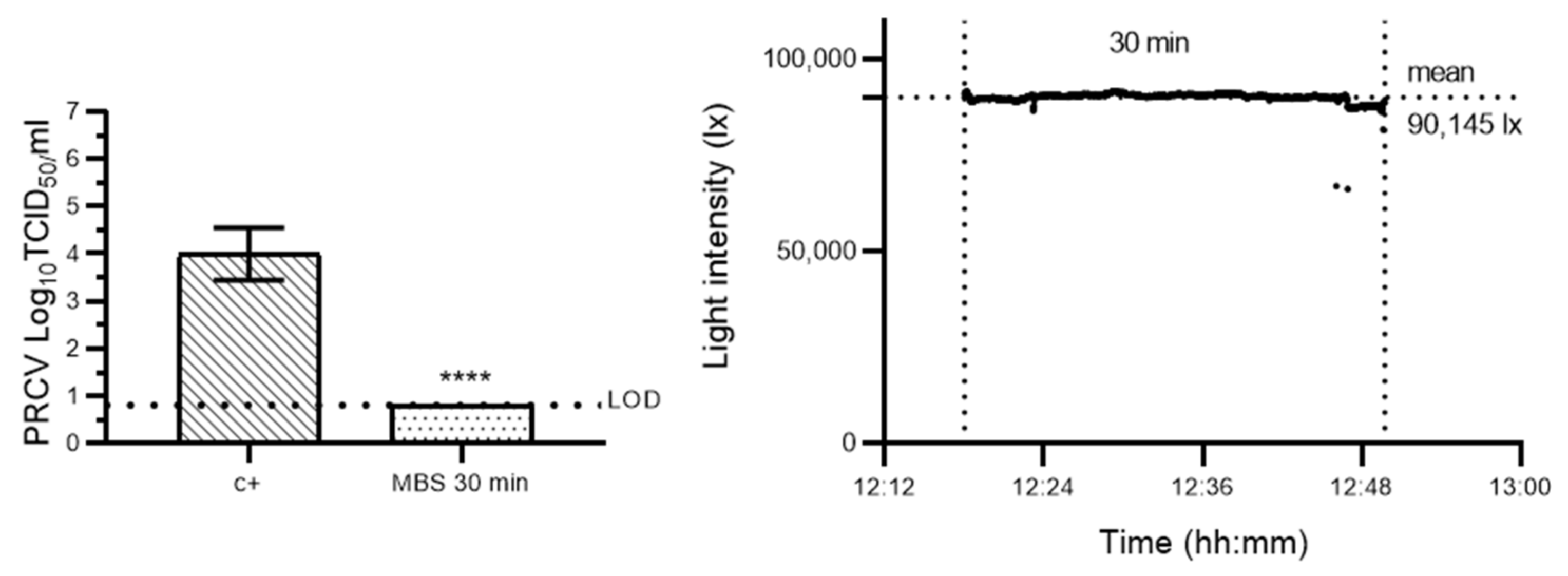
Abstract
1. Introduction
2. Materials and Methods
2.1. Viruses and Cells
2.2. Surgical Masks
2.3. Light Metre
2.4. Porcine Respiratory Coronavirus and Murine Norovirus Inoculation onto Surgical Masks
2.5. Methylene Blue Photochemical Treatment of Surgical Masks
2.6. Virus Elution from Surgical Masks
2.7. Titration of Eluted Virus (Validation of Decontamination Efficacy)
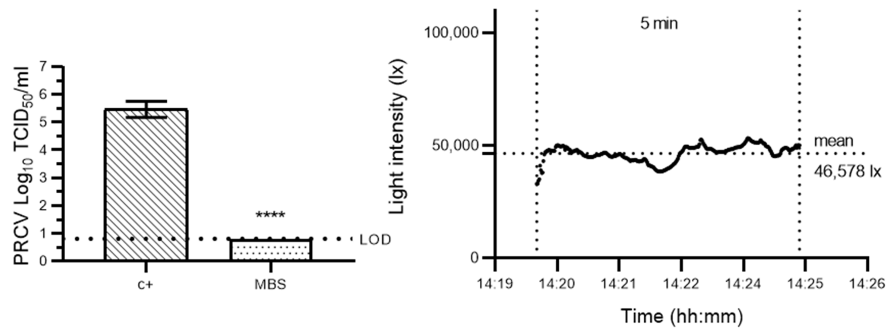
2.8. Evaluation of the Efficacy of Methylene Blue Solar Treatment for Coronavirus Inactivation
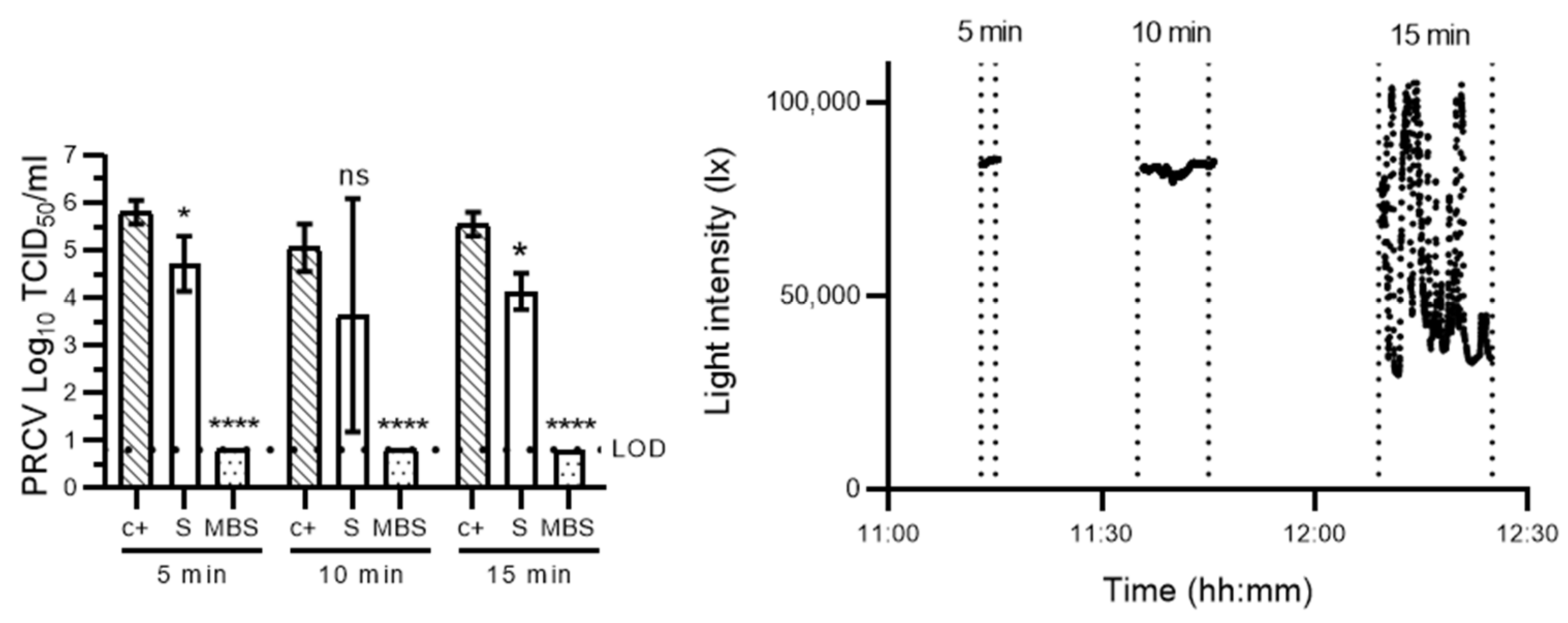
2.9. Evaluation of Time-Dependent Virucidal Kinetics of Methylene Blue Solar Treatment and Sunlight Exposure on Porcine Respiratory Coronavirus
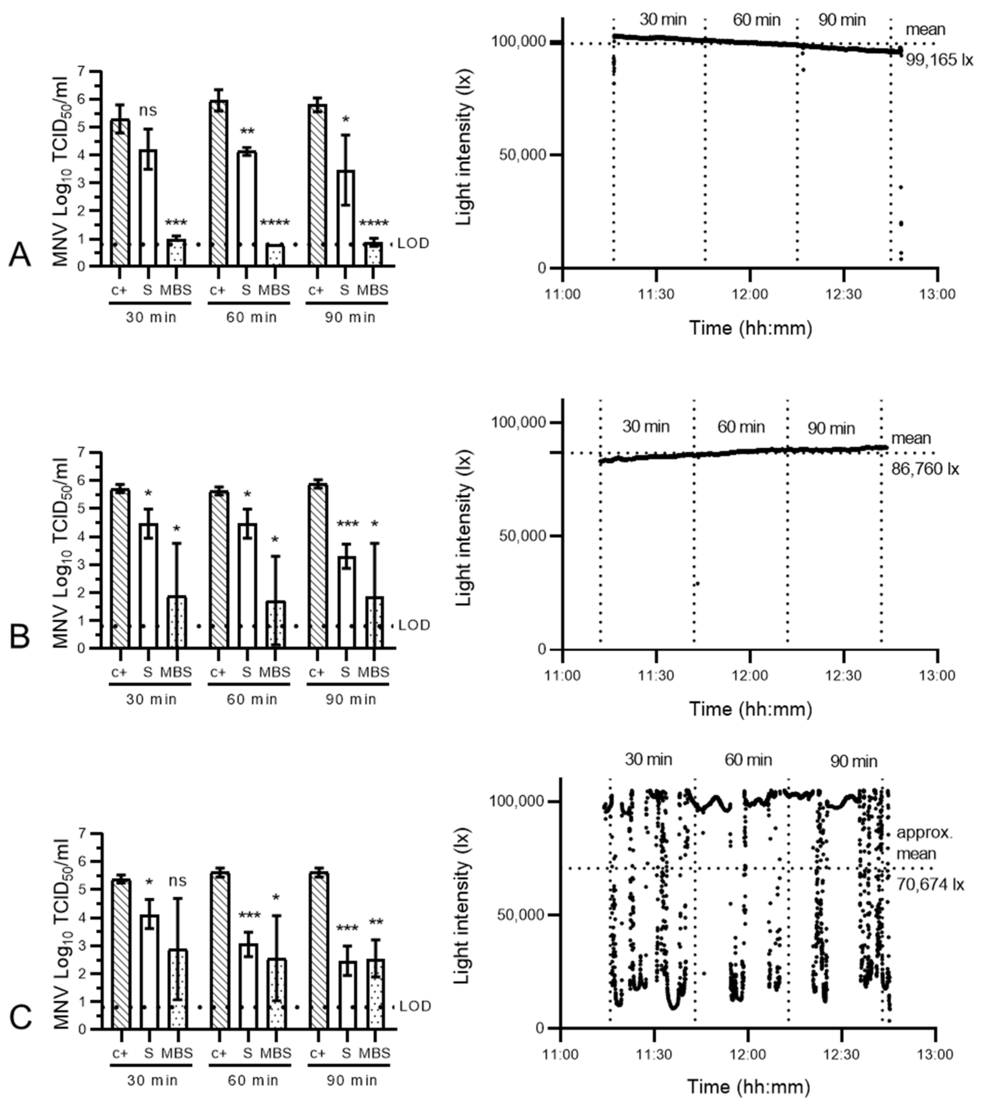
2.10. Evaluation of Time- and Sunlight-Intensity Dependent Virucidal Kinetics of Methylene Blue Solar Treatment and Sunlight Exposure on Murine Norovirus
2.11. Validation of the Efficacy of Two Methylene Blue Solar Photochemical Decontamination Protocols for Norovirus Inactivation
3. Data Analysis
4. Results and Discussion
5. Conclusions
Author Contributions
Funding
Institutional Review Board Statement
Informed Consent Statement
Acknowledgments
Conflicts of Interest
References
- McMahon, D.E.; Peters, G.A.; Ivers, L.C.; Freeman, E.E. Global resource shortages during covid-19: Bad news for low-income countries. PLoS Negl. Trop. Dis. 2020, 14, e0008412. [Google Scholar] [CrossRef]
- World Health Organization (WHO). Rational Use of Personal Protective Equipment for Coronavirus Disease 2019 (COVID-19); WHO: Geneva, Switzerland, 2020; pp. 1–7.
- Leung, N.H.L.; Chu, D.K.W.; Shiu, E.Y.C.; Chan, K.-H.; McDevitt, J.J.; Hau, B.J.P.; Yen, H.-L.; Li, Y.; Ip, D.K.M.; Peiris, J.S.M.; et al. Respiratory virus shedding in exhaled breath and efficacy of face masks. Nat. Med. 2020, 26, 676–680. [Google Scholar] [CrossRef] [PubMed]
- WHO. Strengthening the Health System Response to COVID-19 Recommendations for the WHO European Region Policy Brief; WHO: Geneva, Switzerland, 2020.
- The Lancet. COVID-19: Protecting health-care workers. Lancet 2020, 395, 922. [Google Scholar] [CrossRef]
- Tesfaldet, Y.T.; Ndeh, N.T. Assessing face masks in the environment by means of the DPSIR framework. Sci. Total Environ. 2022, 814, 152859. [Google Scholar] [CrossRef] [PubMed]
- Anastopoulos, I.; Pashalidis, I. Single-use surgical face masks, as a potential source of microplastics: Do they act as pollutant carriers? J. Mol. Liq. 2021, 326, 115247. [Google Scholar] [CrossRef] [PubMed]
- Strasser, B.J.; Schlich, T. A history of the medical mask and the rise of throwaway culture. Lancet 2020, 396, 19–20. [Google Scholar] [CrossRef]
- Ibn-Mohammed, T.; Mustapha, K.B.; Godsell, J.; Adamu, Z.; Babatunde, K.A.; Akintade, D.D.; Acquaye, A.; Fujii, H.; Ndiaye, M.M.; Yamoah, F.A.; et al. A critical analysis of the impacts of COVID-19 on the global economy and ecosystems and opportunities for circular economy strategies. Resour. Conserv. Recycl. 2021, 164, 105169. [Google Scholar] [CrossRef] [PubMed]
- Lee, J.; Bong, C.; Lim, W.; Bae, P.K.; Abafogi, A.T.; Baek, S.H.; Shin, Y.-B.; Bak, M.S.; Park, S. Fast and easy disinfection of coronavirus-contaminated face masks using ozone gas produced by a dielectric barrier discharge plasma generator. Environ. Sci. Technol. Lett. 2020, 8, 339–344. [Google Scholar] [CrossRef]
- Dutch National Institute for Public Health and the Environment (RIVM). Reuse of FFP2 Masks; RIVM: Utrecht, The Netherlands, 2020; pp. 1–5. [Google Scholar]
- Center for Devices and Radiological Health. Enforcement Policy for Face Masks and Respirators during the Coronavirus Disease (COVID-19) Public Health Emergency (Revised) Guidance for Industry and Food and Drug Administration Staff; Center for Devices and Radiological Health: Silver Spring, MD, USA, 2020. [Google Scholar]
- Croke, L. Preparing for the next infectious disease pandemic. Aorn J. 2020, 112, P12–P14. [Google Scholar] [CrossRef] [PubMed]
- Kramer, A.; Schwebke, I.; Kampf, G. How long do nosocomial pathogens persist on inanimate surfaces? A systematic review. BMC Infect. Dis. 2006, 6, 130. [Google Scholar] [CrossRef]
- Ludwig-Begall, L.F.; Wielick, C.; Dams, L.; Nauwynck, H.; Demeuldre, P.-F.; Napp, A.; Laperre, J.; Haubruge, E.; Thiry, E. The use of germicidal ultraviolet light, vaporized hydrogen peroxide and dry heat to decontaminate face masks and filtering respirators contaminated with a SARS-CoV-2 surrogate virus. J. Hosp. Infect. 2020, 106, 577–584. [Google Scholar] [CrossRef]
- Wielick, C.; Ludwig-Begall, L.F.; Dams, L.; Razafimahefa, R.M.; Demeuldre, P.-F.; Napp, A.; Laperre, J.; Jolois, O.; Farnir, F.; Haubruge, E.; et al. The use of germicidal ultraviolet light, vaporised hydrogen peroxide and dry heat to decontaminate face masks and filtering respirators contaminated with an infectious norovirus. Infect. Prev. Pract. 2020, 3, 100111. [Google Scholar] [CrossRef] [PubMed]
- Ludwig-Begall, L.F.; Wielick, C.; Jolois, O.; Dams, L.; Razafimahefa, R.M.; Nauwynck, H.; Demeuldre, P.-F.; Napp, A.; Laperre, J.; Thiry, E.; et al. “Don, doff, discard” to “don, doff, decontaminate”—FFR and mask integrity and inactivation of a SARS-CoV-2 surrogate and a norovirus following multiple vaporised hydrogen peroxide-, ultraviolet germicidal irradiation-, and dry heat decontaminations. PLoS ONE 2021, 16, e0251872. [Google Scholar] [CrossRef] [PubMed]
- Lendvay, T.S.; Chen, J.; Harcourt, B.H.; Scholte, F.E.M.; Lin, Y.L.; Kilinc-Balci, F.S.; Lamb, M.M.; Homdayjanakul, K.; Cui, Y.; Price, A.; et al. Addressing personal protective equipment (PPE) decontamination: Methylene blue and light inactivates SARS-COV-2 on N95 respirators and medical masks with maintenance of integrity and fit. Infect. Control Hosp. Epidemiol. 2021, 43, 876–885. [Google Scholar] [CrossRef]
- Costa, L.; Faustino, M.A.F.; Neves, M.G.P.M.S.; Cunha, Â.; Almeida, A. Photodynamic inactivation of mammalian viruses and bacteriophages. Viruses 2012, 4, 1034–1074. [Google Scholar] [CrossRef]
- Eickmann, M.; Gravemann, U.; Handke, W.; Tolksdorf, F.; Reichenberg, S.; Müller, T.H.; Seltsam, A. Inactivation of Ebola virus and Middle East respiratory syndrome coronavirus in platelet concentrates and plasma by ultraviolet C light and methylene blue plus visible light, respectively. Transfusion 2018, 58, 2202–2207. [Google Scholar] [CrossRef]
- Seghatchian, J.; Walker, W.H.; Reichenberg, S. Updates on pathogen inactivation of plasma using Theraflex methylene blue system. Transfus. Apher. Sci. 2008, 38, 271–280. [Google Scholar] [CrossRef] [PubMed]
- Genina, E.A.; Bashkatov, A.N.; Chikina, E.E.; Knyazev, A.B.; Mareev, O.V.; Tuchin, V.V. Methylene blue mediated laser therapy of maxillary sinusitis. Laser Phys. 2006, 16, 1128–1133. [Google Scholar] [CrossRef]
- Ludwig-Begall, L.F.; Mauroy, A.; Thiry, E. Noroviruses—The State of the Art, Nearly Fifty Years after Their Initial Discovery. Viruses 2021, 13, 1541. [Google Scholar] [CrossRef]
- Zonta, W.; Mauroy, A.; Farnir, F.; Thiry, E. Virucidal Efficacy of a Hydrogen Peroxide Nebulization Against Murine Norovirus and Feline Calicivirus, Two Surrogates of Human Norovirus. Food Environ. Virol. 2016, 8, 275–282. [Google Scholar] [CrossRef]
- Wielick, C.; Fries, A.; Dams, L.; Razafimahefa, R.M.; Heyne, B.; Harcourt, B.H.; Lendvay, T.S.; Willaert, J.F.; de Jaeger, S.; Haubruge, E.; et al. Of masks and methylene blue—The use of methylene blue photochemical treatment to decontaminate surgical masks contaminated with a tenacious small non-enveloped norovirus. Am. J. Infect. Control 2021, 50, 871–877. [Google Scholar] [CrossRef]
- Cozzi, L.; Gould, T.; Bouckart, S.; Crow, D.; Kim, T.Y.; Mcglade, C.; Olejarnik, P.; Wanner, B.; Wetzel, D. World Energy Outlook 2021; IEA: Paris, France, 2021. [Google Scholar]
- Vos, K.A.; Gordon, P.M.K.; Heyne, B. Methylene blue in combination with sunlight as a low cost and effective disinfection method for coronavirus-contaminated PPE. Am. J. Infect. Control 2022, 50, 906–908. [Google Scholar] [CrossRef] [PubMed]
- McClurkin, A.W.; Norman, J.O. Studies on transmissible gastroenteritis of swine. II. Selected characteristics of a cytopathogenic virus common to five isolates from transmissible gastroenteritis. Can. J. Comp. Med. Vet. Sci. 1966, 30, 190–198. [Google Scholar] [PubMed]
- Cox, E.; Hooyberghs, J.; Pensaert, M.B. Sites of replication of a porcine respiratory coronavirus related to transmissible gastroenteritis virus. Res. Vet. Sci. Sci. 1990, 48, 165–169. [Google Scholar] [CrossRef]
- Reed, L.J.; Muench, H. A simple method of estimating fifty percent endpoints. Am. J. Hyg. 1938, 27, 493–497. [Google Scholar]
- Raiteux, J.; Eschlimann, M.; Marangon, A.; Rogée, S.; Dadvisard, M.; Taysse, L.; Larigauderie, G. Inactivation of SARS-CoV-2 by Simulated Sunlight on Contaminated Surfaces. Microbiol. Spectr. 2021, 9, e0033321. [Google Scholar] [CrossRef]
- Zonta, W.; Mauroy, A.; Farnir, F.; Thiry, E. Comparative Virucidal Efficacy of Seven Disinfectants Against Murine Norovirus and Feline Calicivirus, Surrogates of Human Norovirus. Food Environ. Virol. 2015, 8, 1–12. [Google Scholar] [CrossRef]
- Sagripanti, J.L.; Lytle, C.D. Estimated Inactivation of Coronaviruses by Solar Radiation With Special Reference to COVID-19. Photochem. Photobiol. 2020, 96, 731–737. [Google Scholar] [CrossRef]
- Schuit, M.; Ratnesar-Shumate, S.; Yolitz, J.; Williams, G.; Weaver, W.; Green, B.; Miller, D.; Krause, M.; Beck, K.; Wood, S.; et al. Airborne SARS-CoV-2 is rapidly inactivated by simulated sunlight. J. Infect. Dis. 2020, 222, 564–571. [Google Scholar] [CrossRef]
- Bosch, A.; Pintó, R.M.; Abad, F.X. Survival and Transport of Enteric Viruses in the Environment. In Viruses in Foods; Springer: Boston, MA, USA, 2006; pp. 151–187. [Google Scholar]
- Levy, R. NASA Earth Observatory. Incoming Sunlight. 2022. Available online: https://earthobservatory.nasa.gov/features/EnergyBalance/page2.php (accessed on 8 November 2022).
- Scholte, F.E.; Kabra, K.B.; Tritsch, S.R.; Montgomery, J.M.; Spiropoulou, C.F.; Mores, C.N.; Harcourt, B.H. Exploring inactivation of SARS-CoV-2, MERS-CoV, Ebola, Lassa, and Nipah viruses on N95 and KN95 respirator material using photoactivated methylene blue to enable reuse. Am. J. Infect. Control 2022, 50, 863–870. [Google Scholar] [CrossRef]
- Shen, X.; Dong, L.; He, X.; Zhao, C.; Zhang, W.; Li, X.; Lu, Y. Treatment of infected wounds with methylene blue photodynamic therapy: An effective and safe treatment method. Photodiagnosis Photodyn. Ther. 2020, 32, 102051. [Google Scholar] [CrossRef] [PubMed]
- Sellera, F.P.; Barbosa, B.S.; Gargano, R.G.; Ríspoli, V.F.P.; Sabino, C.P.; Ollhoff, R.D.; Baptista, M.S.; Ribeiro, M.S.; de Sá, L.R.M.; Pogliani, F.C. Methylene blue-mediated antimicrobial photodynamic therapy can be a novel non-antibiotic platform for bovine digital dermatitis. Photodiagnosis Photodyn. Ther. 2021, 34, 102274. [Google Scholar] [CrossRef] [PubMed]
- Lendvay, T.S.; Xu, J.; Chen, J.; Clark, T.; Cui, Y. Methylene blue applied to N95 respirators and medical masks for SARS-CoV-2 decontamination: What is the likelihood of inhaling methylene blue? Am. J. Infect. Control 2022, 50, 857–862. [Google Scholar] [CrossRef] [PubMed]


Publisher’s Note: MDPI stays neutral with regard to jurisdictional claims in published maps and institutional affiliations. |
© 2022 by the authors. Licensee MDPI, Basel, Switzerland. This article is an open access article distributed under the terms and conditions of the Creative Commons Attribution (CC BY) license (https://creativecommons.org/licenses/by/4.0/).
Share and Cite
Fries, A.; Dams, L.; Wielick, C.; Heyne, B.; Haubruge, E.; Thiry, E.; Ludwig-Begall, L.F. Here Comes the Sun—Methylene Blue in Combination with Sunlight Sanitises Surgical Masks Contaminated with a Coronavirus and a Tenacious Small Non-Enveloped Virus. Sustainability 2022, 14, 15040. https://doi.org/10.3390/su142215040
Fries A, Dams L, Wielick C, Heyne B, Haubruge E, Thiry E, Ludwig-Begall LF. Here Comes the Sun—Methylene Blue in Combination with Sunlight Sanitises Surgical Masks Contaminated with a Coronavirus and a Tenacious Small Non-Enveloped Virus. Sustainability. 2022; 14(22):15040. https://doi.org/10.3390/su142215040
Chicago/Turabian StyleFries, Allyson, Lorène Dams, Constance Wielick, Belinda Heyne, Eric Haubruge, Etienne Thiry, and Louisa F. Ludwig-Begall. 2022. "Here Comes the Sun—Methylene Blue in Combination with Sunlight Sanitises Surgical Masks Contaminated with a Coronavirus and a Tenacious Small Non-Enveloped Virus" Sustainability 14, no. 22: 15040. https://doi.org/10.3390/su142215040
APA StyleFries, A., Dams, L., Wielick, C., Heyne, B., Haubruge, E., Thiry, E., & Ludwig-Begall, L. F. (2022). Here Comes the Sun—Methylene Blue in Combination with Sunlight Sanitises Surgical Masks Contaminated with a Coronavirus and a Tenacious Small Non-Enveloped Virus. Sustainability, 14(22), 15040. https://doi.org/10.3390/su142215040