Article

Antifungal, Antiviral, and HPLC Analysis of Phenolic and Flavonoid Compounds of Amphiroa anceps Extract

by
Emad H. El-Bilawy
1,
Al-Naji A. Al-Mansori
2,
Seham A. Soliman
3,
Fatimah O. Alotibi
4,
Abdulaziz A. Al-Askar
4,
Amr A. Arishi
5,
Abd El-Naser Sabry
6,
Mohsen Mohamed Elsharkawy
7,
Ahmed A. Heflish
8,
Said I. Behiry
8 and
Ahmed Abdelkhalek
3,*
1
Faculty of Basic Science, King Salman International University, El Tor 46612, Egypt
2
Botany Department, Faculty of Science, Derna University, Derna 99999, Libya
3
Plant Protection and Biomolecular Diagnosis Department, ALCRI, City of Scientific Research and Technological Applications, New Borg El Arab City 21934, Egypt
4
Department of Botany and Microbiology, College of Science, King Saud University, Riyadh 11451, Saudi Arabia
5
School of Molecular Sciences, The University of Western Australia, Perth, WA 6009, Australia
6
Department of Botany and Microbiology, Faculty of Science, Damietta University, New Damietta City 34517, Egypt
7
Department of Agricultural Botany, Faculty of Agriculture, Kafrelsheikh University, Kafr El-Sheikh 33516, Egypt
8
Agricultural Botany Department, Faculty of Agriculture (Saba Basha), Alexandria University, Alexandria 21531, Egypt
*
Author to whom correspondence should be addressed.
Sustainability 2022, 14(19), 12253; https://doi.org/10.3390/su141912253
Submission received: 18 August 2022 / Revised: 15 September 2022 / Accepted: 23 September 2022 / Published: 27 September 2022

Abstract

:
The increasing use of chemical control agents and pesticides to prevent plant disease has resulted in several human and environmental health problems. Seaweeds, e.g., Amphiroa anceps extracts, have significant antimicrobial activities against different human pathogens. However, their anti-phytopathogenic activities are still being investigated. In the present investigation, three fungal isolates were isolated from root rot and grey mold symptomatic strawberry plants and were molecularly identified by ITS primers to Fusarium culmorum, Rhizoctonia solani, and Botrytis cinerea with accession numbers MN398396, MN398398, and MN398400, respectively. In addition, the organic extract of the red alga Amphiroa anceps was assessed for its antifungal activity against the three identified fungal isolates and tobacco mosaic virus (TMV) infection. At 100 µg/mL, the A. anceps extract had the best biological activity against R. solani, B. cinerea, and TMV infection, with inhibition rates of 66.67%, 40.61%, and 81.5%, respectively. Contrarily, the A. anceps extract exhibited lower activity against F. culmorum, causing inhibition in the fungal mycelia by only 4.4% at the same concentration. The extract’s HPLC analysis revealed the presence of numerous phenolic compounds, including ellagic acid and gallic acid, which had the highest concentrations of 19.05 and 18.36 µg/mL, respectively. In this line, the phytochemical analysis also showed the presence of flavonoids, with the highest concentration recorded for catechin at 12.45 µg/mL. The obtained results revealed for the first time the effect of the A. anceps extract against the plant fungal and viral pathogens, making the seaweed extract a promising source for natural antimicrobial agents.

1. Introduction

Seaweeds are a large group of marine algae that inhabit the shallow waters of the sea wherever suitable substrata are available [1]. Seaweeds are a rich and diverse source of metabolites widely known for their biological activities, including laxatives, antimicrobials, and anti-ulcer agents [2,3]. Exposing seaweeds to different biotic and abiotic stressors in their natural habitats results in the production of many bioactive metabolites [4]. Seaweeds can be categorized into three main divisions: Chlorophyceae (green algae), Rhodophyceae (red algae), and Phaeophyceae (brown algae). Among them, red algae are a famous source of many bioactive chemical metabolites, including sulfated polysaccharides like carrageenan and fucoidan, which are known for their antiviral and anticancer activities against many human viruses and cancer cells [5]. Red algae are a source of many other useful products, like agar, alginates, and carrageenan, which are used for different purposes, such as human food and animal feed [6].
Previous phytochemical analysis of different red algae revealed the presence of different bioactive chemical groups, such as steroids, phenols, flavonoids, tannins, saponins, alkaloids, and triterpenoids [5,7]. The red alga Amphiroa anceps is one of the widely distributed seaweeds in Egypt, especially on the coast of Alexandria during the autumn period [2]. Previous studies of the organic extracts of A. anceps showed the presence of many active chemical metabolites that display different bioactivities, including antimicrobial activity [1]. Further, the aqueous extract of A. anceps is used to make nanoparticles, such as silver nanoparticles, using green chemistry techniques [8].
Plant diseases cause many crop losses worldwide, worsening serious food security problems [9]. Plant viruses are among the most significant plant pathogens, causing major threats to sustainable agriculture and productivity [10]. Tobacco mosaic virus (TMV) is one of the most important plant viruses, negatively impacting crop quality and productivity worldwide [11]. TMV is one of the top 10 plant viruses in the field of molecular plant pathology. It is also used as a model virus, and Nicotiana glutinosa is used as a host for TMV-local lesion studies [12]. Soil- and air-borne fungal infections, such as root rots, wilts, and grey mold, are of enormous economic relevance and can result in a substantial production crop. Strawberry is one of Egypt’s most valuable export crops because of its high quality and early market introduction. Root rot infections in strawberries are caused by several fungi, including Rhizoctonia solani, Fusarium culmorum, F. solani, F. oxysporum, and Macrophomina phaseolina [13]. The fungus Botrytis cinerea is to blame for the grey mold that grows on strawberry fruit [13].
Most methods for controlling plant pathogens involve applying chemical pesticides to plants or breeding transgenic plants. However, synthetic pesticides harm the environment and human health, and transgenic crops are not yet universally approved [9,14]. Consequently, there is still a great demand for the discovery of further alternative, eco-friendly, and effective antiviral techniques. Algae have recently been used as biocontrol agents for plant diseases [15]. Seaweed extracts represent a safer fungicide than most synthetic and semi-synthetic fungicides [16]. Many previous reports revealed the presence of compounds derived from seaweed extracts, especially the red algal ones with antifungal activity. The extract of Melanothamnus afaqhusainii is very effective against fungi such as Fusarium moniliforme and Rhizoctonia solani [17]. Moreover, Jiménez et al. [18] investigated the antifungal efficacy of ethanolic extracts of the red algae Gracillaria chilensis against Phytophthora cinnamomi. To our knowledge, no previous studies have revealed the antifungal and antiviral activities of Amphiroa anceps extract against phytopathogenic microorganisms. Thus, this study aims to use HPLC to analyze the A. anceps extract and investigate its antifungal activity against three fungi (Fusarium culmorum, Rhizoctonia solani, and Botrytis cinerea), as well as its antiviral activity against tobacco mosaic virus infection.

2. Materials and Methods

2.1. Pathogen Isolation for Root Rot and Grey Mold

Microbial pathogens were recovered from infected plant samples in Egypt’s most important strawberry-producing zone at the latitude and longitude coordinates 30°34′38.0″ N 30°41′39.0″ E, Behera Governorate. Strawberry root and fruit parts were isolated on PDA media and showed rot and grey mold symptoms. Single spore culture or hyphal tip procedures purified the samples [19]. The isolated fungi were put in slanted tubes and grown at 25 ± 3 °C for 7 days. The pure cultures were inspected, morphologically identified [20], and molecularly described.

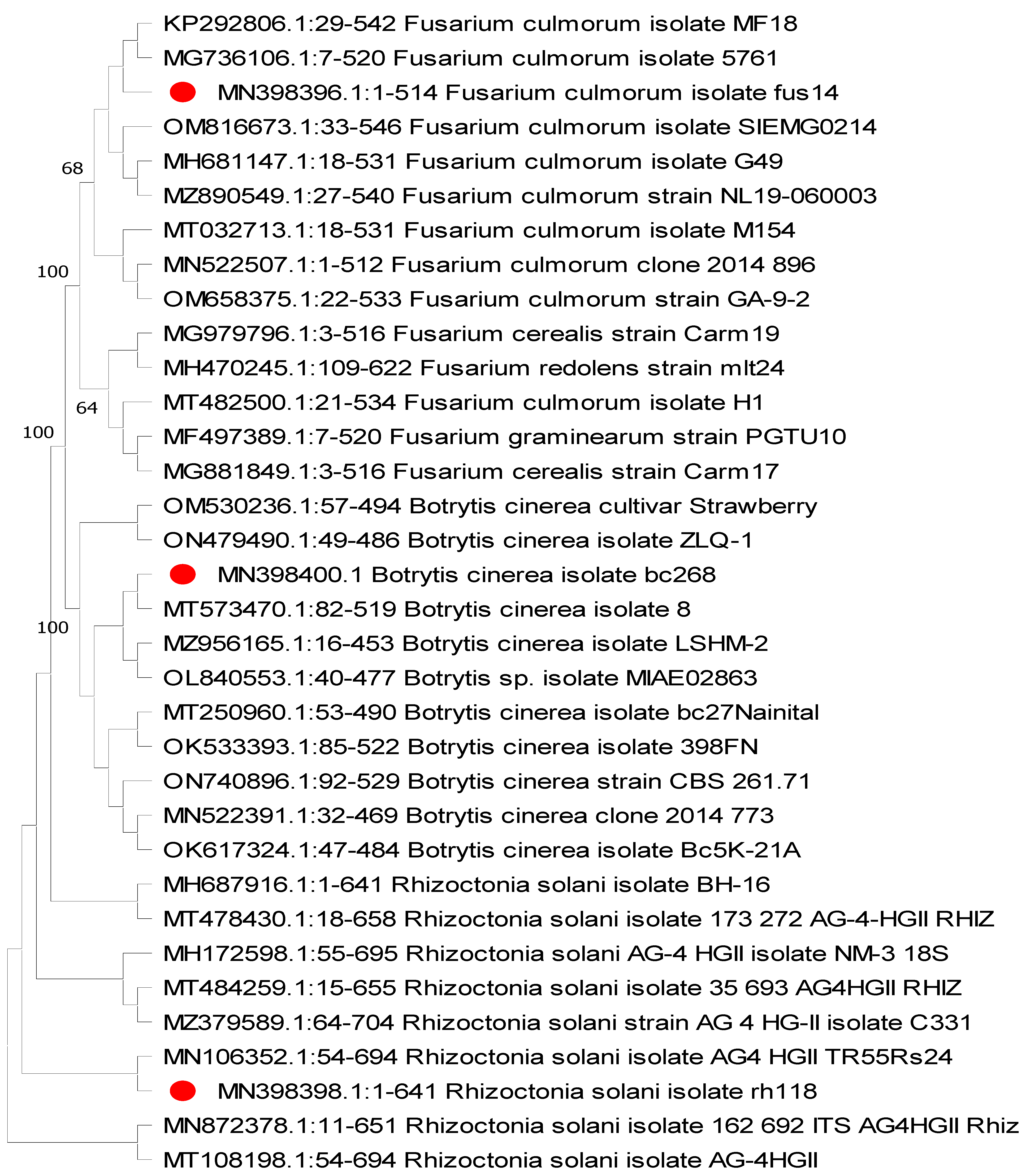
2.2. ITS Sequencing and Identification

The QIAquick PCR purification kit was used to isolate genomic DNA from fresh fungal hyphae (QIAGEN, Manchester, England). The fungi’s internal transcribed spacer (ITS) region was amplified using the PCR technique with ITS1 and ITS4 primers [21]. PCR amplification reaction was performed with a final volume of 50 μL, containing 25 μL of 2x PCR DyeMIX-nTaq (Enzynomics Inc., Ansan, Korea), 50 ng of DNA template, and 2 μL of each primer (10 pmol). PCR cycling was performed with an initial denaturation step at 94 °C for 3 min, followed by 35 cycles (94 °C for 30 s, 56 °C for 30 s, and 72 °C for 40 s) and a final extension step at 72 °C for 7 min. The amplified PCR products were sequenced immediately after removal from the gel and purified with a PCR clean-up column kit (QIAGEN, Hilden, Germany). The sequencing process was carried out using a BigDye Terminator v3.1 Cycle Sequencing kit (Applied Biosystems, Foster City, CA, USA) and a model 3130xl Genetic Analyzer (Applied Biosystems, Foster City, CA, USA). After analyzing DNA nucleotide sequences with NCBI-BLAST (http://blast.ncbi.nlm.nih.gov/Blast.cgi, accessed on 19 September 2020, the annotated sequences were deposited in GenBank to obtain accession numbers. The phylogenetic tree was made using MEGA 11 software and analyzed using the maximum likelihood tree method with bootstrapping of 2000 replicates.

2.3. Sampling Site and Algal Biomass Collection

The preparation of dry biomass was performed according to the method of Valderrama et al. [22]. Biomasses of Amphiroa anceps were gathered in seawater around mid-autumn at a depth of 0.2 m or less from the rocks of Abo-Qir Eastern Harbor (30°4′ E and 30°20′ E and 31°16′ N and 31°28′ N). The A. anceps biomasses were taken directly from the intertidal zone using the Londo scale’s five (1 mm) quadrants [23]. The fresh algal biomass sample size strictly adhered to the Egyptian Environmental Affairs Agency’s (EEAA) regulations for the bio conservation of protected regions. Each sample was rinsed multiple times with saline and then with distilled water to wash away contaminants and epiphytes, and then its species was determined [24,25]. The algal biomass was then dried in the shade, and the sample was sliced, milled, and stored in a tightly sealed dark jar.

2.4. Extraction of the Algal Biomass

Twenty-five grams of A. anceps dry biomass were extracted using a Soxhlet apparatus. The extraction process was performed according to the method of Wang et al. [26], with some modifications. Methanol and hexane were utilized as the extraction solvent combination (1:1). The solvent combination was then added and heated in the flask. The extraction technique was repeated multiple times for 5 h at 65 °C until the vast majority of chemical compounds had been separated. The extract was then chilled, decolored using charcoal filters, and condensed at 30–45 °C with a rotary vacuum evaporator. The dried extract residue was stored at 4 °C until use. The yield of extraction was calculated according to the following formula: Extraction yield (%) = [Dry weight of the extract (g)/Dry weight of algal biomass (g)] × 100.

2.5. Antifungal Activity of the Algal Extract

The activity of the A. anceps extract was tested against the three isolated fungi. For the antifungal activity test, wood blocks of the chinaberry plant were prepared with dimensions of 0.5 × 1 × 2 cm, then sterilized at 121 °C for 15 min. A series of the algal extracts (25, 50, and 100 µg/mL) prepared with dimethyl sulfoxide (DMSO) was applied to each wood block (200 µL of each concentration) against the tested fungal species in triplicate, keeping one sample for each fungus treated only with DMSO to use as a control [27]. All the Petri plates were incubated at 25 ± 3 °C for 7 days. The antifungal activity was recorded by measuring the mycelial growth inhibition percentage as the following formula:
%   inhibition = [ Control     treatment control ] × 100 .

2.6. Source of the Virus, Inoculum Preparation, and Greenhouse Antiviral Activity Assays

The Egyptian TMV (Acc# MG264131, strain KH1) was propagated by tobacco plant and purified using the methods outlined by Gooding and Hebert [28]. Before usage, the purified TMV inoculum concentration was diluted to 20 μg/mL using 0.1 M phosphate buffer, pH 7.2. The A. anceps extract was dissolved in DMSO and diluted to 100 μg/mL concentration using sterile deionized water. As a negative control, an equivalent concentration solution of DMSO was utilized. Using the leaf assay method and Nicotiana glutinosa as a local lesion host for TMV, antiviral efficacy was evaluated based on the percentage of inhibition toward the number of local lesions. The inhibitory effect was determined using the formula: [I = (1 − T/C) × 100], where I represents the inhibition effect, T represents the number of local lesions on the treated leaves, and C represents the number of local lesions on the control leaves. Under greenhouse-controlled conditions, N. glutinosa plants were subjected to the protective treatment assay at the 5–6 leaf stage. After 24 h of applying A. anceps extract to the N. glutinosa leaves, the leaves were dusted with carborundum and mechanically inoculated with TMV as previously described [29]. The number of local lesions was counted 3–4 days after TMV inoculation (dpi). Triplicate runs of each experiment were performed.

2.7. Characterization of Phenolic and Flavonoid Compounds Using HPLC

The phenolic and flavonoid compounds were characterized in the algal biomass using HPLC. Despite the condition of the characterization previously described in previous works, we will briefly mention these conditions in the following lines [30,31]. For characterization of the phenolic compounds, an Agilent 1260 Infinity HPLC Series was used; the HPLC was equipped with a Quaternary pump and a Zorbax Eclipse Plus C18 column (100 mm × 4.6 mm i.d.). The injected volume of the extract was 20 µL. The separation was carried out using gradient elution with (A) HPLC grade water 0.2% H3PO4 (v/v), (B) Methanol, and (C) Acetonitrile at 30 °C. A variable wavelength detector was used at 284 nm. For quantification of flavonoid compounds, a Smart line, Knauer HPLC, was used and equipped with a binary pump in addition to a Zorbax Eclipse plusC18 column (150 mm × 4.6 mm i.d.). For the separation process, an eluent composed of methanol and water containing 0.5% H3PO4 with 50:50 percent and a flow rate of 0.7 mL/min. The injected volume of the extract was 20 µL, a UV detector was set at 273 nm, and data integration was conducted using ClarityChrom@ Version 7.2.0 (Knauer Wissenschaftliche Geräte GmbH, Berlin, Germany). The HPLC investigation was based on the following 18 standard polyphenolic compounds: caffeic acid, ferulic acid, gallic acid, syringic acid, cinnamic acid, salicylic acid, ellagic acid, p-coumaric acid, pyrogallol acid, rutin, quercetin, kaempferol, luteolin, catechin, naringin, 7-oh flavone, apigenin, and myricetin. Merck KGaA supplied all of the chemical substances used in this study (Darmstadt, Germany).

2.8. Statistical Analysis

All data were statistically analyzed using one-way analysis of variance (ANOVA) in CoStat software, with significant differences determined using Tukey’s test post hoc and standard deviation (SD) methods. The differences in data with the same letter are not significant.

3. Results

3.1. Isolation and Preliminary Identification

Three fungal isolates were obtained from 10 infected strawberry plants after isolation from plant organs and tissues exhibiting distinct symptoms; they were placed on PDA medium and incubated at 25 ± 3 °C for 7 days. The first isolated pathogen, Botrytis cinerea (abundance 100%), is responsible for the collapse of water-soaked fruit and foliage and develops brown lesions on unripe fruits. The infected blossoms cause fruit drops and damage growing and mature fruit, such as ridging. The isolated B. cinerea pathogen features a necrotrophic, haploid, and heterothallic ascomycete that produces melanized black spherical sclerotia on the plate in vitro. The fungus produces gray, fuzzy conidiophores on plant tissues with velvet asexual spores.
Root rot disease symptoms and signs showed field plants’ reduced vigor; small fruit, few runners, and many dead leaves and stunted plants revealed a reducing yield. Feeder and main roots might deteriorate and develop black decaying lesions. The central root core was blackened. According to our results, the isolation trails from the root rot symptoms revealed two isolates of the Fusarium (abundance 70%) and Rhizoctonia (abundance 30%) species. Characteristics of the Fusarium pathogen were observed as having a pale pinkish to white colony color. The macroconidia morphology is straight and relatively slender, and 3-septate is the most common. On the other hand, the Rhizoctonia pathogen was seen to have pale white to yellow mycelium that grew faster on PDA. Under a microscope, the fungus was seen to have narrowed branched angle hyphae and no spores.

3.2. ITS Characterization

All fungal isolates were amplified and sequenced for ITS rDNA regions. The known sequences obtained from NCBI confirmed that the initial identifications of F. culmorum, R. solani, and B. cinerea were accurate. The relevant GenBank accession numbers for the three fungal sequences are MN398396, MN398398, and MN398400, respectively. The phylogenetic tree was constructed upon the ITS gene sequences retrieved from the NCBI GenBank database, as shown in Figure 1.

3.3. Invitro Antifungal Activity of the A. anceps Extract

The result of the antifungal activity of the A. anceps extract is clarified in Figure 2 and Figure 3. Generally, the extract of A. anceps exhibited different antifungal activities against different fungal species used in the present work. The highest antifungal activity of the algal extract was observed against B. cinerea using 100 and 50 µg/mL of the algal extract. The extract displayed moderate antifungal activity against R. solani with a percent of inhibition ranging from 39–66.67% for extract concentrations of 25 and 100 µg/mL, respectively. The A. anceps extract showed weak antifungal activity against F. culmorum (Figure 2).

3.4. Inhibitory Effects of A. anceps Extract against TMV

Under greenhouse conditions, the protective activity of A. anceps extract against TMV on N. glutinosa was assessed. The antiviral activity was determined using the leaf assay by counting the number of local lesions on infected leaves at 4 dpi. When comparing treated and untreated tissues, those treated with A. anceps extract showed a significant decrease in local lesion symptoms (Figure 4). Mock-treated plants showed no local lesions on the leaves. The application of A. anceps extract at 100 µg/mL showed the maximum antiviral activity with an inhibition rate of 81.52 ± 2.36% (Figure 4). On the other hand, the 50 and 25 µg/mL showed inhibition rates of 71.01 ± 1.78% and 53.26 ± 3.04%, respectively. Thus, the results showed that A. anceps extract could reduce TMV infection.

3.5. Phenolic and Flavonoid Compounds of the Algal Extract

The results obtained in this study showed a 61% extraction yield. The HPLC chromatograms of phenolic and flavonoid compounds in the A. anceps extract are clearly shown in Figure 5. The phytochemical analysis showed that the algal extract’s most abundant phenolic compound in µg/mL was ellagic acid, with a concentration of 19.05, followed by gallic acid (18.36). At the same time, catechin represents the highest concentration of a flavonoid compound present in the extract, with a concentration of 12.45 µg/mL (Table 1).

4. Discussion

Plant diseases are a major threat to the productivity and sustainability of agriculture around the world. Every year, they cost farmers several billion dollars [32]. Controlling plant diseases requires resistant plant cultivars or intensive insecticide usage, which can harm human health and the environment [33]. Pesticides pollute surface water, exacerbating environmental and ecological issues [34]. Biocontrol agents are safer alternatives to the toxic pesticides currently used to treat plant diseases [35]. As biological control agents, algal extracts are regarded a sustainable and environmentally acceptable alternative to chemical control agents, as they include a variety of physiologically beneficial secondary metabolites that may increase the plant’s systemic resistance and inhibit pathogen development. This study assessed the antifungal and antiviral activities of the Amphiroa anceps extract against Fusarium culmorum, Rhizoctonia solani, and Botrytis cinerea as well as TMV infection. In addition, the main phytochemical constituents of A. anceps extract were analyzed using HPLC.
Seaweeds represent a reservoir of many active metabolites, including vitamins, enzymes, lipids, and antibiotics, in addition to many fine chemicals [36,37]. Many previous studies have shown that the metabolites produced via marine macroalgae and especially extracts of A. anceps exert several bioactive functions, including, for instance, cytotoxic, anti-inflammatory, and antimicrobial activities, making it a promising source for drug design [2,38,39]. HPLC analysis of the A. anceps extract proved the presence of many bioactive phenolic and flavonoid compounds, and these results are consistent with previous studies. Many studies confirm the presence of phenolic compounds qualitatively and quantitatively in the extracts of A. anceps [1,40]. Xiaojun et al. [41] and Nagai and Yukimoto [42] have reported different biological activities of phenolic compounds, such as antioxidant and antimicrobial activities. In the current study, the inhibition rates of fungal hyphae growth demonstrate that the bioactive molecules of algal species can be dissolved in alcohol [43,44]. This may explain why organic extracts have either a strong or weak effect on pathogens. Many previous reports confirm the presence of flavonoid compounds in several red algae [45]. However, our work may be the first report of the presence of flavonoid compounds in the extract of A. anceps. Additionally, flavonoid compounds play different biological activities, including anti-inflammatory, antioxidant, and antimicrobial [46].
In the present work, the extract of A. anceps exhibited potent antifungal activity against R. solani and B. cinerea but showed a lower activity against F. culmorum. These results are consistent with that of Pandian et al. [47], who reported high antifungal activity of the Acanthophora spicifera methanolic extract against Microsporum gypseum. Similarly, Sultana et al. [48] reported a high fungicide effect of the red alga Melanothamnus extract against root infecting fungi F. solani and Macrophomina phaseolina attacking eggplant (Solanum melongena L.) and watermelon (Citrullus lanatus (Thunb.). Khan et al. [17] reported a high antifungal activity against F. moniliforme and R. solani using the extract of M. afaqhusainii. Similarly, Jiménez et al. [18] investigated the antifungal activity of the ethanolic extracts of the red algae Gracillaria chilensis against Phytophthora cinnamomi. On the contrary, Tariq [45] investigated the antifungal efficacy of extracts from four red algae species using the cub plate method. He found that none of the studied extracts inhibited the growth of Aspergillus flavus, A. fumigatus, or Candida albicans.
In contrast, the effects of methanol and chloroform extracts of the red macroalgae Gracilaria confervoides on the mycelial growth of R. solani, Macrophomina phaseolina, and F. solani of cucumber were studied by Soliman et al. [49]. They found that the methanolic extract reduced the R. solani growth by 25.9% and the chloroform extract by 100%, while the solvent and aqueous extracts moderately inhibited mycelia growth in F. solani. Overall, the explanation of different inhibition results suggests that some or all algal extracts added more minerals and nutrients to the medium, which could have hidden the inhibitory effect. As we described previously, many previous works prove different biological activities of the A. anceps extracts. However, the present work may represent the first record of antifungal activity of the red alga A. anceps extract. In addition, there were records before on the antimicrobial activity of different organic extracts of A. anceps against human pathogens. Crude ethanolic extracts of A. anceps demonstrated the greatest antimicrobial activity towards Vibrio parahaemolyticus (13.25 mm) and V. alginolyticus (13.3 mm). Maximum activity against Pseudomonas fluorescens and Proteus mirabilis was seen in the acetone extract of A. anceps. In contrast, the crude benzene extract was the most effective against Streptococcus pneumoniae and V. parahaemolyticus [50].
In recent years, there has been much interest in employing beneficial microbes as a safe and environmentally acceptable approach to controlling viral plant diseases. However, the use of algae for the biological control of viral plant diseases is still limited, and it is unclear how algae might work against viral pathogens. The antiviral activity of A. anceps extract against TMV on N. glutinosa plants was investigated in this research. Under greenhouse conditions, the application of A. anceps extract significantly reduced the local lesion symptoms when N. glutinosa tissues were treated with A. anceps extract at a concentration of either 25 or 50, or 100 µg/mL 24 h before TMV inoculation. The 100 µg/mL of A. anceps extract exhibited a strong inhibitory effect (81.52%), while the 50 and 25 µg/mL concentrations showed an inhibitory effect of 71.01% and 53.26%, respectively. In light of these findings, we hypothesize that A. anceps extract may contain elicitor chemicals that stimulate the immune defense system and/or inhibit TMV replication. In general, antiviral agents can work in two ways: either directly or indirectly to stop viral replication by activating the host’s innate immune system, or by making SAR against viral infection [51].
In the same way, in the HPLC results, we noticed higher concentrations of polyphenolic compounds such as ellagic, gallic, cinnamic acids, and catechin. Previous results suggest that ellagic and gallic acids could be natural antifungal agents [52,53]. Ellagic acid had a broad spectrum of antifungal activity against two fungal Candida strains, with MICs between 25.0 and 75.0 µg/mL and 0.125 to 0.25 µg/mL against C. auris strains [54]. Meanwhile, Li et al. [53] recorded antifungal MICs of gallic acid against three Candida strains ranging between 12.5 and 100.0 μg/mL. The results obtained by Korošec et al. [55] showed high antifungal properties of cinnamic acid derivatives against two pathogens—Cochliobolus lunatus and Aspergillus niger. Moreover, among catechins, pyrogallol catechin showed more potent antifungal activity against Candida sp. than catechol catechin [56]. It was reported that the application of 500 µg/mL of exogenous caffeic acid inhibited the growth of various Fusarium and Saccharomyces species [57]. Numerous polyphenols, including caffeic acid, ellagic acid, catechins, chlorogenic acid, gallic acid, quercetin, ferulic acid, and myricetin, possess antibacterial, antiviral, anti-inflammatory, anticancer, and antioxidant effects [58,59]. Ferulic acid is a common phytochemical found in leaves and seeds, and is free and covalently bound to glycoproteins, polysaccharides, polyamines, hydroxy fatty acids, and lignin. Ferulic acid makes cell walls stiff and works as an antioxidant, an antibacterial, an anticarcinogenic, an antiviral, and a substance that changes how enzymes work [60]. Consequently, we think the polyphenolic compounds we found could be used as elicitor molecules in developing SAR, increasing resistance against TMV infection, and working as potent antifungal agents [52,53].

5. Conclusions

The results obtained from this study proved the antifungal and antiviral activities of the organic extract of A. anceps. The A. anceps extract at 100 µg/mL showed the highest biological properties against R. solani and B. cinerea as well as TMV infection with inhibition rates of 66.67%, 40.61%, and 81.5%, respectively. The biocontrol activities of the A. anceps extract may be attributed to the single or synergetic effect of one or more than one of the compounds. As far as we know, the results obtained from this work may represent the first record of antifungal and antiviral activities of the seaweed A. anceps. The promising results obtained in this study and the simple extraction method determine the tested red algae to be a promising source for producing antifungal and antiviral agents to control plant diseases. Thus, this extract may be useful for protecting plants from diseases.

Author Contributions

Conceptualization, E.H.E.-B., S.I.B. and A.A.; methodology, E.H.E.-B.; A.-N.A.A.-M., S.A.S., A.A., A.E.-N.S. and S.I.B.; software, S.I.B. and A.A.; validation, M.M.E., A.E.-N.S. and E.H.E.-B.; formal analysis, A.A.H. and S.I.B.; investigation, A.A.; resources, E.H.E.-B.; data curation, M.M.E.; writing—original draft preparation, E.H.E.-B.; writing—review and editing, S.I.B. and A.A.; visualization, A.A.A.; supervision, A.A.A.-A.; project administration, F.O.A. and A.A.A.; funding acquisition, F.O.A. and A.A.A.-A. All authors have read and agreed to the published version of the manuscript.

Funding

This research project was supported by a grant from the Researchers Supporting Project number (RSP-2021/114), King Saud University, Riyadh, Saudi Arabia.

Institutional Review Board Statement

Not applicable.

Informed Consent Statement

Not applicable.

Data Availability Statement

Not applicable.

Acknowledgments

The authors would like to extend their appreciation to the Researchers Supporting Project number (RSP-2021/114), King Saud University, Riyadh, Saudi Arabia.

Conflicts of Interest

The authors declare no conflict of interest.

References

  1. Raj, E.D.S. UV–VIS and HPLC studies on Amphiroa anceps (Lamarck) Decaisne. Arab. J. Chem. 2016, 9, S907–S913. [Google Scholar]
  2. Mofeed, J.; Deyab, M.; Mohamed, A.; Moustafa, M.; Negm, S.; El-Bilawy, E. Antimicrobial activities of three seaweeds extract against some human viral and bacterial pathogens. Biocell 2022, 46, 247. [Google Scholar] [CrossRef]
  3. El-Baroty, G.S.; Moussa, M.Y.; Shallan, M.A.; Ali, M.A.; Sabh, A.Z.; Shalaby, E.A. Contribution to the aroma, biological activities, minerals, protein, pigments and lipid contents of the red alga: Asparagopsis taxiformis (Delile) Trevisan. J. Appl. Sci. Res. 2007, 3, 1825–1834. [Google Scholar]
  4. Aslam, A.; Fazal, T.; uz Zaman, Q.; Shan, A.; Rehman, F.; Iqbal, J.; Rashid, N.; Rehman, M.S.U. Biorefinery of Microalgae for Nonfuel Products. In Microalgae Cultivation for Biofuels Production; Elsevier: Amsterdam, The Netherlands; Academic Press: Cambridge, MA, USA, 2020; pp. 197–209. [Google Scholar]
  5. Aziz, E.; Batool, R.; Khan, M.U.; Rauf, A.; Akhtar, W.; Heydari, M.; Rehman, S.; Shahzad, T.; Malik, A.; Mosavat, S.H. An overview on red algae bioactive compounds and their pharmaceutical applications. J. Complement. Integr. Med. 2020, 17. [Google Scholar] [CrossRef]
  6. Sánchez-Machado, D.I.; López-Hernández, J.; Paseiro-Losada, P.; López-Cervantes, J. An HPLC method for the quantification of sterols in edible seaweeds. Biomed. Chromatogr. 2004, 18, 183–190. [Google Scholar] [CrossRef]
  7. König, G.M.; Wright, A.D.; Linden, A. Plocamium hamatum and its monoterpenes: Chemical and biological investigations of the tropical marine red alga. Phytochemistry 1999, 52, 1047–1053. [Google Scholar] [CrossRef]
  8. Roy, S.; Anantharaman, P. Biosynthesis of silver nanoparticle by Amphiroa anceps (Lamarck) decaisne and Its biomedical and ecological implications. J. Nanomed. Nanotechnol. 2018, 9. [Google Scholar] [CrossRef]
  9. Abdelkhalek, A.; Hafez, E. Plant Viral Diseases in Egypt and Their Control. In Cottage Industry of Biocontrol Agents and Their Applications; Springer: Berlin/Heidelberg, Germany, 2020; pp. 403–421. [Google Scholar]
  10. Abdelkhalek, A.; Behiry, S.I.; Al-Askar, A.A. Bacillus velezensis PEA1 Inhibits Fusarium oxysporum Growth and Induces Systemic Resistance to Cucumber Mosaic Virus. Agronomy 2020, 10, 1312. [Google Scholar] [CrossRef]
  11. Abdelkhalek, A. Expression of tomato pathogenesis related genes in response to Tobacco mosaic virus. JAPS J. Anim. Plant Sci. 2019, 29, 1596–1602. [Google Scholar]
  12. Tan, Q.-W.; Gao, F.-L.; Wang, F.-R.; Chen, Q.-J. Anti-TMV activity of malformin A1, a cyclic penta-peptide produced by an endophytic fungus Aspergillus tubingensis FJBJ11. Int. J. Mol. Sci. 2015, 16, 5750–5761. [Google Scholar] [CrossRef]
  13. Ahmed, M.F.A.; El-Fiki, I.A.I. Effect of biological control of root rot diseases of strawberry using Trichoderma spp. Middle East J. Appl. Sci. 2017, 7, 482–492. [Google Scholar]
  14. Abdelkhalek, A.; Al-Askar, A.A.; Elbeaino, T.; Moawad, H.; El-Gendi, H. Protective and Curative Activities of Paenibacillus polymyxa against Zucchini yellow mosaic virus Infestation in Squash Plants. Biology 2022, 11, 1150. [Google Scholar] [CrossRef] [PubMed]
  15. Elsharkawy, M.M.; El-Okkiah, S.; Elsadany, A.Y.; Bedier, M.Y.; Omara, R.I.; Behiry, S.I.; Hassan, S.; Abdelkhalek, A. Systemic resistance induction of tomato plants against tomato mosaic virus by microalgae. Egypt. J. Biol. Pest Control 2022, 32, 1–7. [Google Scholar] [CrossRef]
  16. Baloch, G.N.; Tariq, S.; Ehteshamul-Haque, S.; Athar, M.; Sultana, V.; Ara, J. Management of root diseases of eggplant and watermelon with the application of asafoetida and seaweeds. J. Appl. Bot. Food Qual. 2013, 86, 138–142. [Google Scholar]
  17. Khan, A.M.; Naz, S.; Abid, M. Evaluation of marine red alga Melanothamnus afaqhusainii against Meloidogyne incognita, fungus and as fertilizing potential on okra. Pak. J. Nematol. 2016, 34, 91–100. [Google Scholar]
  18. Jiménez, E.; Dorta, F.; Medina, C.; Ramírez, A.; Ramírez, I.; Peña-Cortés, H. Anti-phytopathogenic activities of macro-algae extracts. Mar. Drugs 2011, 9, 739–756. [Google Scholar] [CrossRef] [Green Version]
  19. Dhingra, O.D.; Sinclair, J.B. Basic Plant Pathology Methods; CRC press: Boca Raton, FL, USA, 2017; ISBN 1315138131. [Google Scholar]
  20. Alexopoulos, C.J.; Mims, C.W.; Blackwell, M. Introductory Mycology; John Wiley and Sons: New York, NY, USA, 1996; ISBN 0471522295. [Google Scholar]
  21. White, T.J.; Bruns, T.; Lee, S.; Taylor, J. Amplification and direct sequencing of fungal ribosomal RNA genes for phylogenetics. PCR Protoc. Guide Methods Appl. 1990, 18, 315–322. [Google Scholar]
  22. Valderrama, D.; Cai, J.; Hishamunda, N.; Ridler, N. Social and Economic Dimensions of Carrageenan Seaweed Farming. 2013. Available online: https://tamug-ir.tdl.org/bitstream/handle/1969.3/29143/a-i3344e.pdf?sequence=1 (accessed on 17 August 2022).
  23. Londo, G. The decimal scale for relevés of permanent quadrats. In Sampling Methods and Taxon Analysis in Vegetation Science: Releve Surveys,’Vegetationsaufnahmen’, Floristic Analysis of Plant Communities; Handbook of vegetation sciences; The Hague (The Netherlands) Junk: The Hague, The Netherlands, 1984; pp. 45–49. [Google Scholar]
  24. Brodie, J.; Wilbraham, J.; Pottas, J.; Guiry, M.D. A revised check-list of the seaweeds of Britain. J. Mar. Biol. Assoc. UK 2016, 96, 1005. [Google Scholar] [CrossRef]
  25. De Clerck, O.; Coppejans, E. Marina Algae of the Jubail Marine Wildlife Sanctuary, Saudi Arabia. 1996. Available online: http://www.vliz.be/imisdocs/publications/239149.pdf (accessed on 17 August 2022).
  26. Wang, T.; Jónsdóttir, R.; Liu, H.; Gu, L.; Kristinsson, H.G.; Raghavan, S.; Ólafsdóttir, G. Antioxidant capacities of phlorotannins extracted from the brown algae Fucus vesiculosus. J. Agric. Food Chem. 2012, 60, 5874–5883. [Google Scholar] [CrossRef]
  27. Salem, M.Z.M.; Mansour, M.M.A.; Elansary, H.O. Evaluation of the effect of inner and outer bark extracts of Sugar Maple (Acer saccharum var. saccharum) in combination with citric acid against the growth of three common molds. J. Wood Chem. Technol. 2019, 39, 136–147. [Google Scholar] [CrossRef]
  28. Gooding, G.V., Jr.; Hebert, T.T. A simple technique for purification of Tobacco mosaic virus in large quantities. Phytopathology 1967, 57, 1285. [Google Scholar] [PubMed]
  29. Abdelkhalek, A.; Ismail, I.A.; Dessoky, E.S.; El-Hallous, E.I.; Hafez, E. A tomato kinesin-like protein is associated with Tobacco mosaic virus infection. Biotechnol. Biotechnol. Equip. 2019, 33, 1424–1433. [Google Scholar] [CrossRef]
  30. Behiry, S.I.; Okla, M.K.; Alamri, S.A.; El-Hefny, M.; Salem, M.Z.M.; Alaraidh, I.A.; Ali, H.M.; Al-Ghtani, S.M.; Monroy, J.C.; Salem, A.Z.M. Antifungal and antibacterial activities of Musa paradisiaca L. peel extract: HPLC analysis of phenolic and flavonoid contents. Processes 2019, 7, 215. [Google Scholar] [CrossRef]
  31. Salem, M.Z.M.; Ibrahim, I.H.M.; Ali, H.M.; Helmy, H.M. Assessment of the use of natural extracted dyes and pancreatin enzyme for dyeing of four natural textiles: HPLC analysis of phytochemicals. Processes 2020, 8, 59. [Google Scholar] [CrossRef]
  32. Strange, R.N.; Scott, P.R. Plant disease: A threat to global food security. Annu. Rev. Phytopathol. 2005, 43. [Google Scholar] [CrossRef]
  33. Aktar, W.; Sengupta, D.; Chowdhury, A. Impact of pesticides use in agriculture: Their benefits and hazards. Interdiscip. Toxicol. 2009, 2, 1–12. [Google Scholar] [CrossRef]
  34. Alengebawy, A.; Abdelkhalek, S.T.; Qureshi, S.R.; Wang, M.-Q. Heavy Metals and Pesticides Toxicity in Agricultural Soil and Plants: Ecological Risks and Human Health Implications. Toxics 2021, 9, 42. [Google Scholar] [CrossRef]
  35. Lahlali, R.; Ezrari, S.; Radouane, N.; Kenfaoui, J.; Esmaeel, Q.; El Hamss, H.; Belabess, Z.; Barka, E.A. Biological control of plant pathogens: A global perspective. Microorganisms 2022, 10, 596. [Google Scholar] [CrossRef]
  36. Deyab, M.; Mofeed, J. Evaluation of Biochemical composition and bioactivities of two Ulva species (Ulva lactuca and Ulva fasciata); a comparative study. Biosci. Res. 2019, 16, 3801–3811. [Google Scholar]
  37. Deyab, M.; Mofeed, J.; El-Bilawy, E.; Ward, F. Antiviral activity of five filamentous cyanobacteria against coxsackievirus B3 and rotavirus. Arch. Microbiol. 2020, 202, 213–223. [Google Scholar] [CrossRef]
  38. Selvin, J.; Lipton, A.P. Biopotentials of Ulva fasciata and Hypnea musciformis collected from the peninsular coast of India. J. Mar. Sci. Technol. 2004, 12, 1. [Google Scholar] [CrossRef]
  39. Lubobi, S.; Matunda, C.; Kumar, V.; Omboki, B. Isolation of bioactive secondary metabolites from seaweeds Amphiroa anceps against chicken meat associated pathogens. J. Antimicrob 2016, 2. [Google Scholar] [CrossRef]
  40. Wang, T.; Jonsdottir, R.; Ólafsdóttir, G. Total phenolic compounds, radical scavenging and metal chelation of extracts from Icelandic seaweeds. Food Chem. 2009, 116, 240–248. [Google Scholar] [CrossRef]
  41. Xiaojun, Y.; Xiancui, L.; Chengxu, Z.; Xiao, F. Prevention of fish oil rancidity by phlorotannins from Sargassum kjellmanianum. J. Appl. Phycol. 1996, 8, 201–203. [Google Scholar] [CrossRef]
  42. Nagai, T.; Yukimoto, T. Preparation and functional properties of beverages made from sea algae. Food Chem. 2003, 81, 327–332. [Google Scholar] [CrossRef]
  43. Kolanjinathan, K.; Stella, D. Antibacterial activity of marine macro algae against human pathogens. Recent Res. Sci. Technol. 2009, 1, 20–22. [Google Scholar]
  44. Omar, H.H.; Gumgumji, N.M.; Shiek, H.M.; El-Kazan, M.M.; El-Gendy, A.M. Inhibition of the development of pathogenic fungi by extracts of some marine algae from the red sea of Jeddah, Saudi Arabia. Afr. J. Biotechnol. 2012, 11, 13697–13704. [Google Scholar]
  45. Kumar, K.S.; Ganesan, K.; Rao, P.V.S. Antioxidant potential of solvent extracts of Kappaphycus alvarezii (Doty) Doty–An edible seaweed. Food Chem. 2008, 107, 289–295. [Google Scholar] [CrossRef]
  46. Robak, J.; Gryglewski, R.J. Bioactivity of flavonoids. Pol. J. Pharmacol. 1996, 48, 555–564. [Google Scholar]
  47. Pandian, P.; Selvamuthukumar, S.; Manavalan, R.; Parthasarathy, V. Screening of antibacterial and antifungal activities of red marine algae Acanthaphora spicifera (Rhodophyceae). J. Biomed. Sci. Res. 2011, 3, 444–448. [Google Scholar]
  48. Sultana, V.; Baloch, G.N.; Ara, J.; Ehteshamul-Haque, S.; Tariq, R.M.; Athar, M. Seaweeds as an alternative to chemical pesticides for the management of root diseases of sunflower and tomato. J. Appl. Bot. Food Qual. 2012, 84, 162. [Google Scholar]
  49. Soliman, A.S.; Ahmed, A.Y.; Abdel-Ghafour, S.E.; El-Sheekh, M.M.; Sobhy, H.M. Antifungal bio-efficacy of the red algae Gracilaria confervoides extracts against three pathogenic fungi of cucumber plant. Middle East J. Appl. Sci 2018, 8, 727–735. [Google Scholar]
  50. Karthick, M.; Balachandar, M.; Raja, M.; Raj, R.A. Antibacterial activity of red alga Amphiroa anceps (rhodophyceae: Lithophyllaceae) against selected human pathogens. Sci. Acta Xaver. 2019, 10, 15–19. [Google Scholar]
  51. Waziri, H.M.A. Plants as antiviral agents. J. Plant Pathol. Microbiol. 2015, 6, 1. [Google Scholar] [CrossRef]
  52. Li, Z.; Guo, X.; Dawuti, G.; Aibai, S. Antifungal activity of ellagic acid in vitro and in vivo. Phyther. Res. 2015, 29, 1019–1025. [Google Scholar] [CrossRef] [PubMed]
  53. Li, Z.; Liu, M.; Dawuti, G.; Dou, Q.; Ma, Y.; Liu, H.; Aibai, S. Antifungal activity of gallic acid in vitro and in vivo. Phyther. Res. 2017, 31, 1039–1045. [Google Scholar] [CrossRef]
  54. Possamai Rossatto, F.C.; Tharmalingam, N.; Escobar, I.E.; d’Azevedo, P.A.; Zimmer, K.R.; Mylonakis, E. Antifungal Activity of the Phenolic Compounds Ellagic Acid (EA) and Caffeic Acid Phenethyl Ester (CAPE) against Drug-Resistant Candida auris. J. Fungi 2021, 7, 763. [Google Scholar] [CrossRef]
  55. Korošec, B.; Sova, M.; Turk, S.; Kraševec, N.; Novak, M.; Lah, L.; Stojan, J.; Podobnik, B.; Berne, S.; Zupanec, N. Antifungal activity of cinnamic acid derivatives involves inhibition of benzoate 4-hydroxylase (CYP 53). J. Appl. Microbiol. 2014, 116, 955–966. [Google Scholar] [CrossRef]
  56. Hirasawa, M.; Takada, K. Multiple effects of green tea catechin on the antifungal activity of antimycotics against Candida albicans. J. Antimicrob. Chemother. 2004, 53, 225–229. [Google Scholar] [CrossRef]
  57. Davidson, P.M.; Taylor, T.M.; Schmidt, S.E. Chemical preservatives and natural antimicrobial compounds. Food Microbiol. Fundam. Front. 2012, 765–801. [Google Scholar] [CrossRef]
  58. Mani, J.S.; Johnson, J.B.; Steel, J.C.; Broszczak, D.A.; Neilsen, P.M.; Walsh, K.B.; Naiker, M. Natural product-derived phytochemicals as potential agents against coronaviruses: A review. Virus Res. 2020, 284, 197989. [Google Scholar] [CrossRef] [PubMed]
  59. Shaygannia, E.; Bahmani, M.; Zamanzad, B.; Rafieian-Kopaei, M. A review study on Punica granatum L. J. Evid. Based. Complement. Altern. Med. 2016, 21, 221–227. [Google Scholar] [CrossRef] [PubMed]
  60. Kumar, N.; Pruthi, V. Potential applications of ferulic acid from natural sources. Biotechnol. Rep. 2014, 4, 86–93. [Google Scholar] [CrossRef] [PubMed] [Green Version]
Figure 1. A degenerated phylogenetic tree based on the nucleotide sequence of the ITS gene for the three fungal isolates F. culmorum (MN398396), B. cinerea (MN398400), and R. solani (MN398398) aligned with the most related sequences downloaded from the NCBI database. The phylogeny was tested for maximum likelihood using the bootstrap method with 2000 replications. The red color cycle indicates three fungal isolates molecularly identified during this study.
Sustainability 14 12253 g001
Figure 2. Antifungal activity of the 25, 50, and 100 µg/mL extract concentrations compared to the control against the fungal pathogen (A) Rhizoctonia solani, (B) Fusarium culmorum, and (C) Botrytis cinerea.
Sustainability 14 12253 g002
Figure 3. Inhibition percentages of 25, 50, and 100 µg/mL extract concentrations compared to control against the fungal isolates R. solani, F. culmorum, and B. cinerea. The small letters on top of the bar graphs in each color mean the significance at a 0.05 probability.
Sustainability 14 12253 g003
Figure 4. A photograph showing the disease symptoms on N. glutinosa leaves infected with tobacco mosaic virus at 4 dpi of the protective activity of A. anceps extract at different concentrations.
Sustainability 14 12253 g004
Figure 5. HPLC chromatograms of phenolic and flavonoid compounds identified in the Amphiroa anceps extract.
Sustainability 14 12253 g005
Table 1. Concentrations of phenolic and flavonoid compound in the extract of Amphiroa anceps using HPLC.
Phenolic CompoundsFlavonoid Compounds
CompoundRTConcentration (µg/mL)CompoundRTConcentration (µg/mL)
Caffeic acid4.28.66Rutin4.80.59
Ferulic acid 5.16.24Quercetin6.84.86
Gallic acid7.018.36Kaempferol8.03.87
Syringic acid8.65.04Luteolin9.14.63
Cinnamic acid10.017.64Catechin11.012.45
Salicylic acid11.05.72
Ellagic acid12.319.05
Publisher’s Note: MDPI stays neutral with regard to jurisdictional claims in published maps and institutional affiliations.

