Article

Characterization of Di-n-Butyl Phthalate Phytoremediation by Garden Lettuce (Lactuca sativa L. var. longifolia) through Kinetics and Proteome Analysis

1
Department of Civil and Ecological Engineering, I Shou University, Kaohsiung 84001, Taiwan
2
Institute of Biotechnology and Chemical Engineering, I Shou University, Kaohsiung 84001, Taiwan
3
Department of Food and Human Health Sciences, Graduate School of Human Life Science, Osaka City University, Sugimoto 558-8585, Japan
*
Author to whom correspondence should be addressed.
Sustainability 2019, 11(6), 1625; https://doi.org/10.3390/su11061625
Submission received: 8 February 2019 / Revised: 12 March 2019 / Accepted: 14 March 2019 / Published: 18 March 2019
(This article belongs to the Special Issue Green and Sustainable Remediation of Contaminated Site)

Abstract

:
Di-n-dutyl phthalate (DBP), an endocrine disruptor, is one of the most widely used phthalate esters (PAEs) in the world. It can be accumulated in seafood or agricultural products and represents a substantial risk to human health via the food chain. Thus, finding a plant which can remediate DBP but have no effects on growth is the main topic of the development of DBP phytoremediation. This study used garden lettuce (Lactuca sativa L. var. longifolia), which has a significant DBP absorption capability, as a test plant to measure phytoremediation kinetics and proteome changes after being exposed to DBP. The results show that DBP accumulated in different parts of the garden lettuce but the physiological status and morphology showed no significant changes following DBP phytoremediation. The optimal condition for the DBP phytoremediation of garden lettuce is one critical micelle concentration (CMC) of non-ionic surfactant Tween 80 and the half-life (t1/2, days), which calculated by first-order kinetics, was 2.686 days for 5 mg L−1 of DBP. This result indicated that the addition of 1 CMC of Tween 80 could enhance the efficiency of DBP phytoremediation. In addition, the results of biotoxicity showed that the median effective concentration (EC50) of DBP for Chlorella vulgaris is 4.9 mg L−1. In this case, the overall toxicity markedly decreased following phytoremediation. In the end, the result of proteome analysis showed six protein spots, revealing significant alterations. According to the information of these proteomes, DBP potentially causes osmotic and oxidative stress in garden lettuce. In addition, since DBP had no significant effects on the morphology and physiological status of garden lettuce, garden lettuce can be recommended for use in the plant anti-DBP toxicity test, and also as the candidate plant for DBP phytoremediation. We hope these findings could provide valuable information for DBP-contaminated water treatment in ecological engineering applications or constructed wetlands.

1. Introduction

Phthalate esters (PAEs) are a group of compounds that have been widely used in plastic manufacturing since the 1930s, and now can be found in many additives, such as cosmetics, paints, pesticides, chemical fertilizer, adhesives and plasticizers [1,2,3,4,5]. In plasticizers, PAEs are characterized by low water solubility and high octanol/water partition coefficients, but are not covalently bound to plastics [6]. The global production of PAEs was approximately 1.8 million tons in 1975, 6.2 million tons in 2009 and increased to more than 8 million tons in 2015 [7,8,9,10]. Due to its higher production and application figures, PAEs are widely distributed in various environmental samples such as the air [11], freshwater [12,13], sediment [14,15] and soil [5,16,17,18]. Several studies have reported that PAEs can cause negative health effects in animals [19,20] and humans [21,22,23]. Therefore, the United States Environmental Protection Agency (USEPA) and government agencies in several other countries have categorized six PAEs as priority environmental pollutants and as endocrine-disrupting compounds (EDCs) [10].
Di-n-butyl phthalate (DBP) is one of the most widely used PAEs in the world and is mainly used as a plasticizer for plastics [24]. The chemical structure of PAEs and DBP are both shown in Figure 1. DBP is an oily liquid that is soluble in fat and slightly soluble in water. It is not very volatile, so it does not readily vaporize into the atmosphere. DBP has a water solubility value of 13 g L−1 and a half-life of 22 years in aqueous solution [25]. The fate of DBP in a natural environment can be biodegradation, hydrolysis and photodecomposition via the attack of free radicals. Recently, the application of DBP has dramatically increased in agricultural soils. DBP and other PAEs may be introduced into agricultural soils from various sources, including waste-water irrigation, fertilizers, pesticides, the use of agricultural plastic mulch bags and some other off-site pollutant sources [26]. After DBP is released into the environment, it can be taken up by vegetables and crops, then entering the food supply chain system. The International Program on Chemical Safety [27], Health Canada [28], and the US Agency for Toxic Substances and Disease Registry (ATSDR) [29] therefore have reported that the largest source of DBP in the general population is food.
In terms of human toxicity, there is no evidence that DBP can cause human cancer, however, DBP is of particular concern because it is known to be a reproductive toxin [30] that aggravates autoimmune thyroid disease [31]. On the other hand, several studies related to the plant toxicity of DBP have been produced. First, the effect of DBP on carotene synthesis during seedling growth has been reported [32,33]. Subsequently, the relative sensitivity of DBP to cabbage (Brassica oleracea) and radish (Raphanus sativus) were also reported [34,35]. Some studies also reported DBP can reduce the capsaicin content in capsicum fruit (Capsicum annum) [36] and affect leaf color in six plant species [37]. A 2016 study indicated increased use of plastic film in greenhouse vegetable production (GVP) could result in PAE contamination in vegetables. The total PAE concentrations ranged from 0.14 to 2.13 mg kg−1 (mean 0.99 mg kg−1) in soils and from 0.15 to 6.94 mg kg−1 (mean 1.49 mg kg−1) in vegetables [38]. The latest study, carried out in 2018, indicated that the toxicity to lettuce of DBP was higher than that of di-(2-ethylhexyl) phthalate (DEHP) in soil, and DBP treatment is associated with a decline in lettuce leaf size, as well as chlorophyll a and carotenoid content when compared to the control lettuce plants [39].
Proteomics analysis is one of the most popular methods to study plant responses to environmental stresses because the extraction of proteins is easy, the obtained two-dimensional electrophoresis gels have great reproducibility and the mass spectrometry (MS) for the sequencing of proteins is very sensitive. Proteomics has been used to study the expression of salt stress related proteins in several plants, which can provide a better indication of cellular activities under salt stress [40,41]. However, only a few reports have focused on proteomics studies of DBP-treated plants. According to our previous study, the morphology, chlorophyll concentration and proteome changes on DBP-treated bok choy and Chinese cabbage were reported [42,43]. In the proteomics analysis of bok choy, stress proteins such as superoxide dismutase (SOD) and the peroxidase 21 precursor were identified. These two proteins were believed to increase in response to free radical exposure as a detoxification mechanism and they might also be used as important molecular markers for DBP tolerance. In addition, three proteins extracted from DBP-treated Chinese cabbage displayed a differential expression. These three proteins were identified as acyl-[acyl-carrier-protein] desaturase (acyl-ACP desaturase), root phototropism protein 3 (RPT3) and ferredoxin-nitrite reductase (Fd-NiR). They are responsible for fatty acid biosynthesis, signal transduction of the phototropic response and nitrate assimilation in plant cells, respectively. These reports illustrate that proteomics analysis could help us to understand the responses of plants under DBP treatment.
Phytoremediation is a kind of bioremediation technique using plants to remove or degrade organic and inorganic pollutants in soils, water or air environments [44]. There are six types of phytoremediation, including phytosequestration, rhizodegradation, phytohydraulics, phytoextraction, phytovolatilization and phytodegradation. Several studies have reported the phytoremediation of PAEs [45,46], however, only a few studies have used the proteomics method to analyze plant responses in the phytoremediation of PAEs [47]. Hence, the aim of this study is to construct the optimal cultural conditions for DBP phytoremediation and use the proteomics technique to analyze plant responses following DBP phytoremediation. First, eleven test plants were used to evaluate the potential capability of DBP phytoremediation. The hydroponic method was used for plant cultivation. Second, the selected plant was used to establish the optimal cultural conditions for phytoremediation. Various cultural conditions, such as the cultural temperature of daytime and night, illumination times of light and dark, initial pH levels, DBP added concentration and surfactants were examined. Meanwhile, the biotoxicity of DBP to the green algae Chlorella vulgaris was also determined. In the end, the proteomics analyses of normal and DBP-treated plants were also examined. We hope the results of this study could provide valuable information for DBP-contaminated water treatment in ecological engineering applications or constructed wetlands.

2. Materials and Methods
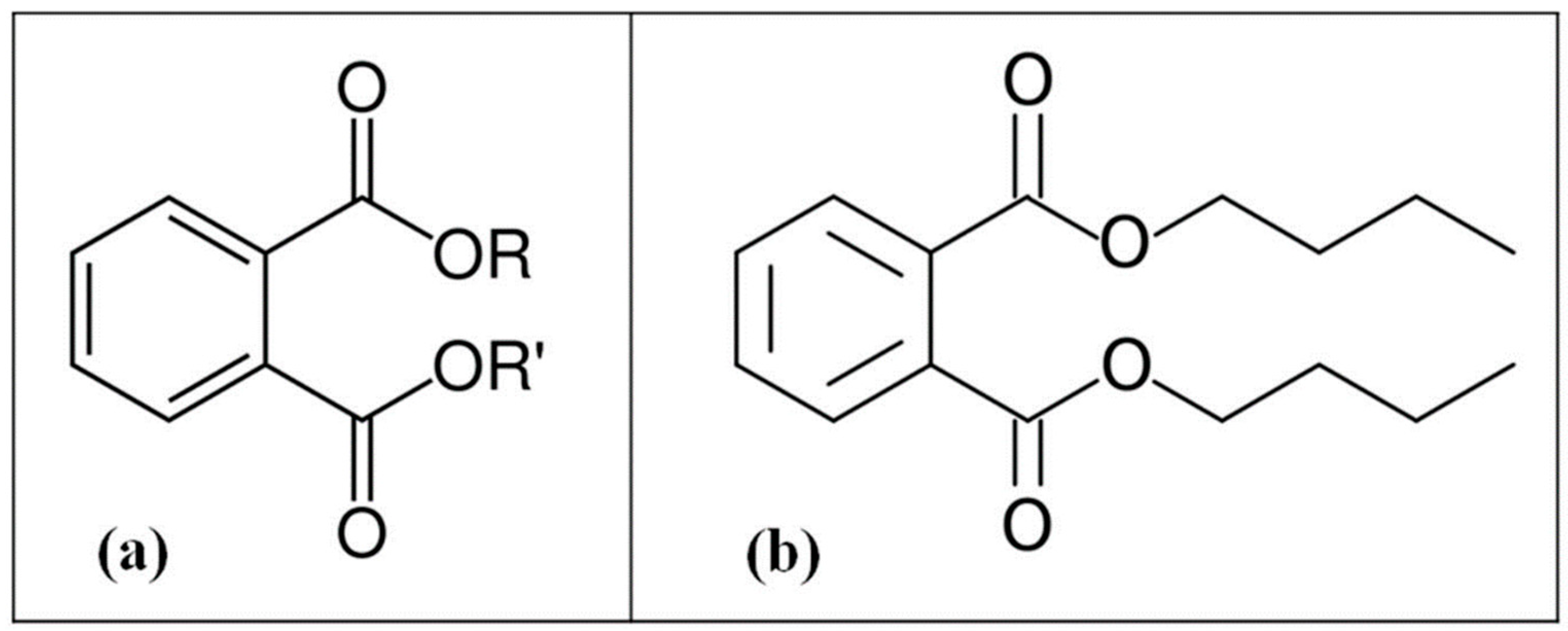
2.1. Chemicals

Di-n-butyl phthalate (DBP) (98.7% purity, CAS: 84-74-2) was purchased from Riedel-deHaën Co, Germany. The solvents used in the experiment, including acetone and n-hexane (HPLC-grade) were purchased from E. Merck, Germany. All other chemicals were purchased from the Sigma Chemical Co, Saint Louis, MI, USA. A stock solution of DBP was dissolved in acetone at a concentration of 100 g L−1. The glassware was thoroughly cleaned to reduce any background contamination of PAEs. All glassware was washed with deionized water and dried overnight in an oven at 80 °C. After cooling, the glassware was rinsed twice with acetone and air-dried for use.

2.2. Cultivation of Plants

The eleven test plants used in this study were all typical plants from North-East Asia and Taiwan. The common and scientific names of them were edible rape (Brassica napus), Chinese cabbage (Brassica rapa var. chinensis), Spinach (Spinacia oleracea L.), Chinese mustard (Brassica rapa L. Chinensis Group), water cabbage (Brassica rapa pekinensis), ceylon spinach (Basella rubra. L.), garden lettuce (Lactuca sativa L. var. longifolia), Chinese celery (Apium graveolens L.), edible amaranth (Amaranthus tricolor L.), cauliflower (Brassica oleracea var. botrytis) and Chinese chive (Allium tuberosum), respectively. After germination and growth for fourteen days, all plant seedlings were each planted in a 3 L pot containing 2.5 L of hydroponic solution. The seedlings were fixed with a styrofoam board and every styrofoam board was used for four seedlings. The hydroponic solution was modified from Hoagland’s solution [48], which consisted of (in g L−1): Ca(NO3)2·4H2O (0.1), KNO3 (0.08), MgSO4·7H2O (0.05), NH4H2PO4 (0.02), Fe-EDTA (3), H3BO3 (3), Cu-EDTA (0.01), Zn-EDTA (0.03), Mn-EDTA (0.4) and NaMoO4·2H2O (0.003). The pH value of the hydroponic solution was adjusted to 6.5. The electrical conductivity (EC) value of the solution was 1.2 mS cm−1. All experiments were carried out in a plant growth chamber and all test plants were cultivated with a controlled relative humidity variation from 70% to 80%, which was carried out to simulate the environmental humidity of Taiwan.

2.3. Experimental Design

First, eleven test plants were used to evaluate the potential capability of DBP phytoremediation. Referring to the DBP data found in the aquatic environment of Taiwan [12], DBP with a concentration of 5 mg L−1 was added to the hydroponic solution. Regarding the average temperature and daylight hours in Taiwan, the cultural temperature of daytime and nighttime were 25 ± 1 °C/23 ± 1 °C, the illumination times of light and dark were 13 h/11 h. The period of cultivation was 21 days and the sampling dates were 0, 7, 14 and 21 days, respectively.
After growing in the DBP-added hydroponic solution for 21 days, the biomass, leaf chlorophyll concentration, residual DBP in hydroponic solution and accumulated DBP in the whole plant were analyzed. The morphology of the plants was also observed and recorded during the period of phytoremediation. Meanwhile, the residual DBP in the hydroponic solution and the accumulated DBP in different parts of test plants were analyzed to represent the phytoremediation ability of test plants. In the end, the plant with the best DBP phytoremediation ability of the eleven test plants was selected for use in this study. The best phytoremediation ability means the plant has the highest rate of DBP removal.
Following the DBP phytoremediation capability evaluation, the selected plant was used to establish the optimal cultural conditions of phytoremediation. The following cultural conditions were modified to study their effects on DBP phytoremediation: Cultural temperature of daytime and night (25 °C /23 °C, 23 °C /21 °C, 21 °C /19 °C), light and dark illumination times (13 h/11 h, 12 h/12 h, 11 h/13 h), initial pH levels (6, 7, 8 or 9), DBP added concentration (1 mg L−1, 3 mg L−1, 5 mg L−1) and surfactants (Brij 35, Brij 30, Triton X-100, Tergitol, Tween 80). The surfactants were all prepared at one critical micelle concentration (CMC). After growing for 21 days, the DBP phytoremediation efficiency was determined by measuring the residual DBP in the hydroponic solution. In the end, the phytoremediation efficiency with all optimal conditions was determined.

2.4. Plant Leaf Chlorophyll (a + b) Concentration Determination

Chlorophyll a and b were measured using the method adapted from our previous study [43]. One gram of fresh plant leaf sample was immersed in a 2 mL sodium phosphate buffer (pH 6.8), then homogenized. After shaking with the buffer, 40 μL of sample solution was taken and extracted with 960 μL absolute ethanol in the dark. The extraction was carried out for 30 min and then centrifuged at 8000 g for 10 min. The absorption of the extracts was measured with a spectrophotometer at 665 and 649 nm and the chlorophyll concentration (mg L−1 FW) was calculated using the relation: 6.1 × A665 nm + 20.04 × A649 nm. All experiments were conducted in triplicate and identified using t-tests.

2.5. DBP Concentration Analyses

The residual DBP in the roots, stems and leaves of the plants was analyzed following the methods of our previous study [43]. The plant samples were rinsed three times with deionized water, then dried at 70 °C, ground into a mortar and sieved to <2 mm in size. Each 1.00 g sample in 2 mL n-hexane was added to the sample bottles and shaken with a rotating shaker at 160 rpm for 1 h. Residual DBP was extracted with n-hexane three times and then the extracts were combined to await analysis.
The analysis of residual DBP in the hydroponic solution followed the methods of our previous study [49] with slight modifications. Briefly, 2 mL of hydroponic solution was added to bottles containing 2 mL of n-hexane and shaken in a rotating shaker at 160 rpm for 10 min. The residual DBP was extracted three times with n-hexane twice, and then the extracts were combined to await analysis.
The DBP extracts and phthalic acid, which is the main metabolite of DBP, were analyzed on a Perkin Elmer Clarus 400 gas chromatograph, coupled with an electron capture detector and an Elite-5ms capillary column (film thickness of 0.25 μm, inner diameter of 0.25 mm, length of 30 m). The injector temperature was set to 250 °C. Nitrogen was used as the carrier gas at a flow rate of 0.8 mL min−1 and a 10:1 split ratio. The initial column temperature was set at 150 °C for 1 min, increasing by a rate of 8 °C min−1 to 220 °C, then increasing by a rate of 4 °C min−1 to 275 °C, where it was then held for 10 min. The detector temperature was set at 320 °C. The recovery percentage of DBP was 96.5% and the method detection limit (MDL) was 0.08 mg l−1. The method of the previous study was that the initial column temperature was set at 160 °C for 1 min, increased by a rate of 10 °C min−1 to 280 °C, then held for 1 min, then increasing by a rate of 10 °C min−1 to 300 °C.

2.6. Statistical Analyses

Statistical analyses were carried out following the equation of our previous study [49]. The residual DBP data collected from each sampling date fit well with the first-order kinetics: S = S0exp (−k1t), t1/2 = ln 2k1−1, where S0 is the initial concentration, S is the substrate concentration, t is the time period and k1 is the phytoremediation rate constant. The remaining percentage was calculated as the DBP residue concentration divided by the original DBP concentration, multiplied by 100. Each experiment was performed in triplicate. Statistical analysis was carried out using an ANOVA.

2.7. Algal Biotoxicity Assays

Algal biotoxicity assays were performed using the freshwater unicellular green alga C. vulgaris Beij. #3001 because of its high sensitivity to contaminants. C. vulgaris was obtained from Professor Wu of the Biodiversity Research Center (Academia Sinica, Taipei, Taiwan). The algae were cultivated in the medium of Chang [50], and all cultures were maintained at 25 °C under an illumination of approximately 400 μmol·m−2·s−1 and a light-dark cycle of 14:10 h. To maintain the log-growing cultures, the density of the cultures was maintained at 5–20 micrograms of chlorophyll a per liter by diluting them every two days.
The chlorophyll a content of C. vulgaris in the hydroponic solution, before and after the phytoremediation experiments, was spectrophotometrically quantified at an absorbance of 680 and 750 nm, respectively. The median effective concentration (EC50) values for the inhibition of cell growth were evaluated to determine the DBP biotoxicity to C. vulgaris. The toxicity calculation was based on the decreased rate of chlorophyll a content following the 24 h test period. The toxicity in a blank test without any DBP was also analyzed after the 24 h test period. The statistical calculation of these values was based on at least three repeated tests. Each test was performed using three replicate cultures.

2.8. Proteomics Analysis of the Selected Plant

2.8.1. Protein Extraction

Plant proteomics analysis of the selected plant followed the methods of Liao [43] with slight modifications. The plant leaves were washed with deionized water and ground to a fine powder in liquid nitrogen. A leaf sample solution was prepared by taking 1.0 g of ground tissue, adding 10 mL of Tris-HCl solution (50 mM Tris-HCl, 1 mM EDTA, 20 mM dithiothreitol (DTT) and 0.01% protease inhibitor; pH 8.0) and kept for 30 min. Then, a 0.2 mL of TCA/acetone solution (10% (w/v) trichloroacetic acid (TCA) and 0.1% DTT in ice-cold acetone) was added to a 1.5 mL plastic tube containing 0.8 mL of leaf sample solution. Followed by incubation at −20 °C for 1 h, the sample solution was centrifuged in a centrifuge (12,000 rpm) for 10 min and the supernatant was discarded. The pellet was washed an additional two times. The pellet was vacuum-dried for 1 h in a desiccator then suspended in 400 μL of buffer (6 M urea, 4% CHAPS, 2 M Thiourea, 40 mM DTT). In the course of time, bovine serum albumin (BSA) was used as standard and the protein concentration was determined using a 2-D Quant Kit (GE Healthcare, 80-6483-56). D-Tube Dialyzer Midi (Merck, 71506-3CN) and a 2-D Clean-Up Kit (GE Healthcare, 80-6484-5) was used for desalting.

2.8.2. Two-Dimensional Gel Electrophoresis (2-DE)

After being dialyzed and desalted, the protein solution was loaded on immobilized pH gradient (IPG) gel strips (pH 3–10, 13 cm in length, GE Healthcare). The IPG strips were rehydrated overnight before use in a rehydration buffer solution (8 M urea, 0.5% Triton X-100, 2% IPG buffer, 65 mM DTT and 0.0002% bromophenol blue). The first dimension, IEF (isoelectric focusing), was carried out using the IPGphor system (GE Healthcare) at 18 °C with 8 kV for a total of 45 kVh. After IEF, the IPG strips were put in equilibration solution A (0.375 M Tris-HCl, 6 M urea, 2% Sodium dodecyl sulfate (SDS), 20% glycerol, 130 mM DTT, pH 8.8) and then in equilibration solution B (6 M urea, 2% SDS, 0.375 M Tris-HCl, 20% glycerol, 135 mM iodoacetamide, pH 8.8), separately, with gentle agitation for 15 min at room temperature. This was then attached with 0.5% agarose to the top of a 12.5% SDS-polyacrylamide gel. The second dimension step was carried out at 45 mA per gel for 5 h with the Hoefer SE 600 Ruby (GE Healthcare) instrument until the bromophenol blue reached the bottom of the gel. Staining was carried out following the method of Hochstrasser [51] with slight modifications. The gels were first fixed in 300 mL of 11.5% Trichloroacetic acid (TCA) and 4.5% sulfosalicylic acid, followed by fixing in a mixture of 300 mL of 40% ethanol and 10% acetic acid. The gels were washed with water for 10 min, incubated in the sensitizer (a mixture of 0.5 M sodium acetate and 0.125% glutaraldehyde, 250 mL) for 20 min and washed twice for 10 min with water. Then, the gels were incubated in 300 mL of silver solution (24 mM AgNO3, 9 mM NaOH, and 0.14% NH3), followed by washing in 500 mL of water for 1 min. The gels were developed in a mixture of citric acid (760 μM) and 0.0037% formaldehyde (300 mL). The silver reaction was stopped by adding 300 mL of solution containing 30% ethanol and 7% acetic acid. After staining, the gel images were obtained by a digital scan. Protein spots were automatically detected and analyzed using the Image-Master software (GE Healthcare).

2.8.3. Digestion of In-Gel Protein

The in-gel protein was digested following the method of Hellman [52]. Each spot of interest in the silver stained gel was sliced into 1 mm cubes and washed three times with 50% (v/v) acetonitrile (ACN) in a 25 mM ammonium bicarbonate buffer (pH 8.0) for 15 min at room temperature. In-gel protein digestion was preformed using porcine trypsin (Promega, Madison, Wisconsin, USA). The gel pieces were soaked in 100% ACN for 5 min, dried in a lyophilizer for 30 min and rehydrated in a 25 mM ammonium bicarbonate buffer (pH 8.0) containing 35 μL of 10 mg L−1 trypsin until the gel pieces were fully immersed. After incubating for 20 h at 37 °C, the remaining trypsin solution was transferred into a new microtube. The gel pieces were resuspended with 50% ACN in 5.0% trifluoroacetic acid (TFA) for 60 min and then concentrated to dryness.

2.8.4. Proteomic Mass Spectrometry Analyses

Following in-gel protein digestion, the lyophilized samples were premixed with a 1:1 matrix solution (5 g L−1 CHCA in 50% acetonitrile, 0.1% v/v TFA and 2% w/v ammonium citrate) and spotted onto the 96-well format matrix assisted laser desorption ionization (MALDI) sample stage. Peptide mass fingerprinting (PMF) analysis was carried out with a dedicated MALDI-TOF-MS (MALDI-time of flight mass spectrometry, Micromass, Manchester, UK). The samples were analyzed in the reflector mode at an accelerating voltage of 20 kV, 70% grid voltage, 0% guide wire voltage, 100 ns delay and a low mass gate of 500 Da. The PMF and individual Tandem mass spectrometry (MS-MS) ion data were saved as Mascot-searchable .txt and .pkl files for independent searches against Swiss-Prot or the NCBI database using the Mascot search engine (http://www.matrixscience.com/) [53]. Also, references of each protein can be accessed in the ExPASy proteomics server (http://tw.expasy.org/) using their accession number.

3. Results and Discussion

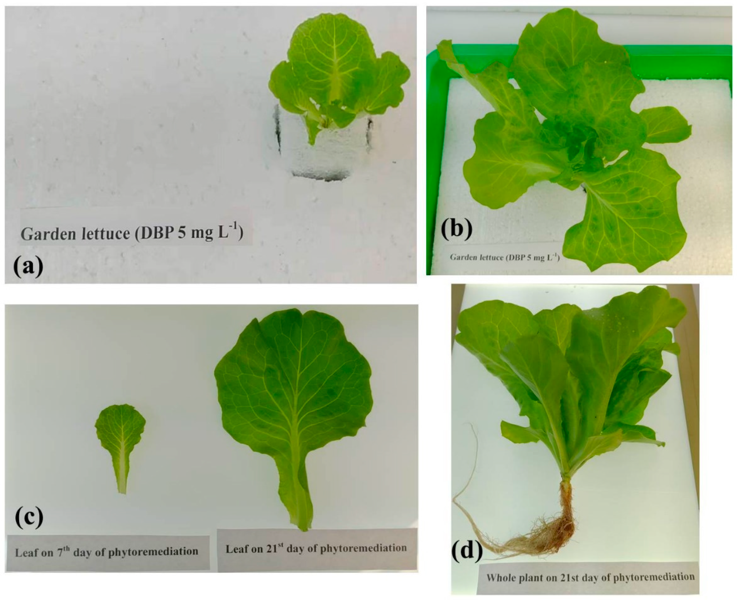
3.1. Evaluation of DBP Phytoremediation Capability in Eleven Test Plants

Eleven popular leaf vegetables in Taiwan were used to evaluate the potential of DBP phytoremediation capability in this study. Table 1 showed the results of biomass, chlorophyll concentration and DBP residue in the hydroponic solution after 21 days of phytoremediation. Garden lettuce showed the best DBP phytoremediation capability of all the test plants (Table 1). Table 1 shows that the biomass and chlorophyll of the plant leaves had all normally increased after DBP phytoremediation. However, the results of residual DBP in the hydroponic solution and accumulated DBP in different parts of the eleven test plants (Table 2) showed garden lettuce had significant DBP absorption capability (paired t-test; p < 0.05). Table 2 reveals that after DBP phytoremediation, the accumulated concentration of DBP in the roots, stems and leaves of garden lettuce was 3.35 ± 0.42, 2.74 ± 0.38 and 4.35 ± 0.42 mg kg−1, respectively. The other test plants all showed no significant DBP absorption capability following DBP phytoremediation. The morphology of garden lettuce during the period of phytoremediation is shown in Figure 2. The result displays the morphology of garden lettuce, such as the leaf shape and color, revealing no significant changes during the exposure to DBP for 21 days.
Previous studies indicated some plants had DBP absorption capability and accumulated distribution in the roots, stems, and leaves of plants [37,38,42,43]. In this study, garden lettuce could accumulate DBP in the root, stem, and leaf in 3.35 ± 0.42, 2.74 ± 0.38 and 4.35 ± 0.42 mg L−1 after 21 days, respectively. This capability was better than any of the other plants reported in previous studies, such as Bok choy [42] and Chinese cabbage [43].

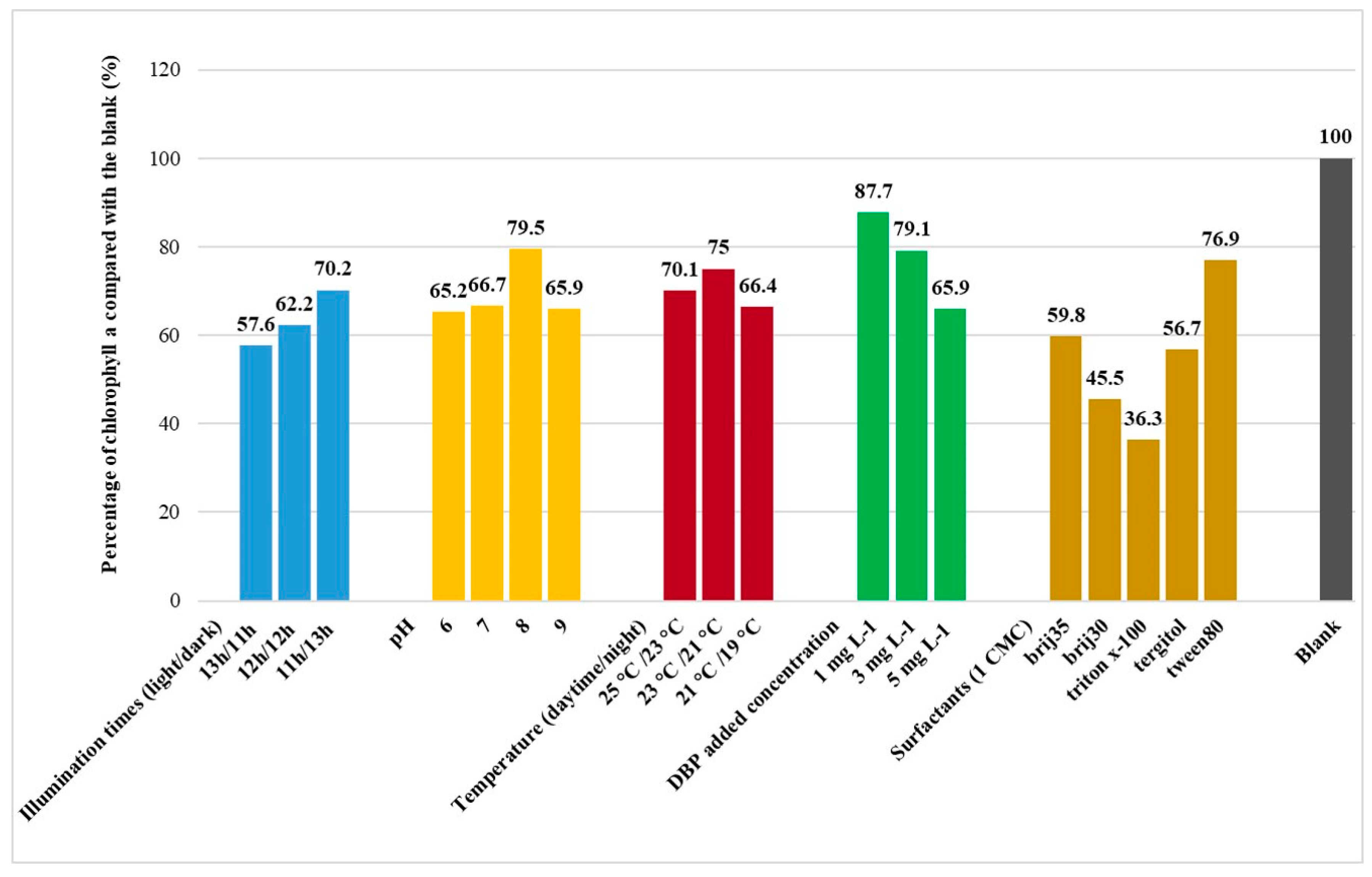
3.2. Cultural Conditions of DBP Phytoremediation

The effects of various illumination times (light/dark) on the phytoremediation of DBP in the culture medium are presented in Table 3. According to the result, the optimal illumination times for DBP phytoremediation were 11 h light and 13 h dark. As shown in Table 3, the DBP phytoremediation rate constants (k1, day−1) of garden lettuce at 11 h light and 13 h dark illumination times calculated by first-order kinetics were 2.283 and had a half-life (t1/2, days) of 5.299 days. The reason this illumination time enhanced the efficiency of phytoremediation is because garden lettuce is a vegetable which can be grown all year round, but the growth rate and quality in spring, autumn and winter is higher than in summer. Garden lettuce is a vegetable species which dislikes a long illumination time.
The effects of various pH levels on the phytoremediation of DBP in the culture medium are presented in Table 3. According to the results, the optimal pH level for DBP phytoremediation was 8. As shown in Table 3, the DBP phytoremediation rate constants (k1, day−1) of garden lettuce at pH 8 were calculated by first-order kinetics and were 4.605, with a half-life (t1/2, days) of 2.887 days. This result indicates that weakly alkaline conditions might improve the absorption capability of garden lettuce and increased the efficiency of DBP phytoremediation.
The effects of various temperatures (daytime/night) on DBP phytoremediation in the culture medium are presented in Table 3. According to the results, the optimal cultural temperature for DBP phytoremediation was 23 °C during daytime and 21 °C at night. As shown in Table 3, the DBP phytoremediation rate constants (k1, day−1) of garden lettuce at a temperature 23 °C in daytime and 21 °C at night were calculated by first-order kinetics, providing a value of 4.609 and a half-life (t1/2, days) of 2.991 days. This result indicates that 23°C during daytime and 21°C at night are the optimal physiological growth temperatures for garden lettuce.
The effects of various DBP added concentrations on DBP phytoremediation in the culture medium are presented in Table 3. According to the results, the DBP added concentration for DBP phytoremediation was 1 mg L−1. As shown in Table 3, the DBP phytoremediation rate constants (k1, day−1) of garden lettuce at a DBP added concentration of 1 mg L−1 were calculated by first-order kinetics, providing a value of 4.741 and a half-life (t1/2, days) of 2.504 days. However, after considering the phytoremediation efficiency and the environmental pollution concentration, we decided on a DBP added concentration of 5 mg L−1 as the recommended optimal concentration of DBP to add. The DBP phytoremediation rate constant (k1, day−1) at a DBP added concentration of 5 mg L−1 was 3.783, with a half-life (t1/2, days) of 3.193 days.
The effects of various non-ionic surfactants on DBP phytoremediation in the culture medium are presented in Table 3. Our previous study found the 1 CMC of the surfactant could enhance the biodegradation efficiency of DBP, but 2 and 5 CMC would inhibit it [54]. At 1 CMC of surfactant, DBP, which partitioned into the micellar phase of the surfactant, was directly available to be degraded on by a microorganism. However, at 2 or 5 CMC of surfactant, cellular toxicity would occur from the interaction of surfactant molecules with the cell membranes or membrane proteins. Therefore, we used 1 CMC as the added concentration of surfactants. According to the results, the optimal non-ionic surfactant condition for DBP phytoremediation was the addition of 1 CMC of Tween 80. The 1 CMC of Tween 80 was 0.012 mM (0.0016%, w/v). As shown in Table 3, the DBP phytoremediation rate constant (k1, day−1) of garden lettuce at 1 CMC of Tween 80 was 4.605, with a half-life (t1/2, days) of 2.686 days. This result indicates that the addition of 1 CMC of non-ionic surfactant Tween 80 is the optimal cultural condition for the DBP-based phytoremediation of garden lettuce. In addition, phthalic acid, which is the main metabolite of DBP, was not be detected in the solution or plant samples after phytoremediation. Therefore, the type of garden lettuce phytoremediation should be the phytoextraction, so that DBP can accumulate in the plant body.

3.3. Algal Biotoxicity of DBP

The biotoxicity effects of DBP before and after phytoremediation were examined using C. vulgaris. The median effective concentration (EC50) of DBP for C. vulgaris was 4.9 mg L−1. The latest study indicated the 96-hour median effective concentration values (96h-EC50) of DBP on two typical freshwater algae (Scenedesmus obliquus and Chlorella pyrenoidosa) were 15.3 mg L−1 and 3.14 mg L−1, respectively [55]. Figure 3 shows the percentage of chlorophyll a of C. vulgaris compared with the blank after DBP phytoremediation. The blank test refers to biotoxicity analysis without DBP treatment. The result showed the chlorophyll a content of C. vulgaris increased after phytoremediation, suggesting after DBP was removed from the hydroponic solution, biotoxicity markedly decreased. Compared with the blank, the chlorophyll a content of C. vulgaris in the DBP solution after phytoremediation with optimal cultural conditions was 76.9%. These results indicate that DBP biotoxicity could be significantly reduced after phytoremediation with optimal cultural conditions.

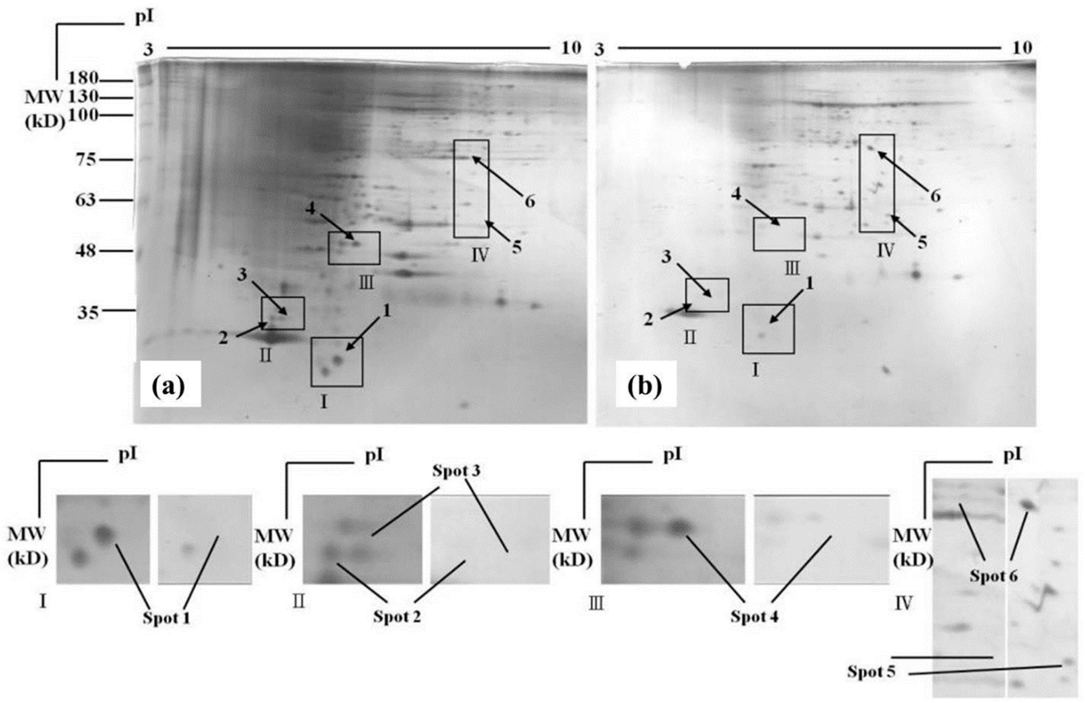
3.4. Proteomic Analysis

After separating with 2-DE, followed by staining with silver, 2-DE images of the garden lettuce leaf proteomes were taken and are shown in Figure 4. There are six protein spots, revealing significant alterations. Four proteins (spots 1, 2, 3 and 4) were identified: Photosystem II reaction center protein H (spot 1), putative Adenosine diphosphate (ADP)-ribosylation factor (spot 2) and chloroplastic 30S ribosomal protein S7 (spot 3). The protection of telomeres 1 protein (spot 2) decreased in amount or disappeared. The other two proteins (spots 5 and 6) identified as Nucleotide-binding site leucine-rich repeat (NBS-LRR) resistance-like protein RGC1F (spot 5) and DNA-directed RNA polymerase subunit beta (spot 6) was found or had increased. In the proteomic 2-DE analysis, the increase or decrease of each spot represented the strength or weakness of a protein activity in an organism. The differentially expressed proteins identified by MALDI-TOF MS and PMF analyses are listed in Table 4.
The reduced photosystem II reaction center protein H (spot 1) suggests the modulation of photosynthesis toward photosystem I (PSI). Since PSI is the terminal electron carrier in the chloroplast, this finding can be correlated to osmotic stress [56]. ADP ribosylation factor (spot 2) is one of the Guanosine triphosphate (GTP)-binding proteins. It is ubiquitous in eukaryotic cells, involved in catalyzing GTP/Guanosine diphosphate (GDP) exchange and acts as the regulator of vesicular traffic [57]. The chloroplastic 30S ribosomal protein (spot 3) and the protection of telomeres 1 protein (spot 4) are both proteins related to transcription and the protein metabolism [56]. Two proteins increased: NBS-LRR resistance-like protein RGC1F (spot 5), which occurs in response to osmotic and oxidative stress [56], and DNA-directed RNA polymerase subunit beta (spot 6), which is the protein related to transcription and the protein metabolism [56]. According to these results, DBP potentially causes osmotic and oxidative stress in garden lettuce. Compared with previous studies, DBP could accumulate in different parts of plants and cause free radical increases within the plants [42,43].

4. Conclusions

In summary, garden lettuce, which has significant DBP absorption capability, was selected as the test plant from eleven plants. The results show that DBP accumulated in different parts of the garden lettuce following DBP phytoremediation. However, the results of plant morphology and physiological status showed no significant changes following DBP phytoremediation. The optimal condition for the DBP phytoremediation of garden lettuce was one critical micelle concentration (CMC) of non-ionic surfactant Tween 80, and the half-life (t1/2, days), which calculated by first-order kinetics, was 2.686 days for 5 mg L−1 of DBP. This result indicated that the addition of 1 CMC of Tween 80 could enhance the efficiency of DBP phytoremediation. In the algae biotoxicity analysis, the median effective concentration (EC50) of DBP for C. vulgaris was 4.9 mg L−1 and DBP biotoxicity could be significantly reduced after phytoremediation. This result indicated that since DBP was absorbed by garden lettuce, the lower concentration of DBP will reduce the biotoxicity to algae. In the end, the proteome analysis showed six protein spots, revealing significant alterations. According to the information of these proteomes, DBP potentially causes osmotic and oxidative stress in garden lettuce. Compared with previous studies, garden lettuce proved again that DBP could accumulate in different parts of plants and cause free radical increases within the plants. This finding has once again proved that plants which can accumulate DBP in the body will also produce some protein or enzymes for free radical resistance. In addition, since DBP had no significant effects on the morphology and physiological status of garden lettuce, garden lettuce can be recommended for use in plant anti-DBP toxicity tests, and also is the ideal plant candidate for DBP phytoremediation. After phytoremediation, garden lettuce can be further processed and used as agricultural waste recycled material. We hope these findings could provide valuable information for DBP-contaminated water treatment in ecological engineering applications or constructed wetlands.

Author Contributions

Conceptualization, C.-S.L. and Y.N.; Data curation, C.-S.L.; Formal analysis, Y.-T.S.; Funding acquisition, C.-S.L.; Investigation, C.-S.L. and Y.-T.S.; Methodology, C.-S.L. and Y.N.; Project administration, C.-S.L.; Resources, C.-S.L.; Supervision, Y.N.; Writing—original draft, C.-S.L.; Writing—review and editing, C.-S.L. and Y.N. All authors read and approved the final manuscript.

Funding

The work was supported by the National Science Council of the Republic of China, Taiwan (grant No.: NSC 99-2313-B-214-001).

Acknowledgments

The authors are grateful to Prof. Jiunn-Tzong Wu, Biodiversity Research Center, Academia Sinica, Taiwan, for providing us with the green algae Chlorella vulgaris Beij. #3001 for toxicity analysis.

Conflicts of Interest

The authors declare no conflict of interest.

References

  1. Abdel Daiem, M.M.; Rivera-Utrilla, J.; Ocampo-Pérez, R.; Méndez-Díaz, J.D.; Sánchez-Polo, M. Environmental impact of phthalic acid esters and their removal from water and sediments by different technologies-A review. J. Environ. Manag. 2012, 109, 164–178. [Google Scholar] [CrossRef] [PubMed]
  2. Peng, X.; Feng, L.; Li, X. Pathway of diethyl phthalate photolysis in sea-water determined by gas chromatography-mass spectrometry and compound-specific isotope analysis. Chemosphere 2013, 90, 220–226. [Google Scholar] [CrossRef] [PubMed]
  3. Zhang, Y.; Wang, P.; Wang, L.; Sun, G.; Zhao, J.; Zhang, H.; Du, N. The influence of facility agriculture production on phthalate esters distribution in black soils of northeast China. Sci. Total Environ. 2015, 506–507, 118–125. [Google Scholar] [CrossRef] [PubMed]
  4. Kaewlaoyoong, A.; Vu, C.T.; Lin, C.; Liao, C.S.; Chen, J.R. Occurrence of phthalate esters around the major plastic industrial area in southern Taiwan. Environ. Earth Sci. 2018, 77, 457. [Google Scholar] [CrossRef]
  5. Ashworth, M.J.; Chappell, A.; Ashmore, E.; Fowles, J. Analysis and assessment of exposure to selected phthalates found in children’s toys in Christchurch, New Zealand. Int. J. Environ. Res. Public Health 2018, 15, 200. [Google Scholar] [CrossRef] [PubMed]
  6. Staples, C.A.; Peterson, D.R.; Parkerton, T.F.; Adams, W.J. The environmental fate of phthalate esters: A literature review. Chemosphere 1997, 35, 667–749. [Google Scholar] [CrossRef]
  7. Peijnenburg, W.J.G.M.; Struijs, J. Occurrence of phthalate esters in the environment of the Netherlands. Ecotoxicol. Environ. Saf. 2006, 63, 204–215. [Google Scholar] [CrossRef] [Green Version]
  8. Meng, X.Z.; Wang, Y.; Xiang, N.; Chen, L.; Liu, Z.; Wu, B.; Dai, X.; Zhang, Y.H.; Xie, Z.; Ebinghaus, R. Flow of sewage sludge-borne phthalate esters (PAEs) from human release to human intake: Implication for risk assessment of sludge applied to soil. Sci. Total Environ. 2014, 476–477, 242–249. [Google Scholar] [CrossRef]
  9. Net, S.; Rabodonirina, S.; Sghaier, R.B.; Dumoulin, D.; Chbib, C.; Tlili, I.; Ouddane, B. Distribution of phthalates, pesticides and drug residues in the dissolved, particulate and sedimentary phases from transboundary rivers (France–Belgium). Sci. Total Environ. 2015, 521–522, 152–159. [Google Scholar] [CrossRef]
  10. Net, S.; Sempéré, R.; Delmont, A.; Paluselli, A.; Ouddane, B. Occurrence, fate, behavior and ecotoxicological state of phthalates in different environmental matrices. Environ. Sci. Technol. 2015, 49, 4019–4035. [Google Scholar] [CrossRef]
  11. Li, J.; Wang, G.; Aggarwal, S.G.; Huang, Y.; Ren, Y.; Zhou, B.; Singh, K.; Gupta, P.K.; Cao, J.; Zhang, R. Comparison of abundances, compositions and sources of elements, inorganic ions and organic compounds in atmospheric aerosols from Xi’an and New Delhi, two megacities in China and India. Sci. Total Environ. 2014, 476–477, 485–495. [Google Scholar] [CrossRef] [PubMed]
  12. Yuan, S.Y.; Liu, C.; Liao, C.S.; Chang, B.V. Occurrence and microbial degradation of phthalate esters in Taiwan river sediments. Chemosphere 2002, 49, 1295–1299. [Google Scholar] [CrossRef]
  13. Selvaraj, K.K.; Sundaramoorthy, G.; Ravichandran, P.K.; Girijan, G.K.; Sampath, S.; Ramaswamy, B.R. Phthalate esters in water and sediments of the Kaveri River, India: Environmental levels and ecotoxicological evaluations. Environ. Geochem. Health 2015, 37, 83–96. [Google Scholar] [CrossRef] [PubMed]
  14. Zheng, X.; Zhang, B.T.; Teng, Y. Distribution of phthalate acid esters in lakes of Beijing and its relationship with anthropogenic activities. Sci. Total Environ. 2014, 476–477, 107–113. [Google Scholar] [CrossRef] [PubMed]
  15. Chen, C.F.; Ju, Y.R.; Lim, Y.; Chang, J.H.; Chen, C.W.; Dong, C.D. Spatial and temporal distribution of di-(2-ethylhexyl) phthalate in urban river sediments. Int. J. Environ. Res. Public Health 2018, 15, 2228. [Google Scholar] [CrossRef] [PubMed]
  16. Plaza-Bolaños, P.; Padilla-Sánchez, J.A.; Garrido-Frenich, A.; Romero-González, R.; Martínez-Vidal, J.L. Evaluation of soil contamination in intensive agricultural areas by pesticides and organic pollutants: South-eastern Spain as a case study. J. Environ. Monit. 2012, 14, 1182. [Google Scholar] [CrossRef]
  17. Ma, T.T.; Christie, P.; Luo, Y.M.; Teng, Y. Phthalate esters contamination in soil and plants on agricultural land near an electronic waste recycling site. Environ. Geochem. Health 2013, 35, 465–476. [Google Scholar] [CrossRef] [PubMed]
  18. Škrbić, B.D.; Ji, Y.; Đurišić-Mladenović, N.; Zhao, J. Occurence of the phthalate esters in soil and street dust samples from the Novi Sad city area, Serbia, and the influence on the children’s and adults’ exposure. J. Hazard. Mater. 2016, 312, 272–279. [Google Scholar] [CrossRef] [PubMed]
  19. Gray, L.E.; Ostby, J.; Furr, J.; Price, M.; Veeramachaneni, D.N.R.; Parks, L. Perinatal exposure to the phthalates DEHP, BBP, and DINP, but not DEP, DMP, or DOTP, alters sexual differentiation of the male rat. Toxicol. Sci. 2000, 58, 350–365. [Google Scholar] [CrossRef]
  20. Saillenfait, A.M.; Sabaté, J.P.; Gallissot, F. Developmental toxic effects of diisobutyl phthalate, the methyl-branched analogue of di-n-butyl phthalate, administered by gavage to rats. Toxicol. Lett. 2006, 165, 39–46. [Google Scholar] [CrossRef]
  21. McKee, R.H.; Butala, J.H.; David, R.M.; Gans, G. NTP center for the evaluation of risks to human reproduction reports on phthalates: Addressing the data gaps. Reprod. Toxicol. 2004, 18, 1–22. [Google Scholar] [CrossRef]
  22. Hansen, J.F.; Bendtzen, K.; Boas, M.; Frederiksen, H.; Nielsen, C.H.; Rasmussen, Å.K.; Feldt-Rasmussen, U. Influence of phthalates on cytokine production in monocytes and macrophages: A systematic review of experimental trials. PLoS ONE 2015, 10, e0120083. [Google Scholar] [CrossRef]
  23. Bui, T.T.; Giovanoulis, G.; Cousins, A.P.; Magnér, J.; Cousins, I.T.; deWit, C.A. Human exposure, hazard and risk of alternative plasticizers to phthalate esters. Sci. Total Environ. 2016, 541, 451–467. [Google Scholar] [CrossRef]
  24. Chemical Manufacturers Association. Comments of the Chemical Manufacturers Association Phthalate Esters Panel in Response to Request for Public Input on Seven Phthalate Esters; Chemical Manufacturers Association: Washington DC, USA, 1999. [Google Scholar]
  25. Wolfe, N.L.; Steen, W.C.; Burns, L.A. Phthalate ester hydrolysis: Linear free energy relationships. Chemosphere 1980, 9, 403–408. [Google Scholar] [CrossRef]
  26. Gao, D.W.; Wen, Z.D. Phthalate esters in the environment: A critical review of their occurrence, biodegradation, and removal during wastewater treatment processes. Sci. Total Environ. 2016, 541, 986–1001. [Google Scholar] [CrossRef]
  27. Long, G.; Meek, E. Environmental Health Criteria 189: Di-n-Butyl Phthalate; World Heal. Organ: Geneva, Switzerland, 1997. [Google Scholar]
  28. Chan, P.K.L.; Meek, M.E. Di-n-Butyl phthalate: Evaluation of risks to health from environmental exposure in Canada. J. Environ. Sci. Heal. Part C 1994, 12, 257–268. [Google Scholar] [CrossRef]
  29. U.S. Department of Health and Human Services. Toxicological Profile for di-n-butyl phthalate. In Public Health Service; Agency for Toxic Substances and Disease Registry: Atlanta, Georgia, USA, 1990. [Google Scholar]
  30. Rudel, R.A.; Camann, D.E.; Spengler, J.D.; Korn, L.R.; Brody, J.G. Phthalates, alkylphenols, pesticides, polybrominated diphenyl ethers, and other endocrine-disrupting compounds in indoor air and dust. Environ. Sci. Technol. 2003, 37, 4543–4553. [Google Scholar] [CrossRef]
  31. Duan, J.; Kang, J.; Deng, T.; Yang, X.; Chen, M. Exposure to DBP and high iodine aggravates autoimmune thyroid disease through increasing the levels of IL-17 and thyroid-binding globulin in Wistar rats. Toxicol. Sci. 2018, 163, 196–205. [Google Scholar] [CrossRef]
  32. Hemming, I.V.; Ann-Marie, H.; Johan, M. Effects of di-n-butyl phthalate on the carotenoid synthesis in green plants. Physiol. Plant. 1981, 53, 158–163. [Google Scholar]
  33. Herring, R.; Bering, C.L. Effects of phthalate esters on plant seedlings and reversal by a soil microorganism. Bull. Environ. Contam. Toxicol. 1988, 40, 626–632. [Google Scholar] [CrossRef]
  34. Hardwick, R.C.; Cole, R.A.; Fyfield, T.P. Injury to and death of cabbage (Brassica oleracea) seedlings caused by vapours of di butyl phthalate emitted from certain plastics. Ann. Appl. Biol. 1984, 105, 97–105. [Google Scholar] [CrossRef]
  35. Hannay, J.W.; Millar, D.J. Phytotoxicity of phthalate plasticisers: 1. Diagnosis and commercial implications. J. Exp. Bot. 1986, 37, 883–897. [Google Scholar] [CrossRef]
  36. Yin, R.; Lin, X.G.; Wang, S.G.; Zhang, H.Y. Effect of DBP/DEHP in vegetable planted soil on the quality of capsicum fruit. Chemosphere 2003, 50, 801–805. [Google Scholar] [CrossRef]
  37. Dueck, T.A.; VanDijk, C.J.; David, F.; Scholz, N.; Vanwalleghem, F. Chronic effects of vapour phase di-n-butyl phthalate (DBP) on six plant species. Chemosphere 2003, 53, 911–920. [Google Scholar] [CrossRef]
  38. Li, C.; Chen, J.; Wang, J.; Han, P.; Luan, Y.; Ma, X.; Lu, A. Phthalate esters in soil, plastic film, and vegetable from greenhouse vegetable production bases in Beijing, China: Concentrations, sources, and risk assessment. Sci. Total Environ. 2016, 568, 1037–1043. [Google Scholar] [CrossRef]
  39. Ma, T.; Zhou, W.; Chen, L.; Wu, L.; Christie, P.; Liu, W. Toxicity of phthalate esters to lettuce (Lactuca sativa) and the soil microbial community under different soil conditions. PLoS ONE 2018, 13, e0208111. [Google Scholar] [CrossRef]
  40. Parker, R.; Flowers, T.J.; Moore, A.L.; Harpham, N.V.J. An accurate and reproducible method for proteome profiling of the effects of salt stress in the rice leaf lamina. J. Exp. Bot. 2006, 57, 1109–1118. [Google Scholar] [CrossRef] [Green Version]
  41. Aghaei, K.; Ehsanpour, A.A.; Komatsu, S. Proteome Analysis of Potato under Salt Stress. J. Proteome Res. 2008, 7, 4858–4868. [Google Scholar] [CrossRef]
  42. Liao, C.S.; Yen, J.H.; Wang, Y.S. Effects of endocrine disruptor di-n-butyl phthalate on the growth of Bok choy (Brassica rapa subsp. chinensis). Chemosphere 2006, 65, 1715–1722. [Google Scholar] [CrossRef]
  43. Liao, C.S.; Yen, J.H.; Wang, Y.S. Growth inhibition in Chinese cabbage (Brassica rapa var. chinensis) growth exposed to di-n-butyl phthalate. J. Hazard. Mater. 2009, 163, 625–631. [Google Scholar] [CrossRef]
  44. He, L.; Gielen, G.; Bolan, N.S.; Zhang, X.; Qin, H.; Huang, H.; Wang, H. Contamination and remediation of phthalic acid esters in agricultural soils in China: A review. Agron. Sustain. Dev. 2015, 35, 519–534. [Google Scholar] [CrossRef]
  45. Cai, Q.Y.; Mo, C.H.; Wu, T.; Zeng, Q.Y. Accumulation of phthalic acid esters in water spinach (Ipomoea aquatica) and in Paddy soil. Bull. Environ. Contam. Toxicol. 2006, 77, 411–418. [Google Scholar] [CrossRef]
  46. Cai, Q.Y.; Mo, C.H.; Zeng, Q.Y.; Wu, Q.T.; Férard, J.F.; Antizar-Ladislao, B. Potential of Ipomoea aquatica cultivars in phytoremediation of soils contaminated with di-n-butyl phthalate. Environ. Exp. Bot. 2008, 62, 205–211. [Google Scholar] [CrossRef]
  47. Zhang, X.; Liu, X.; Chai, W.; Wei, J.; Wang, Q.; Li, B.; Li, H. The use of proteomic analysis for exploring the phytoremediation mechanism of Scirpus triqueter to pyrene. J. Hazard. Mater. 2013, 260, 1001–1007. [Google Scholar] [CrossRef]
  48. Hoagland, D.R.; Arnon, D.I. The water-culture method for growing plants without soil. Circ. Calif. Agric. Exp. Stn. 1950, 347, 22. [Google Scholar]
  49. Liao, C.S.; Chen, L.C.; Chen, B.S.; Lin, S.H. Bioremediation of endocrine disruptor di-n-butyl phthalate ester by Deinococcus radiodurans and Pseudomonas stutzeri. Chemosphere 2010, 78, 342–346. [Google Scholar] [CrossRef]
  50. Chang, R.; Jien, S.H.; Weng, C.H.; Lee, T.W.; Liao, C.S. Fast removal of polybrominated diphenyl ethers from aqueous solutions by using low-cost adsorbents. Sustainability 2017, 9, 102. [Google Scholar] [CrossRef]
  51. Hochstrasser, D.F.; Harrington, M.G.; Hochstrasser, A.C.; Miller, M.J.; Merril, C.R. Methods for increasing the resolution of two-dimensional protein electrophoresis. Anal. Biochem. 1988, 173, 424–435. [Google Scholar] [CrossRef]
  52. Hellman, U.; Wernstedt, C.; Gonez, J.; Heldin, C.-H. Improvement of an “In-Gel” digestion procedure for the micropreparation of internal protein fragments for amino acid sequencing. Anal. Biochem. 1995, 224, 451–455. [Google Scholar] [CrossRef]
  53. Perkins, D.N.; Pappin, D.J.C.; Creasy, D.M.; Cottrell, J.S. Probability-based protein identification by searching sequence databases using mass spectrometry data. Electrophoresis 1999, 20, 3551–3567. [Google Scholar] [CrossRef]
  54. Chang, B.V.; Liao, C.S.; Yuan, S.Y. Anaerobic degradation of diethyl phthalate, di-n-butyl phthalate, and di-(2-ethylhexyl) phthalate from river sediment in Taiwan. Chemosphere 2005, 58, 1601–1607. [Google Scholar] [CrossRef]
  55. Gu, S.; Zheng, H.; Xu, Q.; Sun, C.; Shi, M.; Wang, Z.; Li, F. Comparative toxicity of the plasticizer dibutyl phthalate to two freshwater algae. Aquat. Toxicol. 2017, 191, 122–130. [Google Scholar] [CrossRef]
  56. Heazlewood, J.L.; Jorrín-Novo, J.V.; Agrawal, G.K.; Mazzuca, S.; Lüthje, S. Editorial: International Plant Proteomics Organization (INPPO) world congress 2014. Front. Plant Sci. 2016, 7, 1190. [Google Scholar] [CrossRef]
  57. Pasqualato, S.; Renault, L.; Cherfils, J. Arf, Arl, Arp and Sar proteins: A family of GTP-binding proteins with a structural device for ‘front–back’ communication. EMBO Rep. 2002, 3, 1035–1041. [Google Scholar] [CrossRef]
Figure 1. Chemical structure of phthalate esters (PAEs) (a) and Di-n-dutyl phthalate (DBP) (b).
Sustainability 11 01625 g001
Figure 2. The morphology of garden lettuce during the phytoremediation period. (a) Garden lettuce on 7th day of phytoremediation, (b) garden lettuce on 21st day of phytoremediation, (c) single leaf on the 7th and 21st day of phytoremediation, and (d) the whole plant on the 21st day of phytoremediation.
Sustainability 11 01625 g002
Figure 3. Percentage of chlorophyll a in C. vulgaris compared with the blank after DBP phytoremediation.
Sustainability 11 01625 g003
Figure 4. Two dimensional photographs of normal and DBP phytoremediated garden lettuce. (a) Normal and (b) DBP phytoremediated garden lettuce. Each arrow shows the proteins that are reproducible in expression. The name, accession number, estimated molecular weight and pI values of the proteins are listed in Table 4.
Sustainability 11 01625 g004
Table 1. Biomass, chlorophyll concentration and DBP residue in hydroponic solution after DBP phytoremediation of eleven test plants.
Common NameBiomass (g, dry wt) aChlorophyll Concentration (mg g−1, fresh wt) aResidual DBP in Hydroponic Solution (mg L−1) a
0 day21 days0 day21 days0 day21 days
Edible rape2.64 ± 0.684.32 ± 0.520.227 ± 0.0320.259 ± 0.0485.02 ± 0.153.89 ± 0.18
Control c2.83 ± 0.585.63 ± 0.820.264 ± 0.0340.306 ± 0.066
Chinese cabbage2.82 ± 0.956.58 ± 1.520.132 ± 0.0280.194 ± 0.0425.02 ± 0.163.56 ± 0.13
Control c2.74 ± 0.847.08 ± 1.380.144 ± 0.0320.226 ± 0.064
Spinach2.35 ± 0.763.28 ± 0.850.253 ± 0.0440.268 ± 0.0484.98 ± 0.144.72 ± 0.15
Control c2.39 ± 0.542.98 ± 0.780.244 ± 0.0280.318 ± 0.054
Chinese mustard1.48 ± 0.364.63 ± 1.460.154 ± 0.0350.204 ± 0.0474.99 ± 0.164.79 ± 0.16
Control c1.52 ± 0.265.79 ± 1.240.156 ± 0.0280.417 ± 0.068
Water cabbage2.68 ± 0.287.18 ± 1.440.252 ± 0.0460.309 ± 0.0635.06 ± 0.212.24 ± 0.08
Control c2.57 ± 0.236.92 ± 0.880.264 ± 0.0580.364 ± 0.088
Ceylon spinach1.12 ± 0.253.76 ± 0.880.279 ± 0.0550.319 ± 0.0584.95 ± 0.123.52 ± 0.14
Control c1.15 ± 0.224.21 ± 0.820.282 ± 0.0480.355 ± 0.098
Garden lettuce1.45 ± 0.264.68 ± 0.520.165 ± 0.0370.269 ± 0.0424.99 ± 0.14n.d. b
Control c1.49 ± 0.314.72 ± 0.520.169 ± 0.0370.272 ± 0.042
Chinese celery0.92 ± 0.324.06 ± 0.850.141 ± 0.0320.191 ± 0.0385.03 ± 0.184.72 ± 0.18
Control c1.02 ± 0.294.54 ± 0.920.144 ± 0.0380.255 ± 0.049
Edible amaranth1.54 ± 0.566.13 ± 1.220.153 ± 0.0250.192 ± 0.0355.05 ± 0.243.33 ± 0.16
Control c1.47 ± 0.485.74 ± 0.890.149 ± 0.0230.214 ± 0.042
Cauliflower3.14 ± 0.859.42 ± 1.980.082 ± 0.0160.129 ± 0.0235.02 ± 0.052.16 ± 0.05
Control c3.06 ± 0.629.68 ± 1.420.083 ± 0.0160.133 ± 0.031
Chinese chive0.65 ± 0.223.43 ± 0.340.203 ± 0.0380.249 ± 0.0464.99 ± 0.054.92 ± 0.06
Control c0.66 ± 0.233.42 ± 0.290.205 ± 0.0330.251 ± 0.044
Blank d 5.04 ± 0.124.96 ± 0.08
a The given values are mean ± standard deviation (SD) of the three repeated tests. b n.d. = not detected (below the detection limit of 0.08 mg L−1 in triplicate). c Control: Without DBP treatment. d Blank: Without plant in hydroponic solution.
Table 2. DBP accumulated in different parts of eleven test plants after DBP phytoremediation for 21 days.
Common NameDBP Accumulated Concentration (mg Kg−1) a
RootStemLeaf
0 day21 days0 day21 days0 day21 days
Edible rapen.d. b0.05 ± 0.01n.d. b0.16 ± 0.04n.d. b0.68 ± 0.06
Chinese cabbagen.d. b0.10 ± 0.02n.d. b0.22 ± 0.06n.d. b0.74 ± 0.12
Spinachn.d. b0.03 ± 0.01n.d. bn.d. bn.d. bn.d. b
Chinese mustardn.d. b0.06 ± 0.02n.d. bn.d. bn.d. bn.d. b
Water cabbagen.d. b0.43 ± 0.12n.d. b0.88 ± 0.14n.d. b0.82 ± 0.18
Ceylon spinachn.d. b0.08 ± 0.02n.d. b0.26 ± 0.04n.d. b0.46 ± 0.12
Garden lettucen.d. b3.35 ± 0.42n.d. b2.74 ± 0.38n.d. b4.35 ± 0.42
Chinese celeryn.d. bn.d. bn.d. bn.d. bn.d. bn.d. b
Edible amaranthn.d. b0.22 ± 0.04n.d. b0.12 ± 0.02n.d. b0.25 ± 0.06
Cauliflowern.d. b0.37 ± 0.02n.d. b0.45 ± 0.06n.d. b0.96 ± 0.08
Chinese chiven.d. bn.d. bn.d. bn.d. bn.d. bn.d. b
a The given values are mean ± SD of three repeated tests. b n.d. = not detected (below the detection limit of 0.08 mg L−1 in triplicate).
Table 3. Comparison of the effects of various cultural conditions on DBP phytoremediation rate constants (k1, day−1) and half-lives (t1/2, days).
TreatmentGarden Lettuce (Lactuca sativa L. var. longifolia.)
k1 (day−1)t1/2 (days)r2
Illumination times (light/dark)
13h/11h1.29510.3620.811
12h/12h0.98610.2370.896
11h/13h2.2835.2990.896
pH
61.5756.1550.784
73.5063.5970.921
84.6052.8870.899
93.9123.1730.921
Temperature (daytime/night)
25 °C /23 °C3.6163.2650.942
23 °C /21 °C4.6092.9910.820
21 °C /19 °C2.4774.8340.926
DBP added concentration
1 mg L−14.7412.5040.866
3 mg L−14.2732.8400.905
5 mg L−13.7833.1930.931
Surfactants (1 CMC)
Brij352.4424.7190.963
Brij301.6777.7060.894
Triton x-1001.08510.7300.698
Tergitol2.3545.7970.831
Tween 804.605 2.686 0.937
Table 4. Proteomic characterization of polypeptide differences in expression between normal and DBP treated garden lettuce.
SpotsProtein NameAccession No.Estimated MW (kD)Estimated pIAmounts a
1Photosystem II reaction center protein HQ332U87.76.23-
2Putative ADP-ribosylation factorA8QVJ015.64.89-
3Chloroplastic 30S ribosomal protein S7Q332R917.25.35-
4Protection of telomeres 1 proteinB7T1J450.16.38-
5NBS-LRR resistance-like protein RGC1FQ56P1151.87.95+
6DNA-directed RNA polymerase subunit betaQ91Fl180.47.51+
a + = increase; - = decrease.

