Article

Effect of Fabric Electrode Surface Coating Medium on ECG Signal Quality under Dynamic and Static Conditions

1
College of Textile, Zhongyuan University of Technology, Zhengzhou 450007, China
2
Henan Province Collaborative Innovation Center of Textile and Garment Industry, Zhengzhou 450007, China
3
College of Chemical and Dyeing-Printing Engineering, Henan University of Engineering, Zhengzhou 450007, China
4
College of Automation Engineering, University of Electronic Science and Technology, Chengdu 611731, China
5
Luthai Textile Co., Ltd., Zibo 255100, China
*
Authors to whom correspondence should be addressed.
Coatings 2023, 13(1), 108; https://doi.org/10.3390/coatings13010108
Submission received: 28 November 2022 / Revised: 4 January 2023 / Accepted: 4 January 2023 / Published: 6 January 2023

Abstract

:
This study intends to explore the influence of a fabric electrode skin interface filling medium on ECG signals under dynamic and static conditions, so as to be able to better monitor both stable and dynamic ECG signals. In this paper, cotton fabric electrodes were prepared using electroless silver-plated conductive cotton fabric, and then the electrodes were integrated into a physiotherapy-grade elastic bandage to assemble the fabric ECG tape. The electrode impedance tester and the BIOPAC physiological recorder were used to measure the fabric electrode-skin impedance and electrocardiogram under dynamic and static conditions, respectively. Later, the influence of the fabric electrode coated medium on the acquisition effect of an electrocardiogram signal under dynamic and static conditions is discussed. The results show that in the absence of a dielectric reagent coating, the cotton fabric electrode is easily disturbed by external noise which leads to large electrode-skin contact impedance. However, the electrode-skin contact impedance is greatly reduced after coating the fabric electrode with the dielectric reagent. At the same time, after applying the conducive paste coating, the corresponding change curve of the fabric electrode became more stable, and the impedance value was less than 0.005 MΩ·cm2, which meets the standard requirement for textile dry electrode skin impedance. In a state of rest, a clear and stable ECG can be collected regardless of whether the medium reagent is coated or not. However, only after applying the conductive paste can a stable ECG signal be collected under the three motion modes, or can a P wave, QRS wave group, or T wave be clearly distinguished. In addition, there is a high correlation with the disposable gel electrode, which can satisfy the requirements of the long-term monitoring of ECG signals under dynamic conditions.

1. Introduction

Cardiovascular disease is one of the most fatal diseases in the world, and is characterized by qualities, such as “long-term, accumulative, accidental, high morbidity, high disability rate, high recurrence rate, and more complications” [1,2]. According to the release of the “China Cardiovascular Health and Disease Report 2019” the rate of cardiovascular disease in China has risen continuously and it has maintained the highest mortality rate of all diseases. As of now, the number of patients with cardiovascular disease may be as high as 330 million [3]. These patients require long-term health monitoring to prevent complications or even sudden death [4]. In recent years, researchers have developed a variety of new types of flexible textile ECG electrodes which can be integrated into ECG monitoring clothing, enabling long-term ECG monitoring capabilities [4,5,6]. Although textile electrodes can now be integrated into and worn as normal clothing without causing irritation to the skin, there is a gap between the electrode and the skin when in use, that is susceptible to interference from external noise, resulting in a large electrode-skin contact impedance and a poor ECG signal stability [7,8]. Disposable gel electrodes have a high monitoring ability, due to humid conditions around the gel linking the Ag/AgCl bilayer and the skin, which can improve the ion conductivity and reduce the electrode-skin contact impedance. In this way, the skin-electrode impedance is minimized, thereby obtaining a stable bio-electric signal [9]. However, the conductive gel in the disposable gel electrode will gradually dry up over time, decreasing the quality of the ECG signal [10].
ECG monitoring clothing is mostly worn close to the body. The electrodes are in direct contact with the skin and cannot provide moisture directly. Although the electrode can capture some moisture or sweat from the loss of epidermal moisture or sweating, it is vulnerable to interference from external noise. Due to the unevenness between the surface of the fabric electrode and the skin, a convex interface is formed. Sweat or moisture, in part, contributes to the formation of these air bubbles or gaps. The existence of these gaps changes the effective area of the active electrode reaction, which affects the ECG signal quality [11]. At the same time, the fabric electrodes are used to monitor the dynamic ECG process. Sitting, standing, walking, running, and other actions inevitably produce limb movement, causing the relative position of the electrode and the skin to change. This can result in slippage or deformation of the fabric electrode, forming a “motion artifact” effect, that interferes with the collection of the ECG signals [12,13,14,15]. Zhang et al. used weaving technology to design and produce circular fabric electrodes, and studied and compared the ECG signals of the woven structure electrodes and standard medical electrodes in the swing, squatting, and rotating states. The results showed that the characteristic waveform of the woven fabric electrode with 86.7% improved data was clearer and more obvious than that of the standard medical electrode [16]. Nigusse et al. used silver ink screen printing technology to develop fabric electrodes for the continuous electrocardiogram measurement on knitted cotton and polyester fabrics. The study found that the quality of ECG signals increases with the increase of the tightness of the elastic band. The signals acquired at a 15 mmHg pressure level with the textile electrodes provided a similar quality to those acquired using standard electrodes. The textile electrodes also had an acceptable signal quality even after ten washing cycles [17]. Lou et al. developed a graphene-based flexible electrode and a corresponding wireless ECG acquisition system. The results show that the ECG signal recording can be carried out using a flexible graphene electrode with a high SNR. The electrode has an effective electrical performance, a high flexibility, a satisfactory biocompatibility and wearability, and offers detection capabilities in various states of motion [4].
Changes in the state of the electrode-skin interface seriously affect the quality of the ECG signal monitoring. Filling the contact gap between the electrode and the skin can effectively improve the ion conductivity of the interface, ensuring stable ECG signal monitoring [18]. Satti et al. developed a rigid paraxylene-coated microneedle electrode array and a portable electrocardiogram (ECG) circuit for monitoring ECG and reducing motion artifacts. The results show that the developed MNE showed a stability and durability for the dynamic and long-term ECG monitoring, in comparison to the typical silver-silver chloride (Ag/AgCl) wet electrodes [19]. Lee et al. developed a wearable ECG monitoring garment that employs electrodes made of conductive carbon-based paste. The results showed that the cream was directly applied to the skin to form a detachable and flexible patch electrode. Since the contact area between the carbon-based paste and the skin is almost 100%, the contact impedance between the patch electrode and the skin is very low. The study found that the ECG signal measured using the custom-designed clothing and patch electrodes is very stable, even during actions, such as walking and running [20]. Trindade et al. used gas polymerization technology to synthesize the PEDOT polymer coating in situ on polyester plain weave fabrics and made textile electrodes that can be used for the continuous monitoring of ECG [21]. Ankhili et al. used poly (3,4-ethylene dioxythiophene) polystyrene sulfonate (PEDOT:PSS) as a conductive polymer to make flexible textile electrodes, by the screen printing process. The prepared electrodes, based on PEDOT:PSS coating, have a low surface resistivity and washability [22]. Wang et al. used MPTS to modify the polyester fabric, and then electroless silver plating to obtain the Ag/M-PETF fabric electrode. The stability of the fabric conductive silver coating in the process of elongation, bending, oxidation, and washing, was studied. The electrode has a good electrical conductivity and wash resistance, and can capture the ECG signal of the human body while running [23]. The above-mentioned research is mainly through screen printing or vapor deposition, the conductive polymer coating material and silver layer are deposited on the fabric to impart the electrical conductivity to the fabric, which is used for the ECG signal monitoring. However, there has not yet been much research into what coating can best act as a medium on the contact surface of the fabric electrode and the skin to improve the interfacial ion conductivity, which can effectively fill the gaps caused by exercise and improve the quality of the ECG signals under dynamic conditions.
Therefore, in this paper, a self-made silver-plated cotton conductive fabric is assembled into a cotton fabric electrode and integrated into a bandage. Five different dielectric reagents (deionized water, saline, alcohol, gel, and conductive paste) were then used to coat the contact surface between the electrode and the human body to improve the interfacial ion conductivity. The aim of this experiment is to explore the electrical properties of the fabric electrode after adding various media to the electrode, and to examine the effect they have on the quality of the ECG signals in both static and dynamic states, in order to evaluate which medium is best able to achieve the long-term monitoring of static and dynamic ECG signals in wearable ECG measuring equipment.

2. Materials and Methods

2.1. Materials and Instruments

Materials: Insulation shielding cloth, double-sided adhesive conductive foam, space cotton, double-sided hot melt seamless adhesive, metal concealed buckle; electroless silver-plated conductive fabric (plain cotton conductive fabric).
Dielectric reagents: Deionized water (homemade in the laboratory), alcohol (Tianjin Fuyu Fine Chemical Co., Ltd., Tianjin, China), saline (Hubei Kangsheng Pharmaceutical Co., Ltd., Hubei, China), gels (Guangdong Didi Luxury Pharmaceutical Co., Ltd., Guangdong, China), conductive paste (Weaver Medical Technology Company, Knoxville, TN, USA).
Instruments: IM3533-01 LCR tester (produced by HIOKI, Hiki, Japan), BIOPAC MP160 (BIOPAC, Goleta, CA, USA) multi-channel (16 channels) physiological recorder (BIOPAC, Goleta, CA, USA).

2.2. Structural Design of the Fabric Electrode

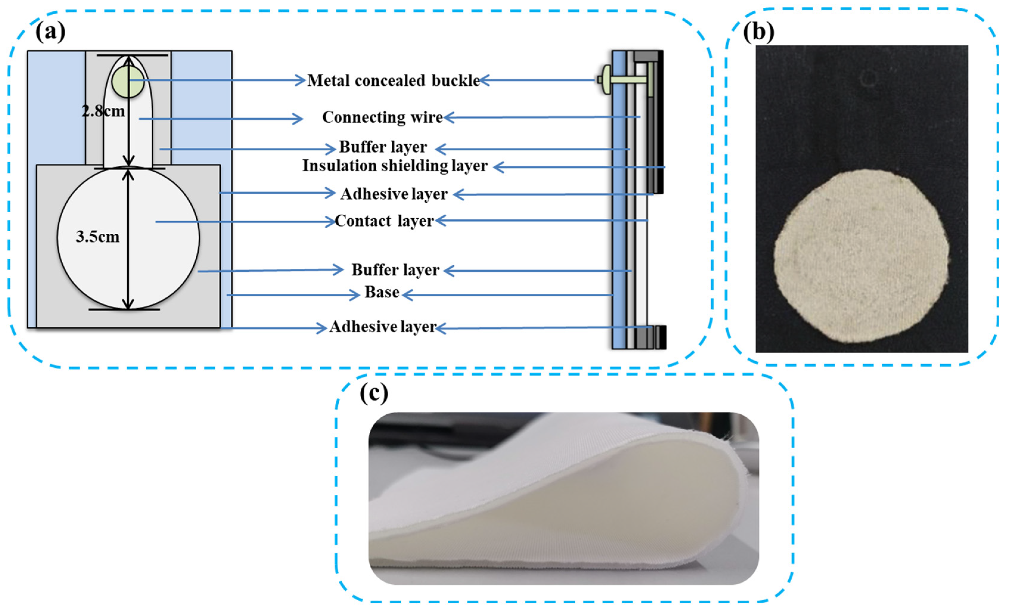
Since the fabric electrode is in direct contact with the skin, the electrode-skin interface is prone to slippage during exercise, which affects the ECG signal. Therefore, in this experiment, space cotton with a hollow structure in the middle, is used as the base support pad, which, not only makes the fabric more comfortable, but also reduces the relative slippage between the electrode and the skin during regular movement. From bottom to top, the silver-plated fabric electrode is composed of a substrate, a paste layer, a buffer layer, a contact surface layer, a paste layer, a metal concealed buckle, and an insulating shielding layer. Figure 1a and 1b display a top view and a side view, respectively, of the schematic diagram of the structure of the fabric electrode, as well as the corresponding physical diagram of the electrode, with each layer being centrally aligned. Figure 1a is a schematic diagram of the overall laminated structure of the fabric electrode with the insulating shielding cloth removed The substrate is composed of hollow space cotton in the middle, and the buffer layer is composed of double-sided adhesive conductive foam that provides a tight seal between the silver-plated conductive fabric and the substrate (Figure 1c). The contact surface layer is composed of a silver-plated conductive fabric, which is also used in the connecting area. The sensing area of the fabric electrode is connected with the concealed metal buckle; the adhesive layer is composed of a double-sided hot melt seamless adhesive to tightly bind the top layer of the insulating cotton fabric with the conductive layer.

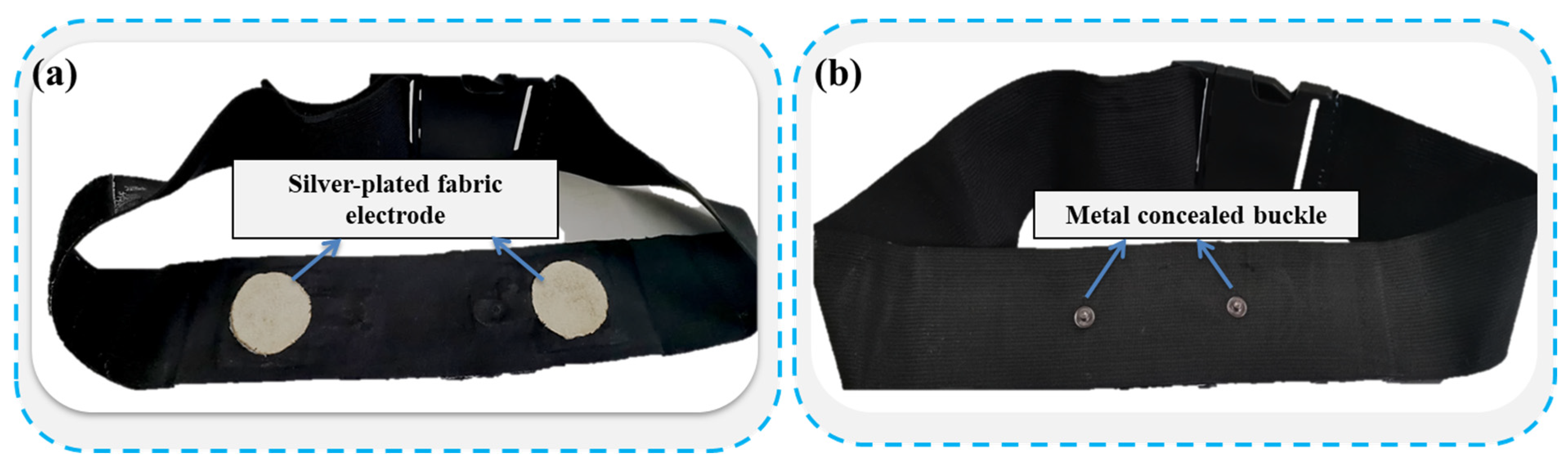
2.3. Preparation of the Fabric ECG Belt

This ECG belt design, in addition to being suitable for daily ECG measurements, has the added benefit of being able to reuse the electrodes on the ECG belt. The fabric electrode structure is shown in Figure 1. The electrode material is an electroless silver-plated fabric. The main body of the ECG belt is a physiotherapy-grade elastic bandage with a width of 6 cm and a total length of 76 cm, and the adjustment buckle is stitched at both ends of the elastic bandage. A length of Velcro is stitched to the back of the adjustment buckle, allowing the belt to be adjusted at any time for the wearer’s comfort, or to adjust the contact pressure during use. The distance between electrodes is 10 cm, and the distance between the concealed metal buckles is 7 cm. When the ECG belt is in use, the fabric electrode is positioned about 10 cm under the clavicle of the chest. In order to study the influence of daily exercise on the signal collected by the ECG band, a layer of space cotton was added between the electrode and the elastic bandage, as a filling material. The overall structure of the fabric ECG belt is shown in Figure 2 below.

2.4. Electrical Performance Testing and Monitoring of the Human ECG Signals

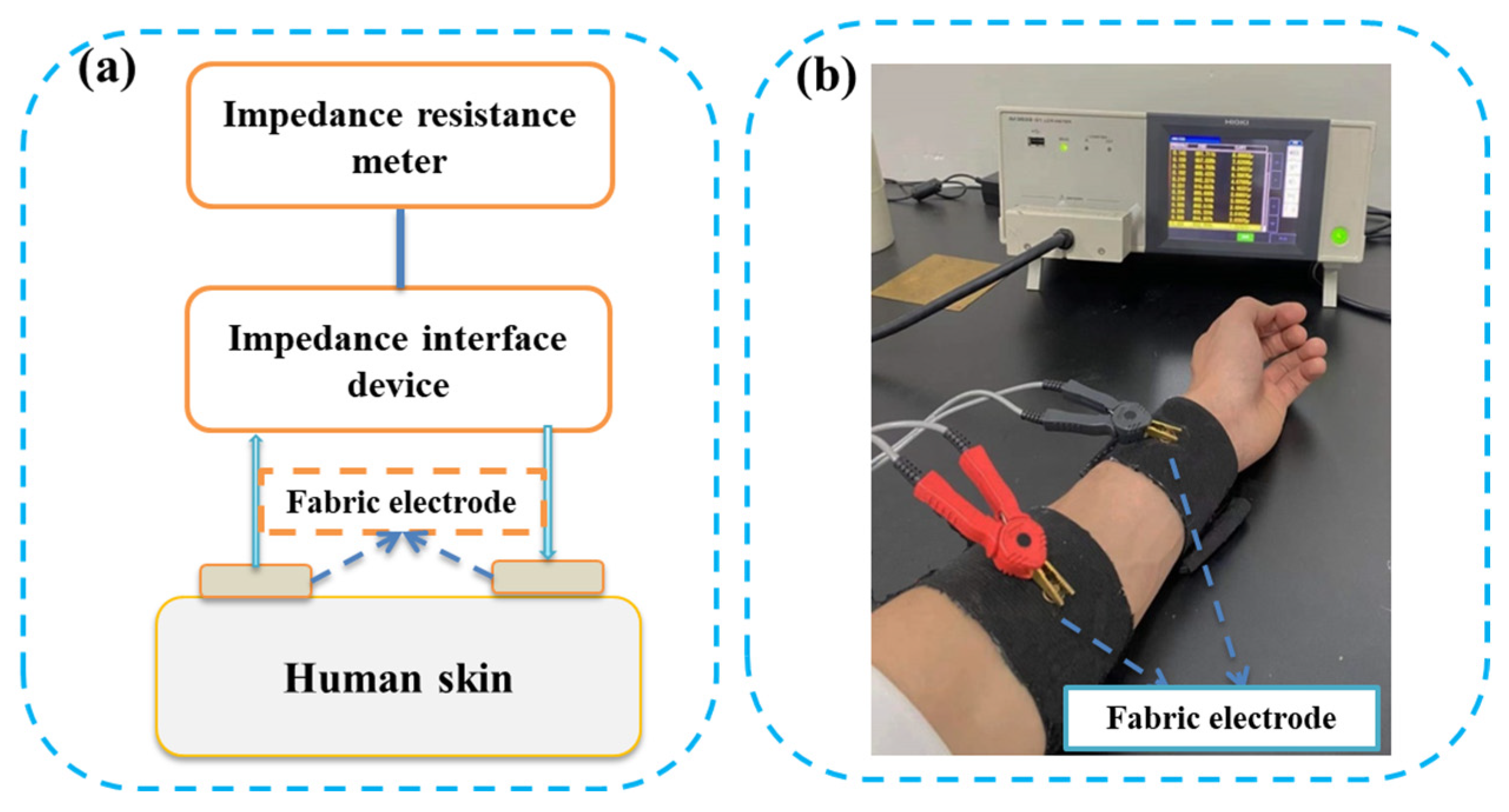
2.4.1. Electrode-Skin Contact Impedance

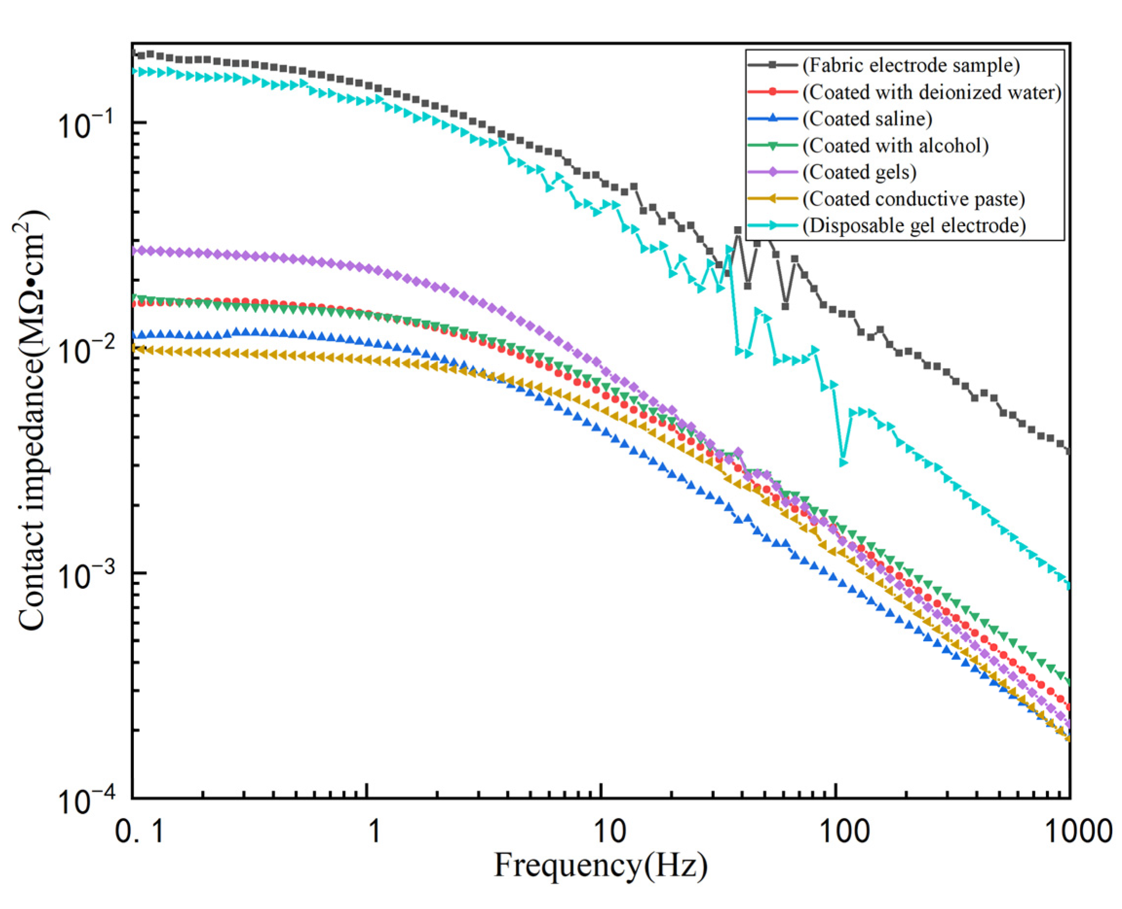
The electrode impedance value directly affects the ECG signal collection accuracy. In order to obtain a clear and stable ECG signal, the contact impedance between the electrode and the skin should be minimized. Refer to the following disposable electrode standard: an electrode pair is composed of two connected electrode conductive adhesives facing each other. When the peak sinusoidal AC current is applied at 100 μA and 10 Hz, the AC impedance of the electrode pair does not exceed 3 KΩ [24]. When using fabric electrodes to measure human ECG signals, the interface between the electrodes and the skin mainly uses sweat as the electrolyte for the electrochemical reaction, which converts the ion flow in the human body into electron flow. The frequency range of the ECG signals is 0.05~100 Hz. In the frequency range of 5~100 Hz, using a typical disposable electrode, compared with a typical disposable electrode, the impedance value between the fabric electrode and the skin should be in the range of 0~5 MΩ·cm2 [25].
As shown in Figure 3, the IM3533-01 LCR tester is used to measure the contact impedance between the skin and the electrodes. During the test, a pair of fabric electrodes are stitched to the inside of the strap, applying pressure to the electrode. The electrode pair is placed on the inside of the arm with the distance between the two being 9 cm and the test voltage set to 50 mV.

2.4.2. Measuring the Human ECG Signals

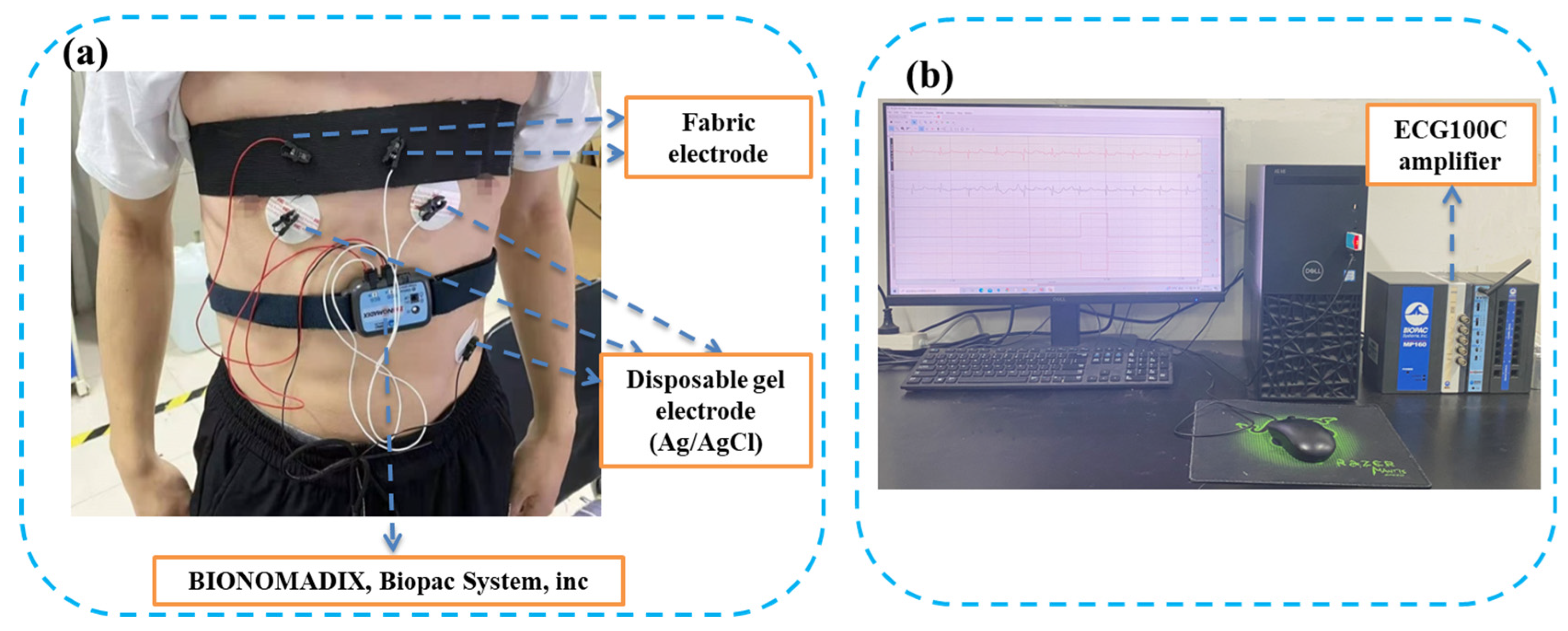
The room temperature was 25 ± 1 °C and the relative humidity was 65 ± 2%, during the ECG signal measurement for this experiment. The BIOPAC MP160 multi-channel (16-channel) physiological recorder is mainly used to measure the human ECG signals. The ECG2-R ECG module then collects those ECG signals, and finally the test results are output by the display screen. As shown in Figure 4a, before the test, an elastic bandage with a width of 6 cm and a length of 76 cm with adjustment buckles at both ends and Velcro stitched to the back, is used to bind the electrode in a specific position on the body, and a pair of disposable gel electrodes (Ag/AgCl) are attached below the corresponding positions of the electrodes. Prior to fixing the ECG belt, the contact surface of the fabric electrode and the skin needs to be coated with different media reagents. During the test, both the bound electrode and the disposable gel electrode (Ag/AgCl) are connected to the ECG signal collector (BIONOMADIX, Biopac System, Inc., Goleta, CA, USA), the ECG signal collector is wirelessly connected to the ECG100C amplifier, and the acquired ECG signal is processed and fed back to the computer (Figure 4b).
This experiment uses a collection of exercises intended to simulate various daily activities, to collect the ECG signals in different states of movement. When measuring an ECG under dynamic conditions, the way the body moves will have an impact on the collected ECG signals. In order to ensure that the pressure of the different ECG belts is consistent throughout the measurement of the ECG signals under dynamic conditions, the tightness of the ECG belt is periodically altered by adjusting the buckle. The electrode is fixed in the center of the torso, in order to understand the effect of coating the different media reagents on the measurement of dynamic and static ECG signals. Taking into account the common daily movements, such as the movements explored in [14], the experimental exercises are designed to mimic sitting down and standing up, walking in place, and expanding the chest. These actions are repeated at a regular speed, each action lasts 60 s followed by 20–30 s of rest, wherein the static ECG is recorded for the first 10 s.

3. Results and Discussion

3.1. Electrode-Skin Contact Impedance

Figure 5 shows the impedance-frequency curve of the cotton fabric electrode without a media reagent and the contact impedance-frequency curve of the skin after coating with different media reagents. As can be seen from the figure, the impedance value of the cotton fabric electrode shows a downward trend with the increase of frequency. At the same time, the corresponding impedance value in the frequency range of 5 to 100 Hz is much less than 1 MΩ·cm2, which satisfies the requirements of the standard skin impedance for dry textile electrodes and therefore can be applied to ECG electrodes, to collect ECG signals. The cotton fabric electrode showed a relatively large impedance value without any media reagent, and the impedance was higher than that of the disposable gel electrode, as the impedance frequency increased. There are several reasons for this: the lack of an electrolyte or gel filling between the fabric electrode and the skin, a rough and uneven contact surface, and the large gap between the fabric electrode and the skin, which is easily disturbed by external noise. These factors combined the result in a large electrode-skin contact impedance. The dielectric reagent coated on the surface of the fabric electrode, not only acts as an electrolyte, but also keeps the skin moist and forms a conductive path. This greatly reduces the contact impedance between the electrode and the skin, although the impedance also changes with frequency. The presence of the dielectric reagent also increased the stability, and the frequency stayed in the range of 5–100 Hz. The observed impedance value was below 0.02 MΩ·cm2. Moreover, the corresponding change curve of the fabric electrodes, after being coated with the physiological brine and conductive paste, were very similar, and their impedance values were below 0.005 MΩ·cm2, which is lower than the impedance value corresponding to the addition of other media. This is due to the fact that saline and conductive paste not only moisten the contact surface, but also act as a conductive medium at the electrode-skin interface, improving the conductivity of the fabric electrode and effectively reducing the skin contact impedance.

3.2. Effect of Coating Different Media on the ECG Signal Quality

The advantage of the fabric electrodes is that they can be integrated into normal clothing, thereby allowing them to monitor the dynamic electrocardiogram throughout the wearer’s daily life [9]. Since the fabric electrode is in direct contact with the skin when in use, no conductive gel is required. However, under dynamic conditions, there will be interaction between the electrode and the skin, causing motion artifacts. The use of different media reagents to coat the surface of the fabric electrode, not only moisturizes the skin, but also acts as an electrolyte to fill the gap between the electrode and the surface of the skin, effectively reducing the movement artifacts generated by the ECG signals during exercise. Figure 6 presents the ECG readings from the disposable medical gel electrodes and fabric electrodes in different states, measured after the electrode surface has been coated with different media reagents (deionized water, saline, alcohol, gel, and conductive paste). The green waveform diagram is the ECG measured by the disposable gel electrode, while the red is the ECG measured by the fabric electrode. The correlation coefficient between the ECG patterns corresponding to the disposable gel electrode and the fabric electrode are shown in Table 1.
As can be seen from the correlation coefficients in Figure 6 and Table 1, regardless of whether the cotton fabric electrode is coated with a dielectric reagent or not, the ECG in the resting state is not significantly disturbed by noise. The characteristic waveforms of the ECG are relatively clear, and the P-wave, T-wave, and QRS wave groups are easily distinguishable. At the same time, the correlation coefficient between the ECG and the disposable gel electrode with the dielectric reagent coating increases, and the correlation coefficient is above 0.5. Among all media reagent coatings, the similarity between the characteristic waveforms collected after applying the physiological saline and conductive paste are the highest, and their observed correlation coefficient with the disposable gel electrode is greater than 0.8.
As shown in Figure 6, when the cotton fabric electrode collects the dynamic ECG without a media reagent, the ECG can be distinguished, even while sitting, standing, or walking, but there is noise interference. In addition, there is a large baseline drift and high-frequency noise in the ECG during the chest expansion exercise. The shape of the ECG is almost indistinguishable during this exercise (shown in Figure 6d), and the correlation coefficient with the disposable gel electrode is negatively correlated. This is due to the fact that the raw cotton fabric electrode has a woven tissue structure and the surface is relatively dry, making it easy to detach from the skin during exercise. This changes the effective area of the electrode, resulting in high-amplitude movement artifacts. During chest expansion exercises, chest muscles produce EMG, which is superimposed on the ECG signal, causing further distortions. As shown in Figure 6, after applying deionized water and alcohol, the characteristic waveforms can hardly be identified from the ECG collected during sitting and standing up, walking in place, or chest expansion, and the correlation coefficients are all 0, as shown in Table 1. Although the coating with deionized water and alcohol provides moisture retention to the fabric electrode, the interfacial electrolyte formed between the electrode and the skin affects the chemical reactions that occur before and after the electron transfer, due to the lack of ions.
Figure 6 demonstrates that after applying a coating of normal saline, gel, and conductive paste, the ECG collected while sitting, standing, and walking, can easily be distinguished from the characteristic waveforms of the ECG, and the correlation coefficient is above 0.4. The fabric electrode surface exhibited a very low noise interference after the coating of saline and conductive paste, and the corresponding correlation coefficients were all above 0.6, which was very similar to the disposable gel electrode. At the same time, after coating the electrode with the conductive paste, the waveform diagram present in the ECG, collected during chest expansion, can still be clearly identified, similar to the diagram from the disposable gel electrode, and the correlation coefficient reaches 0.636. This is because the conductive paste contains a conductive medium, which not only provides the moisturizing properties of the electrode surface, but also acts as an electrolyte to fill the electrode-skin gap, increasing the effective area of contact between the electrode and the skin and reducing the changes in the effective contact area during exercise. In conclusion, the conductive paste coating applied to the contact surface between the fabric electrode and the skin, had the least influence on the ECG signal acquisition under both dynamic and static conditions, allowing for the collection of high quality ECG signals under both conditions.

4. Conclusions

In this paper, materials, such as electroless silver-plated cotton conductive fabric, space cotton, and double-sided adhesive conductive foam were assembled into fabric electrodes, and then the fabric electrodes were integrated into physiotherapy-grade elastic bandages, to prepare the fabric ECG belts. The electrode-skin contact impedance of the fabric electrode and each coated medium reagent were tested separately, and the electrocardiogram of the fabric electrode and the disposable gel electrode were measured with a BIOPAC recorder. The influence of the surface of the fabric electrode on the quality of the electrocardiogram, in both static and dynamic states, after the medium is coated, was discussed. The results show that due to the rough and uneven contact surface of the fabric electrode without a coating, there are large voids on the contact surface that are susceptible to an interference from external noise, resulting in a larger electrode-skin contact impedance. Once the dielectric reagent is coated on the surface of the fabric electrode, the skin is kept moist and a conductive path is formed, which greatly reduces the contact impedance. In addition, the impedance is more stable across frequencies and the frequency stays in the range of 5~100 Hz. The impedance value is below 0.02 MΩ·cm2. When the electrode surface is coated with physiological saline and conductive paste, the corresponding change curve is nearly identical. The impedance values are all below 0.005 MΩ·cm2, which is lower than the impedance values corresponding to the addition of other media and satisfies the standard requirements of the skin impedance when using textile dry electrodes. When measuring the ECG in a state of rest, a clear and stable ECG can be collected, regardless of whether the dielectric reagent is coated or not. However, a stable ECG can only be collected under the three modes of movement after the contact surface of the fabric electrode is coated with conductive paste, wherein the P-wave, T-wave, and QRS wave groups can be clearly identified. The correlation of the conductive paste coating with the disposable gel electrode is very high, offering a new and effective method for the dynamic monitoring of human ECG signals. In future research, the composition of conductive paste should be explored so that it can also achieve a moisturizing effect while avoiding allergic reactions to the skin. This might offer a long-term solution to accurately monitor human ECG signals throughout daily activities.

Author Contributions

Literature research and discussion, Y.Z. and J.Z.; writing, Y.Z.; methodology, J.Z. and Q.L.; supervision, H.Y., L.D. and D.C.; project administration and funding acquisition, H.Y., M.W., F.X., L.D. and D.C. All authors have read and agreed to the published version of the manuscript.

Funding

This work was supported by the National Key R&D Program of China (2016YFB1001401), Scientific and technological project in Henan Province (No. 212102210550), the Science and Technology Guidance Project of China National Textile and Apparel Council (2020060), Zhongyuan University of Technology Subject development program for Young Master’s supervisor (No. SD202216), the Advanced Textile Equipment Technology Provincial and Ministerial Co-construction Collaborative Innovation Center, the Key Laboratory of Functional Textiles of Henan Province.

Institutional Review Board Statement

Not applicable.

Informed Consent Statement

Not applicable.

Data Availability Statement

The raw/processed data required to reproduce these findings cannot be shared at this time as the data also forms part of an ongoing study.

Conflicts of Interest

The authors declare no conflict of interest.

References

  1. Domanski, M.J.; Fuster, V.; Diaz-Mitoma, F.; Grundy, S.; Lloyd-Jones, D.; Mamdani, M.; Roberts, R.; Thorpe, K.; Hall, J.; Udell, J.A.; et al. Next Steps in Primary Prevention of Coronary Heart Disease Rationale for and Design of the ECAD Trial. J. Am. Coll. Cardiol. 2015, 66, 1828–1836. [Google Scholar] [CrossRef] [PubMed]
  2. Yan, N.N.; Zhang, H.; Deng, Y.M. Research status and development trend of wearable medical monitoring clothing. J. Text. 2015, 36, 162–168. [Google Scholar]
  3. China Cardiovascular Health and Disease Report. J. Cardiopulm. Vasc. Dis. 2020, 39, 1157–1162.
  4. Lou, C.; Li, R.; Li, Z.; Liang, T.; Wei, Z.; Run, M.; Yan, X.; Liu, X. Flexible Graphene Electrodes for Prolonged Dynamic ECG Monitoring. Sensors 2016, 16, 1833. [Google Scholar] [CrossRef] [PubMed] [Green Version]
  5. Fernandes, M.S.; Lee, K.S.; Ram, R.J.; Correia, J.H.; Mendes, P.M. Flexible PDMS -based dry electrodes for electro-optic acquisition of ECG signals in wearable devices. In Proceedings of the 2010 Annual International Conference of the IEEE Engineering in Medicine and Biology, Buenos Aires, Argentina, 31 August–04 September 2010; pp. 3503–3506. [Google Scholar] [CrossRef]
  6. Yi, M. NTT invented "intelligent" textile materials. Woolen Technol. 2013, 41, 1–4. [Google Scholar]
  7. Zhen, L.; Liu, X. Progress on Fabric Electrodes Used in Biological Signal Acquisition. J. Miner. Mater. Charac-Terization Eng. 2015, 3, 204–214. [Google Scholar]
  8. Yan, N.N. Research on Wearable Performance of ECG Monitoring Clothing. Master’s Thesis, Xi ’an Engineering University, Xi’an, China, 2015. [Google Scholar]
  9. Paul, G.; Torah, R.; Beeby, S.; Tudor, J. Novel active electrodes for ECG monitoring on woven textiles fabricated by screen and stencil printing. Sens. Actuators A Phys. 2015, 221, 60–66. [Google Scholar] [CrossRef]
  10. Pani, D.; Achilli, A.; Bonfiglio, A. Survey on Textile Electrode Technologies for Electrocardiographic (ECG) Monitoring, from Metal Wires to Polymers. Adv. Mater. Technol. 2018, 3, 1800008. [Google Scholar] [CrossRef]
  11. Xiong, F. Research on the Mechanism and Suppression Algorithm of “Motion Artifacts” Based on Fabric ECG Induction. Master’s Thesis, University of Electronic Science and Technology, Chengdu, China, 2019. [Google Scholar]
  12. Dong, K.; Zhang, L.; Fan, J.X.; Li, M.J.; Mei, L.; Xiao, X.L. Analysis of the mechanism of ECG signal monitoring and wearable pressure by fabric electrodes. J. Text. 2019, 40, 75–82. [Google Scholar]
  13. Xu, P.J. Analysis of the Mechanical Action Between the Textile Structure Electrode and the Skin Used for Surface ECG moni-Toring and Dynamic Noise Research. Master’s Thesis, Donghua University, Shanghai, China, 2012. [Google Scholar]
  14. Boehm, A.; Yu, X.; Neu, W.; Leonhardt, S.; Teichmann, D.A. Novel 12-Lead ECG T-Shirt with Active Electrodes. Electronics 2016, 5, 75. [Google Scholar] [CrossRef] [Green Version]
  15. An, X.; Tangsirinaruenart, O.; Stylios, G.K. Investigating the performance of dry textile electrodes for wearable end-uses. J. Text. Inst. 2018, 110, 151–158. [Google Scholar] [CrossRef]
  16. Zhang, M.; Guo, N.; Gao, Q.; Li, H.; Wang, Z. Design, Characterization, and Performance of Woven Fabric Electrodes for Electrocardiogram Signal Monitoring. Sensors 2022, 22, 5472. [Google Scholar] [CrossRef] [PubMed]
  17. Nigusse, A.; Malengier, B.; Mengistie, D.; Tseghai, G.; Van Langenhove, L. Development of Washable Silver Printed Textile Electrodes for Long-Term ECG Monitoring. Sensors 2020, 20, 6233. [Google Scholar] [CrossRef] [PubMed]
  18. Niu, D.F.; Sun, C.; Wu, X.Y.; Guo, P.P.; Wang, Y.Q.; Yi, W.J. Optimization of fabric electrocardiogram electrode and its performance comparison under different relative humidity. Wool Spinn. Sci. Technol. 2020, 48, 15–20. [Google Scholar]
  19. Satti, A.T.; Park, J.; Park, J.; Kim, H.; Cho, S. Fabrication of Parylene-Coated Microneedle Array Electrode for Wearable ECG Device. Sensors 2020, 20, 5183. [Google Scholar] [CrossRef] [PubMed]
  20. Lee, J.-W.; Yun, K.-S. ECG Monitoring Garment Using Conductive Carbon Paste for Reduced Motion Artifacts. Polymers 2017, 9, 439. [Google Scholar] [CrossRef] [PubMed] [Green Version]
  21. Trindade, I.G.; Martins, F.; Baptista, P. High electrical conductance poly(3,4-ethylenedioxythiophene) coatings on textile for electrocardiogram monitoring. Synth. Met. 2015, 210, 179–185. [Google Scholar] [CrossRef]
  22. Ankhili, A.; Tao, X.; Cochrane, C.; Koncar, V.; Coulon, D.; Tarlet, J.-M. Comparative Study on Conductive Knitted Fabric Electrodes for Long-Term Electrocardiography Monitoring: Silver-Plated and PEDOT:PSS Coated Fabrics. Sensors 2018, 18, 3890. [Google Scholar] [CrossRef] [PubMed] [Green Version]
  23. Wang, L.; Pan, Y.L.; He, D.D.; Qian, L.Y.; Cao, X.H.; He, B.H.; Li, J.R. Conductive Polyester Fabrics with High Washa-bility as Electrocardiogram Textile Electrodes. ACS Appl. Polym. Mater. 2022, 4, 1440–1447. [Google Scholar] [CrossRef]
  24. Muhlsteff, J.; Such, O.; Schmidt, R.; Perkuhn, M.; Reiter, H.; Lauter, J.; Thijs, J.; Musch, G.; Harris, M. Wearable approach for continuous ECG- and activity patient-monitoring. In Proceedings of the 26th Annual International Conference of the IEEE Engineering in Medicine and Biology Society, San Francisco, CA, USA, 1–5 September 2004; pp. 2184–2187. [Google Scholar]
  25. Catrysse, M.; Puers, R.; Hertleer, C.; van Langenhove, L.; van Egmond, H.; Matthys, D. Towards the integration of textile sensors in a wireless monitoring suit. Sens. Actuators A Phys. 2004, 114, 302–311. [Google Scholar] [CrossRef]
Figure 1. (a) Schematic diagram of the structure of the silver-plated fabric electrode. (b) Physical drawings of the silver-plated fabric electrodes. (c) Space cotton fabric.
Coatings 13 00108 g001
Figure 2. (a) ECG with the human body contact surface. (b) ECG belt outer surface.
Coatings 13 00108 g002
Figure 3. The contact impedance test between the fabric electrode and the skin. (a) Schematic diagram. (b) Test diagram.
Coatings 13 00108 g003
Figure 4. (a) Human ECG test diagram. (b) Diagram of the ECG signal acquisition device.
Coatings 13 00108 g004
Figure 5. The contact impedance between the electrode surface of the cotton fabric and the skin after coating with different media reagents.
Coatings 13 00108 g005
Figure 6. Dynamic and static ECGs collected at different states of the fabric electrode interface. (a) Static state. (b) Sitting down and standing up. (c) Walking in place. (d) Expanding the chest.
Coatings 13 00108 g006
Table 1. The correlation coefficient between the ECG pattern corresponding to the cotton fabric electrode and the disposable gel electrode.
State of MotionStatic StateSitting Down and Standing UpWalking in PlaceExpanding the Chest
Electrode Surface State
Fabric Electrode Sample0.4810.3300.209−0.774
Coated with Deionized Water0.530000
Coated Saline0.8210.6240.7760
Coated with Alcohol0.64500−0.687
Coated Gels0.7600.4400.5510
Coated Conductive Paste0.8670.6680.6280.636
Disclaimer/Publisher’s Note: The statements, opinions and data contained in all publications are solely those of the individual author(s) and contributor(s) and not of MDPI and/or the editor(s). MDPI and/or the editor(s) disclaim responsibility for any injury to people or property resulting from any ideas, methods, instructions or products referred to in the content.

