Review

Collagen Injections for Rotator Cuff Diseases: A Systematic Review

1
Department of Orthopaedic and Trauma Surgery, Casa di Cura di Bernardini, 74121 Taranto, Italy
2
Public Health Department, University of Naples Federico II, 80131 Naples, Italy
3
Department of Medicine and Surgery, LUM University, 70010 Casamassima, Italy
4
Physical Therapy and Rehabilitation Unit, Policlinico di Modena, 41125 Modena, Italy
5
Clinical and Experimental Medicine PhD Program, University of Modena and Reggio Emilia, 41125 Modena, Italy
6
Department of Biomedical and Neuromotor Sciences, Alma Mater Studiorum, University of Bologna, 40126 Bologna, Italy
7
Humanitas Research Hospital Department of Biomedical Sciences, 20090 Milan, Italy
8
Department of Orthopedic Rehabilitation, Campolongo Hospital, 84025 Marina di Eboli, Italy
*
Author to whom correspondence should be addressed.
Clin. Pract. 2025, 15(2), 28; https://doi.org/10.3390/clinpract15020028
Submission received: 2 December 2024 / Revised: 17 January 2025 / Accepted: 26 January 2025 / Published: 28 January 2025
(This article belongs to the Special Issue Musculoskeletal Pain and Rehabilitation)

Abstract

:
Background: Because of its anatomy and function, the rotator cuff (RC) is vulnerable to considerable morbidity. The prevalence of RC diseases (RCDs) among the general population is 5–39%, reaching over 30% in patients older than 60. The aim of the present systematic review is to investigate the effects of the use of collagen injections in the treatment of RCDs. Methods: A systematic search of scientific electronic databases (such as PubMed, Scopus and Web of Science) was performed up to November 2024, following the Preferred Reporting Items for Systematic Reviews and Meta-Analyses (PRISMA) guidelines. Two independent authors conducted the search and assessed the articles. The inter-rater reliability for the quality assessment was measured using Cohen’s kappa coefficient, while the Modified Coleman Methodology Score (CMS) was applied to evaluate the methodological quality of the articles included in this systematic review. Results: A total of eight articles were included, with the overall quality of the included articles being evaluated as fair. Despite the use of different types of collagen and injection protocols, as well as the different scores applied, each included study showed clinically relevant improvements. However, given the high degree of heterogeneity of the included studies, we cannot draw conclusions regarding which type of collagen and injection protocol are best for RCD treatment. Discussion: Collagen administration for RCDs seems to be effective at reducing pain and improving function, as well as the tendon structure, especially in partial tears and RC tendinopathy. High-quality, prospective studies with long-term follow-up are necessary to validate the findings of the articles included in this systematic review.

1. Introduction

The rotator cuff (RC) consists of the subscapularis, supraspinatus, and infraspinatus tendons and teres minor muscle, and its function is to dynamically stabilize the glenohumeral joint while avoiding humeral head migration and contributing to shoulder abduction and internal and external rotations [1]. The RC is particularly susceptible to significant morbidity because of its anatomy and function, often resulting from a combination of extrinsic (e.g., anatomical variations, such as acromial morphology, os acromiale, and acromial spurs, which contribute to bony impingement or direct pressure on the surrounding soft tissue) and intrinsic factors (originating within the tendon itself, such as tensile overload, aging, compromised microvascular supply, trauma, or degenerative changes) [2,3].
Rotator cuff diseases (RCDs) are the third most prevalent musculoskeletal pathologies, following lower back pain and knee pain. Their prevalence among the general population ranges from 5% to 39% and increases with age, exceeding 30% among individuals over 60 years old. The majority of cases involve RC tendinopathy (RCTP) or partial- and full-thickness rotator cuff tears (PTRCTs or FTRCTs) [4,5,6,7].
PTRCTs can be divided into bursal, intra-tendinous, and articular tears. They are more common and painful compared to FTRCTs, with a prevalence of 13% to 32% among the adult population [8,9,10]. Several studies have demonstrated that the 80% of PTRCTs progress into FTRCTs despite conservative treatment [11].
The treatment of choice for RCDs is still debated and relies on several factors, including tear degree and size, patient symptoms and needs, and functional loss [12,13].
Arthroscopic cuff repair is the most common and accepted surgical treatment, with proven satisfactory results [14,15], but post-operative re-tears represent an important complication, with an incidence between 34.2% and 40% [16], seriously impacting the rehabilitation process and patients’ quality of life [17,18]. Given the risks related to surgery (such as reduced strength of RC tendons) and the significant risk of recurrence, conservative treatment is usually the first choice, especially for older adults [19,20]. Various studies report good results for the management of both PTRCTs and FTRCTs [4,21], with a surgical option usually being considered when the conservative treatment has not exhibited an effect within the first 5–12 weeks [22,23,24].
Conservative options include pharmacotherapy (such as non-steroidal anti-inflammatory drugs and analgesics), physiotherapy [25,26,27], therapeutic exercise [28,29,30], and injections of various drugs, such as corticosteroids (CS), platelet-rich plasma (PRP), hyaluronic acid (HA), and collagen [31,32,33,34,35].
CS injections provide transient pain relief; however, their use does not modify the natural history of the disease, with recent evidence suggesting a potential role in accelerating the degenerative process in the tendon [36].
PRP alleviates symptoms and slows the degeneration of the tendon better than CS administration and prolotherapy [37,38,39,40]. HA exhibits anti-inflammatory and adhesion-prevention properties [41], and it plays a crucial role in promoting cell differentiation and growth, enhancing type I collagen expression in tendon-derived cells, and supporting tendon and bone healing [42,43,44,45,46]. The use of injectable collagen in the tendon itself or in the subacromial bursa exhibits a positive effect that reduces collagen’s degenerative process [47,48,49].
Several types of collagens (types I, II, III, and V) can be found in fibrous tissues such as tendons, ligaments, and skin. Type I collagen, a major component of the extracellular matrix (ECM) of tendons, directly influences the structural and mechanical properties of tendons [21]. In the RC’s ECM, type I collagen represents more than 95% of the total amount of collagen, whereas the remaining 5% consists of collagen types III and V [50,51].
Atelocollagen, a soluble type I collagen, is the most commonly used injectable collagen because of its excellent biocompatibility, low immunogenicity, prolonged half-life, and high resistance to enzymatic degradation [52,53,54]. Several advantages have been reported with the use of highly purified atelocollagen in terms of collagen–cell interactions and its few adverse effects, such as bruising, redness, swelling, tenderness, or itching, which are, however, very unlikely to occur in patients affected by musculoskeletal disorders, especially RCDs [47,55]. Collagen injections induce regenerative pathways by stimulating tenocyte proliferation and migration, via the synthesis of endogenous collagen, and by the restoration of collagen fibers in damaged tendons [23,36,56,57]. Collagen administration at the target site activates integrin receptors on fibroblast cell membranes [58], triggering a cascade of growth factors that stimulate the synthesis of endogenous collagen [59]. This process ultimately repairs damaged collagen fibers and promotes their proper alignment [60,61,62].
In animal models, the direct administration of atelocollagen at the site of an RC tear improved tendon healing and remodeling, as demonstrated by immunohistochemistry and histopathological analyses [47,63]; a possible explanation of this effect may be a facilitation to re-create tendon continuity at the injured site, decreasing peritendinous adhesions and improving muscle activity.
Collagen injections were also used, with good reported outcomes, for the treatment of large and massive RC tears, with the collagen patches implanted arthroscopically [47,64], as well as for the treatment of tennis elbow [65,66] and plantar fasciitis [59,67].
Evidence of synergic effects between collagen and PRP has been reported, suggesting that PRP can positively influence cellular mitogenic activity, enhance collagen production, and improve the ratio of collagens I/III [68]. Furthermore, injections of combined drugs, such as collagen and PRP or collagen and HA, may promote regeneration of the native insertion site, prevent scar tissue formation, and increase the biomechanical strength [9,69,70,71]. The aim of the present study is to systematically review the use of collagen injections for the treatment of RCDs in humans, given the growing interest and use of infiltrative collagen for treating musculoskeletal disorders in recent years, as well as the need to find a valid, safe, and cost-effective alternative to the currently proposed conservative treatments. We hypothesized that the use of collagen injections allows for the achievement of better clinical and functional outcomes in patients with RCTP, PTRCTs and FTRCTs. To date, this is the first systematic review conducted using PRISMA guidelines regarding the use of collagen injections for RCDs.

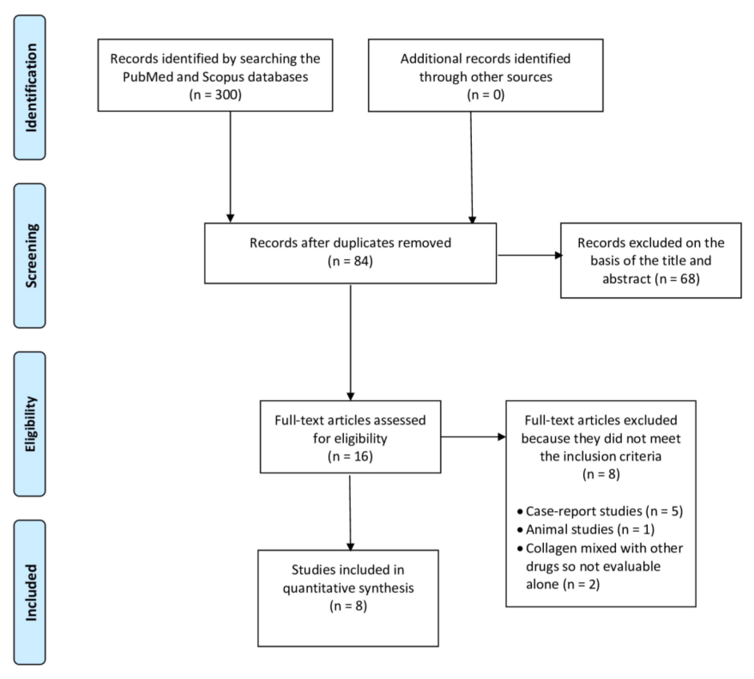
2. Materials and Methods

Study design
This systematic review and its procedures were designed and conducted in accordance with the Preferred Reporting Items for Systematic Reviews and Meta-Analyses (PRISMA) guidelines [72,73,74]. The PRISMA flow diagram is provided in Figure 1, while the PRISMA checklist can be found in the Supplementary Materials [75]. The research protocol has been registered with the International Prospective Register of Systematic Reviews (PROSPERO), under registration number: CRD42023470461.
Eligibility criteria
This review includes randomized clinical trials (RCTs), prospective and case-series studies, with a minimum follow-up of one month. Comparative studies with other injections therapies such as PRP were also included.
Articles such as editorials, technical notes, letters to authors, narrative reviews, systematic reviews, case reports, and animal or cadaveric studies that did not report clinical outcomes of the use of collagen for RCDs were excluded.
Information sources
Potential studies were identified through a search of electronic databases, including the Cochrane Central Register of Controlled Trials (CENTRAL), EMBASE, MEDLINE, PEDro, Web of Science, Scopus, PubMed, and CINAHL. A comprehensive search of all databases was conducted from their inception through to November 2024, with no language restrictions. Additionally, the reference lists of the relevant studies were reviewed for other potential studies, but none were identified.
Search strategy
The strategy had two components including terms for collagen and RCDs. Keywords for population were “Rotator cuff” [MeSH] OR rotator cuff disease*[all fields] OR rotator cuff tendinopathy*[all fields] OR rotator cuff tear*[all fields]. The keywords for the intervention were “collagen” [MeSH] OR “atelocollagen” [MeSH].
Types of participants
This study included patients with a diagnosis of an RCD, defined as RCTP, PTRCTs, or FTRCTs.
Types of interventions
For inclusion, collagen had to be administered to at least one group in the RCTs. Studies in which the effects of collagen alone could not be evaluated (such as a mixture of collagen and another drug compared with collagen alone or another drug) would not be included.
Types of comparison controls
Comparison groups were classified into active and inactive controls according to the “Cochrane Handbook for Systematic Reviews of Interventions” [76]. An inactive control was defined as either no injection or arthroscopic repair without collagen injection. Active control was defined as the use of alternative injection solutions, such as PRP [71] or an acellular dermal matrix allograft [77].
Outcomes measures
The primary outcome was a reduction in pain, measured by the numeric analogue scale (NAS, 0–10) or visual analogue scale (VAS, 0–10). Secondary outcomes included the constant score (CoS) and the American shoulder and elbow surgeons score (ASES), when available. Other scores were evaluated on a case-by-case basis depending on the ones used in the included studies. The outcomes were evaluated at baseline and at final follow-up for each of the included studies.
Study selection and data extraction
Two independent authors (R.A. and D.T.) performed the search and evaluated the articles. Researchers experienced in conducting systematic reviews (E.S., B.C., F.S., and R.P.) resolved any cases of uncertainty. Initially, the investigators reviewed the abstracts of the articles, selecting relevant studies based on the inclusion and exclusion criteria, and then compared their selections with those of other investigators. Two weeks later, the same studies were re-evaluated to confirm agreement. No disagreements were observed among the investigators.
One investigator (R.A.) input data extracted from the full-text articles into structured tables in an Excel spreadsheet for descriptive analysis of each study. The sample sizes, types of management and collagen used, time of follow-up, clinical and functional outcomes before and after treatment, adverse events and complications extracted from the retrieved articles are presented in Table 1. A second investigator (E.S.) independently double-checked the extraction of primary data from all the articles. Doubts and inconsistencies were grouped and solved via another round of revision performed together by the two investigators. All authors participated in drafting the text. All results that were compatible with each outcome domain in each study were sought. A p-value < 0.05 was considered statistically significant. The results are presented in Table 1 for a comparison of the progression from baseline to last follow-up. All results are reported for the baseline and last follow-up, highlighting significant differences. The analysis of the level of evidence was conducted according to the Oxford Centre for Evidence-Based Medicine Levels of Evidence [78].
Quality assessment
The Modified Coleman Methodology Score (MCMS) was used to evaluate the quality of the articles included in this PRISMA-based systematic review [81]. The MCMS was used to assess the quality of the articles found in the present study. The assesment of the methodologies used 10 criteria, with a total score between 0 and 100 (which indicates that the study largely avoids chance, various biases, and confounding factors). The final scores were categorized as excellent (85–100 points), good (70–84 points), fair (55–69 points), or poor (<55 points).
The MCMS’ criteria were modified to make them reproducible and relevant for the present systematic review. We changed, for example, the “description of surgical technique” criterion to “description of injection technique”. More details about the MCMS (such as the definitions for each criterion, along with the scoring system) are reported in the Supplementary Materials [82]. Two authors (D.T. and R.A.) independently applied the MCMS, and a final score was reached by consensus. The MCMS is calculated using ten different criteria (study size, follow-up, number of procedures, type of study, diagnostic certainty, description of the injection technique, rehabilitation and compliance, outcome criteria, outcome assessment, and selection process), with a maximum total possible score of 100 [81]. Then, the agreement on the quality assessment between the two reviewers was evaluated using Cohen’s kappa coefficient.

3. Results

Eligible studies
After the initial literature search, a total of 300 potentially relevant citations were identified. After removal of duplicate records, 84 articles were identified. Then, following a preliminary check of the titles and abstracts, 68 articles were excluded, since they did not investigate outcomes related to the use of collagen injections for RCDs. Finally, after further screening, another eight articles were excluded because they did not meet the inclusion criteria, with a total of eight articles being included in the present systematic review (Figure 1).
Among the eight excluded studies, five were case-report studies, so their outcomes could not be considered reliable. One article was excluded because it was conducted on animals. Finally, two studies were excluded because they combined collagen with other drugs (such as HA); therefore, the effects of only collagen could not be evaluated.
Quality of the included studies
The inter-rater (R.A. and D.T.) reliability for the quality assessment, evaluated using Cohen’s K coefficient, was optimal (0.9). The raters were blinded to the other reviewer’s ratings.
The results of the MCSMS are reported in Table 2.
There was a large range in the MCMS values, from 52 to 76, with a mean of 63.3 ± 8.9, which is regarded as fair (55–69 points). Some of the selected studies involved relatively small patient cohorts, unclear outcome criteria and assessments, suboptimal patient selection processes, and low-quality evidence. Consequently, a meta-analysis was not conducted.
Characteristics of the included studies
Detailed descriptions of the characteristics of the included studies are provided in Table 1. Of the eight articles retrieved, four studies were retrospective [12,36,48,77], three were prospective studies [49,79,80] reporting the results after a different number of collagen injections (from one to four), while a single study was an RCT [71]. The countries in which the included studies were conducted were South Korea [48,77,79], Italy [36,49,80], and Poland [71].
Four studies evaluated the effects of collagen injections for PTRCTs [48,71,79,80], with one of these being an RCT in which patients were treated using collagen injections alone or combined with PRP or PRP alone [71]. One prospective study [79] evaluated the use of collagen for PTRCTs at different concentrations or without injection therapy, while one retrospective [48] study evaluated collagen injections alone.
Two retrospective studies reported the outcomes of patients treated via arthroscopic rotator cuff repair for FTRCTs and who received either a single collagen injection [12], or a single acellular dermal matrix injection [77], or no injection after surgery.
The effects of the collagen injections on RCTP were investigated in one case-series and one prospective study [36,49]. Buda et al. [49] divided patients on the basis of the simple shoulder test (SST), reporting better outcomes in patients treated using two single injections, in approximately two weeks, with the worst SST recorded at baseline.
The follow-ups ranged from a minimum of two months [48] to a maximum of 24 months [79]. The total number of patients enrolled in the included studies was 551, with a minimum of 15 patients [48] and a maximum of 129 patients [77]. The mean age of the patients recruited in the included studies was 57.77 ± 3.93.
Clinical Assessment
The initial assessments of the patients were performed in all of included studies via US or magnetic resonance imaging (MRI) assessments that confirmed the presence of PTRCTs, FTRCTs, or RCTP, and four studies also considered clinical signs and symptoms of RC pathology [36,71,79,80]. Only one study reported the use of specific tests for the clinical assessment of RCDs, such as Neer’s and Jobe’s tests [36].
Injection technique
Six studies used an in-plane, ultrasound (US)-guided injection technique [36,48,49,71,79,80], while in the studies in which collagen injection followed the arthroscopic repair, it was delivered through an arthroscopic visualization [12,77].
In four studies, collagen was injected directly at the tear site [36,48,79,80], in two studies into the subacromial bursa [49,71], and in another two studies at the bone–tendon interface (Figure 2) [12,77].
Comparing the injection techniques, collagen administered into the subacromial bursa for treating PTRCTs [71] and RCTP [49] showed significant improvements in pain and function for both conditions despite the use of various injections protocols and types of collagen administered. Furthermore, no adverse effects related to the injections were observed.
When collagen was injected intra-tendinous for treating PTRCTs [48,79,80] and RCTP [36], improvements in pain and function for both conditions were seen despite different injections protocols and type of collagen administered. However, a higher grade of complications (post-injection pain and progression of the tear size in one patient) compared to subacromial injections was noticed.
In two studies, a single injection was performed at baseline [48,79], in another two, a single injection at baseline was administered after arthroscopic repair [12,77], and in three studies either two [49,80], three [71], or four injections [36] were administered.
Adverse events
A 57% post-injection pain (8/15) score was reported for only a single study [48], while a range from 11.5% [12] to 13.6% [77] for RC re-tears after arthroscopic repair followed by collagen injection was found. Finally, one study [80] reported a progression of the supraspinatus tendon tear with a maximum diameter of >1 cm in a single patient.
Rehabilitation
In one study, the rehabilitation protocol after injection consisted of progressive stretching exercises and posterior capsular stretching, comfortable passive ROM, and strengthening exercises with resistance bands [79], while in another study, pain-free ROM, postural exercises, and scapular stabilization exercise were allowed [71]. In the study in which collagen was injected after the arthroscopic repair [12], the shoulder was immobilized for six weeks using an abduction brace, with early passive ROM permitted within a tolerable range, and active-assisted ROM exercises started after six weeks, while strengthening exercises began after three months.
Primary and secondary outcomes’ evaluation
Various outcomes were assessed, such as range of motion (ROM) [48,79]; VAS; NAS; numeric rating scale (NRS) [12,48,71,79,80]; ASES [48,79]; Korean shoulder score (KSS) [12,48]; CoS and Constant–Murley (CM) scores [36,48]; disabilities of the arm, shoulder, and hand (DASH) and quick-disabilities of the arm, shoulder, and hand (Q-DASH) scores [36,71]; simple shoulder test (SST) [48,49]; shoulder pain and disability index (SPADI) [80]; and the EQ-5D-5L (descriptive part—Utility Index and EQ-VAS 0–100) questionnaire [71].
In five out of the seven studies in which the VAS or NRS were evaluated, the scores decreased at final follow-up compared to the baseline, surpassing the threshold for a minimal clinically important difference (MCID) [83], especially in the short-term [12,36,49,71,80]. Regarding other scores evaluated for shoulder function, such as the DASH, Q-DASH, ASES, CoS, SST, or SPADI, the scores in some studies [36,49,71,79,80] increased significantly from baseline to final follow-up, surpassing the respective MCID [84,85,86,87,88].
In three studies [36,71,79], significant changes in the tendon size and structure were found at the last follow-up using an US or MRI assessment. However, these radiological changes were not observed in the other two studies at the final follow-ups [12,48].
Collagen versus active controls
In the study by Godek et al. [71], collagen injections for PTRCTs were compared to collagen plus PRP and PRP alone. Analysis of the evolution of the NRS among the groups showed a significant reduction in pain intensity, primarily during the first six weeks of follow-up (p < 0.001), but no statistically significant differences were observed among the groups (p = 0.870). Similarly, the Q-DASH results demonstrated a consistent reduction in the mean values at each measurement point (p < 0.001), with no significant differences among the groups (p = 0.997). For the EQ-5D-5L VAS subscore, no statistically significant differences were found among the groups at any measurement point. The most notable changes occurred within the six weeks after the last injection (p < 0.001).
Although no significant differences were detected in the primary outcomes, there was a trend toward greater improvement in the collagen plus PRP and PRP alone groups between 12 and 24 weeks. Additional outcomes included RC discontinuity (n = 3, one case in each group) and rotator cuff regeneration (n = 22 in the collagen plus PRP group, n = 20 in the collagen group, and n = 23 in the PRP group). The authors conclude that combined therapy with collagen and PRP for PTRCTs is no more effective than monotherapy at reducing pain or improving mobility, self-care, and usual activities.
In the study by Aldhafian et al. [77], patients with FTRCTs were treated by arthroscopic repair only or arthroscopic repair together with collagen or acellular dermal matrix allograft injection. Functional outcomes, including the VAS for pain, ASES score, KSS, and CS score, improved for all three groups compared to preoperative assessments at the final follow-up, with the most consistent improvements observed at the 12-month follow-up. However, no significant differences were found among the groups.

4. Discussion

The aim of the present study was to systematically review the use of collagen injections for RCDs, reporting their functional outcomes, alone or in combination with other management modalities. The outcomes of the included studies highlight the paucity of evidence on the effectiveness and safety of collagen injections for RCDs.
Despite variations in collagen types, injection protocols, and assessment scores, all studies included in this systematic review demonstrated that collagen administration effectively reduces pain and improves function in the short to medium term for treating RCDs.
RCDs are among the most prevalent and disabling musculoskeletal disorders, yet their optimal treatment remains a topic of ongoing debate [89,90]. Among conservative treatments, different injection therapies have been proposed over the past few years for the treatment of RCDs, but they have achieved a controversial level of effectiveness according to the scientific literature [91]. Since very few injection therapies have proven effective for RCDs [32,92], collagen injections may represent an effective therapeutic option.
All of the evaluation tools scored differently across the included studies, with statistically significant improvements found for all of scores [36,71,77,79,80] or only for some [12,48].
The US [36,48,71] and MRI [12,79] evaluations were also performed during the follow-up to assess the RC repair’s integrity, with a significant decrease in the tear size reported in three studies [36,71,79].
Five studies [36,49,71,79,80] showed both clinical and radiological improvements for PTRCTs and RCTP with the use of collagen, while non-significant changes were found for only a single article [48] on PTRCTs. When patients treated with collagen injections were compared to non-treated patients, the outcomes were significantly better [77,79]. These outcomes were also observed in two case-reports on the use of injectable collagen for PTRCTs [23,58] in which a complete healing of the tendon tear at the last follow-up, along with improvements in shoulder pain and function, were reported. Therefore, the use of collagen injections for PTRCTs and RCTP appears to be reasonable, especially for patients with worse baselines [49], even if the quality of the studies was relatively low, so several questions still need to be addressed.
Kim et al. [12] compared patients treated with a single collagen injection after RC arthroscopic repair for FTRCTs with patients who underwent arthroscopic repair only, and although patients treated with collagen reported reduced pain at two weeks after surgery, no significant difference in the healing rate of the RC tear at six months post-operatively was found. Surprisingly, a 11.5% re-tear rate was found in the group treated with collagen versus 6.7% in the non-collagen group, even if the difference was not statistically significant.
These outcomes contradict those found by Jeong al. [9], who compared patients treated for FTRCTs with combined collagen and HA after RC arthroscopic repair to patients treated with HA alone or with no injections, reporting no difference in terms of the clinical outcomes at 1-year follow-up. However there was a higher rate of RC tears in the groups treated with HA alone and with no injections, while no re-tears were found in the group treated with combined collagen and HA. Therefore, the authors stated that the co-administration of collagen and HA effectively improved healing of the RC and increased the integrity of the RC repair site. In the same study, at the 3-month follow-up, the authors performed intra-articular CS injection in patients with shoulder pain and ROM limitation; notably, they administered a significantly lower number of CS injections in the group treated with combined collagen and HA. This finding is noteworthy, as emerging evidence suggests that corticosteroids may have tenotoxic effects, including an increased risk of tendon rupture, enhanced tenocyte necrosis, and reduced cell viability [93,94,95].
Despite the good outcomes reported in the included studies in this review, the use of collagen for RCDs remains questionable, since some studies reported conflicting results. For this reason, other types of injections, such as PRP, may be preferable according to two recent systematic review on the efficacy of injections for RC tears [32,92]; however, in one of the included studies [71], patients with a PTRCT were treated with either a combination of collagen and PRP, collagen alone, or PRP alone. No statistically significant differences were observed among the groups for the primary outcomes, with the combined collagen and PRP therapy demonstrating a similar level of effectiveness as monotherapy with either collagen or PRP.
Strengths and limitations
The primary strength of this article is that it is the first systematic review to highlight the potential benefits of collagen injections for RCDs. As no previous reviews on this topic have been published, this work serves as a foundational piece for future research. Furthermore, the extensive scope of this review, encompassing RCTP, as well as PTRCTs and FTRCTs, offers a thorough framework for understanding the clinical and functional improvements following collagen administration for various RCDs. Finally, the comprehensive details on the type of collagen used and the injection protocols adopted in each study included in this review, as reported in Table 1, can help clinicians select the most appropriate treatment option in their clinical practice.
The present review is not without its limitations. First, the quality of the included studies was relatively low, with only one level-I study [71], one level-III [79] study, and six level-IV studies [12,36,48,49,77,80], preventing definitive recommendations regarding the use of collagen injections for RCDs. Furthermore, there was a great heterogeneity in terms of the type of collagen used and the number of injections administered, and even when the same type of collagen was administered, the injection protocol was different [36,49,71]. Even in studies in which the same specific RC issue was addressed [48,71,79], the type of collagen used was different. For this reason, we cannot draw conclusions regarding which collagen and injection protocol is the best choose for the conservative treatment of RCDs.
Finally, the heterogeneity of the studied populations, as well as the absence of a control group, in many of the included studies represent an important limitation.

5. Conclusions

Despite a relatively low number of studies and the low quality of the evidence, collagen administration for the conservative treatment of RCDs exhibited positive trends related to reducing pain and improving function during follow-ups, as well as improving tendon structure, with the most satisfactory results seen for PTRCTs and RCTP when patients experienced worse shoulder symptomatology at baseline.
In each study, all of the evaluated scores, or at least some of them, improved significantly with good, reported outcomes, but given the low level of evidence, recommendations regarding the correct indication for the use of collagen for RCDs cannot be defined.
High-quality studies that include long-term follow-ups, such as RCTs, are needed to confirm the outcomes of the included articles. However, since the best conservative treatment for RCD, especially involving injections, remains debated, collagen injections may represent a viable and safe therapeutic option.

Supplementary Materials

The following supporting information can be downloaded at: https://www.mdpi.com/article/10.3390/clinpract15020028/s1, Table S1: PRISMA checklist; Table S2: The Modified Coleman Methodology Score.

Author Contributions

Conceptualization, D.T. and R.A.; writing—original draft preparation, D.T., R.A., R.M., D.D., R.T. and L.R.; writing—review and editing, D.T., R.A., E.S., R.M., B.C., F.S., D.D., R.T., R.P. and L.R.; supervision, D.T., R.A., E.S., B.C., F.S. and R.P. All authors have read and agreed to the published version of the manuscript.

Funding

This research received no external funding.

Data Availability Statement

The dataset is available upon request from the authors.

Conflicts of Interest

The authors declare no conflict of interest.

References

  1. Dugas, J.R.; Campbell, D.A.; Warren, R.F.; Robie, B.H.; Millett, P.J. Anatomy and dimensions of rotator cuff insertions. J. Shoulder Elb. Surg. 2002, 11, 498–503. [Google Scholar] [CrossRef] [PubMed]
  2. Nho, S.J.; Yadav, H.; Shindle, M.K.; Macgillivray, J.D. Rotator cuff degeneration: Etiology and pathogenesis. Am. J. Sports Med. 2008, 36, 987–993. [Google Scholar] [CrossRef] [PubMed]
  3. Maffulli, N.; Longo, U.G.; Berton, A.; Loppini, M.; Denaro, V. Biological factors in the pathogenesis of rotator cuff tears. Sports Med. Arthrosc. Rev. 2011, 19, 194–201. [Google Scholar] [CrossRef] [PubMed]
  4. Longo, U.G.; Berton, A.; Papapietro, N.; Maffulli, N.; Denaro, V. Epidemiology, genetics and biological factors of rotator cuff tears. Med. Sport. Sci. 2012, 57, 1–9. [Google Scholar] [CrossRef]
  5. Pellegrino, R.; Di Iorio, A.; Del Prete, C.M.; Barassi, G.; Paolucci, T.; Tognolo, L.; Fiore, P.; Santamato, A. Efficacy of Ultrasound-Guided Percutaneous Lavage and Biocompatible Electrical Neurostimulation, in Calcific Rotator Cuff Tendinopathy and Shoulder Pain, A Prospective Pilot Study. Int. J. Environ. Res. Public Health 2022, 19, 5837. [Google Scholar] [CrossRef]
  6. Tuè, G.; Masuzzo, O.; Tucci, F.; Cavallo, M.; Parmeggiani, A.; Vita, F.; Patti, A.; Donati, D.; Marinelli, A.; Miceli, M.; et al. Can Secondary Adhesive Capsulitis Complicate Calcific Tendinitis of the Rotator Cuff? An Ultrasound Imaging Analysis. Clin. Pract. 2024, 14, 579–589. [Google Scholar] [CrossRef]
  7. Farì, G.; Megna, M.; Ranieri, M.; Agostini, F.; Ricci, V.; Bianchi, F.P.; Rizzo, L.; Farì, E.; Tognolo, L.; Bonavolontà, V.; et al. Could the Improvement of Supraspinatus Muscle Activity Speed up Shoulder Pain Rehabilitation Outcomes in Wheelchair Basketball Players? Int. J. Environ. Res. Public Health 2022, 20, 255. [Google Scholar] [CrossRef]
  8. Fukuda, H. Partial-thickness rotator cuff tears: A modern view on Codman’s classic. J. Shoulder Elb. Surg. 2000, 9, 163–168. [Google Scholar] [CrossRef]
  9. Jeong, J.Y.; Khil, E.K.; Kim, T.S.; Kim, Y.W. Effect of co-administration of atelocollagen and hyaluronic acid on rotator cuff healing. Clin. Shoulder Elb. 2021, 24, 147–155. [Google Scholar] [CrossRef]
  10. Ricci, V.; Özçakar, L. The Dodo Bird Is Not Extinct: Ultrasound Imaging for Supraspinatus Tendinosis. Am. J. Phys. Med. Rehabil. 2019, 98, e8–e9. [Google Scholar] [CrossRef]
  11. Yamanaka, K.; Matsumoto, T. The joint side tear of the rotator cuff. A followup study by arthrography. Clin. Orthop. Relat. Res. 1994, 304, 68–73. [Google Scholar] [CrossRef]
  12. Kim, I.B.; Kim, E.Y.; Lim, K.P.; Heo, K.S. Does the Use of Injectable Atelocollagen during Arthroscopic Rotator Cuff Repair Improve Clinical and Structural Outcomes? Clin. Shoulder Elb. 2019, 22, 183–189. [Google Scholar] [CrossRef] [PubMed]
  13. Ricci, V.; Özçakar, L. Looking into the joint when it is frozen: A report on dynamic shoulder ultrasound. J. Back. Musculoskelet. Rehabil. 2019, 32, 663–665. [Google Scholar] [CrossRef] [PubMed]
  14. Jost, B.; Pfirrmann, C.W.; Gerber, C.; Switzerland, Z. Clinical outcome after structural failure of rotator cuff repairs. J. Bone Joint Surg. Am. 2000, 82, 304–314. [Google Scholar] [CrossRef] [PubMed]
  15. Ricci, V.; Chang, K.-V.; Güvener, O.; Mezian, K.; Kara, M.; Leblebicioğlu, G.; Stecco, C.; Pirri, C.; Ata, A.M.; Dughbaj, M.; et al. EURO-MUSCULUS/USPRM Dynamic Ultrasound Protocols for Shoulder. Am. J. Phys. Med. Rehabil. 2022, 101, e29–e36. [Google Scholar] [CrossRef]
  16. Sugaya, H.; Maeda, K.; Matsuki, K.; Moriishi, J. Repair integrity and functional outcome after arthroscopic double-row rotator cuff repair. A prospective outcome study. J. Bone Joint Surg. Am. 2007, 89, 953–960. [Google Scholar] [CrossRef]
  17. Oh, J.H.; Kim, S.H.; Ji, H.M.; Jo, K.H.; Bin, S.W.; Gong, H.S. Prognostic factors affecting anatomic outcome of rotator cuff repair and correlation with functional outcome. Arthroscopy 2009, 25, 30–39. [Google Scholar] [CrossRef]
  18. Ricci, V.; Mezian, K.; Chang, K.-V.; Özçakar, L. Clinical/Sonographic Assessment and Management of Calcific Tendinopathy of the Shoulder: A Narrative Review. Diagnostics 2022, 12, 3097. [Google Scholar] [CrossRef]
  19. Abtahi, A.M.; Granger, E.K.; Tashjian, R.Z. Factors affecting healing after arthroscopic rotator cuff repair. World J. Orthop. 2015, 6, 211–220. [Google Scholar] [CrossRef]
  20. Khazzam, M.; Sager, B.; Box, H.N.; Wallace, S.B. The effect of age on risk of retear after rotator cuff repair: A systematic review and meta-analysis. JSES Int. 2020, 4, 625–631. [Google Scholar] [CrossRef]
  21. Thankam, F.G.; Dilisio, M.F.; Gross, R.M.; Agrawal, D.K. Collagen I: A kingpin for rotator cuff tendon pathology. Am. J. Transl. Res. 2018, 10, 3291–3309. [Google Scholar] [PubMed]
  22. Ryösä, A.; Laimi, K.; Äärimaa, V.; Lehtimäki, K.; Kukkonen, J.; Saltychev, M. Surgery or conservative treatment for rotator cuff tear: A meta-analysis. Disabil. Rehabil. 2017, 39, 1357–1363. [Google Scholar] [CrossRef] [PubMed]
  23. Corrado, B.; Bonini, I.; Alessio Chirico, V.; Rosano, N.; Gisonni, P. Use of injectable collagen in partial-thickness tears of the supraspinatus tendon: A case report. Oxf. Med. Case Rep. 2020, 2020, omaa103. [Google Scholar] [CrossRef] [PubMed]
  24. Solarino, G.; Bortone, I.; Vicenti, G.; Bizzoca, D.; Coviello, M.; Maccagnano, G.; Moretti, B.; D’Angelo, F. Role of biomechanical assessment in rotator cuff tear repair: Arthroscopic vs mini-open approach. World J. Orthop. 2021, 12, 991–1000. [Google Scholar] [CrossRef]
  25. Conaire, E.Ó.; Delaney, R.; Lädermann, A.; Schwank, A.; Struyf, F. Massive Irreparable Rotator Cuff Tears: Which Patients Will Benefit from Physiotherapy Exercise Programs? A Narrative Review. Int. J. Environ. Res. Public Health 2023, 20, 5242. [Google Scholar] [CrossRef]
  26. Dominguez-Romero, J.G.; Jiménez-Rejano, J.J.; Ridao-Fernández, C.; Chamorro-Moriana, G. Exercise-Based Muscle Development Programmes and Their Effectiveness in the Functional Recovery of Rotator Cuff Tendinopathy: A Systematic Review. Diagnostic 2021, 11, 529. [Google Scholar] [CrossRef]
  27. Vita, F.; Pederiva, D.; Tedeschi, R.; Spinnato, P.; Origlio, F.; Faldini, C.; Miceli, M.; Stella, S.M.; Galletti, S.; Cavallo, M.; et al. Adhesive capsulitis: The importance of early diagnosis and treatment. J. Ultrasound 2024, 27, 579–587. [Google Scholar] [CrossRef]
  28. Paraskevopoulos, E.; Plakoutsis, G.; Chronopoulos, E.; Maria, P. Effectiveness of Combined Program of Manual Therapy and Exercise Vs Exercise Only in Patients With Rotator Cuff-related Shoulder Pain: A Systematic Review and Meta-analysis. Sports Health 2023, 15, 727–735. [Google Scholar] [CrossRef]
  29. Ortega-Castillo, M.; Cuesta-Vargas, A.; Luque-Teba, A.; Trinidad-Fernández, M. The role of progressive, therapeutic exercise in the management of upper limb tendinopathies: A systematic review and meta-analysis. Musculoskelet. Sci. Pract. 2022, 62, 102645. [Google Scholar] [CrossRef]
  30. Farì, G.; Megna, M.; Fiore, P.; Ranieri, M.; Marvulli, R.; Bonavolontà, V.; Bianchi, F.P.; Puntillo, F.; Varrassi, G.; Reis, V.M. Real-Time Muscle Activity and Joint Range of Motion Monitor to Improve Shoulder Pain Rehabilitation in Wheelchair Basketball Players: A Non-Randomized Clinical Study. Clin. Pract. 2022, 12, 1092–1101. [Google Scholar] [CrossRef]
  31. DeClercq, M.G.; Fiorentino, A.M.; Lengel, H.A.; Ruzbarsky, J.J.; Robinson, S.K.; Oberlohr, V.T.; Whitney, K.E.; Millett, P.J.; Huard, J. Systematic Review of Platelet-Rich Plasma for Rotator Cuff Repair: Are We Adhering to the Minimum Information for Studies Evaluating Biologics in Orthopaedics? Orthop. J. Sports Med. 2021, 9, 23259671211041971. [Google Scholar] [CrossRef] [PubMed]
  32. Giovannetti de Sanctis, E.; Franceschetti, E.; De Dona, F.; Palumbo, A.; Paciotti, M.; Franceschi, F. The Efficacy of Injections for Partial Rotator Cuff Tears: A Systematic Review. J. Clin. Med. 2020, 10, 51. [Google Scholar] [CrossRef] [PubMed]
  33. Prodromos, C.C.; Finkle, S.; Prodromos, A.; Chen, J.L.; Schwartz, A.; Wathen, L. Treatment of Rotator Cuff Tears with platelet rich plasma: A prospective study with 2 year follow-up. BMC Musculoskelet. Disord. 2021, 22, 499. [Google Scholar] [CrossRef]
  34. Palermi, S.; Gnasso, R.; Belviso, I.; Iommazzo, I.; Vecchiato, M.; Marchini, A.; Corsini, A.; Vittadini, F.; Demeco, A.; De Luca, M.; et al. Stem cell therapy in sports medicine: Current applications, challenges and future perspectives. J. Basic. Clin. Physiol. Pharmacol. 2023, 34, 699–706. [Google Scholar] [CrossRef]
  35. Longo, U.G.; Lalli, A.; Medina, G.; Maffulli, N. Conservative Management of Partial Thickness Rotator Cuff Tears: A Systematic Review. Sports Med. Arthrosc. Rev. 2023, 31, 80–87. [Google Scholar] [CrossRef]
  36. Corrado, B.; Bonini, I.; Chirico, V.; Filippini, E.; Liguori, L.; Magliulo, G.; Mazzuoccolo, G.; Rosano, N.; Gisonni, P. Ultrasound-guided collagen injections in the treatment of supraspinatus tendinopathy: A case series pilot study. J. Biol. Regul. Homeost. Agents 2020, 34, 33–39. [Google Scholar]
  37. Kim, S.J.; Yeo, S.M.; Noh, S.J.; Ha, C.-W.; Lee, B.C.; Lee, H.S.; Kim, S.J. Effect of platelet-rich plasma on the degenerative rotator cuff tendinopathy according to the compositions. J. Orthop. Surg. Res. 2019, 14, 408. [Google Scholar] [CrossRef]
  38. Cross, J.A.; Cole, B.J.; Spatny, K.P.; Sundman, E.; Romeo, A.A.; Nicholson, G.P.; Wagner, B.; Fortier, L.A. Leukocyte-Reduced Platelet-Rich Plasma Normalizes Matrix Metabolism in Torn Human Rotator Cuff Tendons. Am. J. Sports Med. 2015, 43, 2898–2906. [Google Scholar] [CrossRef]
  39. Sari, A.; Eroglu, A. Comparison of ultrasound-guided platelet-rich plasma, prolotherapy, and corticosteroid injections in rotator cuff lesions. J. Back. Musculoskelet. Rehabil. 2020, 33, 387–396. [Google Scholar] [CrossRef]
  40. Vascellari, A.; Demeco, A.; Vittadini, F.; Gnasso, R.; Tarantino, D.; Belviso, I.; Corsini, A.; Frizziero, A.; Buttinoni, L.; Marchini, A.; et al. Orthobiologics Injection Therapies in the Treatment of Muscle and Tendon Disorders in Athletes: Fact or Fake? Muscles Ligaments Tendons J. 2024, 14, 239–246. [Google Scholar] [CrossRef]
  41. Mitsui, Y.; Gotoh, M.; Nakama, K.; Yamada, T.; Higuchi, F.; Nagata, K. Hyaluronic acid inhibits mRNA expression of proinflammatory cytokines and cyclooxygenase-2/prostaglandin E(2) production via CD44 in interleukin-1-stimulated subacromial synovial fibroblasts from patients with rotator cuff disease. J. Orthop. Res. 2008, 26, 1032–1037. [Google Scholar] [CrossRef] [PubMed]
  42. Honda, H.; Gotoh, M.; Kanazawa, T.; Ohzono, H.; Nakamura, H.; Ohta, K.; Nakamura, K.; Fukuda, K.; Teramura, T.; Hashimoto, T.; et al. Hyaluronic Acid Accelerates Tendon-to-Bone Healing After Rotator Cuff Repair. Am. J. Sports Med. 2017, 45, 3322–3330. [Google Scholar] [CrossRef] [PubMed]
  43. Manferdini, C.; Guarino, V.; Zini, N.; Raucci, M.G.; Ferrari, A.; Grassi, F.; Gabusi, E.; Squarzoni, S.; Facchini, A.; Ambrosio, L.; et al. Mineralization behavior with mesenchymal stromal cells in a biomimetic hyaluronic acid-based scaffold. Biomaterials 2010, 31, 3986–3996. [Google Scholar] [CrossRef]
  44. Osti, L.; Berardocco, M.; di Giacomo, V.; Di Bernardo, G.; Oliva, F.; Berardi, A.C. Hyaluronic acid increases tendon derived cell viability and collagen type I expression in vitro: Comparative study of four different Hyaluronic acid preparations by molecular weight. BMC Musculoskelet. Disord. 2015, 16, 284. [Google Scholar] [CrossRef]
  45. Pellegrino, R.; Brindisino, F.; Barassi, G.; Sparvieri, E.; DI Iorio, A.; de Sire, A.; Ruosi, C. Combined ultrasound guided peritendinous hyaluronic acid (500–730 Kda) injection with extracorporeal shock waves therapy vs. extracorporeal shock waves therapy-only in the treatment of shoulder pain due to rotator cuff tendinopathy. A randomized clinical trial. J. Sports Med. Phys. Fit. 2022, 62, 1211–1218. [Google Scholar] [CrossRef]
  46. Pellegrino, R.; Di Iorio, A.; Brindisino, F.; Paolucci, T.; Moretti, A.; Iolascon, G. Effectiveness of combined extracorporeal shock-wave therapy and hyaluronic acid injections for patients with shoulder pain due to rotator cuff tendinopathy: A person-centered approach with a focus on gender differences to treatment response. BMC Musculoskelet. Disord. 2022, 23, 863. [Google Scholar] [CrossRef]
  47. Suh, D.-S.; Lee, J.-K.; Yoo, J.-C.; Woo, S.-H.; Kim, G.-R.; Kim, J.-W.; Choi, N.-Y.; Kim, Y.; Song, H.-S. Atelocollagen Enhances the Healing of Rotator Cuff Tendon in Rabbit Model. Am. J. Sports Med. 2017, 45, 2019–2027. [Google Scholar] [CrossRef]
  48. Chae, S.H.; Won, J.Y.; Yoo, J.C. Clinical outcome of ultrasound-guided atelocollagen injection for patients with partial rotator cuff tear in an outpatient clinic: A preliminary study. Clin. Shoulder Elb. 2020, 23, 80–85. [Google Scholar] [CrossRef]
  49. Buda, M.; Dlimi, S.; Parisi, M.; Benoni, A.; Bisinella, G.; Di Fabio, S. Subacromial injection of hydrolyzed collagen in the symptomatic treatment of rotator cuff tendinopathy: An observational multicentric prospective study on 71 patients. JSES Int. 2023, 7, 799–804. [Google Scholar] [CrossRef]
  50. Thankam, F.G.; Dilisio, M.F.; Agrawal, D.K. Immunobiological factors aggravating the fatty infiltration on tendons and muscles in rotator cuff lesions. Mol. Cell Biochem. 2016, 417, 17–33. [Google Scholar] [CrossRef]
  51. Thankam, F.G.; Evan, D.K.; Agrawal, D.K.; Dilisio, M.F. Collagen type III content of the long head of the biceps tendon as an indicator of glenohumeral arthritis. Mol. Cell Biochem. 2019, 454, 25–31. [Google Scholar] [CrossRef] [PubMed]
  52. Lynn, A.K.; Yannas, I.V.; Bonfield, W. Antigenicity and immunogenicity of collagen. J. Biomed. Mater. Res. B Appl. Biomater. 2004, 71, 343–354. [Google Scholar] [CrossRef]
  53. Banerjee, I.; Mishra, D.; Das, T.; Maiti, S.; Maiti, T.K. Caprine (goat) collagen: A potential biomaterial for skin tissue engineering. J. Biomater. Sci. Polym. Ed. 2012, 23, 355–373. [Google Scholar] [CrossRef]
  54. Tarantino, D.; Mottola, R.; Palermi, S.; Sirico, F.; Corrado, B.; Gnasso, R. Intra-Articular Collagen Injections for Osteoarthritis: A Narrative Review. Int. J. Environ. Res. Public Health 2023, 20, 4390. [Google Scholar] [CrossRef]
  55. Salvatore, L.; Natali, M.L.; Brunetti, C.; Sannino, A.; Gallo, N. An Update on the Clinical Efficacy and Safety of Collagen Injectables for Aesthetic and Regenerative Medicine Applications. Polymers 2023, 15, 1020. [Google Scholar] [CrossRef]
  56. Randelli, F.; Menon, A.; Giai Via, A.; Mazzoleni, M.G.; Sciancalepore, F.; Brioschi, M.; Gagliano, N. Effect of a Collagen-Based Compound on Morpho-Functional Properties of Cultured Human Tenocytes. Cells 2018, 7, 246. [Google Scholar] [CrossRef]
  57. Martinello, T.; Bronzini, I.; Volpin, A.; Vindigni, V.; Maccatrozzo, L.; Caporale, G.; Bassetto, F.; Patruno, M. Successful recellularization of human tendon scaffolds using adipose-derived mesenchymal stem cells and collagen gel. J. Tissue Eng. Regen. Med. 2014, 8, 612–619. [Google Scholar] [CrossRef]
  58. Jo, Y.; Kim, W.-J.; Lee, H. Healing of partial tear of the supraspinatus tendon after atelocollagen injection confirmed by MRI. Medicine 2020, 99, e23498. [Google Scholar] [CrossRef]
  59. Corrado, B.; Bonini, I.; Tarantino, D.; Sirico, F. Ultrasound-guided collagen injections for treatment of plantar fasciopathy in runners: A pilot study and case series. J. Hum. Sport Exerc. 2020, 15, S793–S805. [Google Scholar] [CrossRef]
  60. Canty, E.G.; Kadler, K.E. Procollagen trafficking, processing and fibrillogenesis. J. Cell Sci. 2005, 118, 1341–1353. [Google Scholar] [CrossRef]
  61. Hynes, R.O. Integrins: Bidirectional, allosteric signaling machines. Cell 2002, 110, 673–687. [Google Scholar] [CrossRef] [PubMed]
  62. Massoud, E.I.E. Healing of subcutaneous tendons: Influence of the mechanical environment at the suture line on the healing process. World J. Orthop. 2013, 4, 229–240. [Google Scholar] [CrossRef] [PubMed]
  63. Im, G.-I.; Ahn, J.-H.; Kim, S.-Y.; Choi, B.-S.; Lee, S.-W. A hyaluronate-atelocollagen/beta-tricalcium phosphate-hydroxyapatite biphasic scaffold for the repair of osteochondral defects: A porcine study. Tissue Eng. Part. A 2010, 16, 1189–1200. [Google Scholar] [CrossRef] [PubMed]
  64. Thon, S.G.; O’Malley, L.; O’Brien, M.J.; Savoie, F.H. Evaluation of Healing Rates and Safety With a Bioinductive Collagen Patch for Large and Massive Rotator Cuff Tears: 2-Year Safety and Clinical Outcomes. Am. J. Sports Med. 2019, 47, 1901–1908. [Google Scholar] [CrossRef]
  65. Farkash, U.; Avisar, E.; Volk, I.; Slevin, O.; Shohat, N.; El Haj, M.; Dolev, E.; Ashraf, E.; Luria, S. First clinical experience with a new injectable recombinant human collagen scaffold combined with autologous platelet-rich plasma for the treatment of lateral epicondylar tendinopathy (tennis elbow). J. Shoulder Elb. Surg. 2019, 28, 503–509. [Google Scholar] [CrossRef]
  66. Corrado, B.; Mazzuoccolo, G.; Liguori, L.; Chirico, V.A.; Costanzo, M.; Bonini, I.; Bove, G.; Curci, L. Treatment of Lateral Epicondylitis with Collagen Injections: A Pilot Study. Muscle Ligaments Tendons J. 2019, 09, 584. [Google Scholar] [CrossRef]
  67. Kim, M.; Choi, Y.S.; You, M.-W.; Kim, J.S.; Young, K.W. Sonoelastography in the Evaluation of Plantar Fasciitis Treatment: 3-Month Follow-Up After Collagen Injection. Ultrasound Q. 2016, 32, 327–332. [Google Scholar] [CrossRef]
  68. Baksh, N.; Hannon, C.P.; Murawski, C.D.; Smyth, N.A.; Kennedy, J.G. Platelet-rich plasma in tendon models: A systematic review of basic science literature. Arthroscopy 2013, 29, 596–607. [Google Scholar] [CrossRef]
  69. Kovacevic, D.; Rodeo, S.A. Biological augmentation of rotator cuff tendon repair. Clin. Orthop. Relat. Res. 2008, 466, 622–633. [Google Scholar] [CrossRef]
  70. de Albornoz, P.M.; Aicale, R.; Forriol, F.; Maffulli, N. Cell Therapies in Tendon, Ligament, and Musculoskeletal System Repair. Sports Med. Arthrosc. Rev. 2018, 26, 48–58. [Google Scholar] [CrossRef]
  71. Godek, P.; Szczepanowska-Wolowiec, B.; Golicki, D. Collagen and platelet-rich plasma in partial-thickness rotator cuff injuries. Friends or only indifferent neighbours? Randomised controlled trial. BMC Musculoskelet. Disord. 2022, 23, 1109. [Google Scholar] [CrossRef] [PubMed]
  72. Padulo, J.; Oliva, F.; Frizziero, A.; Maffulli, N. Basic principles and recommendations in clinical and field science research: 2018 update. Muscle Ligaments Tendons J. 2018, 8, 305–307. [Google Scholar] [CrossRef]
  73. Padulo, J.; De Giorgio, A.; Oliva, F.; Frizziero, A.; Maffulli, N. I performed experiments and I have results. Wow, and now? Muscles Ligaments Tendons J. 2017, 7, 403. [Google Scholar] [CrossRef]
  74. Liberati, A.; Altman, D.G.; Tetzlaff, J.; Mulrow, C.; Gøtzsche, P.C.; Ioannidis, J.P.A.; Clarke, M.; Devereaux, P.J.; Kleijnen, J.; Moher, D. The PRISMA statement for reporting systematic reviews and meta-analyses of studies that evaluate health care interventions: Explanation and elaboration. J. Clin. Epidemiol. 2009, 62, e1-34. [Google Scholar] [CrossRef]
  75. Page, M.J.; McKenzie, J.E.; Bossuyt, P.M.; Boutron, I.; Hoffmann, T.C.; Mulrow, C.D.; Shamseer, L.; Tetzlaff, J.M.; Akl, E.A.; Brennan, S.E.; et al. The PRISMA 2020 statement: An updated guideline for reporting systematic reviews. BMJ 2021, 372, n71. [Google Scholar] [CrossRef]
  76. Cochrane Handbook for Systematic Reviews of Interventions. Available online: https://training.cochrane.org/handbook/current (accessed on 25 August 2023).
  77. Aldhafian, O.R.; Choi, K.-H.; Cho, H.-S.; Alarishi, F.; Kim, Y.-S. Outcome of intraoperative injection of collagen in arthroscopic repair of full-thickness rotator cuff tear: A retrospective cohort study. J. Shoulder Elb. Surg. 2023, 32, e429–e436. [Google Scholar] [CrossRef]
  78. Durieux, N.; Vandenput, S.; Pasleau, F. [OCEBM levels of evidence system]. Rev. Med. Liege 2013, 68, 644–649. [Google Scholar]
  79. Kim, J.-H.; Kim, D.-J.; Lee, H.-J.; Kim, B.-K.; Kim, Y.-S. Atelocollagen Injection Improves Tendon Integrity in Partial-Thickness Rotator Cuff Tears: A Prospective Comparative Study. Orthop. J. Sports Med. 2020, 8, 2325967120904012. [Google Scholar] [CrossRef]
  80. Latini, L.; Porta, F.; Maccarrone, V.; Zompa, D.; Cipolletta, E.; Mirza, R.M.; Filippucci, E.; Vreju, F.A. Clinical Efficacy and Safety of Ultrasound-Guided Injection with Low-Molecular-Weight Peptides from Hydrolyzed Collagen in Patients with Partial Supraspinatus Tendon Tears: A Pilot Study. Life 2024, 14, 1351. [Google Scholar] [CrossRef]
  81. Coleman, B.D.; Khan, K.M.; Maffulli, N.; Cook, J.L.; Wark, J.D. Studies of surgical outcome after patellar tendinopathy: Clinical significance of methodological deficiencies and guidelines for future studies. Victorian Institute of Sport Tendon Study Group. Scand. J. Med. Sci. Sports 2000, 10, 2–11. [Google Scholar] [CrossRef]
  82. Mancino, F.; Di Matteo, V.; Mocini, F.; Cacciola, G.; Malerba, G.; Perisano, C.; De Martino, I. Survivorship and clinical outcomes of proximal femoral replacement in non-neoplastic primary and revision total hip arthroplasty: A systematic review. BMC Musculoskelet. Disord. 2021, 22, 933. [Google Scholar] [CrossRef] [PubMed]
  83. Hawker, G.A.; Mian, S.; Kendzerska, T.; French, M. Measures of adult pain: Visual Analog Scale for Pain (VAS Pain), Numeric Rating Scale for Pain (NRS Pain), McGill Pain Questionnaire (MPQ), Short-Form McGill Pain Questionnaire (SF-MPQ), Chronic Pain Grade Scale (CPGS), Short Form-36 Bodily Pain Scale (SF-36 BPS), and Measure of Intermittent and Constant Osteoarthritis Pain (ICOAP). Arthritis Care Res. 2011, 63 (Suppl. 11), S240–S252. [Google Scholar] [CrossRef]
  84. Franchignoni, F.; Vercelli, S.; Giordano, A.; Sartorio, F.; Bravini, E.; Ferriero, G. Minimal clinically important difference of the disabilities of the arm, shoulder and hand outcome measure (DASH) and its shortened version (QuickDASH). J. Orthop. Sports Phys. Ther. 2014, 44, 30–39. [Google Scholar] [CrossRef] [PubMed]
  85. Malavolta, E.A.; Yamamoto, G.J.; Bussius, D.T.; Assunção, J.H.; Andrade-Silva, F.B.; Gracitelli, M.E.C.; Ferreira Neto, A.A. Establishing minimal clinically important difference for the UCLA and ASES scores after rotator cuff repair. Orthop. Traumatol. Surg. Res. 2022, 108, 102894. [Google Scholar] [CrossRef] [PubMed]
  86. Louwerens, J.K.G.; van den Bekerom, M.P.J.; van Royen, B.J.; Eygendaal, D.; van Noort, A.; Sierevelt, I.N. Quantifying the minimal and substantial clinical benefit of the Constant-Murley score and the Disabilities of the Arm, Shoulder and Hand score in patients with calcific tendinitis of the rotator cuff. JSES Int. 2020, 4, 606–611. [Google Scholar] [CrossRef]
  87. Tashjian, R.Z.; Deloach, J.; Green, A.; Porucznik, C.A.; Powell, A.P. Minimal clinically important differences in ASES and simple shoulder test scores after nonoperative treatment of rotator cuff disease. J. Bone Joint Surg. Am. 2010, 92, 296–303. [Google Scholar] [CrossRef]
  88. Dabija, D.I.; Jain, N.B. Minimal Clinically Important Difference of Shoulder Outcome Measures and Diagnoses: A Systematic Review. Am. J. Phys. Med. Rehabil. 2019, 98, 671–676. [Google Scholar] [CrossRef]
  89. Urwin, M.; Symmons, D.; Allison, T.; Brammah, T.; Busby, H.; Roxby, M.; Simmons, A.; Williams, G. Estimating the burden of musculoskeletal disorders in the community: The comparative prevalence of symptoms at different anatomical sites, and the relation to social deprivation. Ann. Rheum. Dis. 1998, 57, 649–655. [Google Scholar] [CrossRef]
  90. Roquelaure, Y.; Ha, C.; Leclerc, A.; Touranchet, A.; Sauteron, M.; Melchior, M.; Imbernon, E.; Goldberg, M. Epidemiologic surveillance of upper-extremity musculoskeletal disorders in the working population. Arthritis Rheum. 2006, 55, 765–778. [Google Scholar] [CrossRef]
  91. Lin, M.-T.; Chiang, C.-F.; Wu, C.-H.; Huang, Y.-T.; Tu, Y.-K.; Wang, T.-G. Comparative Effectiveness of Injection Therapies in Rotator Cuff Tendinopathy: A Systematic Review, Pairwise and Network Meta-analysis of Randomized Controlled Trials. Arch. Phys. Med. Rehabil. 2019, 100, 336–349.e15. [Google Scholar] [CrossRef]
  92. Jiang, X.; Zhang, H.; Wu, Q.; Chen, Y.; Jiang, T. Comparison of three common shoulder injections for rotator cuff tears: A systematic review and network meta-analysis. J. Orthop. Surg. Res. 2023, 18, 272. [Google Scholar] [CrossRef] [PubMed]
  93. Nuelle, C.W.; Cook, C.R.; Stoker, A.M.; Cook, J.L.; Sherman, S.L. In Vivo Toxicity of Local Anesthetics and Corticosteroids on Supraspinatus Tenocyte Cell Viability and Metabolism. Iowa Orthop. J. 2018, 38, 107–112. [Google Scholar] [PubMed]
  94. Khoury, M.; Tabben, M.; Rolón, A.U.; Levi, L.; Chamari, K.; D’Hooghe, P. Promising improvement of chronic lateral elbow tendinopathy by using adipose derived mesenchymal stromal cells: A pilot study. J. Exp. Orthop. 2021, 8, 6. [Google Scholar] [CrossRef] [PubMed]
  95. Zhang, A.Z.; Ficklscherer, A.; Gülecyüz, M.F.; Paulus, A.C.; Niethammer, T.R.; Jansson, V.; Müller, P.E. Cell Toxicity in Fibroblasts, Tenocytes, and Human Mesenchymal Stem Cells—A Comparison of Necrosis and Apoptosis-Inducing Ability in Ropivacaine, Bupivacaine, and Triamcinolone. Arthroscopy 2017, 33, 840–848. [Google Scholar] [CrossRef] [PubMed]
Figure 1. Flowchart of the process for the inclusion of the studies.
Clinpract 15 00028 g001
Figure 2. Examples of US-guided collagen injection techniques. Picture retrieved from Chae et al. [48].
Clinpract 15 00028 g002
Table 1. Main results and outcomes of each study.
Study NamePatient No.Follow-UpGroupsCollagen UsedInterventionScores at BaselineScores at Last Follow-UpAdverse Events
Kim et al.
(2019)
[12]
121VAS: 3 days, 1 and 2 weeks
KSS: 3, 12, 24 months
  • Arthroscopic repair plus collagen injection (Group I, n = 61)
  • Arthroscopic repair alone (Group II, n = 60)
FTRCT
3 mL of 3% porcine type-I atelocollagenSingle injection at baseline (after arthroscopy)Group I
VAS: 5.3 ± 2.1
KSS: 63.0 ± 15.1
Group II
VAS: 6.3 ± 1.7
KSS: 61.5 ± 15.2
Group I
VAS: 1.2 ± 1.0
KSS: 80.1 ± 9.4
Group II
VAS: 3.2 ± 1.7
KSS:82.3 ± 11.2
  • Group I: 7 re-tears (11.5%)
  • Group II: 4 re-tears (6.7%)
Kim et al.
(2020)
[79]
943, 12, and 24 months
  • 0.5 mL collagen injection (Group I, n = 32)
  • 1 mL collagen injection (Group II, n = 30)
  • No injection (Group III, n = 32)
PTRCT
0.5 or 1 mL of 3%, porcine type-I atelocollagenSingle injection at baselineGroup I
VAS: 4.1
ASES: 61.9
CoS: 68.1
Group II
VAS: 3.6
ASES: 63.5
CoS: 65.8
Group III
VAS: 3.4
ASES: 62.9
CoS: 68.4
Group I
VAS: 2.1 ± 1.2
ASES: 82.5 ± 12.3
CoS: 89.0 ± 6.9
Group II
VAS: 1.4 ± 1.1
ASES: 79.3 ± 8.3
CoS: 82.0 ± 10.1
Group III
VAS: 3.3 ± 2.5
ASES: 65.5 ± 8.5
CoS: 62.5 ± 11.5
not reported
Chae et al.
(2020)
[48]
152 monthsCollagen injections
PTRCT
1 mL atelocollagen + 1 mL of lidocaineSingle injection at baselineASES: 57.0
KSS: 64.6
CoS: 56.4
VAS: 4.2
SST: 6.6
FVAS: 6.3
ASES: 60.4
KSS: 68.5
CoS: 58.9
VAS: 3.7
SST: 6.9
FVAS: 7.1
Post-injection pain (57%, 8/15)
Corrado et al. (2020)
[36]
182 weeks, 1 and 3 monthsCollagen injections
RCTP
2 mL, porcine type-I atelocollagen4 injections (one a week for 4 weeks in a row)CoS: 53.11 ± 12.7
DASH: 37.72 ± 19
CoS: 75 ± 12.9
DASH: 18.67 ± 13
Not reported
Godek et al.
(2022)
[71]
826 weeks, 3 and 6 months
  • Collagen plus PRP injections (Group I, n = 28)
  • Collagen injections (Group II, n = 27)
  • PRP injections (Group III, n = 27)
PTRCT
2 mL, porcine type-I atelocollagen3 injections (one a week for 3 weeks in a row)Group I
VAS: 74%
QuickDASH: 37
NRS: 5
Group II
VAS: 68%
QuickDASH: 42
NRS: 5.5
Group III
VAS: 71%
QuickDASH: 41
NRS: 6
Group I
VAS: 82%
QuickDASH: 15
NRS: 1.5
Group II
VAS: 80%
QuickDASH: 20
NRS: 2
Group III
VAS. 86%
QuickDASH: 20
NRS: 1.8
No complications
Aldhafian et al. (2023)
[77]
1293, 6, and 12 months for all groups
Last follow-up (months):
  • Group I: 21.6 ± 5.1
  • Group II: 20 ± 6.3
  • Group III: 18.3 ± 3.2
  • Arthroscopic repair only (Group I, n = 36)
  • Arthroscopic repair plus collagen injection (Group II, n = 44)
  • Arthroscopic repair with acellular dermal matrix allograft injection (Group III, n = 49)
FTRCT
1 mL atelocollagenSingle injection at baseline (after arthroscopy)Group I
VAS: 4
ASES: 58
CoS: 62
KSS: 61
Group II
VAS: 4
ASES: 61
CoS: 68
KSS: 68
Group III
VAS: 4
ASES: 62
CoS: 68
KSS: 68
Group I
VAS: 2
ASES: 80
CoS: 76
KSS: 75
Group II
VAS: 3
ASES: 74
CoS: 79
KSS: 81
Group III
VAS: 3
ASES: 76
CoS: 73
KSS: 73
Re-tear rates after 12 months:
  • Group I: 19.4% (7 of 36)
  • Group II: 13.6% (6 of 44)
  • Group III: 20.4% (10 of 49)
Adverse events were not detected in any groups.
Buda et al.
(2023)
[49]
711 and 6 monthsCollagen injections
Group I (SST < 42, n = 23)
Group II (43 < SST < 74, n = 28)
Group III (SST > 75, n = 20)
RCTP
4 mg/2 mL, bovine collagen, low molecular weight (<3 kDa)2 injections (one at baseline and one between 9 and 17 days after the first one)Overall population
VAS at rest: 4.25 ± 3.10
VAS during movement: 6.56 ± 1.47
VAS at night: 5.33 ± 2.98
CoS: 63.76 ± 12.50
SST: 54.14 ± 20.16
Group I
VAS at rest: 6.35 ± 2.29
VAS during movement: 7.26 ± 4.09
VAS at night: 6.56 ± 4.48
CoS: 51.52 ± 59.17
SST: 30.43 ± 40.58
Group II
VAS at rest: 4.28 ± 2.07
VAS during movement: 6.57 ± 3.96
VAS at night: 5.03 ± 3.04
CoS: 65.32 ± 74.10
SST: 56.79 ± 72.58
Group III
VAS at rest: 1.90 ± 0.95
VAS during movement: 5.85 ± 4.30
VAS at night: 4.55 ± 2.75
CoS: 75.1 ± 81.85
SST: 77.49 ± 81.24
Overall population
VAS at rest: 0.39 ± 0.77
VAS during movement: 1.87 ± 1.85
VAS at night: 0.7 ± 1.32
CoS: 84.07 ± 11.47
SST: 87.15 ± 14.99
Group I
VAS at rest: 0.86 ± 0.99
VAS during movement: 1.77 ± 1.87
VAS at night: 0.91 ± 1.27
CoS: 75.10 ± 10.06
SST: 77.27 ± 16.7
Group II
VAS at rest: 0.18 ± 0.56
VAS during movement: 1.89 ± 1.82
VAS at night: 0.59 ± 1.15
CoS: 85.37 ± 10.24
SST: 90.74 ± 13.34
Group III
VAS at rest: 0.15 ± 0.49
VAS during movement: 1.9 ± 1.97
VAS at night: 0.65 ± 1.63
CoS: 92.45 ± 7.19
SST: 94.18 ± 7.69
No complications
Latini et al.
(2024)
[80]
212 and 12 weeksCollagen injections5 mg/1 mL of hydrolyzed bovine collagen2 injections, at baseline and at 2 weeksVAS: 63 ± 20.5
SPADI: 80.6 ± 21.5
VAS: 37 ± 23.3
SPADI: 50.3 ± 23.5
1 progression to FTRCT
RCTP = rotator cuff tendinopathy; PTRCT = partial-thickness rotator cuff tear; FTRCT = full-thickness rotator cuff tear; CoS = constant score; ASES = American shoulder and elbow surgeons score; VAS = visual analogue score; KSS = Korean shoulder scoring system; SST = simple Shoulder test; FVAS = function—VAS; PVAS = Pain—VAS; NRS = numerical rating scale; DASH = disability of the arm, shoulder, and hand; SPADI= shoulder pain and disability index.
Table 2. The results of the Modified Coleman Methodology Score (MCMS) used to assess the quality of the included articles.
ReferenceStudy SizeFollow-UpN ProceduresType of StudyDiagnostic CertaintyDescription of Injection TechniqueRehabilitation and ComplianceOutcome CriteriaOutcome AssessmentSelection ProcessTotal
Kim et al., 2019 [12]1047051051012568
Kim et al., 2020 [79]7471051051012575
Chae et al., 2020 [48]0010051051012557
Corrado et al., 2020 [36]001005551012552
Godek et al., 2022 [71]707155551012571
Aldhafian et al., 2023 [77]1041005551012554
Buda et al., 2023 [49]7010105501012576
Latini et al., 2024 [80]001010555108558
Maximum Score Possible1010101551010101510100
Mean ± Standard Deviation5.1 ± 4.41.5 ± 28.8 ± 1.55.6 ± 6.25 ± 06.8 ± 2.6 4.3 ± 1.710 ± 011.5 ± 1.45 ± 063.8 ± 9.7
Disclaimer/Publisher’s Note: The statements, opinions and data contained in all publications are solely those of the individual author(s) and contributor(s) and not of MDPI and/or the editor(s). MDPI and/or the editor(s) disclaim responsibility for any injury to people or property resulting from any ideas, methods, instructions or products referred to in the content.

