


Abstract
Background: PTEN hamartoma tumor syndrome (PHTS) has evolved into an umbrella term for a range of syndromes, characterized by loss-of-function variants in the phosphatase and tensin homolog (PTEN) tumor suppressor gene on chromosome 10q23.31. This can result in a lifelong tumor predisposition in patients. Often, the syndrome is diagnosed in early childhood because of macrocephaly, dermatological findings, or development delay. Since the correlation between phenotype and genotype is weak, and the penetrance is age-dependent, this poses the question of the appropriate timing of potentially invasive and burdensome examinations for early cancer detection. Case: The present report describes an infant with cleft palate associated with PHTS, a rare occurrence, though the initial report of Cowden syndrome already pointed to oromaxillofacial abnormalities. The recent pediatric literature is reviewed to assess which clinical symptoms should raise suspicion of PHTS and may then lead to early genetic counseling. Conclusion: Since the amount of prospective data remains limited, and the estimation of tumor risk during infancy and adulthood is very difficult, we advocate for early and broad genetic testing in suspected cases, to gain more insights into this rare disease and allow for better counseling for patients and their families.
1. Introduction
PTEN hamartoma tumor syndrome (PHTS) encompasses a range of syndromes, including Cowden syndrome, Bannayan–Riley–Ruvalcaba syndrome and Lhermitte–Duclos disease. These syndromes are caused by loss-of-function and null variants in the phosphatase and tensin homolog (PTEN) tumor suppressor gene on chromosome 10q23.31, leading to a lifelong predisposition to tumor development [1]. The genotype-phenotype correlation is weak, the penetrance is age-dependent, and the description of the phenotype expression is still evolving [2,3].
The initial description of Cowden syndrome included orofacial dysmorphism, and facial and oral examinations can reveal aberrations in about one third of cases [4,5]. Recent pediatric publications have primarily focused on skin stigmata, macrocephaly, and developmental delay, partly influenced by the subspecialty of the initial examining physician (Table 1). Here, we report on the phenotypic spectrum in a case of cleft palate associated with PHTS in a newborn, a coincidence that has not been reported before.
Cleft palate occurs in about 1 to 2 of 1000 live births, with, in recent years increasing antenatal ultrasonographic detection rates. An association with chromosomal aberrations is only found in about 10% of these cases, without a suggested link with a specific syndrome [6].
The secondary palate is formed by the palate shelves growing out from the maxillary processes, orientating vertically and upward from the tongue around the eighth embryonal week. Fusing and elevation start in the anterior third of the palate, progressing from there during the ninth week. By week ten, mesenchymal differentiation into the hard and soft palate is complete [7]. Though a multitude of genes is involved in these processes, a specific link to PTEN activity has not been found in cleft palate. Theoretically, downstream from PTEN-altered PI3K signaling, pathway cross-talk modifications may be postulated, e.g., on Forkhead Foxf1 or Gsk3beta, which have been shown to be involved during palate forming [8]. The data presented here, has been published as a preprint [9].
Table 1.
Childhood PHTS symptoms reported over the last ten years, listed according to the number of patients examined for each specific symptom. Though the pediatric focus on reporting PHTS symptoms was found to be on cutaneous stigmata, macrocephaly, and cardiovascular changes, the most frequent symptoms were macrocephaly, gastrointestinal polyposis, and developmental delay.
2. Case Description
The male infant was born by planned cesarean section at 38 weeks of gestation due to polyhydramnios and macrocephaly. His birth weight was 3972 g (1.5 z), his length was 52 cm (0.4 z), and his head circumference was 40 cm (3.6 z).
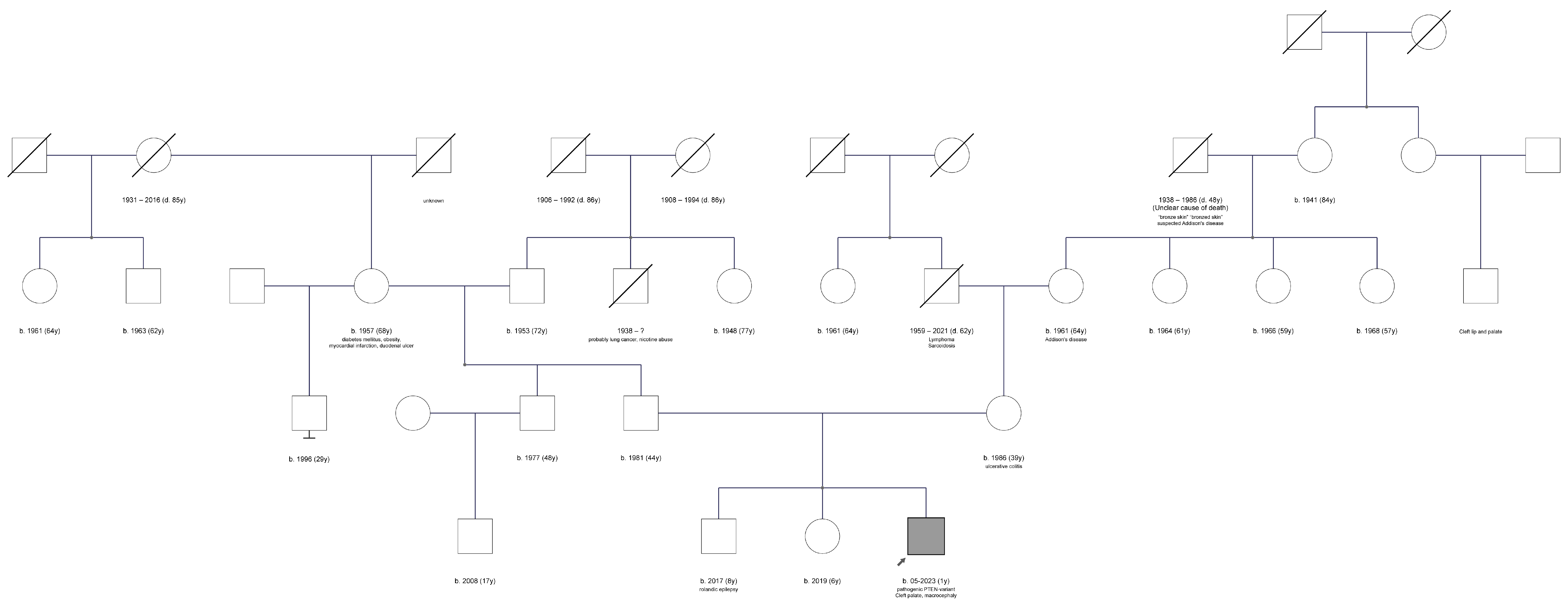
The patient is the third child of non-consanguineous parents. His 6-year-old brother had suffered from a single epileptic seizure with suspected self-limited focal epilepsy of childhood (“Rolandic”). The 4-year-old sister and the father are healthy, while the mother has ulcerative colitis, and a cousin of the maternal grandmother had a cleft palate and lip (Figure 1).
Figure 1.
Pedigree of the family obtained during genetic counseling.
Postnatal respiratory adaptation was prolonged and complicated by hypoglycemic episodes. A cleft soft palate was detected shortly after birth, and, together with macrocephaly, prompted genetic counseling.
Genetic testing was performed due to the heterogeneous symptoms of the patient (e.g., macrocephaly, cleft palate). All protein-coding genes were assessed (i.e., whole-exome sequencing (WES)) with a specific emphasis on genes with a known disease association. Variants were filtered based on impact on protein structure, prevalence in the general population, and evidence in the literature. Whole-exome sequencing with subsequent segregation analysis of the parents revealed a heterozygous, de novo, null variant c.184A>T, p.(Lys62*) in the PTEN gene. The variant has previously been reported as pathogenic (ClinVar-ID: 1069915).
The transcript NM_000314.8 was used. The variant was classified as pathogenic based on the following ACMG criteria: PVS1, PS2_MOD, PS4_SUP, and PM2. The variant has been submitted to ClinVar twice, with one submission being this specific case (SCV004812067). The variant has hitherto not been reported in the literature. However, reports of other loss-of-function/null variants have been published.
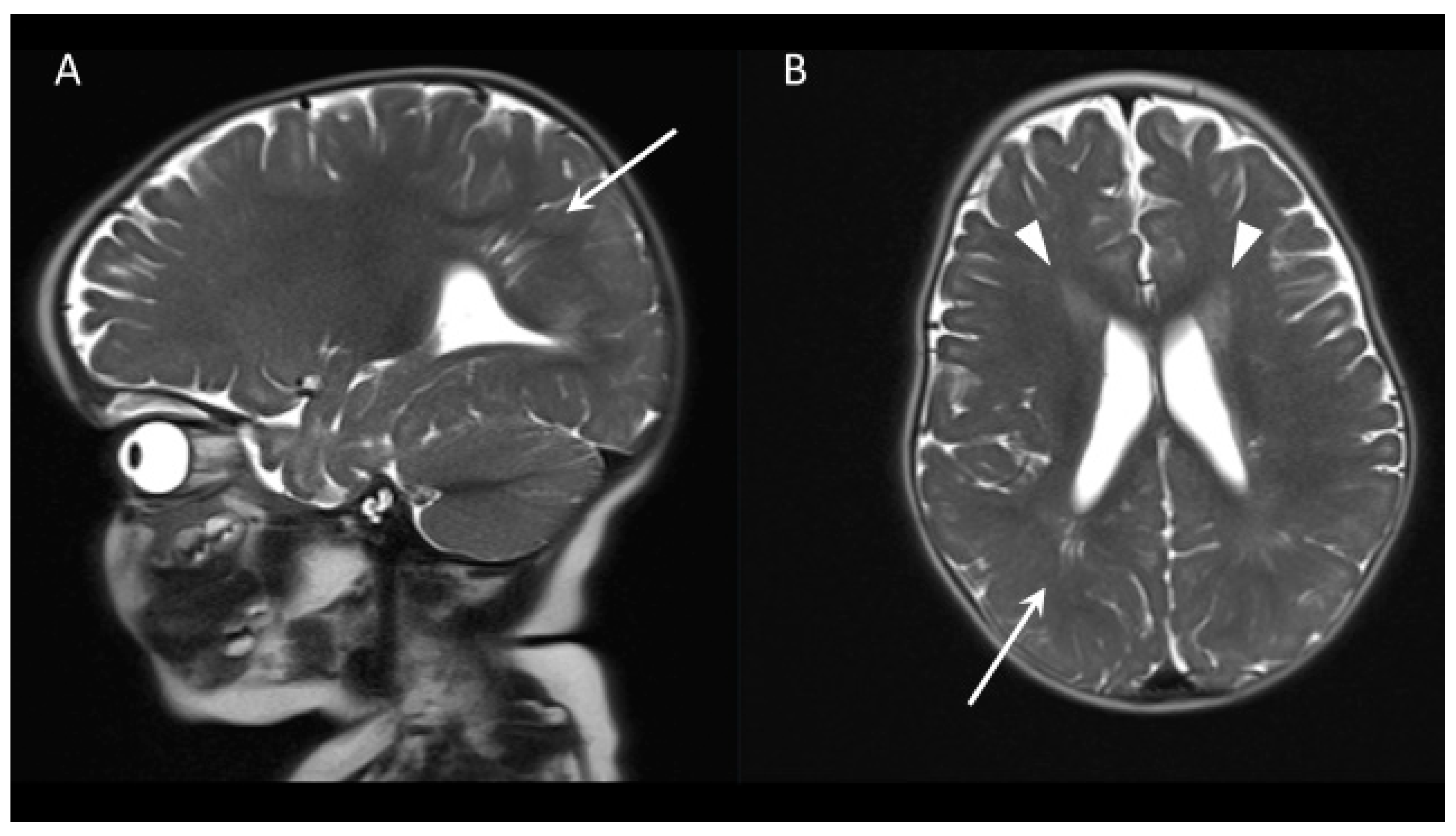
The child was referred to an oromaxillofacial surgeon, who scheduled surgical closure at 10 months and initiated magnetic resonance imaging (MRI) to rule out hydrocephalus before surgery (Figure 2). Examination at 10 months revealed brown skin patches on both knees and soles, without penile freckling. The head circumference was 50 cm (2.8 z), and no partial overgrowth was observed. A persistent foramen ovale and ductus arteriosus, and mild mitral insufficiency were noted. Despite starting to roll over at 6 months of age, the boy was showing mild motor development delay, not yet being able to sit. A detailed diagnostic work-up for PHTS-related symptoms, including thyroid abnormalities, gastrointestinal polyposis, and autism spectrum disorder, was planned according to German guidelines [19].
Figure 2.
Cranial MRI at 10 months of age. T2-weighted fast spin-echo sequence in (A) sagittal and (B) axial orientation. Right occipital stria-like enlarged Virchow–Robin spaces (arrow). T2-hyperdensities rostral to the ventricles (arrowheads).
3. Discussion
PTEN hamartoma tumor syndrome presents a diagnostic challenge. On one hand, patients have a tumor predisposition, with a 6% risk of developing a malignant tumor in childhood. On the other hand, screening for cancer or benign neoplasms in young children poses additional risks due to repeated anesthesia or invasive procedures. Thus, the appropriate timing of routine, guideline-driven diagnostic evaluations remains a matter of debate [18].
Macrocephaly is often an obvious symptom and can be detected during prenatal obstetric ultrasound examinations, whereas, in our case, this was additionally complicated by polyhydramnios. Routine sonographic assessment should be scheduled every 10th week of gestation. From the second trimester onward, fetal MRI can help in detailing anatomic structures when ultrasonography remains equivocal. After birth, transfontanellar ultrasonography and cerebral MRI are indicated to rule out the indication for a neurosurgical intervention, such as in hemorrhage, hydrocephalus, or tumors.
Although MRI findings are characteristic and present in up to 40% of patients, they are not specific to PHTS (Figure 2) [20]. Hydrocephalus in the setting of cranial dysmorphism in pediatric PHTS can result from Chiari malformation or arise as secondary to dysplastic cerebellar gangliocytoma in Lhermitte–Duclos disease. However, in the review of the 159 patients in Table 1, only one ventriculo-peritoneal shunt procedure for pseudotumor cerebri was reported (0.6%) [13].
Skin stigmata, such as lipoma, hemangioma or hamartoma, trichilemmoma, and café au lait spots are detectable by inspection. These signs are generally well documented; in the literature reviewed for Table 1, examination results are given for 155 out of 159 patients. The occurrence of subcutaneous lipoma, which is a rare finding in children, particularly when combined with macrocephaly, should raise suspicion of PHTS and prompt genetic testing [5].
Reports of PTEN-associated cleft palate are scarce, although orofacial dysmorphism and a high-arched palate are present in 27% to 34% of cases, according to the references included in Table 1. A single case of bifid uvula was described in 2007 [14]. Considering that up to 31% of cases present with oral mucosal papillomatosis and that oral hyperkeratosis has been described as part of Cowden syndrome, oral examination is an important adjunct in diagnosing PHTS [21]. However, our overview suggests that oral signs are less frequently described in the diagnostic process compared to the more prominent symptoms, with oral items reported in only 24 cases from 159 observations (Table 1).
It remains elusive whether this case represents a causal or casual coincidence. A genetic link between our patient’s cleft palate and that of the maternal grandmother’s cousin is unlikely due to the distant relationship and the frequency of cleft palates in the general population (1–2:1000). Given the clinical symptoms and standardized variant classification, the identified variant in PTEN was the only variant of clinical significance. It is absent from the general population, and its type is a stop-gain variant, which means that no functional protein product is translated (i.e., null variant).
The development of cleft palate is complex and multifactorial. There are a number of genetic diseases with an increased risk, such as 22q11.2 deletion syndrome, Stickler syndrome, or Beckwith–Wiedemann syndrome. PTEN is a major lipid phosphatase that downregulates the PI3K/AKT pathway to cause G1 cell cycle arrest and apoptosis. When PTEN is absent, decreased, or dysfunctional, the phosphorylation of AKT1 is uninhibited, leading to the inability to activate cell cycle arrest and/or to undergo apoptosis.
In addition, through a lack of protein phosphatase activity, the mitogen-activated protein kinase (MAPK) pathway is dysregulated, leading to abnormal cell survival [22]. This dysregulation could have an influence on the connection of the palatal arches and thus could have contributed to the development of cleft palate. Since this is the first report on the association of a cleft palate with a PTEN mutation, an estimation of the resulting tumor risk or phenotype expression pattern cannot be derived from the literature.
In the present case, genetic testing not only identified the cause of the existing abnormalities but also resulted in recommendations for follow-up examinations, particularly with regard to the increased risk of tumors in the patient (see https://www.ncbi.nlm.nih.gov/books/NBK1488/#phts.Management, accessed on 9 January 2025). By testing the parents, a statement could be made about the risk of PTEN disease in further children. As the variant is probably new (de novo), the risk of recurrence is low (<1%). However, a germ cell mosaic cannot be ruled out. The clinical picture, inheritance, the risk of recurrence, and follow-up examinations were explained to the parents during genetic counseling.
Data collection and analysis in PHTS are limited by the retrospective nature of most case reports and reviews. The data do not allow us to predict tumor risk during infancy or to estimate how many patients live with a pathogenic variant in PTEN without ever requiring medical attention. We advocate for broad genetic testing like WES with a low threshold of suspicion in order to anticipate developmental delays, potentially detect tumor growth at an early stage, and support patients and families in managing this rare, burdensome disease.
Author Contributions
Conceptualization, U.N. and A.M.; methodology, R.-T.J.; software, R.-T.J.; validation, U.N., D.G. and V.S.; formal analysis, F.W.; investigation, A.S. and A.M.; data curation, A.M. and A.S.; writing—original draft preparation, U.N.; writing—review and editing, D.G. and V.S.; visualization, D.G.; supervision, F.W. All authors have read and agreed to the published version of the manuscript.
Funding
This research received no external funding.
Institutional Review Board Statement
Ethical review and approval were not required for this study as per DSGVO (https://dsgvo-gesetz.de/, accessed on 13 November 2024) articles 6(1) a), 9(2) a) and j) and due to the retrospective data collection and anonymized presentation of the report.
Informed Consent Statement
The mother of the patient has given written consent regarding the scientific publication of the case.
Data Availability Statement
The original contributions presented in this study are included in the article material. Further inquiries can be directed to the corresponding author.
Conflicts of Interest
The authors declare no conflicts of interest.
References
- Leibowitz, M.S.; Zelley, K.; Adams, D.; Brodeur, G.M.; Fox, E.; Li, M.M.; Mattei, P.; Pogoriler, J.; MacFarland, S.P. Neuroblastoma and cutaneous angiosarcoma in a child with PTEN hamartoma tumor syndrome. Pediatr. Blood Cancer 2022, 69, e29656. [Google Scholar] [CrossRef] [Scilit]
- Macken, W.L.; Tischkowitz, M.; Lachlan, K.L. PTEN Hamartoma tumor syndrome in childhood: A review of the clinical literature. Am. J. Med. Genet. C Semin Med. Genet. 2019, 181, 591–610. [Google Scholar] [CrossRef] [Scilit] [PubMed]
- Martinez-Rios, C.; De Leon Benedetti, L.S.; Tierradentro-Garcia, L.O.; Kilicarslan, O.A.; Caro-Dominguez, P.; Otero, H.J. Imaging findings of children with PTEN-related hamartoma tumor syndrome: A 20-year multicentric pediatric cohort. Pediatr. Radiol. 2024, 54, 1116–1127. [Google Scholar] [CrossRef] [Scilit] [PubMed]
- Lloyd, K.M., II; Dennis, M. Cowden’s disease. A possible new symptom complex with multiple system involvement. Ann. Intern. Med. 1963, 58, 136–142. [Google Scholar] [CrossRef] [Scilit]
- Martin, H.; Bessis, D.; Bourrat, E.; Mazereeuw-Hautier, J.; Morice-Picard, F.; Balguerie, X.; Chiaverini, C. Cutaneous lipomas and macrocephaly as early signs of PTEN hamartoma tumor syndrome. Pediatr. Dermatol. 2020, 37, 839–843. [Google Scholar] [CrossRef] [Scilit]
- Russell, K.A.; Allen, V.M.; MacDonald, M.E.; Smith, K.; Dodds, L. A population-based evaluation of antenatal diagnosis of orofacial clefts. Cleft Palate Craniofac. J. 2008, 45, 148–153. [Google Scholar] [CrossRef] [Scilit] [PubMed]
- Hammond, N.L.; Dixon, M.J. Revisiting the embryogenesis of lip and palate development. Oral Dis. 2022, 28, 1306–1326. [Google Scholar] [CrossRef] [Scilit]
- Sansal, I.; Sellers, W.R. The biology and clinical relevance of the PTEN tumor suppressor pathway. J. Clin. Oncol. 2004, 22, 2954–2963. [Google Scholar] [CrossRef] [Scilit] [PubMed]
- Nestler, U.; Gräfe, D.; Strehlow, V.; Jauss, R.-T.; Merkenschlager, A.; Schönfeld, A.; Wilhelmy, F. A Newborn with Cleft Palate Associated with PTEN Hamartoma Tumor Syndrome. Authorea 2024. [Google Scholar] [CrossRef] [Scilit]
- Bregvadze, K.; Jabeen, S.; Rafi, S.M.; Tkemaladze, T. The complexity of phosphatase and tensin homolog hamartoma tumor syndrome: A case report. SAGE Open Med. Case Rep. 2024, 12, 2050313X241245317. [Google Scholar] [CrossRef] [Scilit]
- Martín-Valbuena, J.; Gestoso-Uzal, N.; Justel-Rodríguez, M.; Isidoro-García, M.; Marcos-Vadillo, E.; Lorenzo-Hernández, S.M.; Criado-Muriel, M.C.; Prieto-Matos, P. PTEN hamartoma tumor syndrome: Clinical and genetic characterization in pediatric patients. Childs Nerv. Syst. 2024, 40, 1689–1697. [Google Scholar] [CrossRef] [Scilit]
- Ciaccio, C.; Saletti, V.; D’Arrigo, S.; Esposito, S.; Alfei, E.; Moroni, I.; Tonduti, D.; Chiapparini, L.; Pantaleoni, C.; Milani, D. Clinical spectrum of PTEN mutation in pediatric patients. A bicenter experience. Eur. J. Med. Genet. 2019, 62, 103596. [Google Scholar] [CrossRef] [Scilit] [PubMed]
- Plamper, M.; Gohlke, B.; Schreiner, F.; Woelfle, J. Phenotype-Driven Diagnostic of PTEN Hamartoma Tumor Syndrome: Macrocephaly, But Neither Height nor Weight Development, Is the Important Trait in Children. Cancers 2019, 11, 975. [Google Scholar] [CrossRef] [Scilit]
- Yotsumoto, Y.; Harada, A.; Tsugawa, J.; Ikura, Y.; Utsunomiya, H.; Miyatake, S.; Matsumoto, N.; Kanemura, Y.; Hashimoto-Tamaoki, T. Infantile macrocephaly and multiple subcutaneous lipomas diagnosed with PTEN hamartoma tumor syndrome: A case report. Mol. Clin. Oncol. 2020, 12, 329–335. [Google Scholar] [CrossRef] [Scilit]
- Kato, K.; Mizuno, S.; Inaba, M.; Fukumura, S.; Kurahashi, N.; Maruyama, K.; Ieda, D.; Ohashi, K.; Hori, I.; Negishi, Y.; et al. Distinctive facies, macrocephaly, and developmental delay are signs of a PTEN mutation in childhood. Brain Dev. 2018, 40, 678–684. [Google Scholar] [CrossRef] [Scilit]
- Hansen-Kiss, E.; Beinkampen, S.; Adler, B.; Frazier, T.; Prior, T.; Erdman, S.; Eng, C.; Herman, G. A retrospective chart review of the features of PTEN hamartoma tumour syndrome in children. J. Med. Genet. 2017, 54, 471–478. [Google Scholar] [CrossRef]
- Busa, T.; Milh, M.; Degardin, N.; Girard, N.; Sigaudy, S.; Longy, M.; Olshchwang, S.; Sobol, H.; Chabrol, B.; Philip, N. Clinical presentation of PTEN mutations in childhood in the absence of family history of Cowden syndrome. Eur. J. Paediatr. Neurol. 2015, 19, 188–192. [Google Scholar] [CrossRef] [Scilit] [PubMed]
- Smpokou, P.; Fox, V.L.; Tan, W.H. PTEN hamartoma tumour syndrome: Early tumour development in children. Arch. Dis. Child 2015, 100, 34–37. [Google Scholar] [CrossRef] [Scilit] [PubMed]
- Plamper, M.; Gohlke, B.; Woelfle, J. PTEN hamartoma tumor syndrome in childhood and adolescence-a comprehensive review and presentation of the German pediatric guideline. Mol. Cell Pediatr. 2022, 9, 3. [Google Scholar] [CrossRef] [Scilit]
- Plamper, M.; Born, M.; Gohlke, B.; Schreiner, F.; Schulte, S.; Splittstößer, V.; Woelfle, J. Cerebral MRI and Clinical Findings in Children with PTEN Hamartoma Tumor Syndrome: Can Cerebral MRI Scan Help to Establish an Earlier Diagnosis of PHTS in Children? Cells 2020, 9, 1668. [Google Scholar] [CrossRef] [Scilit]
- Fardal, Ø.; Nevland, K.; Johannessen, A.C.; Vetti, H.H. The PTEN hamartoma tumor syndrome: How oral clinicians may save lives. Clin. Adv. Periodontics. 2023, 13, 21–26. [Google Scholar] [CrossRef] [Scilit]
- Yehia, L.; Ni, Y.; Sadler, T.; Frazier, T.W.; Eng, C. Distinct metabolic profiles associated with autism spectrum disorder versus cancer in individuals with germline PTEN mutations. NPJ Genom. Med. 2022, 7, 16. [Google Scholar] [CrossRef] [Scilit]
Disclaimer/Publisher’s Note: The statements, opinions and data contained in all publications are solely those of the individual author(s) and contributor(s) and not of MDPI and/or the editor(s). MDPI and/or the editor(s) disclaim responsibility for any injury to people or property resulting from any ideas, methods, instructions or products referred to in the content. |
© 2025 by the authors. Licensee MDPI, Basel, Switzerland. This article is an open access article distributed under the terms and conditions of the Creative Commons Attribution (CC BY) license (https://creativecommons.org/licenses/by/4.0/).