Case Report

Liver Cirrhosis and Hepatocellular Carcinoma Diagnosed from Chylothorax: A Case Report

1
Department of Respiratory Medicine, Maebashi Red Cross Hospital, 389-1, Asakura-Machi, Maebashi 371-0811, Japan
2
Department of Gastroenterology, Maebashi Red Cross Hospital, 389-1, Asakura-Machi, Maebashi 371-0811, Japan
*
Author to whom correspondence should be addressed.
Clin. Pract. 2021, 11(3), 582-586; https://doi.org/10.3390/clinpract11030073
Submission received: 13 July 2021 / Revised: 24 August 2021 / Accepted: 31 August 2021 / Published: 3 September 2021

Abstract

:
A 71-year-old man visited our hospital with dyspnea and left pleural effusion. Left pleural effusion was diagnosed as chylothorax by thoracentesis. He had no history of trauma or surgery, and there were no findings of malignant lymphoma or thrombosis. Furthermore, he was diagnosed with liver cirrhosis and hepatocellular carcinoma by computed tomography and hematological examinations, and the chylothorax was considered to be caused by liver cirrhosis. We report a review of the literature with this case since it is relatively rare for cirrhosis and hepatocellular carcinoma diagnosed from chylothorax.

1. Introduction

There are many causes of pleural effusion, including infection, malignant tumors, heart failure, and trauma. Chylothorax is a condition caused by a disruption of the thoracic duct, resulting in leakage of chyle into the thoracic cavity [1]. It is characterized by a cloudy nature rich in triglycerides. Chylothorax is caused by trauma or surgery in many cases. However, in the absence of these physical causes, elucidation of the cause is essential.
Here, we report a case of cirrhosis and hepatocellular carcinoma diagnosed from chylothorax and review the relevant literature.

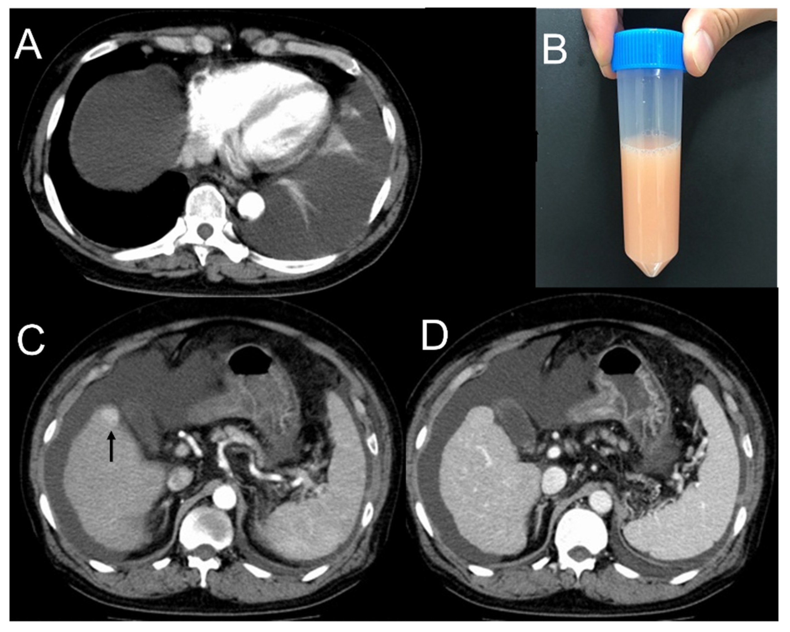
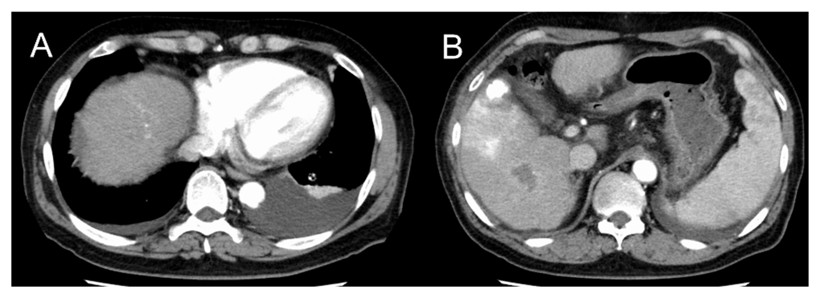
2. Case Presentation

A 71-year-old man visited our hospital with a chief complaint of dyspnea for several days without other symptoms such as fever, cough, phlegm, or chest pain. He is a heavy drinker and had a history of diabetes mellitus and hypertension, but did not have a history of heart disease, respiratory disease, trauma, or surgery. Chest computed tomography (CT) showed a left pleural effusion and ascites (Figure 1A). There was no obvious pulmonary opacity suggestive of a tumor or infection. Serum white blood cell count or brain natriuretic hormone (BNP) were not increased. Laboratory findings did not show any evidence of infection or heart failure (Table 1). He was negative for hepatitis B virus antigen and hepatitis C virus antibody. Pleural effusion from thoracentesis was white and cloudy with high triglyceride levels, consistent with chylothorax (Figure 1B). The nature of pleural effusion was transudative, and cytology was negative for malignancy.
Abdominocentesis was performed, and milky-like ascites were considered to be chylous ascites. Lymphoscintigraphy did not reveal any apparent areas of leakage. Contrast-enhanced chest and abdominal CT revealed an irregular liver surface, consistent with cirrhosis, and a 2-cm-large mass in liver S6 with enhanced arterial phase, diagnosed with hepatocellular carcinoma (Figure 1C,D). There were no other findings suggestive of tumors or thrombosis in the portal vein. He was diagnosed with alcoholic cirrhosis because he was negative for the hepatitis virus and had a heavy alcohol history. The patient was diagnosed with chylothorax and chylous ascites due to liver cirrhosis.
The pleural and ascitic effusion showed a marked reduction with the treatment of furosemide, spironolactone, and propranolol for two months without recurrence (Figure 2). The hepatocellular carcinoma, stage I, was treated with both radiofrequency ablation and transarterial chemoembolization, and the patient has been in remission.

3. Discussion

Chylothorax is a condition caused by a disruption of the thoracic duct and leakage of chyle into the thoracic cavity [1]. The non-traumatic causes of chylothorax include tumors and thrombosis, and malignant lymphoma is the most common cause of malignant disease [2]. Chylothorax due to malignant lymphoma is caused by the reflux of chyle due to the increased pressure in the thoracic duct. It can also be caused by a rupture of the thoracic duct due to vascular invasion of the lymphoma [3]. Liver cirrhosis, congestive heart failure, and nephrotic syndrome are also common causes of transudative chylothorax. In liver cirrhosis, increased portal pressure causes microscopic lymphatic destruction of intra-abdominal organs and the mesentery, resulting in chylous ascites. A mechanism has been proposed in which this fluid flows into the thoracic cavity through a diaphragmatic defect and appears as chylothorax [4]. Most cases of cirrhosis associated with chylothorax have been reported to be caused by viruses or alcohol, and there are few reports of nonalcoholic steato-hepatitis [4]. In a study including patients with cirrhosis-associated hepatic hydrothorax, 77% of the patients had only right-sided effusion, and 17% had only left-sided effusion [5]. The mechanism of the laterality of cirrhosis-associated chylothorax has not been well understood. However, all previously described cases of chylothorax have been reported as laterality of the right side [4,6,7]. This was similar to normal hepatic hydrothorax. The reason for the left-sided pleural effusion, in this case, cannot be clearly explained.
Treatment of non-traumatic chylothorax includes a low-fat diet, thoracic drainage, and thoracic duct embolization. Treatment of the primary disease is also essential in secondary chylothorax. Higuchi et al. reported that propranolol, a β-blocker, was administered to lower portal pressure in patients with cirrhosis. This resulted in an improvement of the chylothorax [8]. In patients with compensated cirrhosis and portal hypertension, it has been reported that lowering the portal-hepatic venous pressure gradient with β-blockers reduces the incidence of ascites. This prevents progression to non-compensated cirrhosis [9]. In our study, propranolol was administered to the patient in accordance with previous reports, and the chylothorax was reduced. Although other drugs may also be effective, β-blockers may effectively treat chylothorax associated with cirrhosis. It has been reported that transjugular intrahepatic portosystemic shunting was effective in the treatment of chylothorax associated with liver cirrhosis [10]. Tolvaptan has also been reported to be effective in the treatment of ascites associated with liver cirrhosis [11]. If his pleural and ascites effusions become more difficult to control, it may be considered as an additional treatment.
In this case, we could diagnose cirrhosis, child-pugh grade B, together with hepatocellular carcinoma as the cause of chylothorax. Romero et al. reported a case of chylothorax diagnosed with hepatocellular carcinoma two months before the patient died of the hepatorenal syndrome [12]. However, there are no reports of hepatocellular carcinoma diagnosed on occasion with chylothorax. Chronic lymphocytic leukemia, primary lung carcinoma, and metastatic lung carcinoma are also known as the causes of chylothorax [2]. However, the coexistence of chylothorax with intra-abdominal parenchymal tumors has not been well known. When hepatocellular carcinoma invades directly into the portal vein, it is considered that the hepatocellular carcinoma itself may cause portal hypertension. However, no such finding was observed in this case. It is not clear to what extent hepatocellular carcinoma affected chylothorax associated with cirrhosis. Although liver cirrhosis rather than hepatocellular carcinoma may be more likely associated with chylothorax in our case, the presence of hepatocellular carcinoma in the case of cirrhosis with chylothorax should be considered.

4. Conclusions

When we diagnose transudative chylothorax, we should consider the complication of liver cirrhosis. In addition, the possibility of the presence of hepatocellular carcinoma should be kept in mind.

Author Contributions

Conceptualization, data curation and visualization, K.I., Y.H.; investigation, K.I., Y.H. and M.S.; writing original draft, K.I., Y.H., M.S., K.E., H.I., A.J., H.A., T.H., A.T.; supervision, H.A., A.T.; All authors have read and agreed to the published version of the manuscript.

Funding

This research received no external funding.

Institutional Review Board Statement

Not applicable.

Informed Consent Statement

Written informed consent has been obtained from the patient to publish this paper.

Conflicts of Interest

The authors declare no conflict of interest.

References

  1. Romero, S. Nontraumatic chylothorax. Curr. Opin. Pulm. Med. 2000, 6, 287–291. [Google Scholar] [CrossRef] [PubMed]
  2. Doerr, C.H.; Allen, M.S.; Nichols, F.C., 3rd; Ryu, J.H. Etiology of chylothorax in 203 patients. Mayo Clin. Proc. 2005, 80, 867–870. [Google Scholar] [CrossRef] [PubMed]
  3. Janjetovic, S.; Janning, M.; Daukeva, L.; Bokemeyer, C.; Fiedler, W. Chylothorax in a patient with Hodgkin’s lymphoma: A case report and review of the literature. Tumori J. 2013, 99, e96–e99. [Google Scholar] [CrossRef]
  4. Tsauo, J.; Shin, J.H.; Han, K.; Yoon, H.-K.; Ko, G.-Y.; Ko, H.-K.; Gwon, D.-I. Transjugular Intrahepatic Portosystemic Shunt for the Treatment of Chylothorax and Chylous Ascites in Cirrhosis: A Case Report and Systematic Review of the Literature. J. Vasc. Interv. Radiol. 2016, 27, 112–116. [Google Scholar] [CrossRef] [PubMed]
  5. Badillo, R.; Rockey, D.C. Hepatic hydrothorax: Clinical features, management, and outcomes in 77 patients and review of the literature. Medicine 2014, 93, 135–142. [Google Scholar] [CrossRef] [PubMed]
  6. Diaz-Guzman, E.; Culver, D.A.; Stoller, J.K. Transudative chylothorax: Report of two cases and review of the literature. Lung 2005, 183, 169–175. [Google Scholar] [CrossRef] [PubMed] [Green Version]
  7. Kinney, T.B.; Ferrara, S.L.; Miller, F.J.; Roberts, A.C.; Hassanein, T. Transjugular intrahepatic portosystemic shunt creation as treatment for refractory chylous ascites and chylothorax in a patient with cirrhosis. J. Vasc. Interv. Radiol. 2004, 15 Pt 1, 85–89. [Google Scholar] [CrossRef]
  8. Higuchi, Y.; Tomizawa, M.; Kakegawa, S.; Suzuki, K.; Ishihara, S.; Kobayashi, H. A case of chylothorax associated with liver cirrhosis successfully treated by β-blocker. AJRS 2014, 3, 70–74. (In Japanese) [Google Scholar]
  9. Villanueva, C.; Albillos, A.; Genescà, J.; Garcia-Pagan, J.C.; Calleja-Panero, J.L.; Aracil, C.; Bañares, R.; Morillas, R.M.; Poca, M.; Peñas, B.; et al. β blockers to prevent decompensation of cirrhosis in patients with clinically significant portal hypertension (PREDESCI): A randomised, double-blind, placebo-controlled, multicentre trial. Lancet 2019, 393, 1597–1608. [Google Scholar] [CrossRef]
  10. Khaliq, M.F.; Noorani, M.M.; Chowdhry, M.; Mohamed, H.; Koirala, A. Transjugular Intrahepatic Portosystemic Shunt (TIPS) in Refractory Transudative Chylothorax due to Liver Cirrhosis. Case Rep. Med. 2020, 2020, 2581040. [Google Scholar] [CrossRef] [PubMed]
  11. Yan, L.; Xie, F.; Lu, J.; Ni, Q.; Shi, C.; Tang, C.-X.; Yang, J. The treatment of vasopressin V2-receptor antagonists in cirrhosis patients with ascites: A meta-analysis of randomized controlled trials. BMC Gastroenterol. 2015, 15, 65. [Google Scholar] [CrossRef] [PubMed] [Green Version]
  12. Romero, S.; Martín, C.; Hernandez, L.; Verdu, J.; Trigo, C.; Perez-Mateo, M.; Alemany, L. Chylothorax in cirrhosis of the liver: Analysis of its frequency and clinical characteristics. Chest 1998, 114, 154–159. [Google Scholar] [CrossRef] [PubMed] [Green Version]
Figure 1. Imaging of chylothorax and computed tomography. (A) Computed tomography showed left pleural effusion and ascites. (B) The color of pleural effusion obtained by thoracentesis was milky white. (C,D) Contrast-enhanced computed tomography showed a 2-cm mass in the liver S6 that was enhanced in the arterial phase and washed out in the portal phase.
Clinpract 11 00073 g001
Figure 2. Imaging after two months of treatment for hepatocellular carcinoma. (A) Left pleural effusion and ascites have decreased compared to pretreatment. (B) There is lipiodol deposition coincident with the mass and no recurrence of hepatocellular carcinoma.
Clinpract 11 00073 g002
Table 1. Laboratory data at the first visit.
Hematology Biochemistry Plural Effusion
White blood cells2700/μLTotal protein5.8g/dLColorMilky white
Neutrophils70.3%Albumin2.9g/dLpH8.5
Lymphocytes13.3%Total bilirubin1.1mg/dLWhite blood cells500/μL
Monocytes12.3%Direct bilirubin0.1mg/dLNeutrophils5.0%
Eosinophils3.7%AST38U/LLymphocytes60.0%
Basophils0.4%ALT33U/LMonocytes33.0%
Red blood cells346 × 104/μLLDH213U/LEosinophils2.0%
Hemoglobin8.7g/dLALP121U/LHematocrit0.1%
Hematocrit28.2%γ-GTP42U/LTotal protein1.6g/dL
Platelets30.4 × 104/μLCK153U/LLDH66U/L
BUN11mg/dLGlucose189mg/dL
Coagulation Creatinine0.74mg/dLTotal cholesterol30mg/dL
PT%64%Sodium139mEq/LTriglyceride227mg/dL
PT-INR1.25 Potassium3.6mEq/LADA9U/L
APTT28.9secChloride107mEq/L
Fibrinogen225mg/dLCalcium7.8mg/dLCytology (−)
D-dimer13.3μg/mLGlucose198mg/dL
HbA1c5.9%Culture
Tumor marker Total cholesterol99mg/dLRoutine (−)
AFP69.7ng/mLLDL-cholesterol70mg/dLMycobacterium (−)
CEA1.5ng/mLHDL-cholesterol26mg/dL
CA19-936.9U/mLTriglyceride56mg/dL
Soluble IL-2 receptor842U/mLBNP34.4pg/mL
CRP1.46mg/dL
Virus marker ANA40titer
HBV antigen(−)
HCV antibody(−)
PT: Prothrombin time, APTT: Activated partial thromboplastin time, AFP: α-fetoprotein, CEA: Carcinoembryonic antigen, CA19-9: Carbohydrate antigen 19-9, HBV: Hepatitis B virus, HCV: Hepatitis C virus, AST: Aspartate aminotransferase, ALT: Alanine aminotransferase, LDH: Lactate dehydrogenase, ALP: Alkaline phosphatase, γ-GTP: γ-glutamyl transpeptidase, CK: Creatine kinase, BUN: Blood urea nitrogen, HbA1c: Hemoglobin A1c, LDL: Low-density lipoprotein, HDL: High-density lipoprotein, BNP: Brain natriuretic peptide, CRP: C-reactive protein, ANA: Anti-nuclear antibody, ADA: Adenosine deaminase.
Publisher’s Note: MDPI stays neutral with regard to jurisdictional claims in published maps and institutional affiliations.

