Review

Virgin Coconut Oil and Its Lauric Acid, Between Anticancer Activity and Modulation of Chemotherapy Toxicity: A Review

by
Debalina Bose
1,
Adetayo Olorunlana
2,
Rania Abdel-Latif
3,
Ademola C. Famurewa
4,5,* and
Eman M. Othman
6,7,*
1
Advanced Technology Development Centre, Indian Institute of Technology Kharagpur, Kharagpur 721302, India
2
Department of Nursing, College of Nursing and Basic Medical Sciences, Caleb University, Lagos 100211, Nigeria
3
Department of Pharmacology and Toxicology, Faculty of Pharmacy, Minia University, Minia 61519, Egypt
4
Department of Medical Biochemistry, Faculty of Basic Medical Sciences, College of Medical Sciences, Alex Ekwueme Federal University, Ndufu-Alike Ikwo, Abakaliki 482131, Nigeria
5
Centre for Natural Products Discovery, School of Pharmacy and Biomolecular Sciences, Faculty of Science, Liverpool John Moores University, Byrom Street, Liverpool L3 3AF, UK
6
Department of Biochemistry, Faculty of Pharmacy, Minia University, Minia 61519, Egypt
7
Cancer Therapy Research Center (CTRC), Department of Biochemistry-I, Biocenter, University of Wuerzburg, Theodor-Boveri-Weg 1, 97074 Wuerzburg, Germany
*
Authors to whom correspondence should be addressed.
J. Xenobiot. 2025, 15(4), 126; https://doi.org/10.3390/jox15040126
Submission received: 16 May 2025 / Revised: 4 June 2025 / Accepted: 17 July 2025 / Published: 5 August 2025

Abstract

Virgin coconut oil (VCO) has emerged as a functional food oil with considerable health benefits and wide applications in the food, pharmaceutical, and cosmetic industries due to its resident bioactive compounds, including lauric acid (LA). LA is the most abundant saturated medium-chain fatty acid in VCO and has been associated with several pharmacological activities. The literatures show the pharmacological effects of VCO and LA on chronic pathologies, infectious diseases, and metabolic disorders. A robust body of evidence shows that LA and other phenolic compounds are responsible for the VCO protection against toxicities and pharmacological efficacies. This review elucidates the anticancer mechanisms of VCO/LA and their modulation of the chemotherapy-induced side effect toxicity. VCO, LA, and their nanomaterial/encapsulated derivatives promote ROS generation, antiproliferation, apoptosis, cell cycle arrest, the inhibition of metastasis, and the modulation of cancer-related signaling pathways for cancer cell death in vivo and in vitro. VCO mitigates oxidative inflammation and apoptosis to block the underlying mechanisms of the side effect toxicity of chemotherapy. However, the possible beneficial effect of LA on the toxicity of chemotherapy is currently unknown. The available evidence emphasizes the anticancer effect and mechanism of VCO and LA, and the VCO potential to combat adverse side effects of chemotherapy. Thus, VCO and LA are potential adjuvant therapeutic agents in the management of various cancers. Nevertheless, future studies should be targeted at elucidating cancer-related molecular mechanisms to bridge the gap in knowledge.

1. Introduction

Cancer continues to pose a significant global health challenge with increasing prevalence despite the Food and Drug Administration (FDA) approval of more than 600 cytotoxic anticancer agents [1]. Chemotherapy is the clinical cornerstone for metastatic cancer treatment globally. It is acknowledged for systemic efficacy, patient improvement, and survival; however, it is increasingly confronted with severe side effects, chemoresistance, and relapses [2,3]. These formidable hurdles hamper clinical chemotherapy effectiveness; therefore, there is an unmet need for alternative therapies, adjuvants, or combination regimens to combat the growing health problem [4]. In this pursuit, there is a growing research interest in exploring the potential therapeutic benefits of natural products for cancer treatment. The multi-target novel adjuvants or treatment agents based on natural products with higher anticancer potency and low toxicity profiles are required for the treatment of cancers [4,5]. Some plant-derived natural products have shown potential anticancer potential in combination therapies, though safety and efficacy must be assessed case by case [6]. Among these potential products is VCO gradually emerging as a potential candidate due to its potential anticancer properties and ability to mitigate the toxicity associated with chemotherapy [7,8]. VCO is extracted from the kernel or meat of mature coconutsand has been recognized for its diverse health benefits [9]. There is a growing demand for VCO in the United Sates and other developed countries, and this demand can be attributed to the increasing number of reports and scientific literature published on the health benefits of VCO [10]. The quantity of VCO exported by India, Philippines, Indonesia, West Malaysia, Sri Lanka, Mexico, and Papua New Guinea, the major countries involved in the large production of VCO, has increased over the years [10,11,12]. VCO is a natural extract of the fresh, mature kernel (meat) of coconuts without the use of heat, without undergoing chemical, refining, bleaching, or deodorizing processes [10]. The literature has shown the analgesic, immunomodulatory, antipyretic, antioxidant, anti-stress, antimicrobial, anti-HIV, antidiabetic, antiviral, anti-inflammatory, anti-obesity, hypocholesterolemia, and anticancer effects of VCO [11,13,14]. A number of studies have also demonstrated the VCO capacity to inhibit the toxicity induced by clinical drugs and various chemicals [15,16,17,18]. In particular, the anticancer drugs used in chemotherapy induce various side effect toxicities in cancer patients [19,20]. Preclinical evidence reveals that it exerts toxicity on the liver, kidney, heart, testis, placenta, brain, lung, ovary, intestine, blood, and thyroid gland [19,20,21,22]. The underlying mechanisms of toxicity strongly reported by experimental studies include oxidative stress, pro-inflammation, apoptosis, and mitochondrial dysfunction [23]. But a number of findings in animal studies indicate that VCO may exert beneficial effects against the toxicity of chemotherapeutic drugs [22,23,24,25]. The beneficial effects are attributed to its unique composition of medium-chain triglycerides (MCTs), medium-chain fatty acids (MCFAs), phenolic compounds, and other bioactive molecules [11,26].
Notably, it is LA that constitutes a substantial portion of the VCO fatty acid profile, and there is a renewed focus on LA in cancer research circles. LA, the most abundant MCFA in VCO, has demonstrated synergistic effects when combined with conventional anticancer drugs; it showcases selective cytotoxicity against cancer cells while sparing normal cells [27,28,29]. Mechanistic investigations have unveiled its capacity to induce apoptosis, inhibit cancer cell proliferation, and interfere with various signaling pathways involved in cancer development and progression [27,30]. Moreover, while the research on VCO’s anticancer effects is still growing, the available evidence suggests potential for this natural oil in cancer prevention and management. By examining the molecular mechanisms of action and evaluating their efficacy in preclinical and clinical settings, a deeper understanding of how VCO and its constituents may complement current cancer treatment regimens can be attained. Additionally, exploring their role in mitigating the toxicity of anticancer drugs offers valuable insights into their potential as adjunctive therapies in cancer management. This ongoing exploration holds the promise of contributing to advancements in oncology research and clinical practice, paving the way for improved cancer treatment and prevention strategies.

2. Extraction and Composition of VCO

VCO is an unprocessed oil extracted from the mature and fresh white kernel of coconut fruit, Cocos nucifera L. VCO is obtained from fresh, mature coconuts by mechanical or natural means without refining, chemicals, preservatives, or high heat [31,32]. The method preserves its nature, color, distinct flavor, and aroma, maintaining marginal differences in the iodine value, saponification value, refractive index, fatty acid profile, specific gravity, and moisture content compared to copra coconut oil extracted under a high temperature ranging from 204OC to 245OC [32]. At a low temperature of about 30OC, VCO is colorless but appears white at a lower temperature and solid form. It is majorly made up of saturated fats (about 94%), and medium-chain fatty acids (above 62%) of which about 45–52% is LA [11,33]. Traditionally, there are two types of coconut oil, the VCO and the copra coconut oil, based on the method adopted (wet or dry process) during extraction from the coconut kernel meat.

2.1. Extraction of VCO (Wet Process)

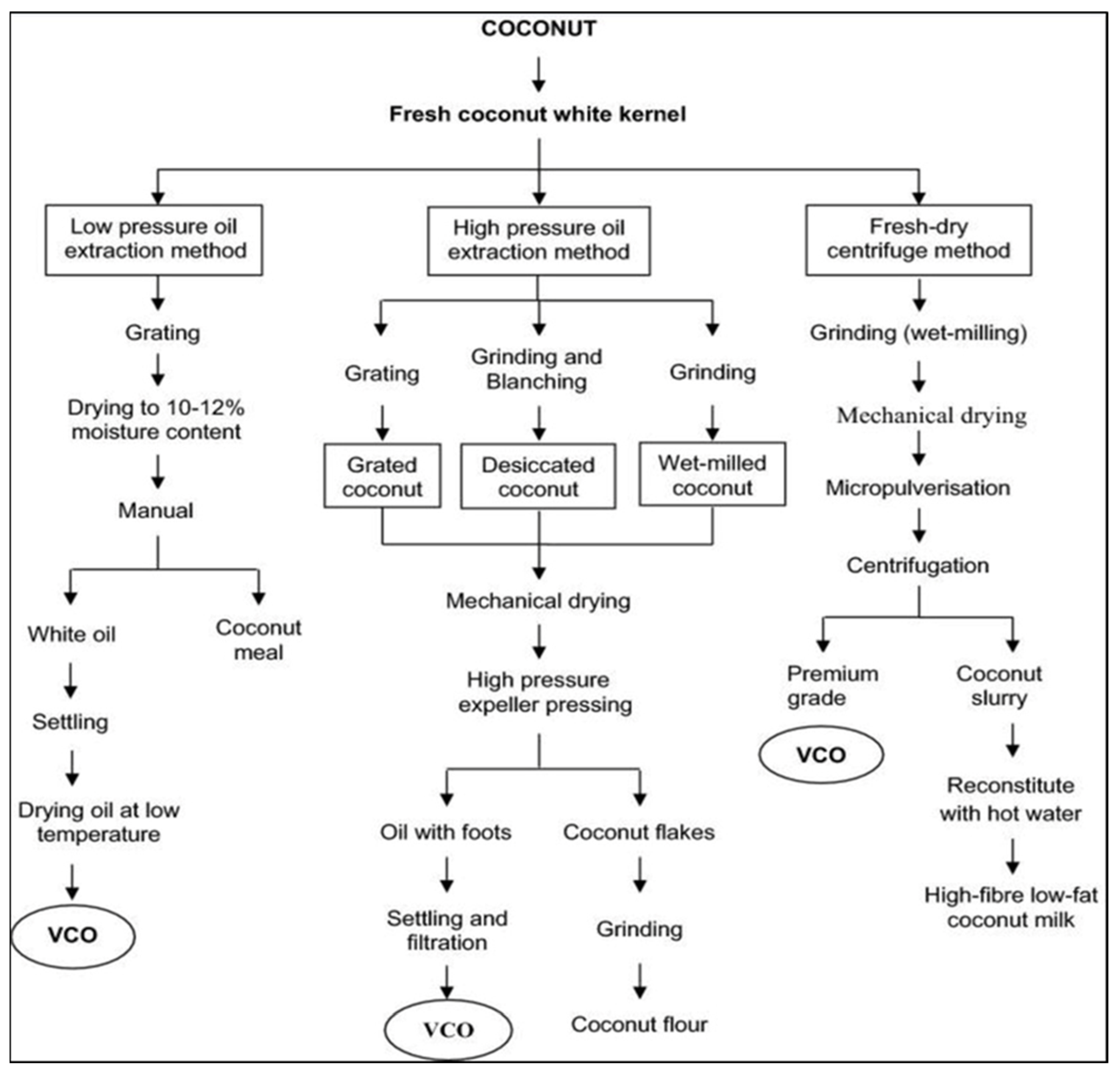
VCO is obtained by the mechanical extraction of coconut kernel without refining or high-temperature treatment, preserving its native properties [34]. Various sub-methods of the wet extraction process exist (Figure 1) in which the production of VCO occurs without RBD, thus preserving the contents and nature of the oil [31,35]. The wet process typically begins with the use of mature coconuts known for their high oil content. The kernel is grated or shredded to facilitate oil extraction. This is followed by either cold-pressing—where mechanical pressure is applied at low temperature or centrifugation, which separates the oil from coconut milk through high-speed spinning. Both methods avoid high heat and chemical processing, preserving the natural composition of the oil, including its lauric acid content. The extracted oil is then clarified through settling and filtration to remove moisture and impurities, resulting in pure VCO (Figure 2). One of the key advantages of natural VCO preparation is the retention of its nutritional integrity. VCO obtained through natural wet methods retains its beneficial fatty acids, medium-chain triglycerides (MCTs), antioxidants, and other micronutrients. This makes natural VCO a preferred choice for those seeking a wholesome, unadulterated source of coconut oil for culinary and wellness purposes. Furthermore, the natural preparation of VCO aligns with sustainable and environmentally friendly practices, as it minimizes the use of chemicals and energy-intensive processes. By exploring the inherent properties of coconuts and employing traditional extraction methods, producers can create a pure and authentic product that embodies the essence of VCO [34]. Other techniques for the optimal removal of the oil from the coconut milk emulsion include enzyme-assisted extraction [36,37,38], chilling, freezing, and thawing techniques [35], and fermentation, a traditional method wherein coconut milk is extracted, allowed to settle, and undergoes natural fermentation for 24–36 h within which the oil phase becomes separated from the aqueous phase. The further slight heating of the oil phase for a short time removes the moisture, leading to oil separation [12,21]. The main demerits of this process are the low oil recovery and fermented odor of VCO. Bawalan [39] detailed this process under wet processing, where the separated oil is filtered and collected as VCO. Likewise, Srivastava et al. [40] employed wet processing with cold extraction, where the coconut milk was left to settle under controlled conditions (35–40 °C, 75% humidity) for 20–24 h. The airborne lactic acid bacteria help in breaking protein bonds, aiding in VCO separation. However, although the method is attractive and produces quality oil, it yields a comparatively low amount of oil, which has discouraged its commercial application.

2.2. Extraction of Copra Coconut Oil (Dry Process)

Traditionally, coconut oil is extracted by pressing copra (dry process) that has been exposed to a very high temperature and/or sunlight to remove moisture [41]. The coconut oil is produced by crushing copra, which contains about 60–65% of the oil [12]. Copra is the dried kernel obtained by smoke-drying, sun-drying, or a combination of both. Then, the clean, ground, and steamed copra is pressed with a screw press expeller or hydraulic press to obtain the copra coconut oil. However, the drying/copra method is traditionally unhygienic; in fact, the handling makes the extracted oil unsafe for human consumption. Therefore, the coconut oil undergoes a refining, bleaching, and deodorizing process. This process requires heating the oil at high temperatures, between 204 °C and 245 °C, which destroys the essential amino acids, tocopherols (e.g., vitamin E), and other valuable phytocompounds present in the coconut oil [10]. Hence, VCO is a healthier oil than commercial copra oil due to its medium-chain saturated fatty acid content and higher amounts of polyphenols [11,42].

2.3. Composition of VCO

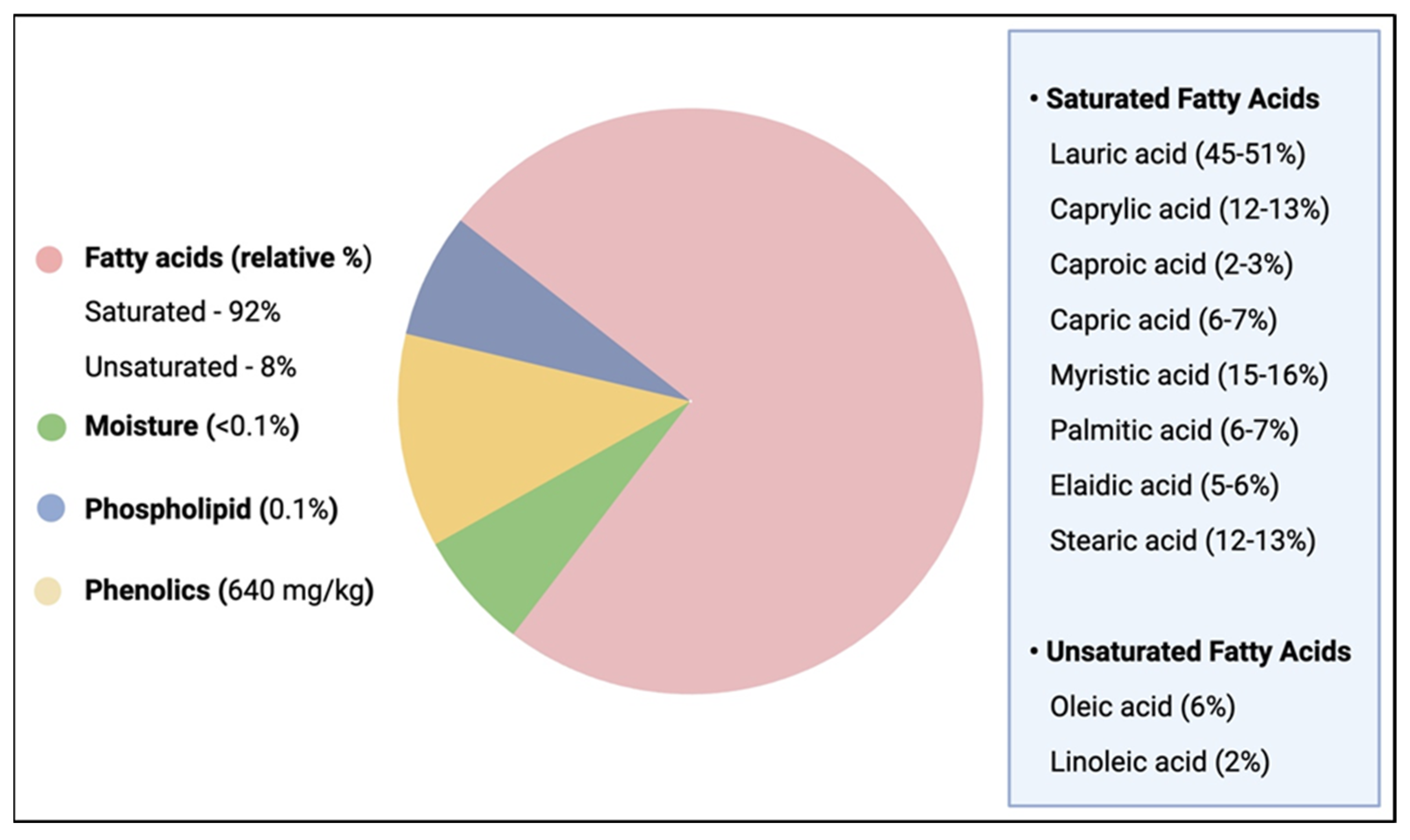
VCO is the pure and healthy form of coconut oil with a coconut aroma and a clear water appearance in contrast to copra coconut oil with a yellow color, which is sometimes due to microbial contaminants or high-temperature processing [10,43]. VCO is rich in natural bioactive compounds including saturated fatty acids (SFAs) (mainly medium-chain FAs), and phospholipids (e.g., tocopherols and sterols), as well as phenolic acids and polyphenols, in addition to minor components like flavonoids [40] (Figure 3).
These bioactive constituents contribute to a variety of biological effects, including antioxidants, anti-inflammatory, antimicrobial, and anticholesterolemic effects (Table 1).
Due to the high content of SFAs, some authors have reported that its continued consumption may increase low-density lipoprotein (LDL) [50,51]. However, unlike the long-chain fatty acids, the physicochemical properties of VCO enhance its intestinal absorption via the hepatic portal vein to hepatic metabolism for the production of energy [41,52] and does not essentially contribute to cholesterol synthesis. Moreover, the medium-chain triglyceride derivatives of MCFAs are bioactive forms with versatile pharmacological properties that can help in boosting the immune system, treating various pathologies such as cardiovascular, gastrointestinal, and inflammatory disorders. They have the capability to fight off numerous bacterial, fungal, and viral infections [53]. In human, the oral ingestion of VCO stimulates the conversion of LA, the most abundant MCFA (45 to 52%), to monolaurin, a key component of breast milk that boosts the baby immune system and has the potential to damage bacteria’s lipid membranes.
Moreover, studies demonstrate that VCO contains nearly seven times more total phenolic content compared to regular coconut oil, underscoring its superior antioxidant capacity and health benefits. This difference arises because the refining process of commercial coconut oil can diminish its biologically active components [54]. While the exact composition of phenolic acids in VCO may vary depending on factors such as the coconut variety, growing conditions, and extraction methods, several phenolic acids are commonly found in this oil.

3. Health Benefits of VCO and LA

3.1. VCO Health Benefits

Functional foods are food substances that provide health benefits beyond basic nutrition and that are capable of preventing diseases (Temple, 2022). VCO is a rapidly emerging functional food oil whose popularity and public awareness is increasing worldwide [35]. It is used in food industries for different purposes; it is an integral part of Sri Lankan and many South Asian diets. Being rich in MCFAs and highly digestible, VCO is increasingly available, especially in Southeast Asia. Its benefits include antioxidant activity and various health advantages, thus making it a desirable addition to culinary practices. Recent research findings support its physicochemical properties and clinical benefits, further boosting its appeal in the food industry [35]. Nevertheless, the use of coconut oil in one’s diet remains controversial owing to its high SFA content. In decades past, high SFA in one’s diet was associated with elevated total cholesterol and low-density lipoprotein (LDL) levels in blood circulation leading to dyslipidemia and cardiovascular diseases (CVDs) [44]. Coconut oil has received an unfavorable reputation because there are claims that it may enhance cholesterol synthesis, and hypercholesterolemia leading to CVDs. However, in the past few years, clinical and non-clinical studies have been conducted on coconut oil and VCO, and positive outcomes were obtained, which might refute those arguments [41,44,55]. The MCFAs abundant in VCO have different chemical and physiological properties when compared to long-chain fatty acids (LCFAs). In human cells, MCFAs and LCFAs are metabolized differently by different lipase enzymes [56,57]. MCTs are hydrolyzed by lingual (mouth) and gastric (stomach) lipases into 2-monoacylglycerol and fatty acids, mostly MCFAs, which are rapidly absorbed by the enterocytes into the portal vein and then directly enter the liver to be quickly metabolized into energy; hence, MCTs do not increase blood triglycerides or LDL levels [56,58]. Additionally, MCTs do not require bile salts for digestion, making them more rapidly absorbed [59,60,61] whereas the pancreatic lipases digest the dietary long-chain triglycerides into free long-chain fatty acids and then become absorbed by the enterocytes and resynthesized into new triglycerides. The new triglycerides enter the lymph system as chylomicrons, and are then transported into the heart and circulation, hence, contributing to blood long-chain triglycerides and the synthesis of LDL. Therefore, VCO does not increase LDL levels, but increases HDL levels and decreases the LDL/HDL ratio and CVD susceptibility [56,58]. However, there are inconsistent reports often arising from different sources and methods of coconut oil production, population differences, and small trial participant size [50,62]. For example, a review on coconut oil and cardiovascular risk factors in humans concluded that the evidence of an association between coconut oil consumption and blood lipids or cardiovascular risk was mostly of poor quality [51]. Interestingly, a randomized clinical trial that compared the effects of VCO (saturated fat), butter (saturated fat), or virgin olive oil (unsaturated fat) on the blood lipid profile shows that the effect of VCO is comparable to that of virgin olive oil, whereas butter significantly increased the LDL-C concentrations [50] The study indicates that VCO significantly increased HDL-C and had a similar effect on the TC/HDL-C ratio as olive oil compared with butter. In another recent randomized trial [63] conducted in the UK, VCO and virgin olive oil had similar beneficial health effects on circulating odd-chain saturated fatty acids and trans-fatty acids by reducing their levels, while butter increased the levels of these fatty acids.
Compared to butter and hydrogenated oils, VCO has a higher content of medium-chain triglycerides, lacks cholesterol and trans fats, and demonstrates faster metabolic oxidation. These distinctions may underline some of the health-related claims attributed to VCO, particularly in the context of lipid metabolism and cardiovascular outcomes (Table 2).
Being rich in MCFAs, VCO prevents infections and promotes health by lowering cholesterol through thyroid stimulation. Its high resistance to oxidative rancidity makes it ideal for cooking [64]. It serves as an excellent carrier for fat-soluble vitamins and has notable antimicrobial properties [64]. VCO is renowned for promoting cholesterol reduction, cardiovascular health, weight management, cognitive function, and antimicrobial effects [65]. VCO has gained popularity for its potential health benefits, especially in cardiovascular health. According to a study by Dumancas et al. (2016) [10], VCO shows comparable effects to other saturated fats on LDL levels but may increase HDL levels. Its antioxidant property suggests potential cardiovascular benefits by reducing oxidative stress, though further research is needed. Research comparing extra virgin coconut oil (EVCO) with extra virgin olive oil (EVOO) indicates that EVCO may be more effective in reducing hunger and the desire to eat, particularly in normal-weight individuals [66]. VCO can aid in weight loss by enhancing satiety and increasing thermogenesis, which promotes fat loss directly from adipose tissue [67]. Its phenolic compounds also contribute to reducing the risk of atherosclerosis by decreasing lipid peroxidation and normalizing lipid levels [65].
Moreover, VCO exhibits antidiabetic, anti-obesity, and antiasthmatic properties by enhancing insulin secretion, and inhibiting fat accumulation, alongside potent antimicrobial activity against diverse pathogens, including bacteria, viruses, fungi, and protozoa [13,68,69,70,71,72,73,74,75,76]. VCO has been shown to be beneficial against ulcerative colitis, experimental infertility, inflammatory bowel disease, antibiotic gentamicin toxicity, and oral cancer [77,78]. VCO increases the absorption of calcium and magnesium, compared to other types of oils with a high content of long-chain fatty acids; it prevents obesity, and, hence, decreases the incidence of or prevents diabetes, and induces insulin sensitivity [56]. The combined topical application of VCO and black cumin oil enhances diabetic wound healing through the increased expression of the vascular endothelial growth factor (VEGF) gene, an essential mediator of angiogenesis and tissue regeneration [79]. The beneficial health effects of VCO in polycystic ovarian syndrome and metabolic syndrome have been reported in the existing literature [80,81]. In pharmaceutical settings, VCO shows promise as a carrier for lipid-soluble drugs when processed under controlled conditions [82]. VCO is being explored for its potential in Alzheimer’s disease (AD) treatment, particularly through formulations designed to increase brain cholesterol levels and mitigate β-amyloid plaque formation, which are characteristics of AD [83]. Aluminum-induced AD-like neurological damage and neurobehavioral dysfunction were alleviated by oral VCO [84]. Aluminum-induced neuroinflammation and oxidative toxicity were suppressed via the regulation of the NLRP3 inflammasome triggered by betaine in combination with VCO [85]. Moreover, a review of the anti-Parkinson-disease effects of VCO is chronicled by Deepika et al. (2024) [86], while the health effects of VCO in enhancing memory and neurobehavioral activity were shown by the study of Rahim et al. (2017) [87] and de Vasconcelos et al. (2023) [88] respectively. These studies highlight VCO’s emerging role in neurological health. The anticancer effects of VCO and its modulation against toxicities of various environmental pollutants or toxicants have been reported [89,90,91,92,93,94,95]. The anticancer effects of VCO have been reported in the literature for a number of cancer cell lines [89]. Both in silico and in vitro analyses show that VCO may inhibit certain hallmarks of cancer development [96,97]. Experimental arthritis induced by an intradermal injection of complete Freund’s adjuvant was suppressed by VCO polyphenolics via the modulation of the antioxidant and anti-inflammatory COX-2/iNOS/TNF-α/IL-6 pathway [98]. Moreover, the anti-HIV/AIDS effect of VCO was also demonstrated in a clinical case–control study that enrolled HIV-positive subjects with a CD4+ T lymphocyte count > 200 cell/µL [99]. In the study, it was found that the VCO supplementation significantly increased the CD4+ T lymphocyte count. In another clinical study on COVID-19-positive patients in a 28-day randomized, single-blind trial, adjunct VCO therapy was found to alleviate symptoms and suppress the C-reactive protein concentration [100]. Clinical studies underscore its ability to alleviate skin disorders through its moisturizing and soothing effects, devoid of skin irritants and phototoxicity, alongside potent anti-inflammatory properties that bolster overall skin health [101,102]. Overall, VCO’s unique FA composition distinguishes it from other oils and contributes to its beneficial effects on body fat reduction, heart health, hyperglycemia, and metabolic disease prevention, and potential antibacterial actions which support its broader role in enhancing overall health [103]. These attributes collectively position VCO as a versatile and potential candidate for further research and development across various healthcare applications.

3.2. LA Health Benefits

LA is a 12-carbon medium-chain saturated fatty acid found abundantly in coconut oil or palm kernel oil, recognized to play a vital role in pathologies and improving immune physiological function, and it comes with multiple beneficial effects [14,104]. Alves et al. (2017) [105] found that LA lowers the experimental blood pressure and oxidative stress in rats. Reports show that LA may modulate genes that underline atherosclerotic plaque formation [106]. In the THP-1 macrophage model, LA ameliorates the insulin resistance via the upregulation of the glucose uptake, mitochondrial membrane potential, ATP generation, and expression of mitochondrial biogenesis regulator genes such as GLUT-1, GLUT-3, TFAM, PGC-1α, and PPAR-γ [14]. In comparative studies, LA exerts more health effects compared to palmitic acid with respect to blood glucose reduction, insulin resistance, metabolic inflammation, and mitochondrial health in human primary myotubes and animal models [104,107,108]. It exhibits significant antimicrobial and antioxidant properties, making it valuable in both nutritional and pharmacological applications [56]. Its effectiveness against microbial growth and oxidative stress enhances its role in promoting health and treating skin conditions. LA improves the antioxidant and immune capacity for the intestinal structure while reducing the microbial population of aquatic organisms [109]. Although this study reveals an immunomodulatory effect of LA in the intestine of swimming crabs, the effect of LA on immunity in animal models should be investigated. LA also shows beneficial health effects by stabilizing the immune status in European Seabass Juvenile fish [110]. LA as monolaurin demonstrates a potential anti-retroviral effect via reductions in the viral load with variable changes in CD4 counts, and shields the body from parasites [33,111]. The antioxidant, anti-inflammatory, and anti-apoptosis mechanisms of LA have been demonstrated in the literature. A recent study reveals that LA modulates ethanol-induced hepatotoxicity via the suppression of hepatic oxidative stress, cytokine inflammation, and apoptosis [112]. The role of LA for improving erectile dysfunction, benign prostatic hyperplasia, and hyperglycemic stroke in diabetic rats and mice, respectively, has been suggested in the existing literature [113,114,115]. Interestingly, the recent study of Kisioglu et al. (2024) [116] indicates that the oral intake of LA abrogates neuroinflammation, systemic inflammation, and anxiety-like behavior, and improves memory in a male C57BL/6 mice model of high-fat/high-fructose diet-triggered neuroinflammation and neurobehavioral deficit. And the study suggests that the downregulation of CD36 expression, which provokes a microglial neuro-inflammatory state, may be linked to the neuroprotective mechanism of LA, as suggested earlier in earlier studies [117,118]. However, in a seemingly contrasting investigation [44], a reduction in the LA level in VCO was associated with the improved lipid profile in high-fat diet-induced hypercholesterolemic mice. The oral consumption of repeatedly heated VCO was found to elevate blood pressure and inflammatory biomarkers in rats [119] confirming the deleterious effect of toxic products in repeatedly heated oils already established in the literature.
Regarding pharmaceutical formulations, a study investigated a nanogel combining chitosan, thiocolchicoside LA (CTLA) for anti-inflammatory, antimicrobial, antioxidant, and cytotoxic effects. CTLA demonstrates substantial anti-inflammatory and antioxidant activity, comparable to diclofenac sodium, and effectively inhibited Streptococcus mutans and Staphylococcus aureus [120]. Additionally, LA has garnered attention in food and pharmaceutical applications for its antimicrobial and antioxidant properties. Found abundantly in VCO, LA contributes to skin health by combating microbial growth and oxidative stress. These applications underscore LA’s versatility and efficacy in promoting skin health and cosmetic formulations.

4. Anticancer Effects of VCO and LA

Although radiation and chemotherapy are the mainstays of cancer treatment, they produce intractable side effects in patients [23]. The emergence of resistance to chemotherapeutic drugs in several cancer types is another formidable hurdle in oncotherapy [3]. Thus, there is a need for effective and safe bioprophylactics and biotherapeutics in cancer therapy. Natural VCO offers a myriad of health benefits, and, interestingly, the existing literature has shown the anticancer effects of VCO and LA.

4.1. Anticancer Effects of VCO

A growing number of published findings indicate the anticancer potential of VCO in breast, lung, colon, oral, and liver cancers (Table 1). In support of this, the in vitro study of Verma et al. (2019) [121], in which three different coconut oils, VCO, Processed Coconut Oil (PCO), and Fractionated Coconut Oil (FCO), were evaluated on the liver and oral cancer cell lines, demonstrates this effect. Although specific anticancer mechanisms were not explored, the study confirmed the anti-proliferative potentials of these coconut oils in the liver and oral cancer cell lines using an MTT cytotoxicity assay within 72 h of incubation. Remarkably, the oils show anticancer effects on the HepG2 and KB cell lines, leading to the conclusion of the authors that the VCO, PCO, and FCO have anticancer efficacy and may be used for the treatment of cancer, especially the liver and oral cancers. However, the anticancer efficacy may be dependent on the type of fatty acid profiles in the coconut oils [121]. The coconut oil nanoemulsion that contains the anticancer drug methotrexate (MTX) enhances the antiproliferative activity of MTX by a cytotoxic effect on A549 non-small-cell lung cancer cells with apoptotic morphological characteristics. The nanoemulsion inhibits A549 proliferation two times more than MTX alone. This suggests the anticancer role of VCO in the nanoemulsion [122]. Interestingly, in this study, the oxidative stress widely reported for the MTX-induced side effect toxicity was significantly ameliorated. Although a number of studies have reported that VCO is able to suppress the oxidative stress associated with chemotherapy toxicity [8,123], the double-edged benefit herein exhibited by the VCO-MTX nanoemulsion is significant. Coconut oil exerts moderate growth inhibition on the skin cancer cell line; however, the lack of a significant difference from normal melanocytes limits its clinical relevance and suggests a non-selective effect [124]. In an earlier study, coconut oil rich in LA has been reported to significantly inhibit the growth of HT-29 malignant human colon cells compared to linoleic acid [125]. A study investigated the impact of the lipid composition (saturated fats) of a high-fat diet on pro-oncogenic processes [97]. This study included diets supplemented with saturated-fatty-acid (SFA)-rich coconut oil against an experimental colon cancer model induced by azoxymethane/dextran sodium sulfate. Interestingly, the SFA-rich coconut oil diet is protective against the induced model of colon cancer via the mechanism of elevated colonic mucin 2 protein content involved in the physiological maintenance of intestinal barrier integrity. The VCO treatment induces reduced cell viability with the induction of apoptosis characterized by morphological alterations, including the appearance of massive cytoplasmic vacuolization and the blebbing of the cell membrane in the A549 and NCI-H1299 lung cancer cell lines [96]. Furthermore, the anticancer effects of coconut oil extracted by different methods were evaluated in human neuroblastoma cells (SH-SY5Y) cultured in vitro [7]. Ramya et al. (2022) [7] shows that VCO and crude coconut oil (ECO) provoke apoptotic characteristics consistent with morphological changes such as nuclear fragmentation, chromatin condensation, and the disintegration of membrane integrity in SH-SY5Y neuroblastoma cells. The VCO and ECO treatments exert greater growth inhibition and nuclear damage leading to apoptotic cell death than the refined coconut oil (RCO) treatment. The mitochondrial membrane potential (∆ψm) determination with JC-1 staining reveals that VCO significantly reduced the ∆ψm than ECO, indicating the mechanism underlying the cell growth inhibition and apoptosis in the study. A similar anticancer effect of VCO inhibits SkBr-3 breast cancer cell proliferation; it also synergizes with trastuzumab treatment to enhance the growth inhibitory effect in vitro [126]. The network pharmacology analysis of the major saturated fatty acids in VCO, like LA, caprylic acid, capric acid, and myristic acid against identifying the target proteins and pathways regulated by VCO, was investigated [89]. Through this computational analysis, 17 cancer-associated proteins and pathways involving various neoplasms were identified. The VCO-gene expression data implicated different neoplasms. However, the experimental validation of the in silico data reveals the anticancer effect of VCO on HepG2 liver cancer cells only (Table 3). It is important to note that the findings in the study of Pruseth et al. (2020) [89] are preliminary data and require further work.

4.2. Anticancer Effect of LA

Virgin coconut oil contains MCFAs, and LA is the most abundant MCFA in all coconut oil samples [7,40,76]. However, LA is not only present in VCO but also in palm kernel oil, fruits, seeds, and breast milk. Lauric acid is 45 g/100 g of the edible portion of coconut oil or palm kernel oil [127]. The literature shows the anticancer properties of LA and suggests it to be a very active agent for cancer treatment [127]. In vitro cell culture studies and findings exist in the published papers underscoring the anticancer mechanisms of LA in colorectal cancer, liver cancer, endometrial cancer, and human skin epidermoid carcinoma. Although there are a few studies currently, the insights from the available data consistently show that LA exposure imparts growth inhibitory effects on several cancer cell lines (Table 4).
To confirm the apoptosis-inducing effect of VCO, SH-SY5Y neuroblastoma cells were exposed to LA [7]. LA exhibits dose-dependent and time-dependent cytotoxic effects on the SH-SY5Y cells demonstrated by a marked growth inhibition. In addition, LA provokes an anticancer mechanism consistent with ROS-mediated apoptosis by reducing the mitochondrial membrane potential in SH-SY5Y cells. A reduction in the potential indicates mitochondrial dysfunction that may lead to low ATP production, which will impart a severe blow on the survival of cancer cells. VCO and crude coconut oil caused the release of ROS, key oxidative stress response genes, and inflammatory genes in SH-SY5Y cells. On this ground, unfolding the redox nexus of antioxidant Nrf2 and the NF-κB signaling pathway may be important in future studies in order to lay a foundation for the effect of VCO in this type of cancer. It is worth noting that the antiproliferative effects observed lacked validation against noncancerous cell lines which may reduce their translational significance. The human hepatocellular carcinoma cell line (HepG2), colorectal cancer (CRC) cell line (HCT-15), and murine macrophages (Raw 264.7) were used to investigate the effects of LA and other MCFAs [30]. Lauric acid demonstrates a dose-dependent inhibition of cell growth. In addition, the LA treatment at 30 and 50 μg/mL in HCT-15 reveals epidermal growth factor receptor (EGFR) downregulation and diminished EGFR levels in the lipid rafts, leading to morphological characteristics of apoptosis [30]. EGFR signaling is crucial to cancer cell survival and progression, and its alteration or downregulation is associated with cytotoxicity, the induction of apoptosis, a anti-metastatic effect, and cancer cell death [138,139]. Therefore, cetuximab, an EGFR inhibitor, is the main targeted chemotherapy against metastatic CRC, although the cetuximab chemotherapy is limited to a subtype of CRC patients without mutated BRAF or KRAS genes. This is because cetuximab chemotherapy is challenged with chemoresistance, and may even trigger uncontrolled cell proliferation, cell growth, and even metastases in CRC patients with a mutation of either BRAF or KRAS genes [140,141]. But, in the study of Weng et al. (2016) [128], it was found that LA could sensitize KRAS/BRAF-mutated CRC cells to cetuximab chemotherapy. The authors observed that LA orchestrates the sensitization to cetuximab via an elevated microRNA-378 expression accompanied by a decreased expression of mitogen-activated protein (MAP)/extracellular signal-regulated kinase-2 (ERK-2). It was also noted that LA could inhibit growth in KRAS/BRAF-mutated CRC cells even without cetuximab [128]. The finding is important for the possible synergistic anticancer effect of LA and shedding new light on overcoming the therapy resistance of the BRAF or KRAS mutation in CRC patients.
Moreover, the effects of LA were observed in a study that indicates the apoptosis induction potential of LA to kill colon cancer cells [129]. In this study, LA was able to induce ROS coupled with a reduced level of glutathione, which mechanistically provokes apoptotic alterations and cell cycle arrest at the G0/G1 and G2/M phases. However, the absence of control comparisons to a normal colon epithelium calls into question the therapeutic selectivity of this effect. Furthermore, LA was also found to inhibit the growth of HT-29 malignant human colon cell line [125]. A mechanistic study indicates that LA triggers a signaling pathway that leads to antiproliferative and pro-apoptotic effects in both endometrial (Ishikawa) and breast (SkBr3) cancer cells [130]. The LA-activated signaling and molecular mechanism stimulates ROS stress and the phosphorylation of c-Jun, ERK1/2, and EGFR, and the elevated expression of c-fos. The Rho-associated kinase was also phosphorylated and could be connected with ROS generation, which is a double-edged sword, activating and or inhibiting biological processes modulated by protein kinases, phosphatases, and several other enzymes [127]. Gemcitabine chemotherapy is the first-line therapy for the treatment of bladder cancer; however, due to chemoresistance linked to the depressed expression of cellular membrane human equilibrative nucleoside transporter 1 (hENT1), the facilitative transporter of gemcitabine (GEM), its use is limited in chemotherapeutic regimens. Although a number of fatty-acid-based GEM derivatives have been developed in order to enhance the transport of GEM into cancer cells [142], the LA-GEM derivative conjugate shows interesting findings against human bladder cancer [27]. The LA-based GEM conjugate (SZY-200) and LA alone were evaluated for anticancer effects. SZY-200 inhibits the proliferation of bladder cancer cells by inducing cell cycle arrest and apoptosis comparable to GEM alone and CP-4126 (an elaidic acid–GEM conjugate), but the interesting finding in this study is that SZY-200’s entry into bladder cancer cells does not rely on hENT1, a transporter protein that promotes GEM resistance. In addition, LA alone could inhibit the proliferation of bladder cancer cells, while SZY-200 downregulates the expressions of the peroxisome-proliferator-activated receptor gamma (PPAR-γ) and prostaglandin-endoperoxide synthase 2 (PTGS2) genes involved in the development of bladder cancer [27]. Clearly, the role of LA is evident in the by-passing of hENT1 for GEM accumulation in bladder cancer cells, triggering antiproliferation, cell cycle arrest, and apoptosis.
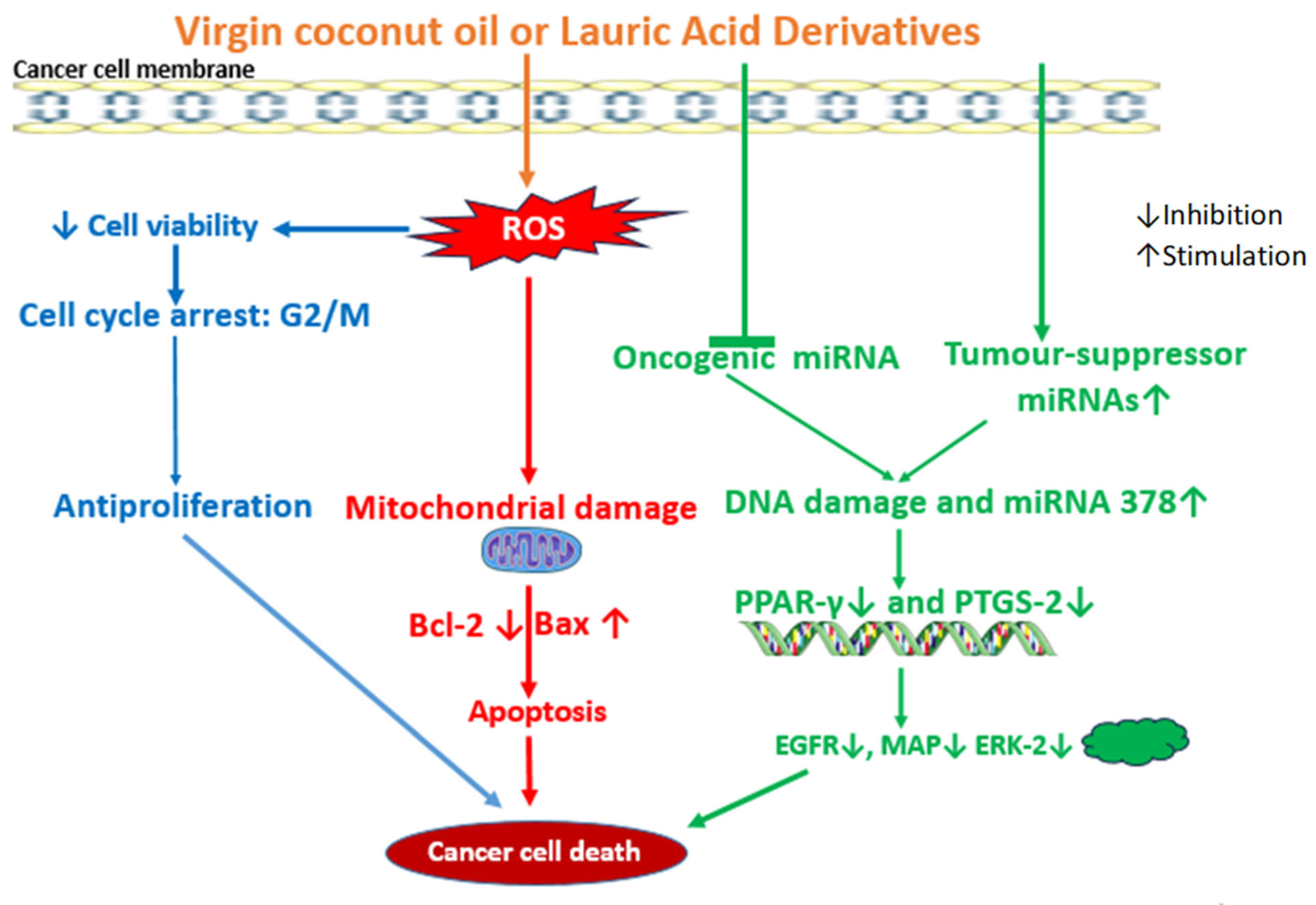
Another interesting study has indicated the influence of LA on microRNA expression in cancer cells. The LA treatment of KB-1 cells and HepG2 cells suppresses the expression of oncogenic microRNA and upregulates the expression of tumor-suppressor microRNAs [137]. These LA-modulated microRNAs are associated with cancer pathways with respect to apoptotic signaling, DNA damage, cell growth, cell cycle phase transition, cell response to stress, p53 mediation. protein signaling pathway, cyclin-dependent protein kinase activity, and epidermal growth factor receptor signaling pathways, according to the metabolic pathways analysis by the in silico GeneMANIA software. However, these associations remain hypothetical and require experimental validation to confirm any direct mechanistic link [137] (Figure 4). MicroRNAs are regulators of major cellular biological processes, including apoptosis, cell division and growth, tissue differentiation, and regeneration, and their dysregulation is a crucial molecular factor that contributes to the development of cancer [143]. For example, there is the upregulation of miR-17-92 and miR-21 in cancer, whereas microRNAs let-7 and miR-34 families are downregulated [137,144]. However, there is a paucity of data on the effect of LA on microRNA expression for the anticancer effect and mechanisms.
Hence, it is suggested that lauric acid exerts anticancer effects in various models mediated by distinct, context-dependent molecular pathways. The observed mechanisms—ranging from ROS-mediated apoptosis to EGFR downregulation and microRNA modulation—highlight the heterogeneity in cellular response, rather than a single universal mechanism.

LA-Based Nanocarriers for Therapeutic Delivery

A report has emerged on a role of LA in thermotherapy or hyperthermia—a technique of anticancer therapy that induces a hyperthermal stress at a temperature range of 41–45 °C in the tumor site. It is currently a widely used method for treating tumors, especially breast cancer without surgery [131]. The physical property of LA as an organic phase change material that absorbs and releases thermal energy during the process of melting (~43 °C) and freezing, respectively, is being explored. This makes LA applicable as a powerful chemotherapeutic agent in combination with thermotherapy. In support of this, a study synthesized novel nanocapsules consisting of an LA core encapsulated in a silica shell (SiO2@LA NPs). The LA-encapsulated nanomaterial was suitable for releasing hyperthermal stress on the breast cancer cells (MCF-7), disrupting the cell membrane and inducing oxidative stress, apoptosis, and morphometric alterations [131]. The study shows that thermal treatment alone did not cause a reduced viability of MCF-7 significantly but did with the LA nanoencapsulation in SiO2@LA NPs. It is widely published that VCO and LA have antibacterial and virucidal effects [56,71,145,146]. With this background, Xin et al. (2023) [132], thus, synthesized a nanocomplex conjugated to the platinum IV Pt(IV)-complex and LA to combat the Gram-negative bacteria, Fusobacterium nucleatum (Fn), that promotes chemoresistance in oxaliplatin chemotherapy against CRC. The bacteria also promote the proliferation and metastasis of CRC. In the study, the nanocomplex was encapsulated with hyaluronic acid to enhance biocompatibility and tumor targeting. Interestingly, the nanocomplex exhibits a notable anti-CRC activity via cytotoxicity mediated by ROS generation, and a high anti-Fn activity. Similar findings were found for LA-based nanodrug systems (5-FU-LA@PPL) which increased chemosensitivity, the combinational compatibility of 5-fluorouracil and oxaliplatin, and the suppressed tumor-resided Fn-associated CRC in the in vitro and in vivo colorectal cancer (HT-29) xenograft model [133]. LA was found to inhibit autophagy and reduce cell viability, demonstrating a higher cytotoxic effect compared with the drug 5-FU against Fn-infected CRC cells.
Considerable findings were obtained when MCFAs, including LA conjugated with 2-hydroxyanthraquinone, a derivative of the anticancer drug doxorubicin, were evaluated for an anticancer effect against breast cancer cell lines MDA-MB-231 and MCF-7, human colon cancer cell line HT-29 and Colo-205, and leukemia cell line K-562 [134]. Among others, the LA conjugate exerts anticancer effects on Colo-205 and MCF-7. According to the findings, the LA conjugate proved to be the most active against human colon and breast cancers among all the MCFA conjugates. The study concludes that the substitution of LA on the anthraquinone ring probably enhances lipophilicity, causing greater cell membrane permeation. In recent investigations, moreover, three LA-based hydrazones inhibit liver cancer cell HepG2 growth after 48 h of exposure in the MTT medium. The cytotoxic effect was ascribed to the LA–hydrazine moiety in the LA-based hydrazones [135]. A chitosan-based nanogel that incorporated LA (CTL) potentially inhibits oral cancer cell (KB-1) proliferation. The cytotoxic inhibition of the CTL nanogel was unfolded in the study and it involves CTL-nanogel-mediated cell cycle arrest and the modulation of the Bax/Bcl-2/caspase gene expression pro-apoptotic pathway [136]. It causes a reduced cell viability and apoptotic morphological changes in KB-1 cells via cell cycle arrest at the G2/M phase and ROS induction leading to increased gene expressions of Bax, Bad, and caspase-3, and tumor-suppressor genes p53 and p21, while the anti-apoptotic gene Bcl-2 exhibited decreased expression. The study provides supportive evidence to support the potential of the chitosan thiocolchicoside lauric acid nanogel as a potential therapeutic candidate for oral cancer treatment and possible application in cancer nanomedicine. While this study highlights one potential mechanism of action involving p21 in KB-1 oral cancer cells, further studies across different models are needed to confirm whether similar pathways are consistently involved.

5. VCO and LA: Protection Against Chemotherapy Toxicity

Traditional cytotoxic chemotherapy primarily targets rapidly proliferating cancer cells vulnerable to being killed by a cytotoxic agent, but, in reality, cytotoxic chemotherapy attacks non-targeted healthy cells due to its lack of specificity for tumor/cancer cells during treatment; therefore, the off-target attack constitutes a long-standing problem of the side effect toxicity in patients undergoing chemotherapy treatment [23,147]. This leads to a high systemic toxicity, and prevents the use of high drug doses that are required for theeffective killing of cancer cells, thus limiting the anticancer efficacy [28]. And oxidative stress, inflammation, and apoptotic mechanisms have been implicated principally to underlie the side effect toxicity mechanism of chemotherapy [3,23]. Notably, a growing body of literature has shown supportive evidence that natural products could mitigate the chemotherapy-induced side effect toxicity [3,23]. Apart from its anticancer properties, VCO has emerged as a functional food oil for mitigating the undesirable side effect toxicity associated with chemotherapy.

5.1. VCO Protects Against Chemotherapy Toxicity

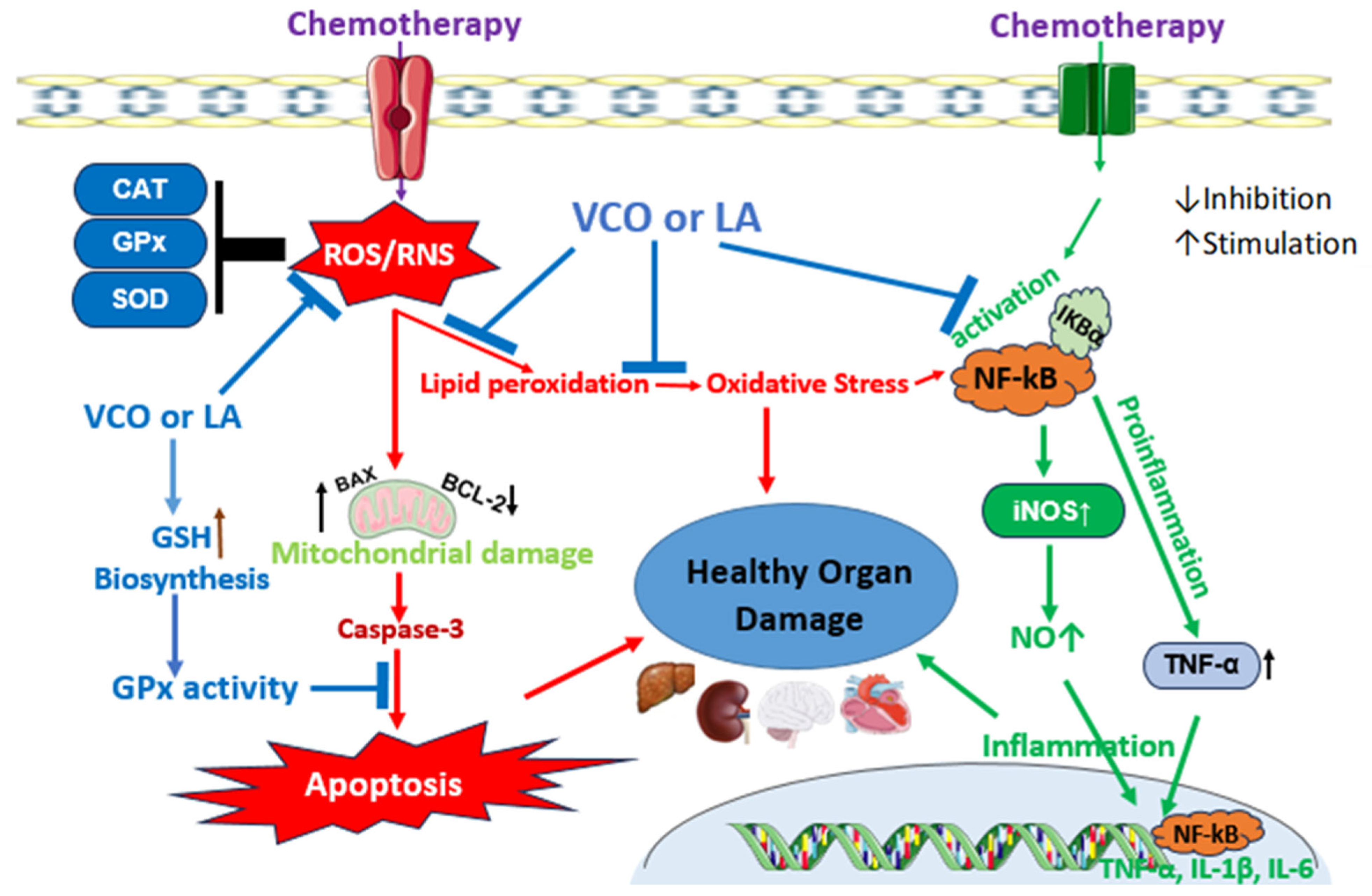
Due to the bioactive natural compounds in VCO, including caffeic acid, ferulic acid, vallinic acid, gallic acid, catechin, rutin, epicatechin, chlorogenic acid, hornavallin, p-coumaric acid, syringic acid, sinapic acid protocatechuic acid, and quercetin [40,45,148]. VCO is reported to combat the underlying toxicity mechanisms of chemotherapy. In male mice and female mice, the anticancer drug doxorubicin (DOX) induced neurobehavioral toxicity via the elevation of acetylcholinesterase (AchE), tumor necrosis factor-α (TNF-α), inducible nitric oxide synthase (iNOS), and altered neurobehavioral factors in both sexes [25]. The oral administration of VCO for 28 days was found to ameliorate some of the markers of neurotoxicity and neuroinflammation. VCO was evaluated for whether it can inhibit immunosuppression associated with DOX chemotherapy, because DOX could adversely modulate the interleukin (IL-2) and interferon-γ production, lymphocyte proliferation, NK cell cytotoxicity, and T lymphocyte CD4+/CD8+ ratio in tumor-bearing mice [149]. In the study, VCO was evaluated for its effect on CD4+ and CD8+ cells against DOX-induced immunosuppression in rats [150]. The CD cells, phagocytosis activity, lymphocyte proliferation, and capacity of macrophages were suppressed by a DOX injection. The DOX also caused a downregulation of IL-10, underscoring the initiation of inflammatory cascades in certain tissues. Interestingly, the oral VCO inhibits the immunosuppression and markedly enhances the CD4+, CD8+, and other immunological indices in the study [150]. It also inhibits the DOX-induced hepatic and cardiac damage and significantly reduces the activities of alanine aminotransferase (ALT), aspartate aminotransferase (AST), lactate dehydrogenase (LDH), and creatine kinase-MB in the serum of Wistar rats [151,152]. The lesions in the tissue histology were observed to be alleviated clearly in comparison to the non-treated DOX group. In a similar study, however, the oxidative inflammatory and apoptotic toxicity effects of DOX leading to liver toxicity were evidently shown. DOX induces significant increases in the serum levels of AST, ALT, and malondialdehyde (MDA), and a decrease in albumin coupled with elevated hepatic levels of pro-inflammatory NF-κB, TNF-α, and IL-6, and pro-apoptotic caspase-3 [24]. The liver levels of anti-apoptotic Bcl-2 as well as the antioxidant apparatus were shown to be considerably reduced in the study. VCO supplementation for 7 days was found to restore the liver function, antioxidant mechanism, and anti-apoptotic effect against the DOX toxicity mechanism in consonance with histological findings [24]. In a comparative investigation, hot-processed VCO (HPVCO) demonstrates a higher antioxidant potential than fermentation-processed VCO in a battery of in vitro antioxidant assays [153]. The authors validated this potential in an in vivo experimental model evaluating the antioxidant and nephroprotective effect of HPVCO and FPVCO against cisplatin (CP)-induced nephrotoxicity. CP is one of the foremost anticancer agents due to its wide efficacy in the treatment of various cancers [154,155]. However, the major side effect of CP chemotherapy is nephrotoxicity [154]. The results from the study show that CP provokes nephrotoxicity which was alleviated by HPVCO or FPVCO [153]. Notably, the HPVCO exerts the higher antioxidant restoration of hepatorenal function, and also inhibits myelosuppression and histological abrasions via the elevated activities of antioxidant enzymes and reduced glutathione. The study highlights the influence of processing methods on antioxidant bioactive compounds in VCO. This is similar to the findings against cyclophosphamide (CYP)-induced hepatorenal toxicity in a murine model. In the study, CYP induced the depression of the antioxidant status in the liver and kidney, followed by deleterious lipid peroxidative reactions and oxidative stress in mice [17]. However, the oral administration of VCO for 20 days alleviated the hematological alterations, lipid peroxidation, hepatorenal function, and oxidative stress. In the toxicity of CYP in lymphoid tissue, VCO prevents alterations in the hematological indices and histological changes both in the spleen and thymus organs [156]. Another anticancer agent of the class antimetabolites called methotrexate (MTX) is well-associated with organ toxicity via oxidative stress and inflammatory alterations. The nanoemulsion of VCO and MTX expressed depressed oxidative stress compared to the free MTX treatment in a model of experimental rats [122]. The VCO in the nanoemulsion was able to elevate the activities of antioxidant enzymes in the brain and lung tissues, while the tissue lipid peroxidation diminished significantly. VCO supplementation to rats in various studies has been shown to possess antioxidant and anti-inflammatory effects against MTX-induced hepatotoxicity [21,22] nephrotoxicity [21] neurotoxicity [8], and systemic toxicity [123]. In these studies, VCO exerts an effect that reverses oxidative stress and also triggers the mechanistic suppression of inflammatory cytokines in the delicate organs (Figure 5). Although there is a paucity of reports on the effect of VCO on the side effects of chemotherapy in cancer patients, the study of Law et al. (2014) [157] was able to show that VCO supplementation improves the quality of life among breast cancer patients in Kelantan, Malaysia. According to the human study, the side effect symptoms, including dyspnea, sleep difficulties, fatigue, and loss of appetite, body image, and sexual function, improved when compared to the patients in the control group. The study reports that there is still a deterioration in sexual enjoyment; the sample size of participants was not large though [157]. However, it is unknown, in this human study, whether VCO could ameliorate the side effect toxicity of the liver and kidney, which are very rampant following chemotherapy. Moreover, VCO reduces mucositis, a common side effect of cancer treatment, in nasopharyngeal carcinoma patients [158]. Bioactive polyphenols in VCO may be responsible for these observations, and more comprehensive studies are needed in order to investigate the anticancer activity and modulatory mechanisms of VCO against chemotherapy side effect toxicity (Table 5).

5.2. LA Protects Against Chemotherapy Toxicity

The effect of LA on the organ-specific side effect toxicity of chemotherapy is currently unknown in existing literature. However, cancer cachexia, characterized by weight and skeletal muscle loss, is an important adverse effect of anticancer chemotherapy, and there are no approved drugs for it [160]. In a mouse cachexia model, combining the oral administration of LA and glucose improves cancer-derived myocardial damage as demonstrated by the recovery of myocardial atrophy and MYL1—a marker of muscle maturity [159].

6. Conclusions and Future Perspectives

VCO and its most abundant saturated MCFA, LA, are natural products with numerous pharmacological applications for human health and industrial uses. VCO is obtained from mature coconut fruits using several traditional methods of extraction devoid of refining and deodorizing. The review of literature herein has revealed that VCO and LA are potential anticancer natural agents. Studies show that VCO, LA, and their synthesized nanocomplex conjugates and nanomaterials exert anticancer effects and also enhance the anticancer efficacy of standard chemotherapeutic agents. In cancer cells, they induce anti-proliferative effect via ROS generation and mitochondrial dysfunction, leading to Bax.Bcl-2/caspase apoptotic cell death, cell cycle arrest, EGFR/c-JUN/ERK1-2/c-fos altered pathway, and microRNA downregulation in various cancers. However, other cancer-related pathways, including ERK/JNK/MAPK, PI3K/Akt/mTOR, Wnt/β-catenin, JAK/STAT, and ferroptosis SLC7A11/GPX-4 axis, remain to be reported. The side effect toxicity of cancer chemotherapy is still an intractable challenge in oncotherapy. The literature reports indicate that VCO could suppress the toxicity of MTX, CYP, CP, and DOX chemotherapies by its antioxidant, anti-apoptotic, and anti-inflammatory activities in preclinical studies. There are no reports on the effect of VCO or LA on the toxicity of other chemotherapeutic agents, such as 5-fluorouracil, paclitaxel, docetaxel, tamoxifen, or topoisomerase inhibitors. It is significant that the effect of LA on chemotherapy toxicity is currently unclear in existing literature. Clinical trials exploring the effectiveness of VCO and LA in the adjuvant treatment and management of cancer are recommended.
As the therapeutic application of VCO and LA continues to expand, it becomes increasingly important to align safety evaluations with established regulatory frameworks. The current evidence mainly focuses on preclinical efficacy and mechanistic insights, while comprehensive safety assessments remain limited. Future research should adopt structured safety assessment protocols such as those outlined by the European Food Safety Authority (EFSA) and other international guidelines for food-related nanomaterials. Incorporating these assessments will be essential for ensuring the translational viability and public health relevance of VCO/LA-based nanoformulations in both functional foods and therapeutic contexts.

Author Contributions

Conceptualization and writing and preparation of original draft, D.B., R.A.-L. and A.O.; conceptualization, supervision, writing of original draft, and reviewing and editing, R.A.-L., A.C.F. and E.M.O. All authors have read and agreed to the published version of the manuscript.

Funding

This research received no external funding.

Institutional Review Board Statement

Not applicable.

Informed Consent Statement

Not applicable.

Data Availability Statement

No new data were created or analyzed in this study.

Conflicts of Interest

The authors declare no conflicts of interest.

References

  1. National Cancer Institute. A to Z List of Cancer Drugs. 2023. Available online: https://www.cancer.gov/about-cancer/treatment/drugs (accessed on 16 July 2025).
  2. Vijayakumar, S.; Dhakshanamoorthy, R.; Baskaran, A.; Krishnan, B.S.; Maddaly, R. Drug resistance in human cancers—Mechanisms and implications. Life Sci. 2024, 352, 122907. [Google Scholar] [CrossRef] [PubMed]
  3. Bose, D.; Famurewa, A.C.; Akash, A.; Othman, E.M. The therapeutic mechanisms of honey in mitigating toxicity from anticancer chemotherapy toxicity: A Review. J. Xenobiotics 2024, 14, 1109–1129. [Google Scholar] [CrossRef] [PubMed]
  4. Figueroa-González, G.; Quintas-Granados, L.I.; Reyes-Hernández, O.D.; Caballero-Florán, I.H.; Peña-Corona, S.I.; Cortés, H.; Leyva-Gómez, G.; Habtemariam, S.; Sharifi-Rad, J. Review of the anticancer properties of 6-shogaol: Mechanisms of action in cancer cells and future research opportunities. Food Sci. Nutr. 2024, 12, 4513–4533. [Google Scholar] [CrossRef] [PubMed]
  5. Cheon, C.; Ko, S.-G. Synergistic effects of natural products in combination with anticancer agents in prostate cancer: A scoping review. Front. Pharmacol. 2022, 13, 963317. [Google Scholar] [CrossRef]
  6. Zhang, X.; Huang, J.; Yu, C.; Xiang, L.; Li, L.; Shi, D.; Lin, F. Quercetin enhanced paclitaxel therapeutic effects towards PC-3 prostate cancer through ER stress induction and ROS production. OncoTargets Ther. 2020, 13, 513–523. [Google Scholar] [CrossRef]
  7. Ramya, V.; Shyam, K.P.; Kowsalya, E.; Balavigneswaran, C.K.; Kadalmani, B. Dual roles of coconut oil and its major component lauric acid on redox nexus: Focus on cytoprotection and cancer cell death. Front. Neurosci. 2022, 16, 833630. [Google Scholar] [CrossRef]
  8. Famurewa, A.C.; Aja, P.M.; Nwankwo, O.E.; Awoke, J.N.; Maduagwuna, E.K.; Aloke, K. Moringa oleifera seed oil or virgin coconut oil supplementation abrogates cerebral neurotoxicity induced by antineoplastic agent methotrexate by suppression of oxidative stress and neuroinflammation in rats. J. Food Biochem. 2019, 43, e12748. [Google Scholar]
  9. Boemeke, L.; Marcadenti, A.; Busnello, F.M.; Gottschall, C.B. Effects of Coconut Oil on Human Health. Open J. Endocr. Metab. Dis. 2015, 5, 84–87. [Google Scholar] [CrossRef]
  10. Dumancas, D.D.; Viswanath, L.C.; de Leon, A.R.; Ramasahayam, S.; Maples, R.; Koralege, R.H.; Perera, U.D.; Langford, J.; Shakir, A.; Castles, S. Health benefits of virgin coconut oil. In Vegetable Oil: Properties, Uses, and Benefits; Holt, B., Ed.; Nova Science Publishers, Inc.: Hauppauge, NY, USA, 2016. [Google Scholar]
  11. Deen, A.; Visvanathan, R.; Wickramarachchi, D.; Marikkar, N.; Nammi, S.; Jayawardana, B.C.; Liyanage, R. Chemical composition and health benefits of coconut oil: An overview. J. Sci. Food Agric. 2021, 101, 2182–2193. [Google Scholar] [CrossRef]
  12. GopalaKrishna, A.G.; Gaurav, R.; Bhatnagar, A.S.; Prasanth Kumar, P.K.; Chandrashekar, P. Coconut Oil: Chemistry, Production and Its Applications—A Review. Indian Coconut J. 2010, 15–27. [Google Scholar]
  13. Kardinasari, E.; Devriany, A. Phytochemical identification of bangka origin virgin green coconut oil: Anti-inflammatory and anti-bacterial potential. Enferm. Clin. 2020, 30, 171–174. [Google Scholar] [CrossRef]
  14. Tham, Y.Y.; Choo, Q.C.; Muhammad, T.S.; Chew, C.H. Lauric acid alleviates insulin resistance by improving mitochondrial biogenesis in THP-1 macrophages. Mol. Biol. Rep. 2020, 47, 9595–9607. [Google Scholar] [CrossRef]
  15. Famurewa, A.C.; Ugwu-Ejezie, C.S.; Iyare, E.E.; Folawiyo, A.M.; Maduagwuna, E.K.; Ejezie, F.E. Hepatoprotective effect of polyphenols isolated from virgin coconut oil against sub-chronic cadmium hepatotoxicity in rats is associated with improvement in antioxidant defense system. Drug Chem. Toxicol. 2021, 44, 418–426. [Google Scholar] [CrossRef]
  16. Famurewa, A.C.; Maduagwuna, E.K.; Folawiyo, A.M.; Besong, E.E.; Eteudo, A.N.; Famurewa, O.A.; Ejezie, F.E. Antioxidant, anti-inflammatory and antiapoptotic effects of virgin coconut oil against antibiotic drug gentamicin-induced nephrotoxicity via suppression of oxidative stress and modulation of iNOS/NF-ĸB/caspase-3 signalling pathway in Wistar rats. J. Food Biochem. 2020, 44, e13100. [Google Scholar] [CrossRef]
  17. Nair, S.S.; Manalil, J.J.; Ramavarma, S.K.; Suseela, I.M.; Thekkepatt, A.; Raghavamenon, A.C. Virgin coconut oil supplementation ameliorates cyclophosphamide-induced systemic toxicity in mice. Hum. Exp. Toxicol. 2016, 35, 205–212. [Google Scholar] [CrossRef]
  18. Otuechere, C.A.; Madarikan, G.; Simisola, T.; Bankole, O.; Osho, A. Virgin coconut oil protects against liver damage in albino rats challenged with anti-folate combination, trimethoprim-sulfamethoxazole. J. Basic Clin. Physiol. Pharmacol. 2014, 25, 249–253. [Google Scholar] [CrossRef]
  19. Wang, Y.; Yuan, A.; Wu, Y.; Wu, L.; Zhang, L. Silymarin in cancer therapy: Mechanisms of action, protective roles in chemotherapy-induced toxicity, and nanoformulations. J. Funct. Food 2023, 100, 105384. [Google Scholar] [CrossRef]
  20. Zhang, Q.; Wang, F.; Jia, K.; Kong, L. Natural product interventions for chemotherapy and radiotherapy-induced side effects. Front. Pharmacol. 2018, 9, 1253. [Google Scholar] [CrossRef]
  21. Famurewa, A.C.; Aja, P.M.; Maduagwuna, E.K.; Ekeleme-Egedigwe, C.A.; Ufebe, O.G.; Azubuike-Osu, S.O. Antioxidant and anti-inflammatory effects of virgin coconut oil supplementation abrogate acute chemotherapy oxidative nephrotoxicity induced by anticancer drug methotrexate in rats. Biomed. Pharmacother. 2017, 96, 905–911. [Google Scholar] [CrossRef]
  22. Famurewa, A.C.; Ufebe, O.G.; Egedigwe, C.A.; Nwankwo, O.E.; Obaje, G.S. Virgin coconut oil supplementation attenuates acute chemotherapy hepatotoxicity induced by anticancer drug methotrexate via inhibition of oxidative stress in rats. Biomed. Pharmacother. 2017, 87, 437–442. [Google Scholar] [CrossRef]
  23. Famurewa, A.C.; Akhigbe, R.E.; George, M.Y.; Adekunle, Y.A.; Oyedokun, P.A.; Akhigbe, T.M.; Fatokun, A.A. Mechanisms of ferroptotic and non-ferroptotic organ toxicity of chemotherapy: Protective and therapeutic effects of ginger, 6-gingerol and zingerone in preclinical studies. Naunyn-Schmiedebergs Arch. Pharmacol. 2024, 398, 4747–4778. [Google Scholar] [CrossRef]
  24. Ezzat, A.; Salem, F.E.; Kassaba, R.B.; Abdel Moneim, A.E.; El-Yamany, N.A. Evaluating the potential protective effect of virgin coconut oil against doxorubicin-mediated hepatotoxicity in rats. Adv. Basic Appl. Sci. 2023, 1, 46–54. [Google Scholar]
  25. Chiamaka, E.A.; Uchechukwu, E.J.; Chioma, D.S.; Asonye, R.; Chijioke, O.K.; Chiemela, D.C. Impact of virgin coconut oil and carvedilol on neurobehavior, apoptotic and inflammatory brain markers in doxorubicin treated mice. Path Sci. 2023, 9, 4020–4049. [Google Scholar] [CrossRef]
  26. Jayathilaka, N.; Seneviratne, K.N. Phenolic antioxidants in coconut oil: Factors affecting the quantity and quality. A review. Grasas Aceites 2022, 73, e466. [Google Scholar] [CrossRef]
  27. Wang, H.; Shao, Z.; Xu, Z.; Ye, B.; Li, M.; Zheng, Q.; Ma, X.; Shi, P. Antiproliferative and apoptotic activity of gemcitabine-lauric acid conjugate on human bladder cancer cells. Iran. J. Basic Med. Sci. 2022, 25, 536–542. [Google Scholar]
  28. Jóźwiak, M.; Filipowska, A.; Fiorino, F.; Struga, M. Anticancer activities of fatty acids and their heterocyclic derivatives. Eur. J. Pharmacol. 2020, 871, 172937. [Google Scholar] [CrossRef]
  29. Chhikara, B.S.; Jean, N.S.; Mandal, D.; Kumar, A.; Parang, K. Fatty acyl amide derivatives of doxorubicin: Synthesis and in vitro anticancer activities. Eur. J. Med. Chem. 2011, 46, 2037–2042. [Google Scholar] [CrossRef]
  30. Sheela, D.L.; Narayanankutty, A.; Nazeem, P.A.; Raghavamenon, A.C.; Muthangaparambil, S.R. Lauric acid induce cell death in colon cancer cells mediated by the epidermal growth factor receptor downregulation: An in silico and in vitro study. Hum. Exp. Toxicol. 2019, 38, 753–761. [Google Scholar] [CrossRef]
  31. Villarino, B.; Dy, L.; Lizada, C. Descriptive sensory evaluation of virgin coconut oil and refined, bleached and deodorized coconut oil. LWT-Food Sci. Technol. 2007, 40, 193–199. [Google Scholar] [CrossRef]
  32. Srivastava, Y.; Semwal, A.D.; Sharma, G.K. Virgin coconut oil as functional oil. In Therapeutic, Probiotic, and Unconventional Foods; Academic Press: Cambridge, MA, USA, 2018; pp. 291–301. [Google Scholar]
  33. Bergsson, G.; Arnfinnsson, J.; Karlsson, S.; Steingrimsson, O.; Chormar, H. In vitro inactivation of Chlamydia trachomatis by fatty acids and monoglycerides. Antimicrob. Agents Chemother. 1998, 42, 2290–2294. [Google Scholar] [CrossRef]
  34. Zeng, Y.-Q.; He, J.-T.; Hu, B.-Y.; Li, W.; Deng, J.; Lin, Q.-L.; Fang, Y. Virgin coconut oil: A comprehensive review of antioxidant activity and mechanisms contributed by phenolic compounds. Crit. Rev. Food Sci. Nutr. 2024, 64, 1052–1075. [Google Scholar] [CrossRef]
  35. Marina, A.M.; Che Man, Y.B.; Amin, I. Virgin coconut oil: Emerging functional food oil. Trend Food Sci. Technol. 2009, 20, 481–487. [Google Scholar] [CrossRef]
  36. Pooi-Pooi, S.; Ali, Y.; Lai, O.M.; Kuan, C.H.; Tang, T.K.; Lee, Y.Y.; Phuah, E.T. Enzymatic and mechanical extraction of virgin coconut oil. Eur. J. Lipid Sci. Technol. 2020, 122, 1900220. [Google Scholar] [CrossRef]
  37. Oseni, N.T.; Fernando, W.M.; Coorey, R.; Gold, I.; Jayasena, V. Effect of extraction techniques on the quality of coconut oil. Afr. J. Food Sci. 2017, 11, 58–66. [Google Scholar] [CrossRef]
  38. Raghavendra, S.N.; Raghavarao, K.S.M.S. Effect of different treatments for the destabilization of coconut milk emulsion. J Food Eng. 2010, 97, 341–347. [Google Scholar] [CrossRef]
  39. Bawalan, D.D. Processing Manual for Virgin Coconut Oil, Its Products and By-Products for Pacific Island Countries and Territories; Secretariat of the Pacific Community: Noumea, New Caledonia, 2011. [Google Scholar]
  40. Srivastava, Y.; Semwal, A.D.; Majumdar, A. Quantitative and qualitative analysis of bioactive components present in virgin coconut oil. Cogent Food Agric. 2016, 2, 1164929. [Google Scholar] [CrossRef]
  41. Nevin, K.G.; Rajamohan, T. Wet and dry extraction of coconut oil: Impact on lipid metabolic and antioxidant status in cholesterol co-administered rats. Can. J. Physiol. Pharmacol. 2009, 87, 610–616. [Google Scholar] [CrossRef]
  42. Srivastava, Y.; Semwal, A.D.; Swamy, M.S.L. Hypocholesterimic effects of cold and hot extracted virgin coconut oil (vco) in comparison to commercial coconut oil: Evidence from a male wistar albino rat model. Food Sci. Biotechnol. 2013, 22, 1501–1508. [Google Scholar] [CrossRef]
  43. Dayrit, F.; Buenafe, O.E.; Chainani, E.; de Vera, I.M.; Dimzon, I.K.; Gonzales, E.; Santos, J.E. Standards for essential composition and quality factors of commercial virgin coconut oil and its differentiation from RBD coconut oil and copra oil. Philipp. J. Sci. 2007, 136, 121–131. [Google Scholar]
  44. Chatturong, U.; Palang, I.; To-on, K.; Deetud, W.; Chaiwong, S.; Sakulsak, N.; Sonthi, P.; Chanasong, R.; Chulikorn, E.; Kanprakobkit, W.; et al. Reduction of lauric acid content in virgin coconut oil improved plasma lipid profile in high-fat diet-induced hypercholesterolemic mice. J. Food Sci. 2023, 88, 4305–4315. [Google Scholar] [CrossRef]
  45. Marina, A.M.; Che Man, Y.B.; Nazimah, S.A.H.; Amin, I. Antioxidant capacity and phenolic acids of virgin coconut oil. Int. J. Food Sci. Nutr. 2009, 60, 114–123. [Google Scholar] [CrossRef] [PubMed]
  46. German, G.B.; Dillard, C.J. Saturated fats: What dietary intake? Am. J. Clin. Nutr. 2004, 80, 550–559. [Google Scholar] [CrossRef] [PubMed]
  47. Dia, V.P.; Garcia, V.V.; Mabesa, R.C.; Tecson-Mendoza, E.M. Comparative physicochemical characteristics of virgin coconut oil produced by different methods. Philipp. Agric. Sci. 2005, 88, 462–475. [Google Scholar]
  48. Somade, O.T.; Oyinloye, B.E.; Ajiboye, B.O.; Osukoya, O.A. Methyl cellosolve-induced hepatic oxidative stress: The modulatory effect of syringic acid on Nrf2-Keap1-Hmox1-NQO1 signaling pathway in rats. Phytomedicine Plus 2023, 3, 100434. [Google Scholar] [CrossRef]
  49. Cordeiro, M.L.; Martins, V.G.; Silva, A.P.; Rocha, H.A.; Rachetti, V.P.; Scortecci, K.C. Phenolic acids as antidepressant agents. Nutrients 2022, 14, 4309. [Google Scholar] [CrossRef]
  50. Khaw, K.-T.; Sharp, S.J.; Finikarides, L.; Afzal, I.; Lentjes, M.; Luben, R.; Forouhi, N.G. Randomised trial of coconut oil, olive oil or butter on blood lipids and other cardiovascular risk factors in healthy men and women. BMJ Open 2018, 8, e020167. [Google Scholar] [CrossRef]
  51. Eyres, L.; Eyres, M.F.; Chisholm, A.; Brown, R.C. Coconut oil consumption and cardiovascular risk factors in humans. Nutr. Rev. 2016, 74, 267–280. [Google Scholar] [CrossRef]
  52. Gomes, H.M.; Silveira, A.K.; Gasparotto, J.; Bortolin, R.C.; Terra, S.R.; Brum, P.O.; Gelain, D.P.; Moreira, J.C. Effects of coconut oil long-term supplementation in Wistar rats during metabolic syndrome-regulation of metabolic conditions involving glucose homeostasis, inflammatory signals, and oxidative stress. J. Nutr. Biochem. 2023, 114, 109272. [Google Scholar]
  53. Jack, K.S.; Asaruddin, M.R.; Bhawani, S.A. Pharmacophore study, molecular docking and molecular dynamic simulation of virgin coconut oil derivatives as anti-inflammatory agent against COX-2. Chem. Biol. Technol. Agric. 2022, 9, 73. [Google Scholar] [CrossRef]
  54. Seneviratne, K.N.; Dissanayake, M.S. Variation of phenolic content in coconut oil extracted by two conventional methods. Int. J. Food Sci. Technol. 2008, 43, 597–602. [Google Scholar] [CrossRef]
  55. Vogel, C.É.; Crovesy, L.; Rosado, E.L.; Soares-Mota, M. Effect of coconut oil on weight loss and metabolic parameters in men with obesity: A randomized controlled clinical trial. Food Funct. 2020, 11, 6588–6594. [Google Scholar] [CrossRef]
  56. Dayrit, F.M. The properties of lauric acid and their significance in coconut oil. J. Am. Oil Chem. Soc. 2015, 92, 1–15. [Google Scholar] [CrossRef]
  57. Jeukendrup, A.E.; Aldred, S. Fat supplementation, health, and endurance performance. Nutrition 2004, 20, 678–688. [Google Scholar] [CrossRef] [PubMed]
  58. Silalahi, J.; Silalahi, Y.C.E. Virgin coconut oil in ketogenic diet. Ind. J. Pharm. Clin. Res. 2022, 5, 36–46. [Google Scholar] [CrossRef]
  59. Bach, A.C.; Babayan, V.K. Medium-chain triglycerides: An update. Am. J. Clin. Nutr. 1982, 36, 950–962. [Google Scholar] [CrossRef] [PubMed]
  60. Man, Y.B.C.; Manaf, M.A. Medium-chain triacylglycerols. In Nutraceutical and Specialty Lipids and Their Co-Products; Shahidi, F., Ed.; Taylor & Francis Group, LLC: New York, NY, USA, 2006; pp. 27–56. [Google Scholar]
  61. Jaarin, K.; Norliana, M.; Kamisah, Y.; Nursyafoza, M.; Qodriyah, M.S. Potential role of virgin coconut oil in reducing cardiovascular risk factors. Exp. Clin. Cardiol. 2014, 20, 3399–3410. [Google Scholar]
  62. Sacks, F.M.; Lichtenstein, A.H.; Wu, J.H.Y.; Appel, L.J.; Creager, M.A.; Kris-Etherton, P.M.; Miller, M.; Rimm, E.B.; Rudel, L.L.; Robinson, J.G.; et al. Dietary fats and cardiovascular disease: A presidential advisory from the American Heart Association. Circulation 2017, 136, e1–e23. [Google Scholar] [CrossRef]
  63. Sowah, S.A.; Koulman, A.; Sharp, S.J.; Imamura, F.; Khaw, K.-T.; Forouhi, N.G. Effects of coconut oil, olive oil, and butter on plasma fatty acids and metabolic risk factors: A randomized trial. J. Lipid Res. 2024, 65, 100681. [Google Scholar] [CrossRef]
  64. Suryani, S.; Sariani, S.; Earnestly, F.; Marganof, M.; Rahmawati, R.; Sevindrajuta, S.; Mahlia, T.M.I.; Fudholi, A. A comparative study of virgin coconut oil, coconut oil and palm oil in terms of their active ingredients. Processes 2020, 8, 402. [Google Scholar] [CrossRef]
  65. Babu, S.; Arena, R.; Veluswamy, S.K.; Lavie, C. Virgin coconut oil and its potential cardioprotective effects. Postgrad. Med. 2014, 126, 76–83. [Google Scholar] [CrossRef]
  66. Metin, Z.E.; Bilgic, P.; Tengilimoğlu Metin, M.M.; Akkoca, M. Comparing acute effects of extra virgin coconut oil and extra virgin olive oil consumption on appetite and food intake in normal-weight and obese male subjects. PLoS ONE 2022, 17, e0274663. [Google Scholar] [CrossRef]
  67. Gao, Y.; Liu, Y.; Han, X.; Zhou, F.; Guo, J.; Huang, W.; Zhan, J.; You, Y. Coconut oil and medium-chain fatty acids attenuate high-fat diet-induced obesity in mice through increased thermogenesis by activating brown adipose tissue. Front. Nutr. 2022, 9, 896021. [Google Scholar] [CrossRef] [PubMed]
  68. Jadhav, P.; Kasar, G.; Rasal, P.; Mahajan, M.; Upaganlawar, A.; Upasani, C. Virgin coconut oil solubilised curcumin protects nephropathy in diabetic rats. J. Pharm. Res. 2023, 22, 87–92. [Google Scholar] [CrossRef]
  69. Machado, M.; Rodriguez-Alcalá, L.M.; Pintado, M.; Gomes, A.M. Insights into coconut oil’s anti-obesity potential: In vitro modulation of lipidic accumulation, adipolysis, and immune response. Eur. J. Lipid Sci. Technol. 2023, 125, 230003. [Google Scholar] [CrossRef]
  70. Shetty, S.S.; Roopashree, P.G.; Ramesh, S.V.; Singh, A.; Arivalagan, M.; Manikantan, M.R. Virgin coconut oil (VCO) ameliorates high fat diet (HFD)-induced obesity, dyslipidemia and bestows cardiovascular protection in rats. Proc. Natl. Acad. Sci. India Sect. B Biol. Sci. 2022, 92, 249–259. [Google Scholar] [CrossRef]
  71. Bhojraj, N.; Shankarguru, G.M.; Bhaskaran, R.M.; Devraj, I.M. Evaluation of antimicrobial efficacy of coconut oil and low-fluoride mouthwashes against Streptococcus mutans in children: A comparative clinicomicrobiological study. World J. Dent. 2022, 13, 562–567. [Google Scholar] [CrossRef]
  72. de Vasconcelos, M.H.; Tavares, R.L.; Torres Junior, E.U.; Dorand, V.A.; Batista, K.S.; Toscano, L.T.; Silva, A.S.; Cordeiro, A.M.T.d.M.; Meireles, B.R.L.d.A.; Araujo, R.d.S.; et al. Extra virgin coconut oil (Cocos nucifera L.) exerts anti-obesity effect by modulating adiposity and improves hepatic lipid metabolism, leptin and insulin resistance in diet-induced obese rats. J. Funct. Foods 2022, 94, 105122. [Google Scholar] [CrossRef]
  73. Van, A.; Thi, N. Antibacterial activity of hydrolyzed virgin coconut oil by immobilized lipase. Vietnam J. Sci. Technol. 2018, 54, 227. [Google Scholar] [CrossRef]
  74. Shino, B.; Peedikayil, F.C.; Jaiprakash, S.R.; Ahmed Bijapur, G.; Kottayi, S.; Jose, D. Comparison of antimicrobial activity of chlorhexidine, coconut oil, probiotics, and ketoconazole on candida albicans isolated in children with early childhood caries: An in vitro study. Scientifica 2016, 2016, 7061587. [Google Scholar] [CrossRef]
  75. Iranloye, B.; Oludare, G.; Olubiyi, M.; Oludare, G.; Olubiyi, M. Anti-Diabetic and antioxidant effects of virgin coconut oil in alloxan induced diabetic male Sprague Dawley rats. J. Diabetes Mellit. 2013, 3, 221–226. [Google Scholar] [CrossRef]
  76. Vasconcelos, L.H.; Silva, M.C.; Costa, A.C.; de Oliveira, G.A.; de Souza, I.L.; Araujo, R.D.; Alves, A.F.; Cavalcante, F.D.; da Silva, B.A. Virgin coconut oil prevents airway remodeling and recovers tracheal relaxing reactivity by reducing transforming growth factor β expression on asthmatic guinea pig. J. Funct. Foods 2024, 122, 106544. [Google Scholar] [CrossRef]
  77. Weerapol, Y.; Manmuan, S.; Chuenbarn, T.; Limmatvapirat, S.; Tubtimsri, S. Nanoemulsion-based orodispersible film formulation of guava leaf oil for inhibition of oral cancer cells. Pharmaceutics 2023, 15, 2631. [Google Scholar] [CrossRef] [PubMed]
  78. Sabra, M.S.; Allam, E.A.; Abd El-Aal, M.; Hassan, N.H.; Mostafa, A.M.; Ahmed, A.A. A novel pharmacological strategy using nanoparticles with glutathione and virgin coconut oil to treat gentamicin-induced acute renal failure in rats. Naunyn-Schmiedebergs Arch. Pharmacol. 2025, 398, 933–950. [Google Scholar] [CrossRef] [PubMed]
  79. Arman, E.; Almahdy, A.; Dafriani, P.; Almasdy, D. Combined effect of topical application of virgin coconut oil (VCO) and black cumin oil (Nigella sativa) on the upregulation of VEGF gene expression and wound healing in diabetic ulcerated rats. Int. J. Appl. Pharm. 2024, 16, 35–40. [Google Scholar] [CrossRef]
  80. Mansouri, E.; Asghari, S.; Nikooei, P.; Yaseri, M.; Vasheghani-Farahani, A.; Hosseinzadeh-Attar, M.J. Effects of virgin coconut oil consumption on serum brain-derived neurotrophic factor levels and oxidative stress biomarkers in adults with metabolic syndrome: A randomized clinical trial. Nutr. Neurosci. 2024, 27, 487–498. [Google Scholar] [CrossRef]
  81. Akintoye, O.O.; Ajibare, A.J.; Omotuyi, I.O. irgin coconut oil reverses behavioral phenotypes of letrozole-model of PCOS in Wistar rats via modulation of Nrf2 upregulation. J. Taibah Univ. Med. Sci. 2023, 18, 831–841. [Google Scholar]
  82. Santos, H.O.; Howell, S.; Teixeira, F.J. Coconut oil as a vehicle for lipophilic drug administration. J. Diabetes Obes. 2019, 6, 8–12. [Google Scholar]
  83. Tripathi, A.; Kar, S.K.; Shukla, R. Cognitive Deficits in Schizophrenia: Understanding the Biological Correlates and Remediation Strategies. Clin. Psychopharmacol. Neurosci. 2018, 16, 7–17. [Google Scholar] [CrossRef]
  84. Aldemh, O.; Al-Nefeiy, F.A.; Bawazir, E. A comparative Study of the Effects of virgin coconut oil and vitamin d on Alzheimer’s disease induced by aluminium chloride in male albino rats hippocampus. Egypt. J. Vet. Sci. 2025, 56, 559–573. [Google Scholar] [CrossRef]
  85. Thawkar, S.; Kaur, G. Betanin combined with virgin coconut oil inhibits neuroinflammation in aluminum chloride-induced toxicity in rats by regulating NLRP3 inflammasome. J. Tradit. Compliment. Med. 2024, 14, 287–299. [Google Scholar] [CrossRef]
  86. Deepika, N.P.; Kondengadan, M.S.; Sweilam, S.H.; Rahman, H.; Muhasina, K.M.; Ghosh, P.; Bhargavi, D.; Palati, D.J.; Maiz, F.; Duraiswamy, B. Neuroprotective role of coconut oil for the prevention and treatment of Parkinson’s disease: Potential mechanisms of action. Biotechnol. Genet. Eng. Rev. 2024, 40, 3346–3378. [Google Scholar]
  87. Rahim, N.S.; Lim, S.M.; Mani, V.; Abdul Majeed, A.B.; Ramasamy, K. Enhanced memory in Wistar rats by virgin coconut oil is associated with increased antioxidative, cholinergic activities and reduced oxidative stress. Pharm. Biol. 2017, 55, 825–832. [Google Scholar] [CrossRef]
  88. Vasconcelos, M.H.; Tavares, R.L.; Dutra, M.L.; Batista, K.S.; D’Oliveira, A.B.; Pinheiro, R.O.; Pereira, R.D.; Lima, M.D.; Salvadori, S.M.; de Souza, E.L.; et al. Extra virgin coconut oil (Cocos nucifera L.) intake shows neurobehavioural and intestinal health effects in obesity-induced rats. Food Funct. 2023, 14, 6455–6469. [Google Scholar] [CrossRef] [PubMed]
  89. Pruseth, B.; Banerjee, S.; Ghosh, A. Integration of In Silico and In Vitro approach to reveal the anticancer efficacy of virgin coconut oil. Cord 2020, 36, 1–9. [Google Scholar] [CrossRef]
  90. Illam, S.P.; Kandiyil, S.P.; Narayanankutty, A.; Veetil, S.V.; Babu, T.D.; Uppu, R.M.; Raghavamenon, A.C. Virgin coconut oil complements with its polyphenol components mitigate sodium fluoride toxicity in vitro and in vivo. Drug Chem. Toxicol. 2022, 45, 2528–2534. [Google Scholar] [CrossRef] [PubMed]
  91. Akintoye, O.O.; Ajibare, A.J.; Folawiyo, M.A.; Asuku, A.O.; Jimoh-Abdulghaffar, H.O.; Akintoye, A.O.; Babalola, K.T.; Idowu, O.O.; Oyiza, Y.G. Virgin coconut oil-supplemented diet reverses behavioural phenotypes of sodium benzoate-model of acetylcholinesterase dysfunction and cognitive impairment: Role of Nrf2/NfKb signaling pathway. Niger. J. Physiol. Sci. 2022, 37, 225–233. [Google Scholar]
  92. Akintunde, O.W.; Joseph, O.O.; Afolabi, H.O.; Alamu, O.A.; Abdulrahaman, A. Protective and ameliorative effects of virgin coconut oil on cadmium-damaged wistar rats’ testes. World J. Surg. Surg. Res. 2023, 6, 1440. [Google Scholar]
  93. Allam, E.A.; Darwish, M.H.; Abou Khalil, N.S.; Abd El-Baset, S.H.; Abd El-Aal, M.; Elrawy, A.; Ahmed, A.A.; Sabra, M.S. Evaluation of the therapeutic potential of novel nanoparticle formulations of glutathione and virgin coconut oil in an experimental model of carbon tetrachloride-induced liver failure. BMC Pharmacol. Toxicol. 2024, 25, 74. [Google Scholar] [CrossRef]
  94. Ajibare, A.J.; Akintoye, O.O.; Folawiyo, M.A.; Babalola, K.T.; Omotuyi, O.I.; Oladun, B.T.; Aransi-ola, K.T.; Odetayo, A.F.; Olayaki, L.A. Therapeutic potential of virgin coconut oil in mitigating sodium benzoate-model of male infertility: Role of Nrf2/Hmox-1/NF-kB signaling pathway. Iran. J. Basic Med. Sci. 2024, 27, 543–551. [Google Scholar]
  95. Ajeigbe, K.O.; Oladokun, O.O. Exploring the trichloroacetic acid-induced toxicity on the hepato-renal system and intervention by virgin coconut oil-rich diet. Acta Biol. Slov. 2024, 67, 16–30. [Google Scholar] [CrossRef]
  96. Kamalaldin, N.A.; Yusop, M.R.; Sulaiman, S.A.; Yahaya, B.H. Apoptosis in lung cancer cells induced by virgin coconut oil. Regen. Res. 2015, 4, 30–36. [Google Scholar]
  97. Enos, R.T.; Velázquez, K.T.; McClellan, J.L.; Cranford, T.L.; Nagarkatti, M.; Nagarkatti, P.S.; Davis, J.M.; Murphy, E.A. High-fat diets rich in saturated fat protect against azoxymethane/dextran sulfate sodium-induced colon cancer. Am. J. Physiol. Gastrointest. Liver Physiol. 2016, 310, G906–G919. [Google Scholar] [CrossRef] [PubMed]
  98. Vysakh, A.; Ratheesh, M.; Rajmohanan, T.P.; Pramod, C.; Premlal, S.; Girishkumar, B.; Sibi, P.I. Polyphenolics isolated from virgin coconut oil inhibits adjuvant induced arthritis in rats through antioxidant and anti-inflammatory action. Int. Immunopharmacol. 2014, 20, 124–130. [Google Scholar] [CrossRef] [PubMed]
  99. Widhiarta, K.D. Virgin coconut oil for HIV-positive people. Cord 2016, 32, 50–57. [Google Scholar]
  100. Angeles-Agdeppa, I.; Nacis, J.S.; Dayrit, F.M.; Tanda, K.V. Virgin coconut oil (VCO) supplementation relieves symptoms and inflammation among COVID-19 positive adults: A single-blind randomized trial. J. Nutr. Sci. 2024, 13, e5. [Google Scholar] [CrossRef]
  101. Varma, S.R.; Sivaprakasam, T.O.; Arumugam, I.; Dilip, N.; Raghuraman, M.; Pavan, K.B.; Rafiq, M.; Paramesh, R. In vitro anti-inflammatory and skin protective properties of Virgin coconut oil. J. Tradit. Complement. Med. 2018, 9, 5–14. [Google Scholar] [CrossRef]
  102. Zainodin, E.L.; Rashidi, N.F.; Mutalib, H.A.; Ishak, B.; Ghazali, A.R. Antioxidant properties of virgin coconut oil and its cytotoxicity towards human keratinocytes. Nat. Prod. Commun. 2024, 19, 1–9. [Google Scholar] [CrossRef]
  103. Silalahi, J. Nutritional values and health protective properties of coconut oil. Indones. J. Pharm. Clin. Res. 2020, 3, 1–12. [Google Scholar] [CrossRef]
  104. Sergi, D.; Luscombe-Marsh, N.; Naumovski, N.; Abeywardena, M.; O’Callaghan, N. Palmitic acid, but not lauric acid, induces metabolic inflammation, mitochondrial fragmentation, and a drop in mitochondrial membrane potential in human primary myotubes. Front. Nutr. 2021, 8, 663838. [Google Scholar] [CrossRef]
  105. Alves, N.F.B.; de Queiroz, T.M.; de Almeida Travassos, R.; Magnani, M.; de Andrade Braga, V. Acute treatment with lauric acid reduces blood pressure and oxidative stress in spontaneously hypertensive rats. Basic Clin. Pharmacol. Toxicol. 2017, 120, 348–353. [Google Scholar] [CrossRef]
  106. Ong, M.H.L.; Wong, H.K.; Tengku-Muhammad, T.S.; Choo, Q.-C.; Chew, C.-H. Proatherogenic proteoglycanase ADAMTS-1 is down-regulated by lauric acid through PI3K and JNK signaling pathways in THP-1 derived macrophages. Mol. Biol. Rep. 2019, 46, 2631–2641. [Google Scholar] [CrossRef]
  107. Saraswathi, V.; Kumar, N.; Gopal, T.; Bhatt, S.; Ai, W.; Ma, C.; Talmon, G.A.; Desouza, C. Lauric acid versus palmitic acid: Effects on adipose tissue inflammation, insulin resistance, and non-alcoholic fatty liver disease in obesity. Biology 2020, 9, 346. [Google Scholar] [CrossRef]
  108. Alex, E.A.; Dubo, A.B.; Ejiogu, D.C.; Iyomo, K.W.; Jerome, K.V.; Aisha, N.D.; Daikwo, A.; Yahaya, J.; Osiyemi, R.; Yaro, J.D. Evaluation of oral administration of lauric acid supplement on fasting blood glucose level and pancreatic histomorphological studies in high fat diet/streptozotocin-induced type 2 diabetic male wistar rats. J. Diabetes Metab. 2020, 11, 849. [Google Scholar]
  109. Zhan, W.; Peng, H.; Xie, S.; Deng, Y.; Zhu, T.; Cui, Y.; Cao, H.; Tang, Z.; Jin, M.; Zhou, Q. Dietary lauric acid promoted antioxidant and immune capacity by improving intestinal structure and microbial population of swimming crab (Portunus trituberculatus). Fish Shellfish Immunol. 2024, 151, 109739. [Google Scholar] [CrossRef] [PubMed]
  110. Fontinha, F.; Martins, N.; Magalhães, R.; Peres, H.; Oliva-Teles, A. Dietary lauric acid supplementation positively affects growth performance, oxidative and immune status of European seabass juveniles. Fishes 2025, 10, 190. [Google Scholar] [CrossRef]
  111. Tayag, E.A.; Santiago, E.G.; Manado, M.A.; Alban, P.N.; Agdamag, D.M.; Lazo, S.; Adel, A.; Tactacan, R.; Caspellan, A.O.; Dayrit, C.; et al. A study on the safety and efficacy of monoglyceride of lauric acid as adjuvant treatment in persons living with HIV infection. Cord 2000, 16, 34. [Google Scholar] [CrossRef]
  112. Namachivayam, A.; Gopalakrishnan, A.V. Effect of lauric acid against ethanol-induced hepatotoxicity by modulating oxidative stress/apoptosis signalling and HNF4α in Wistar albino rats. Heliyon 2023, 9, e21267. [Google Scholar] [CrossRef]
  113. Olubiyi, M.V.; Kawu, M.U.; Magaji, M.G.; Salahdeen, H.M.; Magaji, R.A. Influence of lauric acid on the relaxation of corpus cavernosum in streptozotocin-induced diabetic male Wistar rats. Future J. Pharm. Sci. 2022, 8, 60–67. [Google Scholar] [CrossRef]
  114. Shaheryar, Z.A.; Khan, M.A.; Hameed, H.; Zaidi, S.A.; Anjum, I.; Rahman, M.S. Lauric acid provides neuroprotection against oxidative stress in mouse model of hyperglycaemic stroke. Eur. J. Pharmacol. 2023, 956, 175990. [Google Scholar] [CrossRef]
  115. Babu, S.V.; Veeresh, B.; Patil, A.A.; Warke, Y.B. Lauric acid and myristic acid prevent testosterone induced prostatic hyperplasia in rats. Eur. J. Pharmacol. 2010, 626, 262–265. [Google Scholar] [CrossRef]
  116. Kisioglu, B.; Onal, E.; Karabulut, D.; Onbasilar, I.; Akyol, A. Neuroprotective roles of lauric acid and resveratrol: Shared benefits in neuroinflammation and anxiety, distinct effects on memory enhancement. Food Sci. Nutr. 2024, 12, 9735–9748. [Google Scholar] [CrossRef] [PubMed]
  117. Dobri, A.M.; Dudău, M.; Enciu, A.M.; Hinescu, M.E. CD36 in Alzheimer’s Disease: An Overview of Molecular Mechanisms and Therapeutic Targeting. Neuroscience 2021, 453, 301–311. [Google Scholar] [CrossRef] [PubMed]
  118. Treviño, S.; Díaz, A.; González-López, G.; Guevara, J. Differential biochemical-inflammatory patterns in the astrocyte-neuron axis of the hippocampus and frontal cortex in wistar rats with metabolic syndrome induced by high fat or carbohydrate diets. J. Chem. Neuroanat. 2022, 126, 102186. [Google Scholar] [CrossRef]
  119. Hamsi, M.A.; Othman, F.; Das, S.; Kamisah, Y.; Thent, Z.C.; Qodriyah, H.M.; Zakaria, Z.; Emran, A.; Subermaniam, K.; Jaarin, K. Effect of consumption of fresh and heated virgin coconut oil on the blood pressure and inflammatory biomarkers: An experimental study in Sprague Dawley rats. Alex. J. Med. 2015, 51, 53–63. [Google Scholar] [CrossRef]
  120. Ameena, M.; Arumugham, M.; Ramalingam, K.; Shanmugam, R. Biomedical applications of lauric acid: A narrative review. Cureus 2024, 16, e62770. [Google Scholar] [CrossRef]
  121. Verma, P.; Naik, S.; Nanda, P.; Banerjee, S.; Naik, S.; Ghosh, A. In vitro anticancer activity of virgin coconut oil and its fractions in liver and oral cancer cells. Anticancer Agents Med. Chem. 2019, 19, 2223–2230. [Google Scholar] [CrossRef]
  122. Alkhatib, M.H.; Alyamani, S.H.; Abdu, F. Incorporation of methotrexate into coconut oil nanoemulsion potentiates its antiproliferation activity and attenuates its oxidative stress. Drug Deliv. 2020, 27, 422–430. [Google Scholar] [CrossRef]
  123. Famurewa, A.C.; Folawiyo, A.M.; Enohnyaket, E.B.; Azubuike-Osu, S.O.; Abi, I.; Obaje, S.G.; Famurewa, O.A. Beneficial role of virgin coconut oil supplementation against acute methotrexate chemotherapy-induced oxidative toxicity and inflammation in rats. Integr. Med. Res. 2018, 7, 257–263. [Google Scholar] [CrossRef]
  124. Smith, D.E.; Salerno, J.W. Selective growth inhibition of a human malignant melanoma cell line by sesame oil in vitro. Prostaglandins Leukot. Essent. Fat. Acids 1992, 46, 145–150. [Google Scholar] [CrossRef]
  125. Salerno, J.W.; Smith, D.E. The use of sesame oil and other vegetable oils in the inhibition of human colon cancer growth in vitro. Anticancer Res. 1991, 11, 209–215. [Google Scholar]
  126. Calderon, J.; Brillantes, J.; Buenafe, M.; Cabrera, N.; Campos, E.; Canoy, I.; Capili, C.; Carasco, M.; Cielo, P.; Co, M.; et al. Virgin Coconut Oil Inhibits skbr-3 breast cancer cell proliferation and synergistically enhances the growth inhibitory effects of trastuzumab (herceptin™). Eur. J. Med. Res. 2009, 14 (Suppl. II), I–XXII, 1–208. [Google Scholar]
  127. Roopashree, P.G.; Shetty, S.S.; Kumari, N.S. Effect of medium chain fatty acid in human health and disease. J. Funct. Foods 2021, 87, 104724. [Google Scholar] [CrossRef]
  128. Weng, W.-H.; Leung, W.-H.; Pang, Y.-J.; Hsu, H.-H. Lauric acid can improve the sensitization of Cetuximab in KRAS/BRAF mutated colorectal cancer cells by retrievable microRNA-378 expression. Oncol. Rep. 2016, 35, 107–116. [Google Scholar] [CrossRef] [PubMed]
  129. Fauser, J.K.; Matthews, G.M.; Cummins, A.G.; Howarth, G.S. Induction of apoptosis by the medium-chain length fatty acid lauric acid in colon cancer cells due to induction of oxidative stress. Chemotherapy 2014, 59, 214–224. [Google Scholar] [CrossRef]
  130. Lappano, R.; Sebastiani, A.; Cirillo, F.; Rigiracciolo, D.C.; Galli, G.R.; Curcio, R.; Malaguarnera, R.; Belfiore, A.; Cappello, A.R.; Maggiolini, M. The lauric acid-activated signaling prompts apoptosis in cancer cells. Cell Death Discov. 2017, 3, 17063. [Google Scholar] [CrossRef]
  131. Matteis, V.D.; Cascione, M.; De Giorgi, M.L.; Leporatti, S.; Rinaldi, R. Encapsulation of thermo-sensitive lauric acid in silica shell: A green derivate for chemo-thermal therapy in breast cancer cell. Molecules 2019, 24, 2034. [Google Scholar] [CrossRef]
  132. Xin, Y.T.; Wang, L.Y.; Chang, H.H.; Ma, F.H.; Sun, M.L.; Chen, L.; Gao, H. Construction of PAMAM-based nanocomplex conjugated with Pt(IV)-complex and lauric acid exerting both anti-tumor and antibacterial effects. Chin. J. Polym. Sci. 2023, 41, 887–896. [Google Scholar] [CrossRef]
  133. Su, M.; Wen, X.; Yu, Y.; Li, N.; Li, X.; Qu, X.; Elsabahy, M.; Gao, H. Engineering lauric acid-based nanodrug delivery systems for restoring chemosensitivity and improving biocompatibility of 5-FU and OxPt against Fn-associated colorectal tumor. Mater. Chem. B 2024, 12, 3947. [Google Scholar] [CrossRef]
  134. Mishra, B.; Acharya, P.C.; De, U.C. Synthesis and antineoplastic efficacy of anthraquinone and saturated fatty acid conjugates. ChemistrySelect 2023, 8, e202301502. [Google Scholar] [CrossRef]
  135. Assiri, M.A.; Ali, A.; Ibrahim, M.; Khan, M.U.; Ahmed, K.; Akash, M.S.; Abbas, M.A.; Javed, A.; Suleman, M.; Khalid, M.; et al. Potential anticancer and antioxidant lauric acid-based hydrazone synthesis and computational study toward the electronic properties. RSC Adv. 2023, 13, 21793. [Google Scholar] [CrossRef]
  136. Mustafa, A.; Indiran, M.A.; Ramalingam, K.; Perumal, E.; Shanmugham, R.; Karobari, M.I. Anticancer potential of thiocolchicoside and lauric acid loaded chitosan nanogel against oral cancer cell lines: A comprehensive study. Sci. Rep. 2024, 14, 9270. [Google Scholar] [CrossRef]
  137. Verma, P.; Ghosh, A.; Ray, M.; Sarkar, S. Lauric acid modulates cancer-associated microRNA expression and inhibits the growth of the cancer cell. Anticancer Agents Med. Chem. 2020, 20, 834–844. [Google Scholar] [CrossRef] [PubMed]
  138. Chen, Y.; Henson, E.S.; Xiao, W.; Huang, D.; McMillan-Ward, E.M.; Israels, S.J.; Gibson, S.B. Tyrosine kinase receptor EGFR regulates the switch in cancer cells between cell survival and cell death induced by autophagy in hypoxia. Autophagy 2016, 12, 1029–1046. [Google Scholar] [CrossRef] [PubMed]
  139. Crook, M.; Upadhyay, A.; Ido, L.J.; Hanna-Rose, W. Epidermal growth factor receptor cell survival signaling requires phosphatidylcholine biosynthesis. G3 2016, 6, 3533–3540. [Google Scholar] [CrossRef] [PubMed]
  140. Karapetis, C.S.; Khambata-Ford, S.; Jonker, D.J.; O’Callaghan, C.J.; Tu, D.; Tebbutt, N.C.; Simes, R.J.; Chalchal, H.; Shapiro, J.D.; Robitaille, S.; et al. K-ras mutations and benefit from cetuximab in advanced colorectal cancer. N. Engl. J. Med. 2008, 359, 1757–1765. [Google Scholar] [CrossRef]
  141. Amado, R.G.; Wolf, M.; Peeters, M.; Van Cutsem, E.; Siena, S.; Freeman, D.J.; Juan, T.; Sikorski, R.; Suggs, S.; Radinsky, R.; et al. Wild-type KRAS is required for panitumumab efficacy in patients with metastatic colorectal cancer. J. Clin. Oncol. 2008, 26, 1626–1634. [Google Scholar] [CrossRef]
  142. Bergman, A.M.; Adema, A.D.; Balzarini, J.; Bruheim, S.; Fichtner, I.; Noordhuis, P.; Fodstad, Ø.; Myhren, F.; Sandvold, M.L.; Hendriks, H.R.; et al. Antiproliferative activity, mechanism of action and oral antitumor activity of CP-4126, a fatty acid derivative of gemcitabine, in in vitro and in vivo tumor models. Investig. New Drugs 2011, 29, 456–466. [Google Scholar] [CrossRef]
  143. Guo, F.; Li, H.; Wang, J.; Wang, J.; Zhang, J.; Kong, F.; Zhang, Z.; Zong, J. MicroRNAs in hepatocellular carcinoma: Insights into regulatory mechanisms, clinical significance, and therapeutic potential. Cancer Manag. Res. 2024, 16, 1491–1507. [Google Scholar] [CrossRef]
  144. Esquela-Kerscher, A.; Slack, F.J. Oncomirs—microRNAs with a role in cancer. Nat. Rev. Cancer 2006, 6, 259–269. [Google Scholar] [CrossRef]
  145. Fischer, C.L.; Blanchette, D.R.; Brogden, K.A.; Dawson, D.V.; Drake, D.R.; Hill, J.R.; Wertz, P.W. The roles of cutaneous lipids in host defense. Biochim. Biophys. Acta 2014, 1841, 319–322. [Google Scholar] [CrossRef]
  146. Lieberman, S.; Enig, M.G.; Preuss, H.G. A review of monolaurin and lauric acid: Natural virucidal and bactericidal agents. Focus Altern. Complement. Ther. 2006, 12, 310–314. [Google Scholar] [CrossRef]
  147. Ferah Okkay, I.; Famurewa, A.C.; Bayram, C.; Okkay, U.; Mendil, A.S.; Sezen, S.; Ayaz, T.; Gecili, I.; Ozkaraca, M.; Senyayla, S.; et al. Arbutin abrogates cisplatin-induced hepatotoxicity via upregulating Nrf2/HO-1 and suppressing genotoxicity, NF-κB/iNOS/TNF-α and caspase-3/Bax/Bcl2 signaling pathways in rats. Toxicol. Res. 2024, 13, tfae075. [Google Scholar] [CrossRef] [PubMed]
  148. Arunima, S.; Rajamohan, T. Effect of virgin coconut oil enriched diet on the antioxidant status and paraoxonase 1 activity in ameliorating the oxidative stress in rats—A comparative study. Food Funct. 2013, 4, 1402–1409. [Google Scholar] [CrossRef] [PubMed]
  149. Zhang, X.Y.; Li, W.G.; Wu, Y.J. Amelioration of doxorubicin-induced myocardial oxidative stress and immunosuppression by grape seed proanthocyanidins in tumour-bearing mice. J. Pharm. Pharmacol. 2005, 57, 1043–1052. [Google Scholar] [CrossRef]
  150. Silalahi, J.; Yuandani, R.; Satria, D. Virgin coconut oil modulates TCD4+ AND TCD8+ cell profile of doxorubicin-induced immune-suppressed rats. Asian J. Pharm. Clin. Res. 2018, 11, 37–38. [Google Scholar] [CrossRef]
  151. Girsang, Y.E.; Nasution, A.N.; Lister, I.N. Comparison Hepatoprotective effect of virgin coconut oil and Curcuma longa linn against doxorubicin induced hepatotoxicity in Wistar rats. In Proceedings of the 2020 3rd International Conference on Mechanical, Electronics, Computer, and Industrial Technology (MECnIT), Medan, Indonesia, 25–27 June 2020; pp. 99–103. [Google Scholar]
  152. Utari, A.U.; Djabir1, Y.Y.; Palinggi, B.P. Combination of virgin coconut oil and extra virgin olive oil elicits superior protection against doxorubicin cardiotoxicity in rats. Turk. J. Pharm. Sci. 2022, 19, 138–144. [Google Scholar] [CrossRef]
  153. Narayanankutty, A.; Illam, S.P.; Rao, V.; Shehabudheen, S.; Raghavamenon, A.C. Hot-processed virgin coconut oil abrogates cisplatin-induced nephrotoxicity by restoring redox balance in rats compared to fermentation-processed virgin coconut oil. Drug Chem. Toxicol. 2022, 45, 1373–1382. [Google Scholar] [CrossRef]
  154. Famurewa, A.C.; Mukherjee, A.G.; Wanjari, U.R.; Sukumar, A.; Murali, R.; Renu, K.; Vellingiri, B.; Dey, A.; Gopalakrishnan, A.V. Repurposing FDA-approved drugs against the toxicity of platinum-based anticancer drugs. Life Sci. 2022, 305, 120789. [Google Scholar] [CrossRef]
  155. Zhang, X.; Peng, X.; Wang, C.; Olatunji, O.J.; Famurewa, A.C. Tiliacora triandra attenuates cisplatin triggered hepatorenal and testicular toxicity in rats by modulating oxidative inflammation, apoptosis and endocrine deficit. Front. Biosci. 2022, 27, 44–53. [Google Scholar] [CrossRef]
  156. Senin, M.M.; Al-Ani, I.M.; Mahmud, A.M.; Muhammad, N.; Kasmuri, H.M. Protective effect of virgin coconut oil on cyclophosphamide-induced histological changes in lymphoid tissues. Int. Med. J. Malays. 2018, 17, 65–74. [Google Scholar] [CrossRef]
  157. Law, K.S.; Azman, N.; Omar, E.A.; Musa, M.Y.; Yusoff, N.M.; Sulaiman, S.A.; Hussain, N.H. The effects of virgin coconut oil (VCO) as supplementation on quality of life (QOL) among breast cancer patients. Lipids Health Dis. 2014, 13, 139. [Google Scholar] [CrossRef] [PubMed]
  158. Cruz, M.; Tangco, E.; Habana, M.A.; Cordero, C.; Mantaring, J.; Banuelos, G.; Sarmiento, T.; Olvina, M.; Aguilar, C.; Tan-Pusag, C. PO-0659: Prevention of mucositis in nasopharyngeal carcinoma using virgin coconut oil and salt and soda mouthwash. Radiother. Oncol. 2014, 111, 4–5. [Google Scholar] [CrossRef]
  159. Nukaga, S.; Mori, T.; Miyagawa, Y.; Fujiwara-Tani, R.; Sasaki, T.; Fujii, K.; Mori, S.; Goto, K.; Kishi, S.; Nakashima, C.; et al. Combined administration of lauric acid and glucose improved cancer-derived cardiac atrophy in a mouse cachexia model. Cancer Sci. 2020, 111, 4605–4615. [Google Scholar] [CrossRef]
  160. Setiawan, T.; Sari, I.N.; Wijaya, Y.T.; Julianto, N.M.; Muhammad, J.A.; Lee, H.; Chae, J.H.; Kwon, H.Y. Cancer cachexia: Molecular mechanisms and treatment strategies. J. Hematol. Oncol. 2023, 16, 54–79. [Google Scholar] [CrossRef]
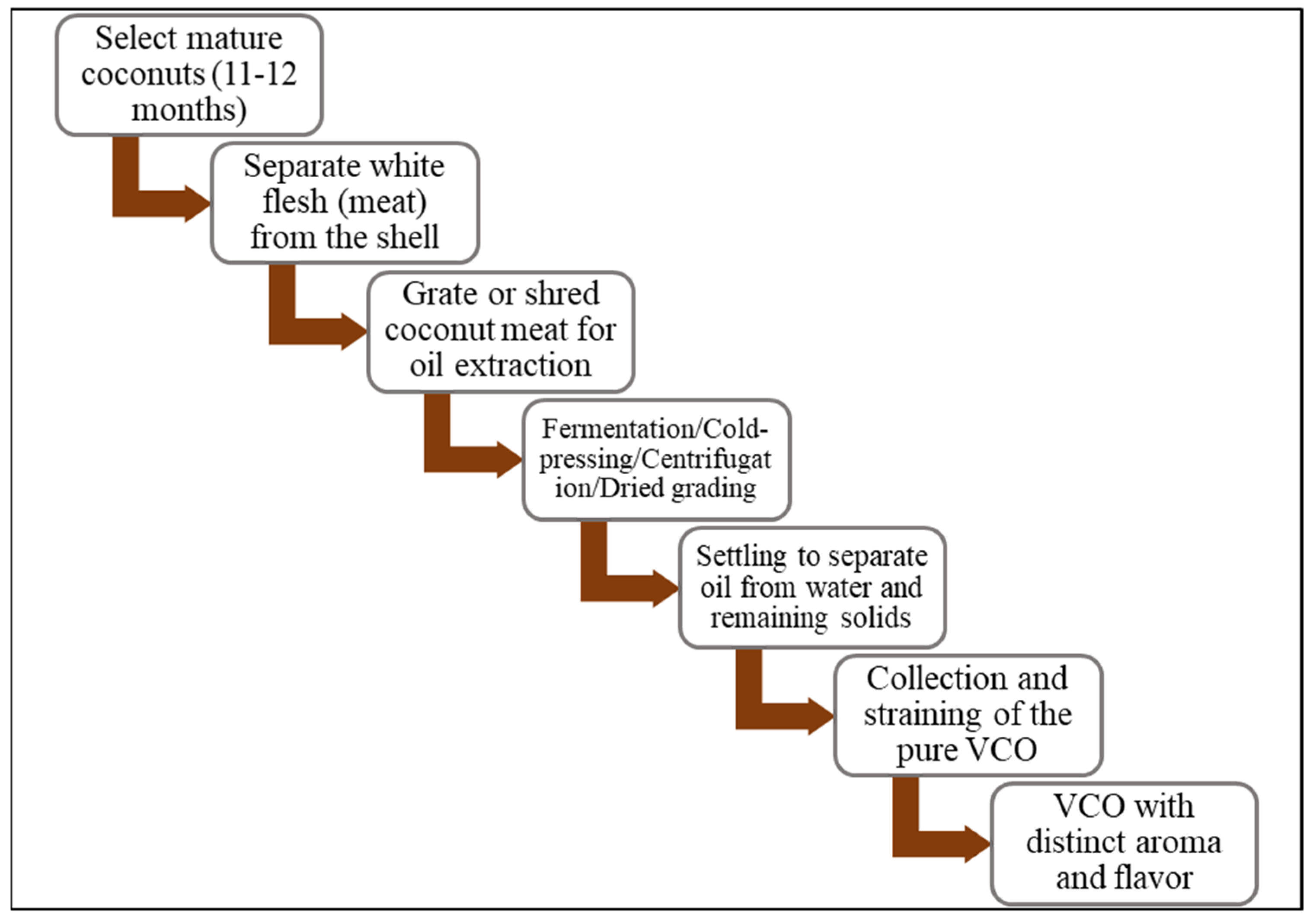
Figure 1. A schematic process flowchart for the extraction of VCO.
Jox 15 00126 g001
Figure 2. A schematic flowchart of methods and processing stages for VCO production.
Jox 15 00126 g002
Figure 3. Composition and percentage component of VCO.
Jox 15 00126 g003
Figure 4. Molecular mechanisms associated with anticancer effects of VCO or LA and their derivatives. VCO, LA, and their derivatives, upon entering the cell, interact directly with molecules and organelles to generate ROS and/or oxidative stress, which leads to depressed cancer cell viability, anti-proliferation, mitochondrial damage, and modulation of cancer-cell-death-related miRNAs. These alterations cause apoptotic cascades via cell cycle arrest, depressed expression of proteins, and genes that provoke cancer cell death.
Jox 15 00126 g004
Figure 5. VCO or LA protective mechanisms against chemotherapy-induced organ toxicity. Chemotherapeutic drugs centrally induce ROS generation and oxidation stress for promotion of apoptosis and inflammatory mechanisms in healthy organs. VCO or LA inhibits these mechanisms at various key mechanistic points, leading to elevation of antioxidant mechanism and dampening or blockage of apoptosis and inflammatory process.
Jox 15 00126 g005
Table 1. Bioactive components in VCO and their reported effects.
Bioactive ClassBioactive Constituents Reported Effect References
Medium-chain fattyLA (C12), capric acid (C10), caprylic acid (C8), caproic acid (C6)Antimicrobial, immunomodulatory, antiviral[40,44,45,46]
PolyphenolFerulic acid, catechins, p-coumaric acid, Antioxidant, anti-inflammatory[42]
Tocopherolsα-TocopherolAntioxidant[45,47]
Phytosterolsβ-SitosterolHypocholesterolemia[43]
Phenolic acidsCaffeic acid, gallic acidFree radical scavenging, anti-inflammatory, blood glucose regulation, chemoprevention, and immunomodulation[48,49]
FlavonoidsQuercetinAntioxidant, vascular protection[40]
Table 2. Compositional and metabolic overview of VCO, butter, and hydrogenated oils.
Component/FeatureVCOButter Hydrogenated Oil
Medium-chain triglycerides HighLow Absent
PolyphenolsModerateLowAbsent
Trans-fatsAbsentAbsentHigh
CholesterolAbsentHighVariable
Absorption rateFastSlowSlow
Metabolic oxidationFastSlowSlow
LDL/HDL effectIncreases HDL, decreases LDL/HDL ratioIncrease LDLHighly increase LDL
Table 3. Studies on anticancer effects of VCO and its derivatives.
VCO NatureCancer TypeModel/Cell LineEffect/ActivityDoseTimingReferences
VCOLiver and oral cancersHepG2, KB Antiproliferation 50–100 µg/mL72 h[121]
VCO nanoemulsionLung cancerA549, EACAntiproliferationNot specified48 h[122]
VCOSkin cancerMelanoma cell lineAntiproliferation10–300 µg/mL48 h[124]
VCOColon cancerColon adenocarcinoma cell lineAntiproliferation30 µg/mL72 h[125]
COColon cancerIn vivo↑ Colonic mucin 2 proteinHigh-fat SFA-rich diet28 days[97]
VCOLung cancerNCI-H1299, A549Antiproliferation
↑ Apoptosis
25–100 µg/mL24–72 h[96]
CONeuroblastomaSH-SY5YAntiproliferation
↑ Apoptosis
Mitochondrial damage
50–150 µg/mL24–72 h[7]
VCO-trastuzumabBreast cancerSKBR-3AntiproliferationNot specified48 h[126]
↑ = Stimulation.
Table 4. Studies on the anticancer effect of LA and its derivatives.
LA/DerivativeCancer TypeModel/Cell LineEffect/ActivityDoseTimingNormal Cell Control UsedReferences
LANeuroblastomaISH-SY5Y↑ ROS, apoptosis,
mitochondrial damage
20–100 µg/mL24–72 hNo[7]
LALiver and colorectal cancersHepG2, HT-29Antiproliferation, ↓ epidermal growth factor receptor (EGFR), and ↑ apoptosis, anti-metastatic effect30–50 µg/mL24–48 hNo[30]
LA-cetuximab or LA aloneKRAS/BRAF-mutated colorectal cancerHCT116Antiproliferation, ↑ microRNA-378,
↓ MAP, ↓ ERK-2
25 µM48 hNo[128]
LAColon cancerHCT116Antiproliferation,
↑ ROS, ↓ GSH, ↑ apoptosis, G2/M cell cycle arrest
20–60 µM24–48 hYes[129]
LAEndometrial and breast cancersIshikawa, MCF-7Antiproliferation, ↑ apoptosis, ↑ EGFR/ERK25–100 µM24–48 hYes[130]
LAColon cancerHT-29Antiproliferation30 µM72 hYes[125]
LA-Gemcitabine or LA aloneBladder cancerT24Antiproliferation, ↑ apoptosis, ↓ PPARG and ↓ COX2 genes,
cell cycle arrest
10–40 µM48 hNo[27]
LA nanocapsule (SiO2@LA)Breast cancerMCF-7↓ Cell viability, ↑ ROS, ↑ apoptosisNot specified24 hYes[131]
LA-platinum nanocomplex Colorectal cancerHT-29↓ Cell viability, ↑ ROS5 µM24 hNo[132]
LA-nanodrug systems (5-FU-LA@PPL)Colorectal cancerXenograft (BALB/c nude mice)↓ Cell viability
↓ autophagy
5 µM48 hNo[133]
LA-conjugated 2-hydroxyanthraquinoneBreast, colon and leukemia cancersHL-60, HCT116,↓ Cell viability10–25 µM24–48 hNo[134]
LA-based hydrazonesLiver cancerHepG2Antiproliferation50 µM48 hNo[135]
LA-chitosan-based nanogelOral cancerGa9-22Antiproliferation, ↓ cell viability, ↑ ROS, G2/M, cell cycle arrest, and ↑ Bad/Bax/p53 apoptosis20 µg/mL24 hYes[136]
LAOral and liver cancersHepG2, SCC-15↓ Oncogenic microRNA, ↑ tumor-suppressor microRNA, cell cycle arrest, ↑ apoptosis20 µg/mL24–48 hNo[137]
↑ = Stimulation. ↓ = Inhibition.
Table 5. Protective effects/mechanisms of VCO or LA against chemotherapy toxicity.
VCO or LAAnticancer DrugType of ToxicityAnimal Model DetailsMechanistic Outcomes (% Improvement)Dose and TimingReferences
VCODoxorubicin (DOX)Neurobehavioral toxicitySwiss albino mice, DOX-induced, post-treatment, behavior assays↓ AchE, TNF-α, iNOS; improved behavior; ↓ oxidative stress (approx. 35–50% change in markers)5 mL/kg/day VCO × 28 days, DOX 5 mg/kg i.p.[25]
VCODoxorubicin (DOX)ImmunosuppressionBalb/c mice, DOX-induced, pre-treatment, immune assays↑ CD4+, CD8+, lymphocyte proliferation, macrophage activity (up to 45%)10 mL/kg/day VCO × 14 days, DOX 10 mg/kg i.p.[150]
VCODoxorubicin (DOX)Hepatic and cardiac toxicityWistar rats, DOX-induced, post-treatment, histopathological evaluation↓ ALT, AST, LDH, CK-MB (~30–40%); restored histology5 mL/kg/day VCO × 21 days, DOX 5 mg/kg i.p.[151,152]
VCODoxorubicin (DOX)Liver toxicityWistar rats, DOX-induced, post-treatment, cytokine, and antioxidant evaluation↓ AST, ALT, MDA, inflammatory cytokines (30–60%), ↑ Bcl-2, antioxidant enzymes5 mL/kg/day VCO × 21 days, DOX 5 mg/kg i.p.[24]
VCOCisplatin (CP)NephrotoxicityWistar rats, CP-induced nephrotoxicity, comparison of hot/cold VCO↓ Creatinine, urea, ↑ SOD, CAT; higher in hot-processed VCO (20–50% difference vs. CP-only)5 mL/kg/day VCO × 10 days, CP 5 mg/kg i.p.[153]
VCOCyclophosphamide (CYP)Hepatorenal toxicitySwiss albino mice, CYP-induced, pre-treatment, hepatorenal function assessed↑ GSH, ↓ MDA, restored organ function (30–45%)10 mL/kg/day VCO × 20 days, CYP 200 mg/kg i.p.[17]
VCOCyclophosphamide (CYP)Lymphoid toxicity (altered hematological indices, histological spleen/thymus changes)Swiss mice, CYP-induced, spleen/thymus histology, hematological changesImproved spleen/thymus histology and hematology (qualitative only)5 mL/kg/day VCO × 10 days, CYP 100 mg/kg[156]
VCOMethotrexate (MTX)Oxidative stress & inflammationWistar rats, MTX-induced, pre-treatment with VCO nanoemulsion, antioxidant evaluation↑ SOD, CAT, ↓ lipid peroxidation (35–60%)5 mL/kg/day VCO nanoemulsion × 7 days, MTX 20 mg/kg[122]
Methotrexate (MTX)Hepatotoxicity, nephrotoxicity, neurotoxicity, systemic toxicityWistar rats, MTX-induced, systemic toxicity markers assessed↓ ALT, creatinine, TNF-α, IL-6; ↑ SOD, CAT (~40–60%)5 mL/kg/day VCO × 10 days, MTX 20 mg/kg[8,21,22,123]
LALA and glucoseCachexia (muscle atrophy, weight loss)C57BL/6 mice, cancer cachexia model, LA + glucose supplementation↑ Muscle mass, ↑ MYL1 gene (approx. 30%)250 mg/kg LA + 1 g/kg glucose × 14 days[159]
↑ = Stimulation. ↓ = Inhibition.
Disclaimer/Publisher’s Note: The statements, opinions and data contained in all publications are solely those of the individual author(s) and contributor(s) and not of MDPI and/or the editor(s). MDPI and/or the editor(s) disclaim responsibility for any injury to people or property resulting from any ideas, methods, instructions or products referred to in the content.