Article

Anterior Column Reconstruction of the Thoracolumbar Spine with a Modular Carbon-PEEK Vertebral Body Replacement Device: Single-Center Retrospective Case Series of 28 Patients

by
Samuel F. Schaible
1,*,
Fabian C. Aregger
1,
Christoph E. Albers
1,2,
Lorin M. Benneker
1,2 and
Moritz C. Deml
1,*
1
Department of Orthopaedic Surgery and Traumatology, University Hospital Bern, Inselspital, University of Bern, CH-3010 Bern, Switzerland
2
Sonnenhofspital Bern, University of Bern, CH-3006 Bern, Switzerland
*
Authors to whom correspondence should be addressed.
Surg. Tech. Dev. 2025, 14(4), 35; https://doi.org/10.3390/std14040035
Submission received: 1 May 2025 / Revised: 22 August 2025 / Accepted: 22 September 2025 / Published: 10 October 2025

Abstract

Background: Carbon-fiber-reinforced polyetheretherketone (CFR-PEEK) vertebral-body replacements (VBRs) aim to mitigate subsidence, minimize imaging artifacts, and facilitate radiation planning while preserving fusion potential. We assessed the safety and efficacy of a novel modular, titanium-coated CFR-PEEK VBR (Kong®) for anterior column reconstruction (ACR) in the thoracolumbar spine. Primary question: Does the implant safely and effectively achieve and maintain kyphosis correction after ACR for trauma and neoplasms? Methods: A single-center retrospective case series was performed on 28 patients who underwent thoracolumbar ACR with the Kong® VBR for fractures or tumors (2020–2021). The primary outcome was the bi-segmental kyphotic angle (BKA). Secondary outcomes were screw loosening, cage height loss, fusion rate, subsidence, and tilting. Clinical status was recorded with Odom criteria, Karnofsky Performance Status (KPS), and AOSpine PROST. Results: Twenty-eight patients (mean age, 61 yr; 33% female; mean follow-up, 17.7 mts) were studied. Mean postoperative BKA correction was 16.5° (p = 0.006) and remained 14.5° at final follow-up (p = 0.008); loss of correction was 2.0° (p = 0.568). Subsidence, cage height, and sagittal tilt were unchanged. Fusion (Bridwell grade I/II) was observed in 95% on CT. One deep surgical-site infection occurred. At final follow-up, 91% of patients were graded “excellent” or “good” by Odom. KPS improved by 20 points (p = 0.031), and mean AOSpine PROST was 56.9. Conclusions: Single-center early results indicate that the modular titanium-coated CFR-PEEK VBR is a safe, effective adjunct for thoracolumbar ACR in trauma and neoplasm, providing durable kyphosis correction, mechanical stability and high fusion rates and grants for improved follow-up imaging quality.

1. Introduction

The reconstruction of the anterior and middle spinal columns following corpectomy is essential for restoring spinal stability; however, stabilizing the anterior column remains challenging due to complications such as construct failures, including cage dislocation, non-fusion, segment degeneration, and iatrogenic deformity [1,2]. Early attempts at anterior column reconstruction (ACR) employed bone autografts and femoral allograft struts and rings, but these methods often failed to maintain long-term construct stability [3,4]. The advent of titanium implants, initially as mesh cages [5] and later as expandable devices, led to significant improvements in stabilization, fusion rates, and kyphosis correction. Modular implants have further enhanced applicability and adaptability [2].
Polyetheretherketone (PEEK) implants have gained popularity in interbody fusion and vertebral body replacement (VBR) due to their elastic modulus comparable to cancellous bone, potentially reducing subsidence. Moreover, their radiolucency allows for enhanced follow-up imaging and facilitates postoperative radiation therapy [6,7]. Conversely, PEEK lacks direct osseointegration potential due to its lower bioactivity with bone, leading to proposals for adding bioactive coatings [6]. Titanium-coated carbon fiber-reinforced PEEK (CFR-PEEK) implants theoretically provide improved osseointegration capabilities while retaining the advantages of a reduced elastic modulus and radiolucency. In clinical practice, pedicle screws made from CFR-PEEK have shown a risk profile and rates of implant failure similar to those of traditional metallic implants, with the added benefits of enhanced postoperative imaging quality and greater accuracy in calculating radiotherapy doses compared to titanium counterparts [8,9,10]. Although vacuum plasma-sprayed titanium-coated CFR-PEEK cages have proven effective in lumbar interbody fusion [11], only one study with limited follow-up has evaluated an expandable titanium-coated CFR-PEEK VBR device for ACR to date, underscoring the need for further assessment in different clinical settings [12].
The primary objective of this study was to assess the safety and efficacy of a new modular, expandable, titanium-coated CFR-PEEK VBR (Kong®) in preserving local kyphosis correction in the thoracolumbar spine for spinal defects due to trauma and spinal neoplasms. Secondary objectives included radiographic evaluation of cage and construct stability, fusion rate, complications, artifacts in post-op CT imaging and clinical outcomes.

2. Materials and Methods

2.1. Study Design and Patient Selection

With approval from the Cantonal Ethics Committee (Kantonale Ethikkommission Bern [KEK]), we conducted a retrospective case series analysis of demographic, perioperative, and radiographic data on patients aged 18 years and older who underwent spinal surgery, using the Kong® VBR (icotec ag, Altstätten, Switzerland; Figure 1) at our institution. This analysis included our first 28 consecutive cases treated for trauma or tumors in the T9 to L4 region between January 2020 and December 2021. We excluded patients with infectious conditions, those without general consent, those with incomplete medical records, and those with follow-up periods shorter than six months.

2.2. Radiographic Evaluation

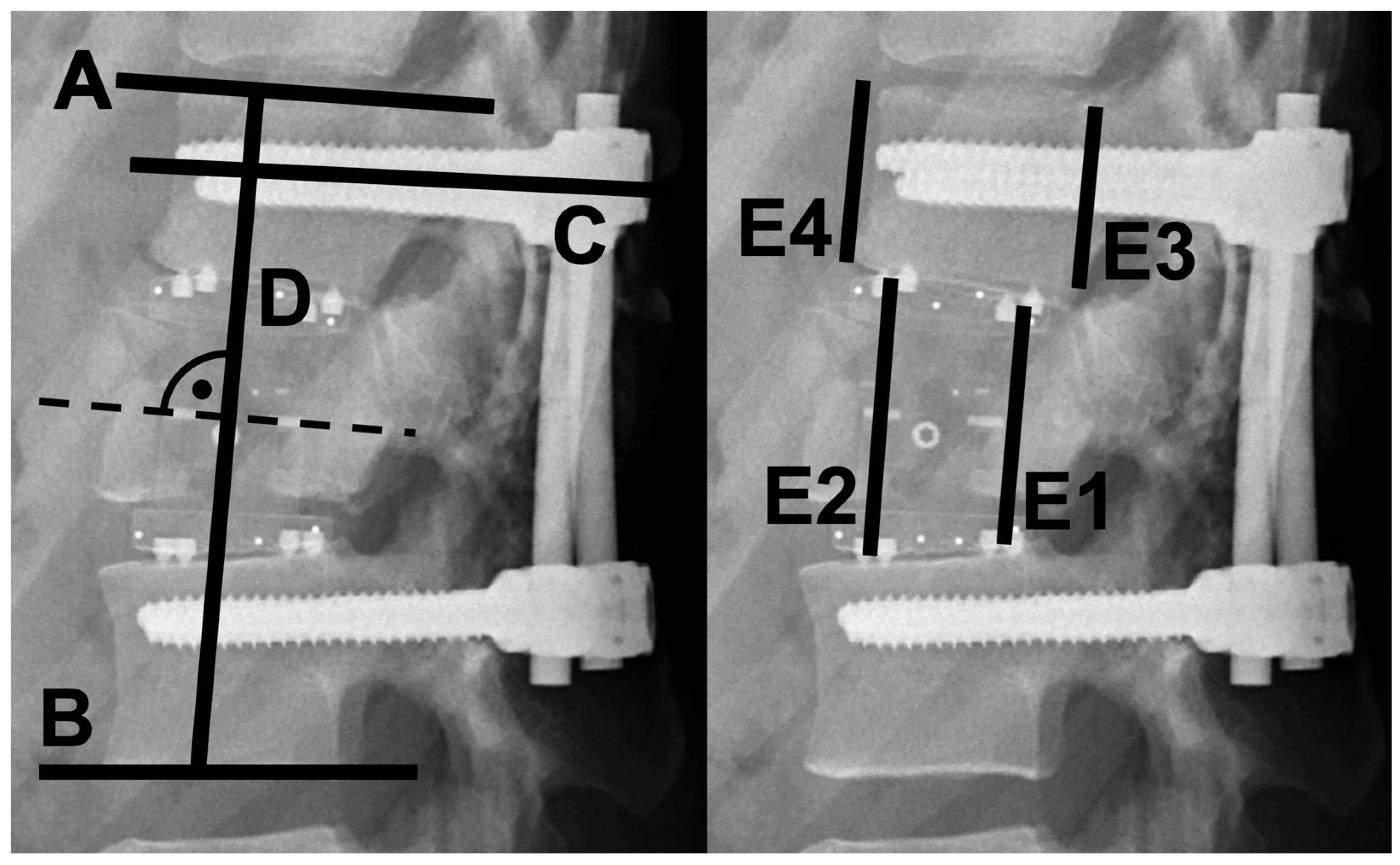
Standing plain radiographs were obtained preoperatively, immediately postoperatively, at 6 weeks, 3 months, 1 year, and 2 years, with annual follow-up thereafter if indicated. After 12 months, most patients underwent computed tomography (CT). Kyphosis correction was evaluated by measuring the bi-segmental kyphotic angle (BKA) with the Cobb method; the angle was defined between the upper endplate of the cranial intact vertebra and the lower endplate of the caudal intact vertebra [13]. Measurements were recorded preoperatively, postoperatively, and at the last available follow-up (≥6 months). For single-segment stabilization (hemicorpectomy), only the affected segment was evaluated, whereas in 3-segment stabilization all treated segments were assessed. Subsidence (decrease in construct height) and sagittal cage angulation were determined with the method of Schnake et al. [2]. Screw loosening was evaluated with the Epsilon-angle method of Aghayev et al. to minimize measurement bias [14] (Figure 2). Cage-expansion integrity was assessed with the previously described cage-height coefficient [15]. Anterior bony fusion was graded on available CT scans and plain radiographs according to the Bridwell I–IV scale [16,17].
To evaluate metallic artifacts from carbon cages versus titanium screws, we performed artifact quantification on all available CT scans (Siemens Biograph Vision Quadra PET/CT with iMAR; Siemens Healthineers, Forchheim, Germany) using axial bone-window reconstructions (1 mm slice thickness). We adapted methods from Guggenberger et al. [18] and Bamberg et al. [19]. For titanium screws, we selected the slice with the greatest hypodense streak and placed a circular ROI in the artifact to measure mean Hounsfield units (HU), standard deviation (SD), and diameter (mm). We obtained reference HU/SD from an unaffected slice in identical tissue (e.g., muscle/vertebral body). For carbon cages, we obtained measurements at the cage center in the axial mid-slice, with reference values from adjacent tissue. The rationale is that visually, due to the central locking mechanism, this was the only location where artifacts were uniquely produced by the cage, since the screws are usually above and below its center. While the tantalum spikes at the endplates also cause artifacts, in their corresponding slices, the chief artifacts are produced by screws. We quantified noise as SD in background air. Qualitative evaluation included artifact extent (0 = none to 4 = massive) and diagnostic value (0 = fully diagnostic to 4 = non-diagnostic) scores on a 0–4 Likert scale [18,19].

2.3. Clinical Outcomes

Intraoperative complications were extracted from operative reports to evaluate implantation safety. Perioperative and postoperative complications were recorded from follow-up notes. Clinical outcome was assessed postoperatively with the Odom criteria, a 4-point scale (excellent, good, fair, poor) widely used after spinal surgery [20,21]. At the final follow-up, trauma patients completed the AOSpine Patient-Reported Outcome Spine Trauma (PROST) questionnaire [22]. In tumor patients, the Karnofsky Performance Status (KPS) was documented preoperatively and at the final follow-up [23].

2.4. Surgical Technique

All patients first underwent posterior stabilization with a pedicle screw–rod system, with or without decompression, depending on the underlying pathology and clinical presentation. Corpectomy was performed via mini-open thoracotomy for thoracic pathologies and via lateral mini-open lumbotomy for lumbar pathologies. The extent of corpectomy was tailored to the pathology: hemicorpectomy was performed for mono-segmental cases (e.g., A3-type fractures according to the AO Spine Trauma Classification with severe upper endplate destruction and kyphosis > 30°), whereas complete corpectomy was necessary for bi- or tri-segmental pathologies. The cage size and endplate configuration were intraoperatively customized to the individual defect (see Figure 1). In trauma cases, after corpectomy and adjacent endplate preparation, the cage bed was filled with local cancellous autograft; in tumor cases, allograft bone chips were used. Correct cage positioning was confirmed intraoperatively with fluoroscopy.

2.5. Statistical Analyses

All analyses were carried out in R (v 4.3.1; R Foundation for Statistical Computing) within RStudio (2023.03.0 + 386, Boston, MA, USA). Continuous variables were examined for normality with the Shapiro–Wilk test. Normally distributed data (bi-segmental kyphotic angle, sagittal tilt, Epsilon angle, construct height) are expressed as mean ± SD and were compared between paired time points with two-tailed paired Student t-tests. Non-normal continuous data (cage-height coefficient, Karnofsky Performance Status, AOSpine PROST) are reported as median (IQR) and were analyzed with two-tailed Wilcoxon signed-rank tests. Ordinal and categorical variables (Odom grades, Bridwell fusion grades, complications) are presented as counts and percentages without inferential statistics. Statistical significance was defined as p < 0.05. All R code was independently audited by a biostatistician to confirm accuracy and reproducibility.

3. Results

3.1. Cohort Characteristics

Thirty-three consecutive cases were screened; five were excluded—three for inadequate follow-up and two for infection—yielding a case series of twenty-eight patients: fifteen with high-energy fractures, seven with osteoporotic fractures, and six with tumors (Table 1). The cohort included ten women (36%); the mean age was 60.5 ± 18.6 years (range, 19 to 89 years), and the mean radiographic follow-up was 17.7 ± 7.7 months (range, 9 to 36 months).
Eleven reconstructions (39%) were completed in a single stage and seventeen (61%) in two stages. All trauma patients received titanium pedicle screws configured as two percutaneous monoaxial, four percutaneous polyaxial, six open monoaxial, and three open polyaxial constructs. Every osteoporotic case was treated with titanium polyaxial screws—three percutaneously and four via an open approach—with cement augmentation in all seven patients. All tumor cases were instrumented through an open approach; one patient received titanium monoaxial screws and five received carbon-fiber-reinforced PEEK polyaxial screws. Titanium rods were used in every case except three tumor patients. Screw cement augmentation was performed in eight patients (seven osteoporotic and one tumor). In the trauma cohort, posterior hardware was electively removed in nine patients (32%) after at least six months, once fusion had been confirmed on CT.

3.2. Radiographic Outcomes

Postoperatively, the bi-segmental kyphotic angle (BKA) improved by 16.5° ± 23.6° (p = 0.006; preoperative value, −11.6° ± 20.4°) (Figure 3, Table 2). At the final review the loss of correction was 2.0° ± 15.0° (p = 0.568), leaving a net gain of 14.5° ± 22.0° over baseline (p = 0.008). Sagittal tilt remained unchanged (mean difference, 0.83 ± 4.61°; p = 0.385). The Epsilon angle showed no interval change (mean difference, −0.17° ± 1.47°; p = 0.793), and no screw lysis > 2 mm was detected on radiographs or CT.
Construct height was stable (mean change, −0.5 ± 0.71 mm; p = 0.052), and the cage-height coefficient did not shift (median change, 0.02; p = 0.140) (Table 2). No cage collapse, expansion-mechanism failure, or loosening at the endplate–core interface occurred.
Twelve-month CT was obtained in eighteen patients and demonstrated Bridwell grade I or II fusion in seventeen (94.4%); one osteoporotic patient was grade III. On the latest plain radiographs, 26 of 28 patients (92.9%) were grade I or II and 2 (7.1%) were grade III, findings that mirrored the CT assessments (Table 3).
Artifact analysis was conducted on CT scans from all patients with available imaging. Quantitative metrics for carbon-fiber-reinforced PEEK cages and titanium screws are summarized in Table 4. Artifact width (ROI diameter) was 1.4 ± 0.5 mm for cages and 2.6 ± 0.8 mm for screws (p = 0.0004). Mean artifact HU was −46.8 ± 39.7 HU for cages and −360.0 ± 104.1 HU for screws (p < 0.0001). Artifact SD was 57.2 ± 49.8 for cages and 127.7 ± 47.7 for screws (p = 0.0010). Absolute HU difference from reference tissue was 109.4 ± 56.6 HU for cages and 409.5 ± 107.0 HU for screws (p < 0.0001). Relative density was −87.6 ± 95.1% for cages and −946.2 ± 581.3% for screws (p = 0.0001). Implant HU was 352.7 ± 23.4 HU for cages and 3068.6 ± 3.9 HU for screws (p < 0.0001). Reference HU was 62.6 ± 33.7 for cages and 49.5 ± 22.7 for screws (p = 0.2638). Reference SD was 48.2 ± 32.8 for cages and 45.7 ± 28.7 for screws (p = 0.6734). Noise SD was 50.6 ± 42.3 HU for both (p = NaN).
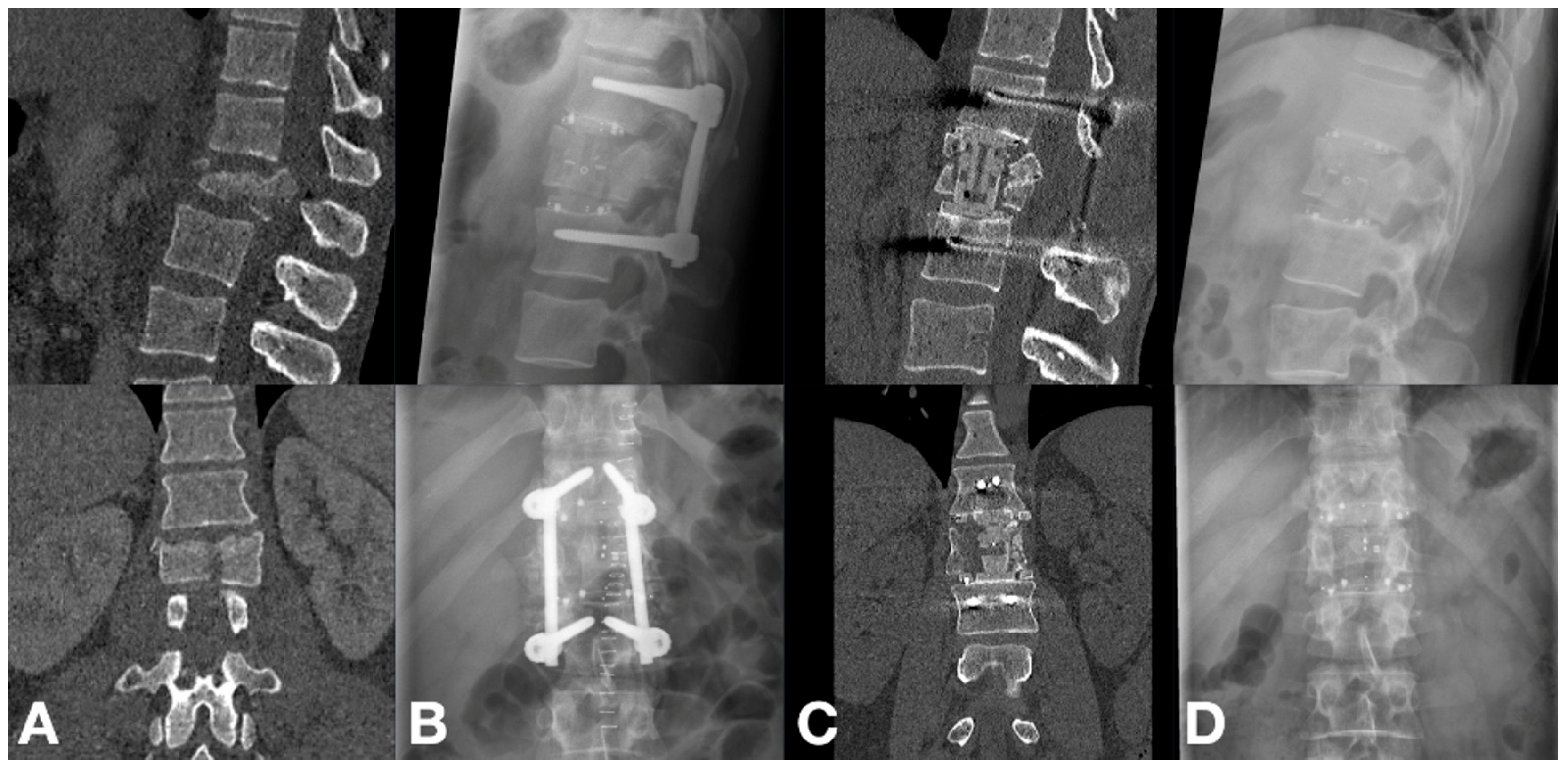
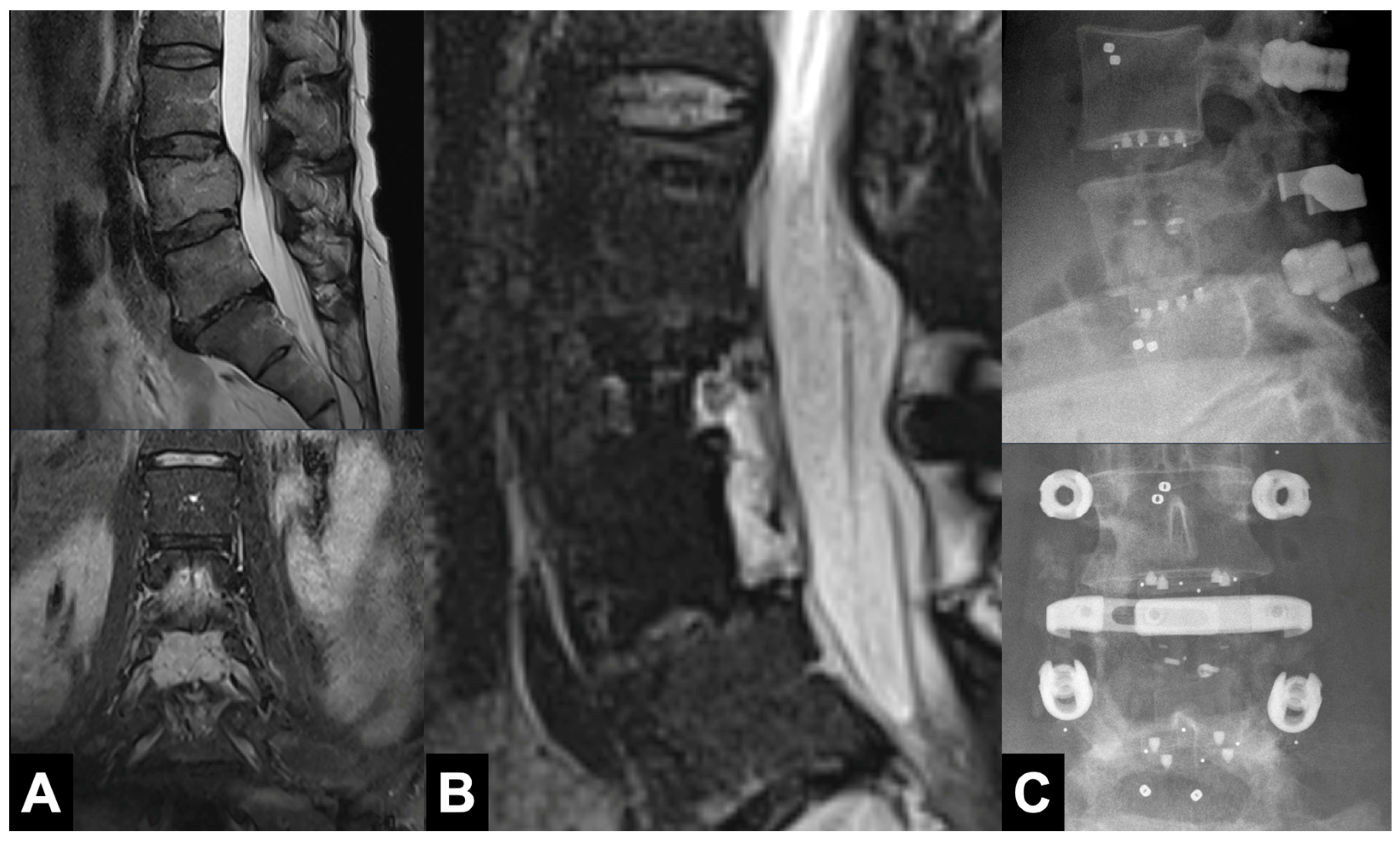
Qualitative assessment was performed for cages only. Artifact extent score was 0.2 ± 0.4 on a 0–4 scale. Diagnostic value score was 0.0 ± 0.0 on a 0–4 scale. Figure 4 and Figure 5 illustrate the excellent CT and MRI image quality afforded by the low-artifact carbon-fiber PEEK implants.

3.3. Clinical Outcomes

Median Karnofsky Performance Status in the six oncologic cases improved by 20 points from baseline to final review (p = 0.031; Table 4). Among the 22 non-tumor patients, Odom grades improved serially, and 20 (91%) rated the result “excellent” or “good” at last follow-up (Table 4). The 15 high-energy–trauma patients recorded a mean AOSpine PROST of 56.9 ± 21.3, indicating moderate postoperative function (Table 4). Neurologic status improved in seven of the eight patients who presented with a deficit (four traumatic, four neoplastic; Table 5).

3.4. Complications

No intraoperative nerve or major vascular injuries occurred, and no secondary cage migration or construct failure was observed. Construct-adjacent vertebral fractures developed in 3 patients (10.7%), all >70 years. One tumor patient (3.6%) experienced a deep surgical-site infection 2 weeks postoperatively; the infection resolved after a single debridement without hardware exchange.

4. Discussion

This retrospective pilot case series evaluated a modular, expandable, titanium-coated CFR-PEEK vertebral-body replacement (Kong®) for anterior column reconstruction after thoracolumbar trauma or tumor. The bi-segmental kyphotic angle improved by a mean 14.5° ± 22.0° from baseline (p = 0.008) and was preserved at final review. Subsidence was negligible, sagittal tilt unchanged, and no screw loosening or implant failure occurred. The device therefore provided durable kyphosis correction, high fusion rates, and a low complication burden, supporting its use as an alternative to conventional titanium cages.

4.1. Comparison with Existing Literature

Our radiographic results mirror those reported for expandable titanium cages. Schnake et al. noted a 2.2° loss of bi-segmental kyphotic angle (BKA) at 12 months and 6.5° at 60 months, with 2.2 mm of subsidence, after thoracolumbar fracture reconstruction with titanium cages [2]. Lange et al. recorded a 2.3° BKA loss and 2.9 mm subsidence at 12 months in a mixed trauma-tumor cohort treated with expandable titanium vertebral-body replacements [24]. The present series showed comparable maintenance of alignment and similarly minimal subsidence.
The lower elastic modulus of PEEK may further mitigate subsidence. In a randomized trial of cervical spondylotic myelopathy, Chen et al. reported subsidence >3 mm in 34.5% of titanium versus 5.4% of PEEK cage [25]. Schwendner et al. evaluated the same titanium-coated CFR-PEEK (Kong®) cage in 17 thoracolumbar tumor cases and found a 3.3° BKA loss and 3.8% subsidence at 0.8-year mean follow-up [12], corroborating our findings.

4.2. Technical Aspects

Due to the better load distribution, subsidence is least when the cage spans the apophyseal ring and maximizes end-plate contact, as previously demonstrated in biomechanical experiments [26]. The Kong® system supplies interchangeable endplates with varied footprints for intraoperative, anatomy-matched sizing. Its carbon-fiber PEEK core approximates the elastic modulus of cancellous bone, a particular advantage in osteoporotic bone. Reflecting these features, mean subsidence in our series was only −0.5 ± 0.7 mm (p = 0.052).

4.3. Osseointegration and Fusion Rates

PEEK’s bio-inert nature has historically raised concerns about its osseointegration potential [27]. Prior studies evaluating uncoated PEEK VBRs for thoracolumbar reconstruction have reported similarly high success rates, such as the 96.3% fusion rate observed by Brandão et al. [28]. The current series observed a Bridwell grade I/II fusion rate of 95% on CT imaging with the titanium-coated CFR-PEEK cage. This result is favorable and is in the same ballpark as reported fusion rates reported for all-titanium constructs [29]. However, this study was not designed for direct comparison, and given the absence of a control group with titanium implants, conclusions on osseointegration performance relative to all-titanium devices cannot be drawn. In theory, the titanium layer supplies an osteoconductive surface. Conversely, preclinical studies have described gradual coating loss [30,31], and a meta-analysis reported no fusion advantage for coated versus uncoated PEEK implants [6]. In summary, the high rate of fusion observed here is a promising finding, but it requires validation through direct comparative studies.

4.4. Rationale for Use and Economic Considerations

The selection of a vertebral body replacement is guided by its clinical performance and economic impact. While the radiolucency of CFR-PEEK offers a clear advantage in spinal oncology and infection-related indications, facilitating imaging and radiation planning, its application in trauma and osteoporotic fractures in this cohort was primarily driven by mechanical properties, including footprint modularity for anatomical contact and a lower elastic modulus compared to titanium, which theoretically reduces subsidence risk in compromised bone. Outside oncologic or infection-related cases, where radiolucency is the primary benefit, the higher device cost necessitates selective use. Although a formal cost–utility analysis is beyond this study’s scope, the convergence of mechanical, oncologic, and institutional fiscal advantages justified adopting the CFR-PEEK device across our heterogeneous pathologies. This low-artifact profile may enable more accurate detection of complications such as subsidence or loosening. Formal cost–utility data are lacking and should be addressed prospectively.

4.5. Implantation Safety and Complications

The Kong® cage showed an excellent safety profile: no intraoperative implant-related events and no hardware failures during follow-up. One deep surgical-site infection required debridement. The cage-height coefficient remained stable, confirming construct integrity [15]. From our early experiences, insertion of the cage in narrow anatomic situation (e.g., hemicorpectomy) can be challenging because of the pronounced anchoring spikes; precise sizing and gentle impaction are essential to avoid neural or vascular injury, especially for first-time users.

4.6. Limitations

The conclusions of this study must be interpreted in the context of several limitations. First, as a retrospective, single-center case series, the study is subject to selection bias and lacks a concurrent control group. The absence of a comparative arm—such as patients treated with all-titanium VBRs—precludes any definitive statements regarding the superiority of this CFR-PEEK device.
Second, the cohort was small and heterogeneous, comprising patients with distinct pathologies (trauma, osteoporosis, tumors). This heterogeneity may introduce confounding factors affecting outcomes, such as varying bone quality or healing potential across groups. Although subgroup data are presented, the small sample size limits statistical power for subgroup analyses. We combined tumor and osteoporotic fracture patients for certain outcome measures based on the shared clinical characteristic of compromised, osteopenic bone; however, this pragmatic grouping does not overcome the fundamental limitation of low statistical power.
Third, the mean radiographic follow-up of 17.7 months is adequate for assessing early outcomes such as implant stability and fusion initiation, but it is insufficient to evaluate long-term phenomena like delayed implant subsidence, adjacent segment disease, or hardware fatigue. Consequently, conclusions on long-term durability are preliminary and should be interpreted cautiously. Nevertheless, the goal of the investigated procedure is a fusion of minimum two vertebra, which was achieved in 95% of the cases. A late subsidence or failure of the VBR after solid fusion is very unlikely.
Finally, our commentary on the implant’s favorable imaging characteristics is based on qualitative radiological assessment. A quantitative analysis of imaging artifact was not performed but would be a valuable addition in future prospective studies. Ultimately, the promising results of this series require validation through larger, prospective, randomized controlled trials.

5. Conclusions

This study suggests that the modular, expandable, titanium-coated CFR-PEEK Kong® VBR system is a safe and effective option for ACR in the thoracolumbar spine due to trauma or neoplasia. It achieves significant kyphosis correction, maintains sagittal alignment with minimal subsidence, and demonstrates high fusion rates. The system’s adaptability and favorable imaging characteristics due to low artifact formation offer additional advantages. While these single-arm data compare favorably with historical titanium-cage series, controlled trials are required to verify any superiority to titanium implants.

Author Contributions

S.F.S. contributed to the methodology, investigation, formal analysis, data curation, and writing—original draft preparation. F.C.A. contributed to the investigation, formal analysis, data curation, and writing—original draft preparation. M.C.D. performed the surgeries, contributed to the investigation, formal analysis, and data curation. C.E.A. and L.M.B. performed the surgeries. All authors contributed to writing—review and editing. All authors have read and agreed to the published version of the manuscript.

Funding

This research received no external funding. The APC was funded by the authors.

Institutional Review Board Statement

Ethical review and approval were waived for this study by conthe Cantonal Ethics Commission Bern (KEK Bern) because it involved a retrospective analysis of fully anonymized clinical and radiographic data and imposed no additional risk or intervention for the patients.

Informed Consent Statement

All patients had previously provided institutional general consent for the use of their anonymized clinical and radiographic data in research. Accordingly, study-specific consent was waived because this retrospective analysis imposed no additional intervention or risk.

Data Availability Statement

The anonymized clinical and imaging datasets generated and analyzed during the present study are not publicly available due to patient-privacy regulations. They can be obtained from the corresponding author for reasonable academic purposes upon receipt of a data-sharing agreement approved by the institutional data protection office.

Acknowledgments

During the preparation of this manuscript, the authors used Grok 4 (xAi Inc., August 2025 version) for language refinement and clarity. The authors have reviewed and edited the output and take full responsibility for the content of this publication.

Conflicts of Interest

M.C.D. and C.E.A. have received research support from icotec ag outside the submitted work. M.C.D holds advisory roles within the AO Spine. C.E.A. has also obtained grant funding from the Bangerter-Rhyner Stiftung and Swiss Orthopaedics. L.M.B. serves as a consultant to icotec ag and contributed to the development of the Kong® cage; he holds advisory roles within AO Spine. All other authors report no relevant conflicts.

Abbreviations

The following abbreviations are used in this manuscript:
ACRAnterior Column Reconstruction
AO SpineArbeitsgemeinschaft für Osteosynthesefragen Spine
APCArticle Processing Charge
ASIAAmerican Spinal Injury Association impairment scale
BKABi-segmental Kyphotic Angle
CFR-PEEKCarbon-Fiber-Reinforced Polyether-Ether-Ketone
CTComputed Tomography
IQRInterquartile Range
KEKKantonale Ethikkommission (Cantonal Ethics Commission, Bern)
KPSKarnofsky Performance Status
MRIMagnetic Resonance Imaging
PEEKPolyether-Ether-Ketone
PROSTPatient-Reported Outcome Spine Trauma (AO Spine questionnaire)
SDStandard Deviation
SINSSpinal Instability Neoplastic Score
STIRShort Tau Inversion Recovery
VBRVertebral-Body Replacement

References

  1. Eleraky, M.; Papanastassiou, I.; Tran, N.D.; Dakwar, E.; Vrionis, F.D. Comparison of polymethylmethacrylate versus expandable cage in anterior vertebral column reconstruction after posterior extracavitary corpectomy in lumbar and thoraco-lumbar metastatic spine tumors. Eur. Spine J. 2011, 20, 1363–1370. [Google Scholar] [CrossRef]
  2. Schnake, K.J.; Stavridis, S.I.; Kandziora, F. Five-year clinical and radiological results of combined anteroposterior stabilization of thoracolumbar fractures. J. Neurosurg. Spine 2014, 20, 497–504. [Google Scholar] [CrossRef]
  3. Sacks, S. Anterior Interbody Fusion of the Lumbar Spine. J. Bone Jt. Surg. Br. Vol. 1965, 47-B, 211–223. [Google Scholar] [CrossRef]
  4. Lewandrowski, K.-U.; Hecht, A.C.; DeLaney, T.F.; Chapman, P.A.; Hornicek, F.J.; Pedlow, F.X. Anterior Spinal Arthrodesis With Structural Cortical Allografts and Instrumentation for Spine Tumor Surgery. Spine 2004, 29, 1150–1158. [Google Scholar] [CrossRef]
  5. Grob, D.; Daehn, S.; Mannion, A.F. Titanium mesh cages (TMC) in spine surgery. Eur. Spine J. 2005, 14, 211–221. [Google Scholar] [CrossRef]
  6. Kumar, N.; Ramakrishnan, S.A.; Lopez, K.G.; Madhu, S.; Ramos, M.R.D.; Fuh, J.Y.H.; Hallinan, J.; Nolan, C.P.; Benneker, L.M.; Vellayappan, B.A. Can Polyether Ether Ketone Dethrone Titanium as the Choice Implant Material for Metastatic Spine Tumor Surgery? World Neurosurg. 2021, 148, 94–109. [Google Scholar] [CrossRef] [PubMed]
  7. Muthiah, N.; Yolcu, Y.U.; Alan, N.; Agarwal, N.; Hamilton, D.K.; Ozpinar, A. Evolution of polyetheretherketone (PEEK) and titanium interbody devices for spinal procedures: A comprehensive review of the literature. Eur. Spine J. 2022, 31, 2547–2556. [Google Scholar] [CrossRef] [PubMed]
  8. Neal, M.T.; Richards, A.E.; Curley, K.L.; Patel, N.P.; Ashman, J.B.; Vora, S.A.; Kalani, M.A. Carbon fiber–reinforced PEEK instrumentation in the spinal oncology population: A retrospective series demonstrating technique, feasibility, and clinical outcomes. Neurosurg. Focus 2021, 50, E13. [Google Scholar] [CrossRef] [PubMed]
  9. Hubertus, V.; Wessels, L.; Früh, A.; Tkatschenko, D.; Nulis, I.; Bohner, G.; Prinz, V.; Onken, J.; Czabanka, M.; Vajkoczy, P.; et al. Navigation accuracy and assessability of carbon fiber-reinforced PEEK instrumentation with multimodal intraoperative imaging in spinal oncology. Sci. Rep. 2022, 12, 15816. [Google Scholar] [CrossRef]
  10. Nevelsky, A.; Borzov, E.; Daniel, S.; Bar-Deroma, R. Perturbation effects of the carbon fiber-PEEK screws on radiotherapy dose distribution. J. Appl. Clin. Med. Phys. 2017, 18, 62–68. [Google Scholar] [CrossRef]
  11. Li, S.; Li, X.; Bai, X.; Wang, Y.; Han, P.; Li, H. Titanium-coated polyetheretherketone cages vs. polyetheretherketone cages in lumbar interbody fusion: A systematic review and meta-analysis. Exp. Ther. Med. 2023, 25, 305. [Google Scholar] [CrossRef]
  12. Schwendner, M.; Ille, S.; Kirschke, J.S.; Bernhardt, D.; Combs, S.E.; Meyer, B.; Krieg, S.M. Clinical evaluation of vertebral body replacement of carbon fiber-reinforced polyetheretherketone in patients with tumor manifestation of the thoracic and lumbar spine. Acta Neurochir 2023, 165, 897–904. [Google Scholar] [CrossRef]
  13. Sadiqi, S.; Verlaan, J.-J.; Lehr, A.M.; Chapman, J.R.; Dvorak, M.F.; Kandziora, F.; Rajasekaran, S.; Schnake, K.J.; Vaccaro, A.R.; Oner, F.C. Measurement of kyphosis and vertebral body height loss in traumatic spine fractures: An international study. Eur. Spine J. 2017, 26, 1483–1491. [Google Scholar] [CrossRef]
  14. Aghayev, E.; Zullig, N.; Diel, P.; Dietrich, D.; Benneker, L.M. Development and validation of a quantitative method to assess pedicle screw loosening in posterior spine instrumentation on plain radiographs. Eur. Spine J. 2014, 23, 689–694. [Google Scholar] [CrossRef] [PubMed]
  15. Deml, M.C.; Mazuret Sepulveda, C.; Albers, C.; Hoppe, S.; Bigdon, S.; Häckel, S.; Milavec, H.; Benneker, L.M. Anterior column reconstruction of the thoracolumbar spine with a new modular PEEK vertebral body replacement device: Retrospective clinical and radiologic cohort analysis of 48 cases with 1.7-years follow-up. Eur. Spine J. 2020, 29, 3194–3202. [Google Scholar] [CrossRef] [PubMed]
  16. Bridwell, K.H.; Lenke, L.G.; McEnery, K.W.; Baldus, C.; Blanke, K. Anterior fresh frozen structural allografts in the thoracic and lumbar spine. Do they work if combined with posterior fusion and instrumentation in adult patients with kyphosis or anterior column defects? Spine 1995, 20, 1410–1418. [Google Scholar] [CrossRef]
  17. Eck, K.R.; Lenke, L.G.; Bridwell, K.H.; Gilula, L.A.; Lashgari, C.J.; Riew, K.D. Radiographic assessment of anterior titanium mesh cages. Clin. Spine Surg. 2000, 13, 501–509. [Google Scholar] [CrossRef]
  18. Guggenberger, R.; Winklhofer, S.; Osterhoff, G.; Wanner, G.; Fortunati, M.; Andreisek, G.; Alkadhi, H.; Stolzmann, P. Metallic artefact reduction with monoenergetic dual-energy CT: Systematic ex vivo evaluation of posterior spinal fusion implants from various vendors and different spine levels. Eur. Radiol. 2012, 22, 2357–2364. [Google Scholar] [CrossRef]
  19. Bamberg, F.; Dierks, A.; Nikolaou, K.; Reiser, M.F.; Becker, C.R.; Johnson, T.R. Metal artifact reduction by dual energy computed tomography using monoenergetic extrapolation. Eur. Radiol. 2011, 21, 1424–1429. [Google Scholar] [CrossRef] [PubMed]
  20. Odom, G.L.; Finney, W.; Woodhall, B. Cervical disk lesions. J. Am. Med. Assoc. 1958, 166, 23–28. [Google Scholar] [CrossRef]
  21. Shi, L.; Su, Y.; Yan, T.; Wang, H.; Wang, K.; Liu, L. Early microsurgery on thoracolumbar spinal extradural arachnoid cysts: Analysis of a series of 41 patients. J. Clin. Neurosci. 2021, 94, 257–265. [Google Scholar] [CrossRef]
  22. Häckel, S.; Oswald, K.A.C.; Koller, L.; Benneker, L.M.; Benneker, L.A.; Sadiqi, S.; Oner, F.C.; Deml, M.C. Reliability and Validity of the German Version of the AO Spine Patient Reported Outcome Spine Trauma Questionnaire. Glob. Spine J. 2023, 14, 1771–1777. [Google Scholar] [CrossRef] [PubMed]
  23. Péus, D.; Newcomb, N.; Hofer, S. Appraisal of the Karnofsky Performance Status and proposal of a simple algorithmic system for its evaluation. BMC Med. Inform. Decis. Mak. 2013, 13, 72. [Google Scholar] [CrossRef]
  24. Lange, U.; Edeling, S.; Knop, C.; Bastian, L.; Oeser, M.; Krettek, C.; Blauth, M. Anterior vertebral body replacement with a titanium implant of adjustable height: A prospective clinical study. Eur. Spine J. 2007, 16, 161–172. [Google Scholar] [CrossRef]
  25. Chen, Y.; Wang, X.; Lu, X.; Yang, L.; Yang, H.; Yuan, W.; Chen, D. Comparison of titanium and polyetheretherketone (PEEK) cages in the surgical treatment of multilevel cervical spondylotic myelopathy: A prospective, randomized, control study with over 7-year follow-up. Eur. Spine J. 2013, 22, 1539–1546. [Google Scholar] [CrossRef]
  26. Lowe, T.G.; Hashim, S.; Wilson, L.A.; O’Brien, M.F.; Smith, D.A.B.; Diekmann, M.J.; Trommeter, J. A Biomechanical Study of Regional Endplate Strength and Cage Morphology as It Relates to Structural Interbody Support. Spine 2004, 29, 2389–2394. [Google Scholar] [CrossRef] [PubMed]
  27. Torstrick, F.B.; Lin, A.S.P.; Safranski, D.L.; Potter, D.; Sulchek, T.; Lee, C.S.D.; Gall, K.; Guldberg, R.E. Effects of Surface Topography and Chemistry on Polyether-Ether-Ketone (PEEK) and Titanium Osseointegration. Spine 2020, 45, E417–E424. [Google Scholar] [CrossRef] [PubMed]
  28. Brandao, R.C.S.; Martins, W.d.S.; Arantes, A.; Gusmão, S.S.; Perrin, G.; Barrey, C. Titanium versus polyetheretherketone implants for vertebral body replacement in the treatment of 77 thoracolumbar spinal fractures. Surg. Neurol. Int. 2017, 8, 191. [Google Scholar] [CrossRef]
  29. Seaman, S.; Kerezoudis, P.; Bydon, M.; Torner, J.C.; Hitchon, P.W. Titanium vs. polyetheretherketone (PEEK) interbody fusion: Meta-analysis and review of the literature. J. Clin. Neurosci. 2017, 44, 23–29. [Google Scholar] [CrossRef]
  30. Kienle, A.; Graf, N.; Wilke, H.-J. Does impaction of titanium-coated interbody fusion cages into the disc space cause wear debris or delamination? Spine J. 2016, 16, 235–242. [Google Scholar] [CrossRef]
  31. Torstrick, F.B.; Klosterhoff, B.S.; Westerlund, L.E.; Foley, K.T.; Gochuico, J.; Lee, C.S.; Gall, K.; Safranski, D.L. Impaction durability of porous polyether-ether-ketone (PEEK) and titanium-coated PEEK interbody fusion devices. Spine J. 2018, 18, 857–865. [Google Scholar] [CrossRef] [PubMed]
Figure 1. Modular components of the Kong® titanium-coated carbon-fiber-reinforced polyetheretherketone (CFR-PEEK) vertebral body replacement system, including five core body sizes, an extension option, and various endplate sizes. The system’s modularity enables intraoperative customization to patient anatomy, with expandable bodies and adjustable endplates for different surgical approaches. Radiopaque tantalum spikes improve fluoroscopic visibility, primary implant stability, and postoperative imaging quality.
Std 14 00035 g001
Figure 2. Standing lateral radiograph illustrating reference lines and angles (left panel) and marker distances for the cage-height coefficient (right panel). Sagittal alignment is quantified by the bi-segmental kyphotic angle (lines A–B). Pedicle-screw stability is monitored with the Epsilon angle (lines A–C). Cage orientation is given by the cage angulation (lines D–B). Subsidence is assessed as change in construct height (length of line D) over time. Cage-expansion integrity is expressed as the cage-height coefficient: (E1 + E2)/2 divided by (E3 + E4)/2, where E1–E2 are distances from the cage’s central tantalum markers to its superior endplate and E3–E4 to its inferior endplate.
Std 14 00035 g002
Figure 3. Mean bi-segmental kyphotic angle (BKA) for the whole cohort at preoperative, postoperative, and last follow-up time points. Points represent the mean BKA (°); error bars indicate mean ± standard error (SE). ** p < 0.01, ns = not significant (p ≥ 0.05), based on paired t-tests.
Std 14 00035 g003
Figure 4. Sequential imaging of an L3 burst fracture managed with a modular titanium-coated CFR-PEEK vertebral-body replacement. (A) Preoperative computed tomography (CT) showing an AO Spine Type A4, N1, M1 burst fracture of L3. (B) Immediate postoperative lateral radiograph demonstrating restoration of sagittal alignment after insertion of the titanium-coated CFR-PEEK cage and supplemental posterior pedicle-screw instrumentation. (C) Sagittal CT reconstruction at 12 months confirming Bridwell Grade I fusion, with continuous trabeculation across the graft–end-plate interface. (D) Standing lateral radiograph at 24 months, obtained after removal of posterior hardware, illustrating maintained alignment and stability; the radiolucent CFR-PEEK cage permits unobstructed assessment of the fusion grade.
Std 14 00035 g004
Figure 5. Pre- and postoperative imaging of an L4 osteolytic metastasis from adrenocortical carcinoma. (A) Preoperative sagittal MR images—T2-weighted (upper) and T2-weighted STIR (lower)—show a pathologic burst fracture of L4 with high-grade canal stenosis. (B) Postoperative sagittal T2-weighted STIR MR image obtained after dorsal L4 corpectomy with tumor debulking, L3–L5 posterior spondylodesis (allograft), and ventral cage support demonstrates complete decompression; the titanium-coated CFR-PEEK cage generates minimal susceptibility artifact. (C) Standing lateral (upper) and anteroposterior (lower) radiographs confirm accurate alignment of the ventral cage and posterior instrumentation.
Std 14 00035 g005
Table 1. Patient demographics and clinical characteristics, including fracture classifications, neurological status, and surgical details, stratified by pathology (trauma, osteoporotic fractures, tumors) and overall cohort.
VariableTrauma (n = 15)Osteoporotic (n = 7)Tumors (n = 6)Total (n = 28)
Age, mean ± SD a (years)54.5 ± 18.477.7 ± 9.862.5 ± 14.060.5 ± 18.6
Female sex, n (%)5 (33.3%)3 (42.9%)2 (33.3%)10 (35.7%)
Fracture Classifications
AO Spine Classification
Type A99
Type B44
Type C22
AO Spine OF b Classification
OF 311
OF 466
SINS c Classification
Score 0–611
Score 7–1233
Score 13–1822
Initial Neurological Impairment (ASIA d Grade)
ASIA A1001
ASIA B0000
ASIA C3025
ASIA D0022
ASIA E117220
Number of Spinal Levels Instrumented
Less than 3 levels0101
3 levels83213
More than 3 levels73414
Staging of Surgeries
1-stage surgery25411
2-stage surgery132217
a SD, standard deviation; b AO, Arbeitsgemeinschaft für Osteosynthesefragen (Association for the Study of Internal Fixation); OF, Osteoporotic Fracture; c SINS, Spinal Instability Neoplastic Score; d ASIA, American Spinal Injury Association; n, number of patients.
Table 2. Changes in radiographic measurements over time. Bisegmental kyphotic angle and other measurement outcomes at preoperative, post-operative, and last follow-up, including statistical comparisons between time points.
Bisegmental Kyphotic Angle, Mean ± SD, in Degrees
Group T1 a T2 b T3 cp-Values d
Whole−11.6 ± 20.44.9 ± 20.52.9 ± 18.0T1 vs. T2: 0.006 *
T2 vs. T3: 0.568
T1 vs. T3: 0.008 *
Trauma−10.8 ± 16.7−2.2 ± 16.7−1.1 ± 18.7T1 vs. T2: 0.234
T2 vs. T3: 0.837
T1 vs. T3: 0.178
Osteoporotic−16.3 ± 23.85.5 ± 13.22.8 ± 13.6T1 vs. T2: 0.009 *
T2 vs. T3: 0.072
T1 vs. T3: 0.021 *
Tumor−2.5 ± 41.745.0 ± 14.427.0 ± 4.2T1 vs. T2: 0.248
T2 vs. T3: 0.236
T1 vs. T3: 0.466
Sagittal tilt, mean ± sd, in degrees
GroupT1T3p-values d
Whole88.9 ± 9.788.1 ± 10.4T1 vs. T3: 0.39
Epsilon angle, mean ± sd, in degrees
GroupT1T3p-values d
Whole4.8 ± 4.05.0 ± 3.5T1 vs. T3: 0.80
Construct height, mean ± sd, in mm
GroupT1T3p-values d
Whole106.2 ± 12.9105.7 ± 13.2T1 vs. T3: 0.052
Cage height coefficient, median (IQR)
GroupT1T3p-values e
Whole1.7 (0.36)1.6 (0.35)T1 vs. T3: 0.140
a T1: Preoperative; b T2: before postoperative discharge; c T3: last follow-up; d paired student’s t-test for normally distributed variables; e Wilcoxon signed-rank test for non-normally distributed values; * indicates significance.
Table 3. Distribution of fusion grades among patients evaluated using Bridwell’s classification on computed tomography (n = 18) and plain radiographs (n = 28).
GradeDescription of Fusionn (%)
Bridwell classification (computed tomography)Total n = 18
IDefinite (fused with remodeling and trabeculae)14 (78%)
IIProbable (graft intact, not fully remodeled and incorporated through; no lucencies)3 (17%)
IIIProbably no (graft intact, but a definite lucency at the top or bottom of the graft)1 (5%)
IVNo (definitely not fused with resorption of bone graft and with collapse)0
VCould not be assessed0
Bridwell Classification (plain radiograph)Total n = 28
IDefinite (fused with remodeling and trabeculae)18 (64%)
IIProbable (graft intact, not fully remodeled and incorporated through; no lucencies)8 (29%)
IIIProbably no (graft intact, but a definite lucency at the top or bottom of the graft)2 (7%)
IVNo (definitely not fused with resorption of bone graft and with collapse)0
VCould not be assessed0
Table 4. Summary of CT Artifact Metrics for Carbon Cages and Titanium Screws.
MetricCarbon Cage (Mean ± SD a)Titanium Screw (Mean ± SD)p-Value
Artifact Width (mm)1.4 ± 0.52.6 ± 0.80.0004
Artifact HU b−46.8 ± 39.7−360.0 ± 104.1<0.0001
Artifact SD57.2 ± 49.8127.7 ± 47.70.0010
Absolute Difference (HU)109.4 ± 56.6409.5 ± 107.0<0.0001
Relative Density (%)−87.6 ± 95.1−946.2 ± 581.30.0001
Implant HU352.7 ± 23.43068.6 ± 3.9<0.0001
a SD: standard deviation; b HU: Hounsfield unit.
Table 5. Serial outcome measures recorded preoperatively and at 2-, 6-, and final-month follow-ups: Odom grades (non-tumor, n = 22), AOSpine PROST (trauma, n = 15), Karnofsky Performance Status (tumor, n = 6), and ASIA grade evolution (neurologic deficit, n = 8).
Outcome MeasurePreoperative2 Months6 MonthsLast
A. Odom’s Criteria (Non-Tumor Patients, n = 22)
Excellent, n (%)4 (18.2%)10 (45.5%)13 (59.1%)
Good, n (%)11 (50.0%)10 (45.5%)7 (31.8%)
Fair, n (%)6 (27.3%)2 (9.1%)1 (4.5%)
Poor, n (%)1 (4.5%)0 (0.0%)1 (4.5%)
B. AOSpine PROST a Score (Trauma Patients, n = 15)
Mean ± SD56.9 ± 21.3
C. Karnofsky Performance Status (Tumor Patients, n = 6)
Median (IQR)60.0 (15.0)80.0 (15.0)
D. ASIA b Impairment Scale (Patients with Initial Deficits, n = 8)
Patient 1 (Trauma)AAAA
Patient 2 (Trauma)CDDD
Patient 3 (Trauma)CDDD
Patient 4 (Trauma)CDDE
Patient 5 (Tumor)CDDD
Patient 6 (Tumor)CEEE
Patient 7 (Tumor)DEEE
Patient 8 (Tumor)DEEE
a AOSpine Patient Reported Outcome Spine Trauma; b American Spinal Injury Association impairment scale.
Disclaimer/Publisher’s Note: The statements, opinions and data contained in all publications are solely those of the individual author(s) and contributor(s) and not of MDPI and/or the editor(s). MDPI and/or the editor(s) disclaim responsibility for any injury to people or property resulting from any ideas, methods, instructions or products referred to in the content.