Case Report

Three-Dimensional Computed Tomography-Assisted Complex Lung Segmentectomies for Challenging Oncological Cases

1
Department of Thoracic Surgery, University of Milan, 20122 Milan, Italy
2
Department of Thoracic Surgery, Cochin Hospital, AP-HP Centre—University of Paris, 75014 Paris, France
3
Department of Thoracic Surgery, IRCCS Istituto Nazionale dei Tumori Foundation, 20133 Milan, Italy
*
Author to whom correspondence should be addressed.
Surg. Tech. Dev. 2024, 13(3), 269-277; https://doi.org/10.3390/std13030020
Submission received: 1 May 2024 / Revised: 4 July 2024 / Accepted: 30 July 2024 / Published: 1 August 2024

Abstract

:
Background: anatomic lung segmentectomies allow accurate resection of pulmonary lesions, maximizing healthy tissue preservation, and reducing unnecessary loss of lung function. In this setting, accurate preoperative planning is crucial. We present our early experience, detailing the successful use of 3D-CT models in tailoring therapeutic strategies for three patients undergoing complex anatomical lung resections due to neoplastic diseases. Case Presentation: (1) 60-year-old male patient with significant pulmonary functional impairment underwent successful right lower lobe bi-segmentectomy (S7–S8) for carcinoid, stage IA1. (2) 65-year-old female patient with previous left lung resection and functional impairment underwent uneventful right upper lobe bi-segmentectomy (S1–S2) for double lung adenocarcinoma, stage IIb. (3) 67-year-old male with previous ipsilateral lung resection underwent left lower lobe segmentectomy (S8) for metastatic colic adenocarcinoma without any complications. Conclusion: 3D-CT imaging, particularly through VPTM platform, enhances the safety and precision of complex lung segmentectomy, providing a valuable surgical map for improved outcomes.

1. Introduction

Worldwide, lung cancer is still the leading cause of cancer-related mortality [1], and surgical resection remains the key therapeutic tool. Since the results of the main randomized trials on sub-lobar resection were published [2,3], lung segmentectomy has been recognized as the standard of care in very early-stage non-small cell lung cancer (NSCLC). Nowadays lung cancer surgery is pushing its own boundaries: complex segmentectomies, bilateral anatomical lung resections, ipsilateral re-do surgeries, all in more and more elderly patients or with further compromised lung function [4]. In such complex cases the keyword should be “spare”, preserving as much lung parenchyma as possible, avoiding unnecessary loss of pulmonary function, and spending as little operative time as possible. Meticulous preoperative planning is mandatory to reach the above-mentioned tenets, which can no longer rely on standard two-dimensional computed tomography (CT). Recently, three-dimensional computed tomography (3D-CT) images reconstructing anatomic structures have been reported to be useful for safe and effective lung anatomical resections [5], allowing to precisely localize the tumor, to highlight the segmentation and its structures, to identify surgical margins, to reveal anatomical variations, and ultimately to plan the best surgical approach. Surprisingly, literature is still lacking reports on application of such innovation on oncologically complex and surgically challenging cases. We implemented 3D-CT models, by combining multidetector contrast-enhanced CT and advanced 3D volume-rendering reconstruction software, Visible PatientTM (VP) (version number: V 1.0.17, Strasbourg, France), to enhance preoperative planning for patients undergoing complex anatomical lung resections due to neoplastic diseases, in order to tailor the best therapeutic approach for each frail patient. The scanner used was a multidetector CT unit (Siemens Healthcare, Forchheim, Germany), while the scanning parameters were the following: tube voltage 120 kVp, 70 keV, effective tube current 100–150 mAs, collimator thickness 0.6 mm, and reconstruction thickness 1 mm. The aim of this study was to share the early experience of our center in the application of preoperative 3D reconstruction software for surgical planning of complex lung segmentectomies for challenging oncological cases.

2. Case Presentation

2.1. Case 1

A 60-year-old male patient, heavy former smoker, was referred to our Department due to accidental finding of basal right lung opacity at a chest X-ray after chest trauma. Total body CT-scan detected a pulmonary nodule of 13 × 8 mm in the basal anterior segment of the right lower lobe [Figure 1A–D]. Fluorodeoxyglucose-Positron Emission Tomography (18FDG-PET) scan identified significant tracer uptake in the nodule (SUVmax 4.5). Pulmonary function testing (PFT) revealed impaired lung function, with Forced Espiratory Volume in the 1st second (FEV1) of 65% and Diffusing Capacity of the Lung for Carbon monoxide (DLCO) of 45%. The CT scan was uploaded on VP-platform, and 3D planning was provided [Figure 1E–H]. The patient underwent a successful bi-segmentectomy (S7–S8) of the right lower lung lobe, and lymphadenectomy through mini-thoracotomic approach [Figure 1I–L]. The postoperative course was uneventful, and the patient was discharged on 4th postoperative day. Histologic examination of the sample revealed a typical carcinoid, pT1aN0 (stage IA1). 36 months later, the patient is alive, without evidence of disease.

2.2. Case 2

A 65-year-old female patient, former smoker, was referred to our Department due to accidental finding of three pulmonary nodules during routinary check-up: a nodule was in the left upper lobe, whereas the other 2 nodules were in the right upper lobe [Figure 2A–D]. 18FDG-PET scan detected the three lesions as metabolically active, with SUVmax of 6, 2.5, and 3.2, respectively. After multidisciplinary discussion, up-front 2-step surgical strategy was planned: left culmenectomy and right upper lobectomy. The left lesion was firstly excised through culmenectomy and lymphadenectomy by biportal Video-Assisted Thoracic Surgery (VATS). The histological examination revealed a lung adenocarcinoma, acinar-cribriform variant, T1bN0 (stage IA2). 1 month later, the patient was re-assessed for the second surgical step, but a more-than-expected reduction in DLCO prevented her from undergoing right upper lobectomy, due to DLCO value of 53%. Therefore, a tailored solution was adopted, by uploading her CT scan on VP-platform [Figure 2E–H]. A bi-segmentectomy (S1–S2) of the right upper lobe and lymphadenectomy was successfully performed through mini-thoracotomic approach [Figure 2I–J]. The patient was discharged on 3rd postoperative day without any complication. Histological examination revealed double lung adenocarcinoma, acinar-papillary variant with focal micropapillary aspects, T3N0 (stage IIb). 30 months later, the patient is alive without evidence of disease.

2.3. Case 3

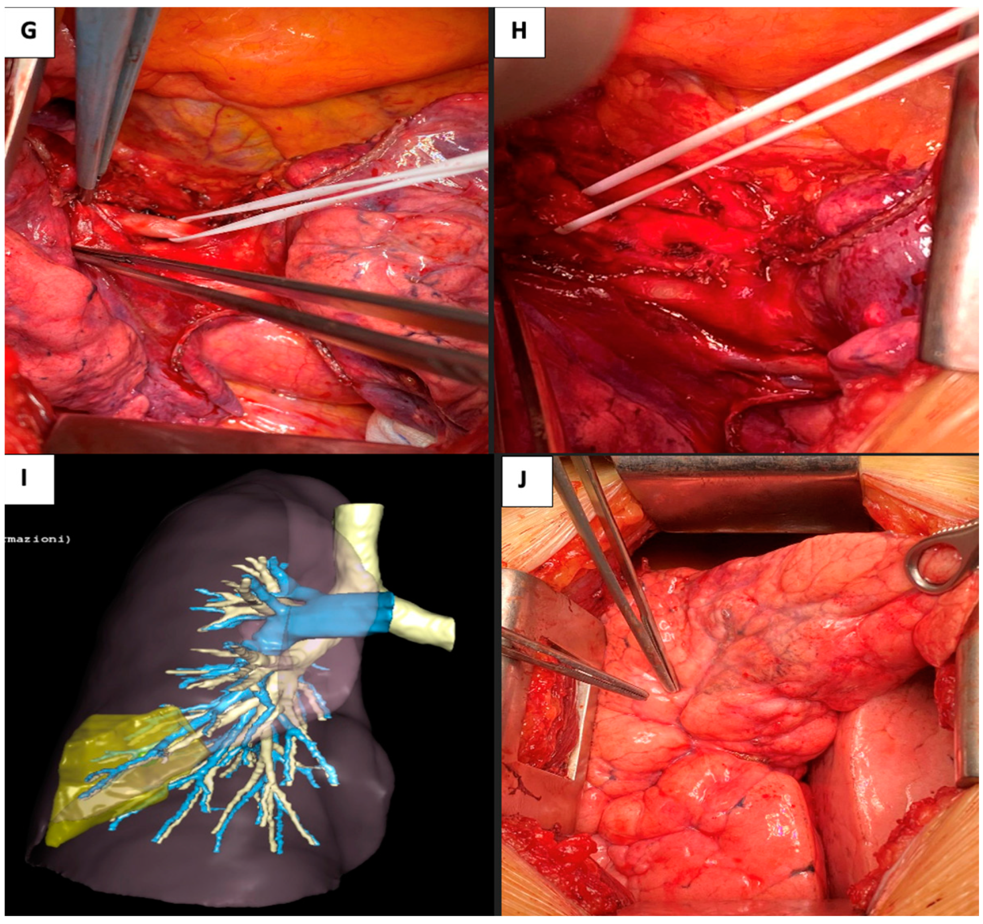
A 67-year-old male patient, non-smoker, affected by colorectal adenocarcinoma surgically treated through sigma-rectum resection 4 years before, was referred to our Department after finding a slowly growing left lower lobe pulmonary nodule [Figure 3A–C], FDG-PET positive (SUVmax 2.3), during oncological follow-up. 1 year earlier he had undergone left upper lobe wedge resection through biportal VATS, for a single lung metastasis of his known adenocarcinoma. The new lesion was radiologically suggestive for a metastasis, but a non-anatomic resection was not feasible due to its location within the pulmonary parenchyma. Aiming to spare lung tissue and considering the previous lung resection and the potential need for further resections, a tailored solution was planned. The CT scan was uploaded on VP-platform [Figure 3D–F]. A segmentectomy (S8) of the left lower lobe was successfully performed through left completely-muscle-sparing thoracotomy [Figure 3G–J]. The patient was discharged on 3rd postoperative day, after regular course. Histological examination confirmed the lesion was a metastasis from the previously known colic adenocarcinoma. 33 months later, the patient is alive without evidence of disease.

3. Discussion

This study presents three challenging real-life oncological cases, successfully managed using preoperative 3D reconstruction platform VPTM, which has allowed us to perform complex segmentectomies without any other intraoperative aid, such as indocyanine or indigo blue. VPTM has allowed us to overcome main criticalities in complex segmentectomies, providing clear preoperative insight in anatomical variation of broncho-vascular elements, assessment of intersegmental plan and definition of safe margins of resection. Without the aid of such preoperative 3D reconstructions, we would have not being able to perform these complex segmentectomies, probably falling back onto more traditional anatomical resections at the cost of higher chances of postoperative respiratory complications.
Anatomic segmentectomy enables minimal resection of the lesion, maximizes the preservation of healthy lung tissue, and mitigates unnecessary loss of lung function [3]. In contrast to wedge resection, segmentectomy guarantees ample surgical margins and facilitates lymph node sampling, ensuring precise tumor staging [6]. Nonetheless, segmentectomy may elevate the risk of locoregional recurrence along the resection line in comparison to lobectomy [7]. The primary factors attributed to this recurrence are inadequate safety surgical margins and incomplete lymph node clearance.
Indeed, executing an anatomical segmentectomy poses a considerable technical challenge for surgeons, demanding a comprehensive knowledge of segmental anatomy to overcome the risk of inadvertently eliminating blood supply or drainage from certain areas of the remaining pulmonary parenchyma, resulting in preserved structures lacking proper function [8], which may also explain the lower-than-expected differences in FEV1 and DLCO reduction between patients undergoing segmentectomy compared to those undergoing lobectomy.
Numerous studies have explored the viability of employing various imaging software programs in thoracic surgery [9,10]. Nonetheless, most of these 3D software applications are both costly and challenging for a surgeon to navigate on a personal computer, besides providing low-detailed models.
Instead, in our experience the outsourcing of 3D-model rendering is entrusted to the private company VPTM, without any additional task on the referring surgeon, given the potential complexity and time-intensive nature of the work. After proper data anonymization, CT scan images are transferred to VPTM, where radiological technicians undertake the rendering process, meticulously contouring all anatomical structures, up to the fifth level of division of arteries, veins, and bronchi; then, the software itself automatically performs the segmentation [9].
VP models have been previously demonstrated to be reliable and accurate [10], allowing for examining individual broncho-vascular elements, conducting virtual resections, calculating the volume of each segment to analyze its impact on predicted postoperative respiratory function, and simulating safety margins. Another crucial element is the possibility to review 3D models on any device, even on portable supports.
At our Institution, the visualization of 3D images is currently confined to a 2D window. A forthcoming challenge involves integrating augmented reality to merge live 3D-models with thoracoscopic images [11]. The primary complexity in thoracic surgery to accomplish this target arises from the fact that the lung undergoes changes in anatomical configuration during surgery, since CT scans are conducted with the lung fully inflated, whereas the surgery is consistently carried out with the lung in a deflated state (single lung ventilation).
Our experience, although being encouraging and interesting, has some limitations: high costs (800 to 900 euros per reconstruction) still prevent spreading of VPTM throughout all thoracic centers, even more in those countries where there is a national healthcare system; 3D reconstructions should be ordered and scheduled beforehand since delivery can take up to 2 weeks; 3D modeling requires top quality CT scans, with proper injection phase of contrast medium, and without artefacts.

4. Conclusions

In conclusion, according to our early and limited experience, complex lung segmentectomy could be accurately performed using preoperative 3D-planning offered by VPTM software, which provides a detailed surgical map to improve safety and precision of pulmonary resection by facilitating the achievement of suitable resection margins while ensuring the vascularization of the residual parenchyma.
Prospective and randomized studies are required to validate these findings and to define the actual potential of this technology.

Author Contributions

Conceptualization: A.P., R.O., L.G. and M.F.; Methodology: A.P., R.O., L.G. and M.F.; Project administration: A.P. and M.A.; Supervision: A.P. and M.A.; writing—original draft: All authors; writing—review & editing: all authors. All authors have read and agreed to the published version of the manuscript.

Funding

This research received no external funding.

Institutional Review Board Statement

The approval of a research committee was deemed unnecessary by local laws, due to the nature of the study (case report without any change to usual intra e and postoperative management of the patient).

Informed Consent Statement

Informed consent was obtained from all subjects involved in the study.

Data Availability Statement

The original contributions presented in the study are included in the article, further inquiries can be directed to the corresponding author.

Conflicts of Interest

The authors declare no conflicts of interest.

References

  1. Bray, F.; Laversanne, M.; Sung, H.; Ferlay, J.; Siegel, R.L.; Soerjomataram, I.; Jemal, A. Global cancer statistics 2022: GLOBOCAN estimates of incidence and mortality worldwide for 36 cancers in 185 countries. CA Cancer J. Clin. 2024, 74, 229–263. [Google Scholar] [CrossRef] [PubMed]
  2. Saji, H.; Okada, M.; Tsuboi, M.; Nakajima, R.; Suzuki, K.; Aokage, K.; Aoki, T.; Okami, J.; Yoshino, I.; Ito, H.; et al. Segmentectomy versus lobectomy in small-sized peripheral non-small-cell lung cancer (JCOG0802/WJOG4607L): A multicentre, open-label, phase 3, randomised, controlled, non-inferiority trial. Lancet 2022, 399, 1607–1617. [Google Scholar] [CrossRef] [PubMed]
  3. Altorki, N.; Wang, X.; Kozono, D.; Watt, C.; Landrenau, R.; Wigle, D.; Port, J.; Jones, D.R.; Conti, M.; Ashrafi, A.S.; et al. Lobar or Sublobar Resection for Peripheral Stage IA Non-Small-Cell Lung Cancer. N. Engl. J. Med. 2023, 388, 489–498. [Google Scholar] [CrossRef]
  4. Nomori, H.; Mori, T.; Ikeda, K.; Yoshimoto, K.; Iyama, K.; Suzuki, M. Segmentectomy for selected cT1N0M0 non-small cell lung cancer: A prospective study at a single institute. J. Thorac. Cardiovasc. Surg. 2012, 144, 87–93. [Google Scholar] [CrossRef]
  5. Oizumi, H.; Endoh, M.; Takeda, S.-I.; Suzuki, J.; Fukaya, K.; Sadahiro, M. Anatomical lung segmentectomy simulated by computed tomographic angiography. Ann. Thorac. Surg. 2010, 90, 1382–1383. [Google Scholar] [CrossRef] [PubMed]
  6. Hu, W.; Zhang, K.; Han, X.; Zhao, J.; Wang, G.; Yuan, S.; He, B. Three-dimensional computed tomography angiography and bronchography combined with three-dimensional printing for thoracoscopic pulmonary segmentectomy in stage IA non-small cell lung cancer. J. Thorac. Dis. 2021, 13, 1187–1195. [Google Scholar] [CrossRef] [PubMed]
  7. Iwano, S.; Yokoi, K.; Taniguchi, T.; Kawaguchi, K.; Fukui, T.; Naganawa, S. Planning of segmentectomy using three-dimensional computed tomography angiography with a virtual safety margin: Technique and initial experience. Lung Cancer 2013, 81, 410–415. [Google Scholar] [CrossRef] [PubMed]
  8. Yao, F.; Wang, J.; Yao, J.; Hang, F.; Lei, X.; Cao, Y. Three-dimensional image reconstruction with free open-source OsiriX software in video-assisted thoracoscopic lobectomy and segmentectomy. Int. J. Surg. 2017, 39, 16–22. [Google Scholar] [CrossRef] [PubMed]
  9. Baste, J.M.; Soldea, V.; Lachkar, S.; Rinieri, P.; Sarsam, M.; Bottet, B.; Peillon, C. Development of a precision multimodal surgical navigation system for lung robotic segmentectomy. J. Thorac. Dis. 2018, 10 (Suppl. S10), S1195–S1204. [Google Scholar] [CrossRef] [PubMed]
  10. Le Moal, J.; Peillon, C.; Dacher, J.-N.; Baste, J.-M. Three-dimensional computed tomography reconstruction for operative planning in robotic segmentectomy: A pilot study. J. Thorac. Dis. 2018, 10, 196–201. [Google Scholar] [CrossRef] [PubMed]
  11. Wu, Z.; Huang, Z.; Qin, Y.; Jiao, W. Progress in three-dimensional computed tomography reconstruction in anatomic pulmonary segmentectomy. Thorac. Cancer 2022, 13, 1881–1887. [Google Scholar] [CrossRef]
Figure 1. Radiologic imaging, 3D-model reconstructions, and intraoperative views of Case 1. (A): Axial slice Chest CT; (B): Coronal slice Chest CT; (C): Axial slice Chest CT with contouring; (D): Coronal slice Chest CT with contouring; (E): 3D-model showing the nodule within arterial, venous, and bronchial branches; (F): 3D-model showing the nodule within venous and bronchial branches; (G): 3D-model showing the nodule within arterial and bronchial branches; (H): 3D-model showing the nodule within bronchial branches; (I): Intraoperative view, showing arterial segmental branching; (J): Intraoperative view, showing bronchial segmental branching; (K): 3D-model showing S7–S8; (L): Surgical sample.
Std 13 00020 g001aStd 13 00020 g001b
Figure 2. Radiologic imaging, 3D-model reconstructions and intraoperative views of Case 2. (A): Axial slice Chest CT; (B): Coronal slice Chest CT; (C): Axial slice Chest CT with contouring; (D): Coronal slice Chest CT with contouring; (E): 3D-model showing the nodules within arterial, venous, and bronchial branches; (F): 3D-model showing the nodules within venous and bronchial branches; (G): 3D-model showing the nodules within arterial and bronchial branches; (H): 3D-model showing the nodules within bronchial branches; (I): 3D-model showing S1–S2; (J): Surgical sample.
Std 13 00020 g002aStd 13 00020 g002b
Figure 3. Radiologic imaging, 3D-model reconstructions and intraoperative views of Case 3. (A): Axial slice Chest CT; (B): Coronal slice Chest CT; (C): Axial slice Chest CT with contouring; (D): 3D-model showing the nodule within arterial, venous, and bronchial branches; (E): 3D-model showing the nodule within arterial and bronchial branches; (F): 3D-model showing the nodule within bronchial branches; (G): Intraoperative view, showing arterial segmental branching; (H): Intraoperative view, showing bronchial segmental branching; (I): 3D-model showing S8; (J): Intraoperative view, showing deflated target segment.
Std 13 00020 g003aStd 13 00020 g003b
Disclaimer/Publisher’s Note: The statements, opinions and data contained in all publications are solely those of the individual author(s) and contributor(s) and not of MDPI and/or the editor(s). MDPI and/or the editor(s) disclaim responsibility for any injury to people or property resulting from any ideas, methods, instructions or products referred to in the content.