Case Report

Achalasia Post-Bariatric Surgery, Placement Roux-En-Y Gastric Bypass: Case Report

by
Juan Pablo Landeros-Ruiz
1,
Lourdes Marlene Zúñiga-Ramos
1,
Daniela Cárdenas-Guerrero
1 and
Quitzia Libertad Torres-Salazar
2,*
1
Hospital Regional Valentín Gómez Farías, Instituto de Seguridad y Servicios Sociales de los Trabajadores del Estado, Zapopan 45100, JA, Mexico
2
Alpha 0.01 Biomedical Research Institute, Continuing Education Department, Calle Paloma No.812, Durango 34060, Dgo., Mexico
*
Author to whom correspondence should be addressed.
Surg. Tech. Dev. 2023, 12(3), 119-125; https://doi.org/10.3390/std12030011
Submission received: 14 April 2023 / Revised: 7 July 2023 / Accepted: 21 July 2023 / Published: 25 July 2023

Abstract

:
Introduction: Achalasia is a pathology with an incidence of 1 in 100,000 inhabitants per year. There are very limited data on achalasia in the obese population, especially in those undergoing bariatric surgery. The approach of choice for cases of achalasia is the procedure partial fundoplication to correct the reflux; however, lacking a fundus due to a previous gastrectomy, an alternative that offers optimal results should be chosen. Here, we present the surgical approach in a case of esophageal achalasia and a history of vertical sleeve gastrectomy, where we performed a simultaneous Heller’s cardiomyotomy and laparoscopic Roux-en-Y gastric bypass, as well as the results obtained. Case Presentation: A 44-year-old woman with no chronic degenerative diseases, who had a vertical sleeve gastrectomy carried out 5 years ago. Her first symptoms manifested 17 months before, and they were dysphagia to liquids and then to solids, in addition to weight loss of 10 kg in 4 months. Her body mass index before the vertical sleeve gastrectomy was 32 kg/m2; her body mass index at the time of admission was 20 kg/m2; she also presented regurgitation and generalized weakness. After analyzing the surgical options, it was decided to perform a Heller cardiomyotomy and a Roux-en-Y gastric bypass. Discussion and Conclusions: The procedure turned out to be safe and successful in treating achalasia symptomatology, in addition to completely resolving the reflux symptoms.

1. Introduction

Obesity is a complex multifactorial disease; its worldwide prevalence has doubled since 1980 to the point that almost one-third of the world’s population during 2019 was classified as overweight or obese [1]. Severe obesity is related to various other medical issues, such as heart disease, elevated glycaemia (diabetes), high blood pressure (hypertension), liver problems caused by fat build-up (nonalcoholic fatty liver disease), gastroesophageal reflux, and uncommonly observed movement disorders like achalasia. Sleeve gastrectomy (SG) or sleeve gastrectomy was first performed by Hess in 1988 as a modification of the duodenal crossover procedure; the procedure was adapted from Scopinaro’s biliopancreatic diversion [2]. Michael Gagner performed the first laparoscopic SG in 1998 as the first part of the biliopancreatic diversion procedure in super-obese and high-risk patients, and, since then, it has been considered to be a primary bariatric technique that has seen great diffusion and acceptance by patients and surgeons, and it is considered to be the most performed bariatric technique worldwide [3]. Despite the simplicity of the procedure, it is not without complications. A study by Giuliani found an incidence in complications of 4.95% in 101 procedures performed between 2012 and 2017, most of them related to gastric leakage [4]. A much less common complication following bariatric surgery is achalasia. Achalasia is the most common motility disorder of the esophagus, with an incidence of 1 case per 100,000 inhabitants per year [5]. This pathology is characterized by relaxation failure or incomplete relaxation of the lower esophageal sphincter (LES) after swallowing, accompanied by the absence of peristalsis in the gastric body. Although the prevalence of esophageal motility disorders in patients with obesity is important, there are very limited data on achalasia in this population, especially in those undergoing bariatric surgery [6]. The standard surgical treatment for the resolution of this problem is usually anterior cardiomyotomy of the LES fibers, extending 2–3 cm into the stomach and several into the esophagus, associated with partial fundoplication to treat reflux. However, the absence of gastric fundus represents a challenge at the time of the surgical approach, and to guarantee an adequate prevention of reflux, it is necessary to opt for another alternative to conventional procedures. Laparoscopic Roux-en-Y gastric bypass (RYGBP) is a procedure characterized by performing a small size gastric reservoir over which a Roux-en-Y gastrojejunal anastomosis is performed [7]. Here, we present the surgical technique performed in a case of an association of esophageal achalasia and a history of vertical sleeve gastrectomy in which Heller’s cardiomyotomy and RYGBP were performed simultaneously, as well as the results obtained.

2. Case Presentation

We present the case of a 44-year-old female patient with no chronic degenerative diseases, with a surgical history of laparoscopic cholecystectomy 13 years ago, vertical sleeve gastrectomy 5 years ago, and a cesarean section and an appendectomy 4 years ago. The patient reported that 1 year and 5 months ago she started experiencing dysphagia to liquids and later to solids, and she also presented a weight loss of 10 kg (kg) in 4 months. Her body mass index (BMI) before vertical sleeve gastrectomy was 39 kg/m2; her current BMI is 20 kg/m2. The patient mentioned, in addition to the symptoms already described, to have regurgitation and generalized weakness.
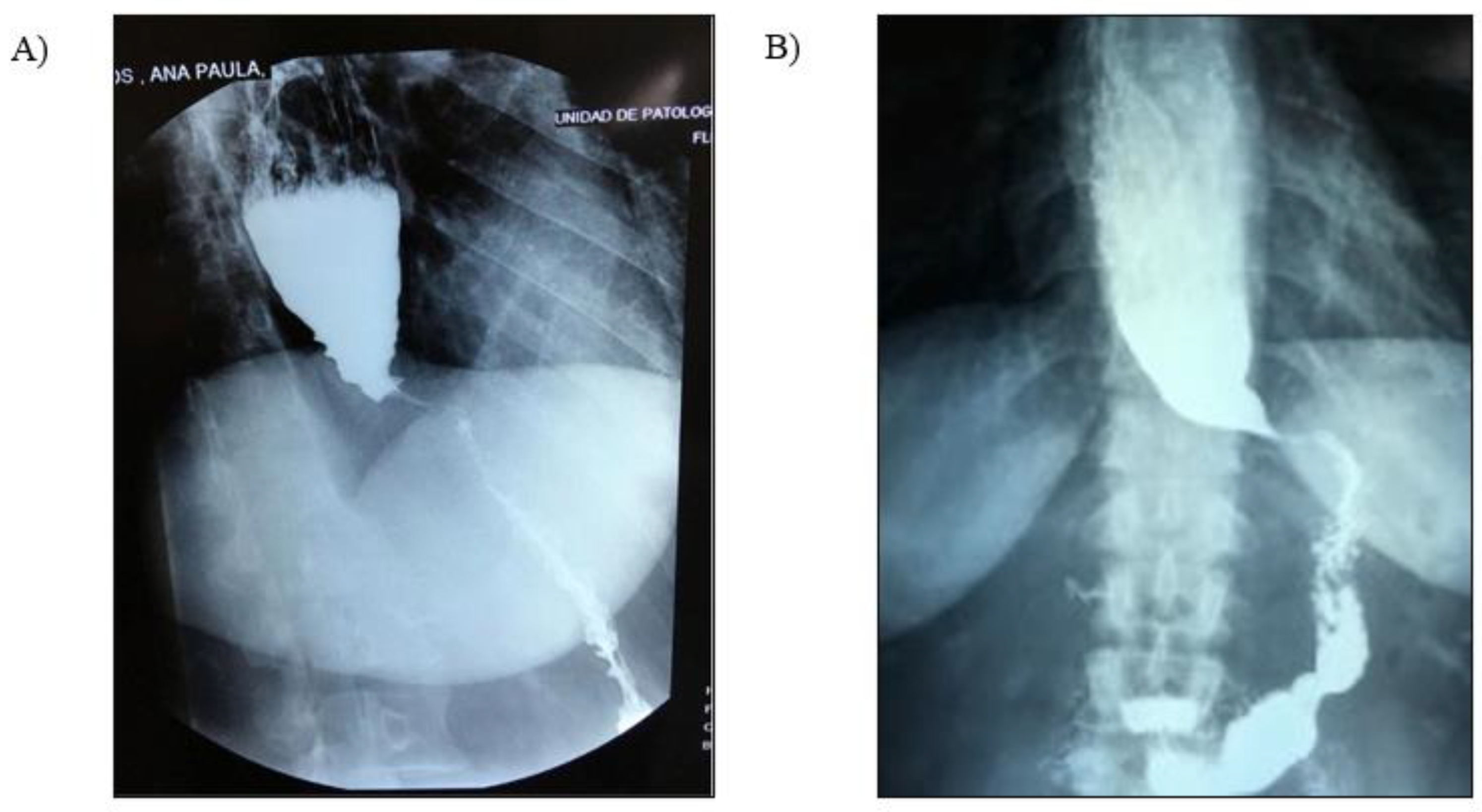
First, a panendoscopy was performed (Figure 1A,B), showing an increase in the esophageal diameter in each of its three thirds with abundant food debris. A retentive esophagus with a diagnostic impression of probable achalasia was reported. Afterwards, manometry was indicated, identifying a mean residual pressure in the lower esophageal sphincter of 29.1 mmHg. Regarding the upper esophageal sphincter, a mean basal pressure of 133.9 mmHg was reported, with a relaxation time of 1092 ms and a recovery time of 849 ms, which is compatible with a diagnosis of achalasia subtype II of the Chicago classification version 3.0. Finally, an esophagogastroduodenal series was performed (Figure 2A,B) where typical pencil-point images were found. She was scheduled by the surgical service for the procedure of a Heller cardiomyotomy with laparoscopic Roux-en-Y bypass, who chose this technique instead of partial fundoplication due to the surgical history of gastric sleeve.

3. Surgical Technique

The procedure was performed under balanced general anesthesia, a supraumbilical incision was made and pneumoperitoneum was insufflated with the Veress technique, a 12 mm trocar was introduced, and under direct vision four more trocars were introduced, including one of 10 mm and three of 5 mm.
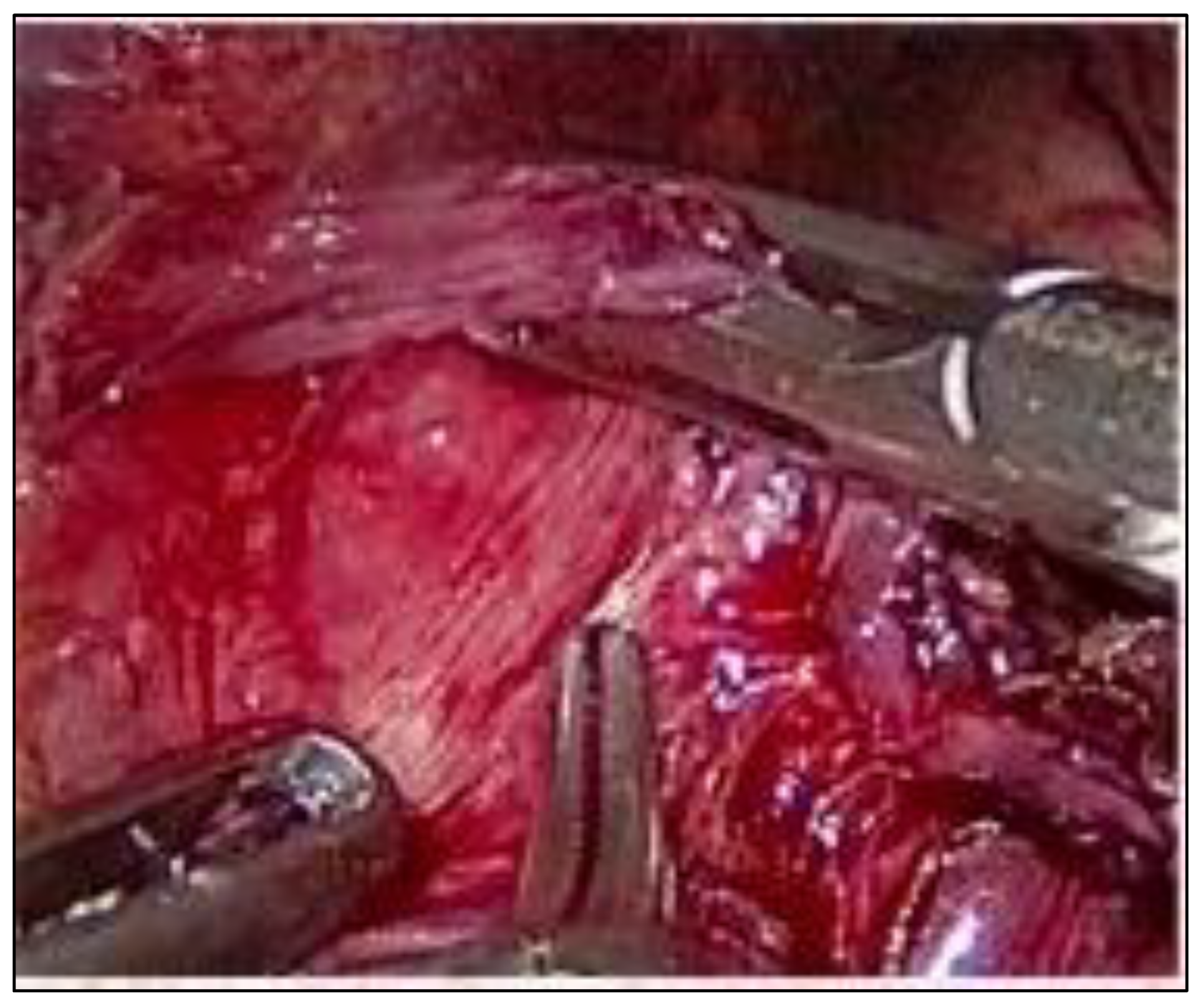
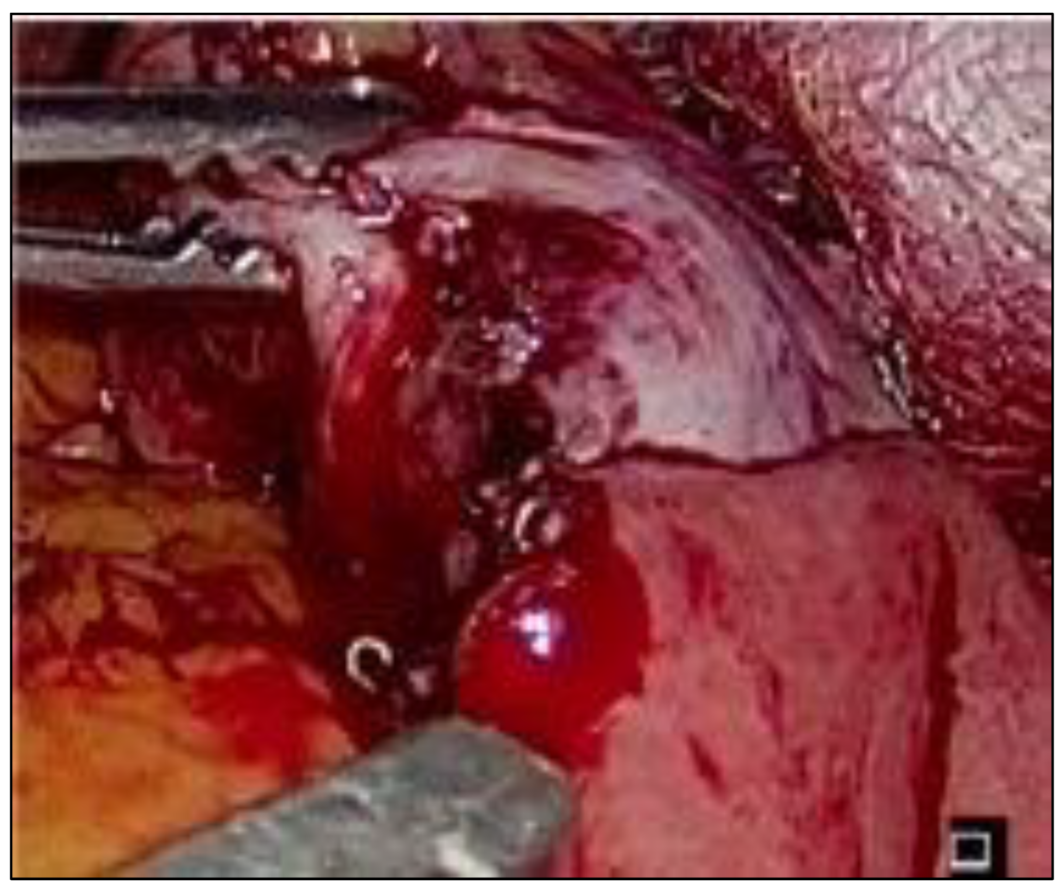
The hepatic lift was performed, the fat pad of the gastroesophageal junction was resected, and the esophagus was freed from the pillars. A retroesophageal tunnel was created, and the muscle fibers were cut 4 cm above the junction and 2 cm below the junction (Figure 3). Permeability was checked with a caliper probe without finding evidence of perforation. From the ligament of Treitz, 60 cm of jejunum were counted, and a linear stapler cut was made; at 100 cm from this cut, a mechanical jejuno-duodenal anastomosis was performed on the lateral side of the jejunum (Figure 4).
The gastric pouch was performed with linear staplers. The jejunal loop was brought up through an opening in the mesocolon, and a mechanical gastrojejunal anastomosis was performed with the linear stapler. The surgical procedure was completed without eventualities, the surgical time was 150 min, and there was no conversion to open surgery. The patient was left fasting for 24 h, the oral route was started with a liquid diet with good tolerance and evolved to a soft diet after 36 h, and there was no dysphagia or odynophagia. The postoperative evolution was satisfactory, with no episodes of regurgitation or reflux, and a weight gain of 8 kg in 6 months.

4. Discussion

Achalasia is an esophageal pathology that causes alterations in the life quality of patients. It is characterized by a motility disorder of the esophagus that produces progressive dysphagia, regurgitation, weight loss, retrosternal pain, and chronic cough. In addition, it also causes malnutrition due to the difficulty of the passage of food into the stomach. Achalasia affects the way the esophagus functions, with a recorded occurrence of around 1 in every 100,000 individuals [4]. Common symptoms of this disorder include difficulty swallowing and discomfort in the chest. It is identified by the inability of the lower esophageal sphincter (LES) to relax during swallowing, resulting in a consistently normal or elevated LES pressure. The lack of peristalsis, or involuntary contractions, in the esophagus is a definitive characteristic of this condition [8].
The standard procedure for achalasia is usually Heller cardiomyotomy with partial fundoplication for the prevention of gastroesophageal reflux disease [9]. However, our patient debuted with achalasia 5 years after having undergone a vertical sleeve gastrectomy, making conventional surgical treatment, which is a Heller cardiomyotomy with partial fundoplication, difficult. In 2005, Kauffman described the first case of a patient treated with esophageal myotomy and RYGBP simultaneously [10]. Since then, very few studies have published cases of the association between a Heller myotomy and a bariatric procedure such as gastric bypass, duodenal switch, gastric banding (GB), vertical gastrectomy, and even a non-bariatric procedure such as total gastrectomy with esophagojejunal and jejunojejunal anastomosis [11]. In 2015, Fisichella performed an evidence-based review on the treatment of achalasia in patients with obesity, analyzing three therapeutic options: laparoscopic Heller myotomy associated with RYGBP, associated with biliopancreatic diversion, and associated with vertical gastrectomy. The authors concluded that Heller myotomy associated with RYGBP provides excellent symptom relief and reflux control, as well as adequate treatment of obesity and comorbidities [12]. In 2016, Boules et al. conducted a study on patients with achalasia who had previously undergone bariatric surgery. The diagnosis of achalasia was confirmed by endoscopy and manometry. Eight patients had undergone RYGBP and two had vertical banded gastroplasty (VBG). The average time between bariatric surgery and achalasia diagnosis was 6 years. Two patients received Botox treatment and five underwent esophageal dilations. All patients received surgical intervention for achalasia, including Heller myotomy (HM) for RYGBP patients (four open and four laparoscopic) and other procedures for VBG patients. All patients showed a significant decrease in BMI after bariatric surgery. Of the eight patients who underwent HM, six achieved resolution of achalasia symptoms within 1.6 months and remained asymptomatic for 36 months. One patient developed recurrent achalasia and received a peroral endoscopic myotomy. One HM patient was lost to follow-up, but the two patients who underwent esophagectomies were symptom-free at 36 months. These patients were classified as having end-stage achalasia [5]. More recently, Donatelli presented a video case of percutaneous endoscopic myotomy performed in a patient with multiple previous bariatric surgeries. In her past medical history, she underwent laparoscopic banding, sleeve gastrectomy, and Roux-en-Y gastric bypass. Peroral endoscopic myotomy was performed without complications. Myotomy was performed only 1 cm below the cardia due to the presence of the gastrojejunal anastomosis. The postoperative period was uneventful and an oral diet was restarted after one day. At 2 months follow-up, the patient was asymptomatic and weight had not been regained [13]. Low intragastric pressure after a Roux-en-Y gastric bypass helps control appetite, prevents complications, and can have beneficial effects on the metabolism. These factors make intragastric pressure an important consideration when choosing Roux-en-Y gastric bypass as a treatment option for obesity. Most of the written works available discuss cases where people developed achalasia after RYGBP. Ramos et al. presented a case about a patient who experienced achalasia about four years following their RYGBP procedure. The patient had common symptoms of regurgitation and difficulty swallowing solid foods. A barium esophagogram and esophageal manometry confirmed the diagnosis, revealing a widened esophagus with the characteristic “bird beak” appearance, as well as achalasia with aperistalsis [14].
Bariatric surgery is a reliable and efficient way to treat severe obesity, leading to positive changes in related health conditions and overall well-being. However, there is scarce information on the occurrence of achalasia in obese individuals who undergo bariatric surgery, despite the high prevalence of esophageal motility disorders in this group. Currently, the most frequently used bariatric procedures include RYGBP, laparoscopic sleeve gastrectomy (LSG), and laparoscopic adjustable gastric banding (LAGB). The surgical considerations that we must not lose sight of are those associated with the BMI of the patient to be operated on, especially for the placement and determination of the size of the trocars, since the obese patient has a greater thickness of the abdominal wall and, consequently, there are variations in its degree of rigidity. On the other hand, the possibility of great thickness and size of the liver must be considered, as well as the presence of adhesions due to previous surgeries, which will also modify the initial and subsequent location of the trocars [15].
The decision of the appropriate surgical approach to treat achalasia in our patient represented a challenge for our team, since the surgery could not be performed using the conventional technique due to the history of vertical sleeve gastrectomy. Considering the condition above, it was decided, as a successful alternative, to perform the extramucosal Heller cardiomyotomy in addition to a Roux-en-Y bypass. We must point out that it is not possible to perform a partial fundoplication due to the absence of the gastric fundus. Myotomy resolved the dysphagia, and RYGBP was an effective antireflux measure.

5. Conclusions

Although the occurrence rate of achalasia in people who have undergone bariatric surgery is not known, it can still occur and needs to be treated when detected. It is not uncommon for dysmotility to occur, and achalasia is often not diagnosed prior to bariatric surgery. Paying more attention to identifying motility diseases before surgery and recognizing that achalasia can develop after bariatric surgery can improve patient care. Esophageal motility disorders in obese patients are not usually evaluated before a bariatric surgical procedure unless they present severe symptomatology. The search for motility disorders should be included as part of the presurgical protocol to choose the optimal surgical procedure for the patient. The treatment of these patients represents a challenge for the surgeon. RYGBP associated with Heller cardiomyotomy performed by an experienced surgeon is a safe technique that allows the control of achalasia symptomatology and the avoidance of gastroesophageal reflux.

Author Contributions

Conceptualization, J.P.L.-R.; methodology, L.M.Z.-R.; data curation, D.C.-G.; writing, review and editing, Q.L.T.-S. All authors have read and agreed to the published version of the manuscript.

Funding

This research received no external funding.

Institutional Review Board Statement

Protection of humans and animals. The authors declare that no experiments on humans or animals have been performed for this research.

Informed Consent Statement

Right to privacy and informed consent. The authors have obtained the informed consent of the patients and/or subjects referred to in the article.

Data Availability Statement

Confidentiality of data. The authors declare that no patient data appear in this article. This document is in the possession of the corresponding author.

Conflicts of Interest

There are no financial or personal relationships that could give rise to conflict of interests in connection with this item.

References

  1. Chooi, Y.; Ding, C.; Magkos, F. The epidemiology of obesity. Metabolism 2019, 92, 6–10. [Google Scholar] [CrossRef] [PubMed] [Green Version]
  2. Hess, D.; Hes, D. Biliopancreatic diversión with a duodenal switch. Obes. Surg. 1988, 8, 267–282. [Google Scholar] [CrossRef] [PubMed]
  3. Pacheco-García, J.; Mayo-Osorio, M.; Bengoechea-Trujillo, A.; Fornell-Ariza, M.; Vílchez-López, F.; Aguilar-Diosdado, M. Gastrectomía vertical: La técnica quirúrgica bariátrica más utilizada en la actualidad. Cir. Andal. 2019, 30, 455–464. [Google Scholar] [CrossRef]
  4. Giuliani, A.; Romano, L.; Papale, E.; Puccica, I.; Di Furia, M.; Salvatorelli, A.; Cianca, G.; Schietroma, M.; Amicucci, G. Complications of postlaparoscopic sleeve gastric resection: Review of surgical technique. Minerva Chir. 2019, 74, 213–217. [Google Scholar] [CrossRef]
  5. Schlottmann, F.; Patti, M. Esophageal achalasia: Current diagnosis and treatment. Expert. Rev. Gastroenterol. Hepatol. 2018, 12, 711–721. [Google Scholar] [CrossRef]
  6. Boules, M.; Corcelles, R.; Zelisko, A.; Batayyah, E.; Froylich, D.; Rodriguez, J.; Brethauer, S.; El-Hayek, K.; Kroh, M. Achalasia After Bariatric Surgery. J. Laparoendosc. Adv. Surg. Tech. 2016, 26, 428–432. [Google Scholar] [CrossRef] [PubMed]
  7. Chapman, R.; Rotundo, A.; Carter, N.; George, J.; Jenkinson, A.; Adamo, M. Laparoscopic Heller’s myotomy for achalasia after gastric bypass: A case report. Int. J. Surg. Case Rep. 2013, 4, 396–398. [Google Scholar] [CrossRef] [PubMed] [Green Version]
  8. Sadowski, D.C.; Ackah, F.; Jiang, B.; Svenson, L.W. Achalasia: Incidence, prevalence and survival. A population-based study. Neurogastroenterol. Motil. 2010, 22, e256–e261. [Google Scholar] [CrossRef]
  9. Koppman, J.S.; Poggi, L.; Szomstein, S.; Ukleja, A.; Botoman, A.; Rosenthal, R. Esophageal motility disorders in the morbidly obese population. Surg. Endosc. 2007, 21, 761–764. [Google Scholar] [CrossRef]
  10. Kaufman, J.A.; Pellegrini, C.A.; Oelschlager, B.K. Laparoscopic Heller myotomy and Roux-en-Y gastric bypass: A novel operation for the obese patient with achalasia. J. Laparoendosc. Adv. Surg. Tech. A. 2005, 15, 391–395. [Google Scholar] [CrossRef]
  11. Lutrzykowski, M. Tratamento cirúrgico do paciente com obesidade mórbida com Acalasia. BMI 2011, 153, 361–364. [Google Scholar]
  12. Wesp, J.A.; Farrell, T.M. The surgical management of achalasia in the morbid obese patient. J. Gastrointest. Surg. 2015, 19, 1139–1143. [Google Scholar]
  13. Donatelli, G.; Cereatti, F.; Soprani, A. Per Oral Endoscopic Myotomy for the Management of Achalasia in a Patient with Prior Lap Band, Sleeve Gastrectomy, and Roux-en-Y Gastric Bypass. Obes. Surg. 2021, 31, 2843–2844. [Google Scholar] [CrossRef]
  14. Ramos, A.C.; Murakami, A.; Lanzarini, E.G.; Neto, M.G.; Galvão, M. Achalasia and laparoscopic gastric bypass. Surg. Obes. Relat. Dis. 2009, 5, 132–134. [Google Scholar] [CrossRef] [PubMed]
  15. Asociación Mexicana de Cirugía General. Tratado de Cirugía General, 3rd ed.; Morales-Saavedra, J., Ed.; Manual: Ciudad de México, Mexico, 2017. [Google Scholar]
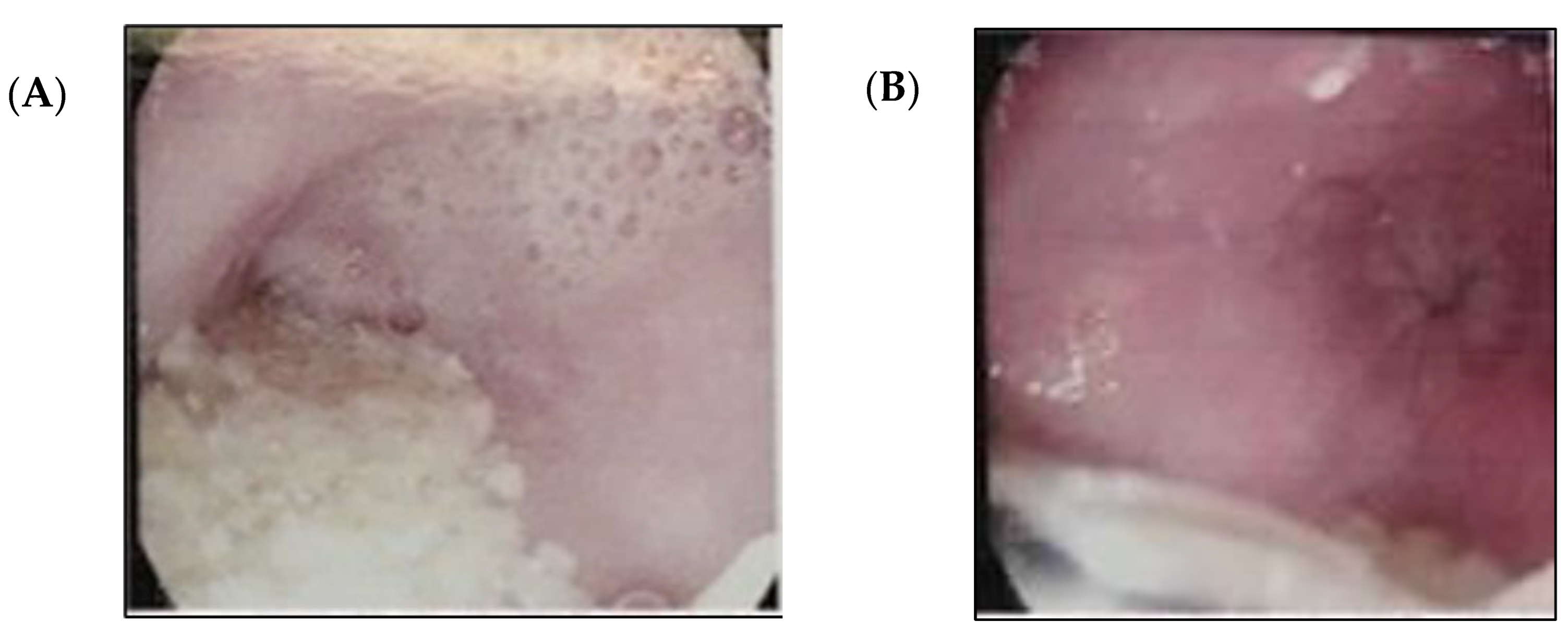
Figure 1. (A,B). Panendoscopy. There is an increase in esophageal diameter and abundant food debris.
Std 12 00011 g001
Figure 2. (A) Esophageal dilatation and (B) pencil-point termination of the gastroesophageal junction.
Std 12 00011 g002
Figure 3. Heller’s cardiomyotomy: dissection of the esophageal muscle fibers.
Std 12 00011 g003
Figure 4. Roux-en-Y bypass gastric bypass: gastrojejunal anastomosis.
Std 12 00011 g004
Disclaimer/Publisher’s Note: The statements, opinions and data contained in all publications are solely those of the individual author(s) and contributor(s) and not of MDPI and/or the editor(s). MDPI and/or the editor(s) disclaim responsibility for any injury to people or property resulting from any ideas, methods, instructions or products referred to in the content.