Viewpoint

Plant Tissue Culture In Vitro: A Long Journey with Lingering Challenges

by
Taras Pasternak
1,* and
Douglas Steinmacher
2,*
1
Independent Researcher, 03202 Elche, Spain
2
AlfaPalm Agrociências, Marechal Cândido Rondon 85960-148, PR, Brazil
*
Authors to whom correspondence should be addressed.
Int. J. Plant Biol. 2025, 16(3), 97; https://doi.org/10.3390/ijpb16030097 (registering DOI)
Submission received: 10 July 2025 / Revised: 10 August 2025 / Accepted: 16 August 2025 / Published: 21 August 2025
(This article belongs to the Section Plant Reproduction)

Abstract

In recent years, plant tissue culture has become a crucial component of the modern bioeconomy. From a commercial perspective, plant micropropagation remains one of its most valuable applications. Plants exhibit remarkable developmental plasticity; however, many species still remain recalcitrant in tissue culture. While the term recalcitrant is commonly used to describe plants with poor in vitro regeneration capacity, from a biological point of view it suggests that the minimal culture requirements for this species were unmet. Despite evidence that the Skoog–Miller exogenous hormonal balance theory and Murashige–Skoog medium were species-limited in applicability, generations of plant biotechnologists applied these tools indiscriminately. This led to systemic propagation of ineffective protocols, publication of misleading standards, and a culture of scientific inertia—costing both time and resources. The field must now move beyond historical dogma toward data-driven, species-specific innovation based on multiple endogenous auxin biosynthesis pathways, epigenetic reprogramming of competent cells, and further modern biotechnologies that are evolving. In this short viewpoint, we describe possible solutions in plant biotechnology to significantly improve the effectiveness of it.

1. Auxin: The Main Force in Micropropagation (Shoot Formation and Rooting)

The scientific foundation of plant micropropagation is traditionally anchored in two seminal concepts: the theory of cellular totipotency, postulated by Haberlandt in 1902 [1,2], and the discovery by Skoog and Miller in 1957, which used tobacco stem cultures as a plant model [3]. Both of these foundations continue to be applied in recent years [4,5] and even in 2025 [6]. Skoog and Miller’s theory posits that the ratio of exogenous auxin to cytokinin is the key regulator of plant morphogenesis in tobacco stem-derived callus—where high levels of exogenous cytokinin induce shoot formation, while high levels of exogenous auxin promote root formation. In other words, their work showed that altering the exogenous auxin and cytokinin treatments in plant tissue culture medium could influence the type of organized tissue formed.
Although their work on the exogenous hormonal control of organogenesis was foundational, research has expanded over the years and has revealed that their concept is misleading and that more complex mechanisms based on endogenous hormone production are responsible for organogenesis [7,8]. Both shoot and root formation in plants involve a common mechanism known as the de novo induction of polar auxin gradients [9]. Recent in situ data revealed phytohormone auxin as the central regulator of organogenesis at the shoot apical meristem (SAM) [10]. Auxin is transported through cells via specific transport proteins (PINs) [11], leading to the establishment of gradients—monodirectional (single direction) for roots and multidirectional (multiple directions) for shoots—as well as auxin-induced cell fate specification (Figure 1A). However, auxin transport/canalization requires endogenous auxin synthesis. The plants are equipped with multiple auxin biosynthesis genes (now up to 11 in Arabidopsis and up to 14 in rice and maize), with each gene located in a specific cell type and induced/regulated only by a specific process and requiring a specific chromatin status [12,13,14,15,16]. Despite that all pathways at the end produce a single molecule—IAA—the resulting function and response of it is different. It is interesting that only the TAA1 gene is linked with a specific cell type in planta—the shoot apical meristem with a specific chromatin structure [17,18,19]. The key role of chromatin modifications in the regulation of IAA synthesis and metabolism and the link of specific auxin pathways with cell types have been reviewed by Wang et al. [13]. Correspondingly, exogenous cytokinin can only induce shoot formation if it triggers auxin biosynthesis via the TAA1 pathway (precursors for shoot organogenesis), leading to shoot stem cell formation (Figure 1B). Inducing another auxin biosynthesis pathway like YUCCA1, or other YUCCA pathways, has resulted in soft callus formation [20,21]. The role of multiple auxin biosynthetic pathways, and therefore sources, each with specific functions ranging from shoot induction to cell differentiation, is often overlooked from a broader perspective [22,23].
Therefore, revisiting the first foundational pillar, Haberlandt’s (1902) [1,2] theory of cellular totipotency, while universally accepted, requires a critical qualification. The long-standing assertion that “all plant cells are totipotent” is challenged by practical observations. We argue that totipotency is not an absolute state but rather a potential that is contingent upon the cell’s ability to perform specific chromatin modifications and activate its endogenous auxin biosynthesis pathways. Consequently, only cells capable of activating specific auxin biosynthesis pathways can express a new morphogenetic fate, leading to either callus formation (partial totipotency) or whole plant regeneration (full totipotency) [17,23,24,25]. Nevertheless, plants maintain niches of totipotent cells throughout their entire vegetative life cycle.
The primary determinant of callus type or cell morphogenetic response is the epigenomic status of the initial cell and its ability to modify its epigenome [26,27]. These modifications are key in determining which genes are expressed and, therefore, the resulting morphology and development. In in vitro culture, a cell’s ability to divide and acquire a new morphogenetic pathway is directly linked to the production of endogenous (within a cell) auxin. In this context, the type of auxin-producing enzymes (Figure 1A) is the determination factor, as these enzymes are associated with specific cell types and their epigenomic status in planta and have local effects (e.g., mesophyll cell vs. embryo induction) [28,29].
Each specific auxin biosynthesis pathway is linked with certain types of morphogenesis, such as YUC2 with callus formation, YUC4 with root development, and YUC9 and TAA1 with shoot formation [22]. Yet, comprehensive in situ analyses confirming these associations in vitro across different cell types and developmental stages are still lacking.
Advancements in spatial transcriptomics and single-cell RNA sequencing hold promise for filling these gaps. Such technologies could provide high-resolution maps of YUC gene expression and auxin distribution, offering deeper insights into their roles in plant morphogenesis. It is important to point out that epigenetic factors, such as DNA methylation and histone modifications, play a key role in the induction of auxin biosynthesis and callus formation [20,21]. A root only exhibits unidirectional endogenous polar auxin transport and therefore root formation cannot be induced by exogenous auxin alone. Root induction requires auxin synthesis through a specific pathway, followed by xylem differentiation to establish directional auxin canalization routes [30]. Only after these conditions are met can exogenous auxins support de novo root formation [31].
In addition to endogenous auxin, other hormones may play specific roles as regulators of auxin signaling/polarity. Cytokinin is a hormone which can induced auxin synthesis, while gibberellins (GAs) induce cell polarization and polar elongation [32]. This hormonal cross-talk involves diverse mechanisms, where GAs biosynthesis is enhanced by auxin [33], which in turn induce polar auxin canalization through auxin efflux PIN proteins [34].
An additional piece of evidence supporting this point is the direct link between genes regulating morphogenesis and auxin biosynthesis, such as BABY BOOM [35].

2. Nutrients Balance as a Key Factor in Plant Growth Regulation and the Limitations of MS Medium

Plant growth is classified into two main types: vegetative and generative. Vegetative growth is driven by cell differentiation (expansion) and characterized by large leaves (enhancing photosynthesis) and elongated stems. In contrast, “generative growth” (organogenesis) involves compact shoots with a higher number of adventives buds or increased organogenesis from explants. Soft callus with rapid post-mitotic cell expansion requires a specific nutrient balance [36].
Such formulation, known as MS, found earlier support due to the plasticity of plant development and showed positive outcomes in early studies within certain plant groups; however, its applicability for mass multiplication is very species-limited and may cause issues such as soft callus formation and excessive water uptake [37]. In in vitro tissue culture, endogenous carbon production is not a limiting factor, and excessive vegetative growth can negatively impact plant quality and its multiplication. Plants regulate their growth pathway based on nutrient balance in the medium. Specifically, high nitrogen (N) and chloride (Cl) levels promote rapid cell expansion [38].
A major issue in micropropagation is the widespread use of LCL medium (MS salt [36] + B5 vitamins [39] + MES), a widespread formula that is utilized in commercial applications as well, which favors vegetative development over generative development. LCL medium has a N:P:K ratio of 48:1:20 known to induce rapid vegetative growth, and it contains 6 mM Cl, which promotes post-mitotic cell expansion. However, these conditions are suboptimal for auxin cofactors, leading to excessive vegetative growth, hyperhydricity, and poor shoot formation.
Hyperhydricity is a wide problem in plant micropropagation. A “common” suggestion to solve this problem is aeration, low nitrogen, etc. [40]. However, the real nature of the phenomenon is different: after shoot induction, some auxin biosynthesis gene overexpression in mesophyll cell produces IAA that cannot be canalized for rooting and, therefore, induce rapid mesophyll growth and expansion resulting in excess water uptake. Therefore, such an imbalance between the cell expansion of different cell types in de novo-formed shoots causes more rapid growth of mesophyll cells by vacuolation because of excess auxin synthesis in these cells (also induced by excess exogenous cytokinin). High N contents promote mesophyll cell growth by vacuolation [41] and Cl as a “ballast” element serves as a macro-ion, leading to rapid uptake by vacuoles accompanied by acidification and water uptake [42,43].
As a result, LCL medium is unsuitable for commercial micropropagation, causing economic losses due to reduced shoot multiplication, high hyperhydricity rates, and poor adaptation of the regenerated plants. Conservative estimates suggest that misapplications based on these publications may have cost the industry over 200 million USD due to failed protocols, misguided commercialization efforts, and slowed innovation [37,44]. An optimized medium with balanced nutrient composition is essential for improving micropropagation efficiency and plant quality [37].
The basic principles of medium preparation should be based on the function of each component in plant growth regulation and its role in hormonal signaling, which have been provided in [45]. For example, this approach is demonstrated in the description of TK medium optimized for several plant species [20,45].

3. Conclusions

Based on the information above, we can conclude the following: endogenous auxin and its related biosynthetic enzymes are key factors responsible for de novo morphogenesis, including both roots and shoots. Nutrient balance is also a crucial factor that influences endogenous hormone levels. The traditional theory, which emphasizes the role of exogenous auxin/cytokinin balance in plant regeneration as a key factor, should be re-evaluated. Instead, endogenous auxin synthesis, with a specific pathway and related epigenomic status, appears to be the primary driving mechanisms of plant morphogenesis (Figure 1B).

Funding

T.P. thanks the Universidad Miquel Hernandez for the Maria Zambrano fellowship (2022–2024); D.S. thanks FINEP—Financiadora de Estudos e Projetos for funding via the InovaDoc FINEP Project.

Data Availability Statement

No new data was described here.

Acknowledgments

The authors thank Serhii Kondratenko and Denes Dudits for fruitful discussion.

Conflicts of Interest

Douglas Steinmacher was employed by the company AlfaPalm Agrociências. The remaining authors declare that the research was conducted in the absence of any commercial or financial relationships that could be construed as potential conflicts of interest.

Abbreviations

The following abbreviations are used in this manuscript:
TAATRYPTOPHAN AMINOTRANSFERASE OF ARABIDOPSIS
YUCCAindole-3-pyruvate monooxygenase gene–auxin biosynthesis gene
SAMshoot apical meristem

References

  1. Haberlandt, G. Culturversuche mit isolierten pflanzenzellen. Sitzungsber. Akad. Wiss. Math.-Naturwiss. Kl. 1902, 111, 69–91. [Google Scholar]
  2. Haberlandt, G. Experiments on the culture of isolated plant cells. Bot. Rev. 1969, 35, 68–88. [Google Scholar] [CrossRef]
  3. Skoog, F. Miller CO Chemical regulation of growth and organ formation in plant tissues cultured in vitro. Symp. Soc. Exp. Biol. 1957, 11, 118–130. [Google Scholar]
  4. Melnyk, C.W. Quantitative regeneration: Skoog and Miller revisited. Quant. Plant Biol. 2023, 4, e10. [Google Scholar] [CrossRef]
  5. Welsch, R.; Touraev, A.; Palme, K. Small molecules mediate cellular reprogramming across two kingdoms. J. Exp. Bot. 2021, 72, 7645–7647. [Google Scholar] [CrossRef]
  6. Li, Y.; Li, C.; Shahzad, A. Regeneration of plant organs in vitro and its mechanistic basis. Front. Plant Sci. 2025, 16, 1568614. [Google Scholar] [CrossRef]
  7. De Klerk, G.J.; Van Der Krieken, W.; de Jong, J.C. Review the formation of adventitious roots: New concepts, new possibilities. Vitr. Cell. Dev. Biol.–Plant 1999, 35, 189–199. [Google Scholar] [CrossRef]
  8. Abdalla, N.; El-Ramady, H.; Seliem, M.K.; El-Mahrouk, M.E.; Taha, N.; Bayoumi, Y.; Shalaby, T.A.; Dobránszki, J. An academic and technical overview on plant micropropagation challenges. Horticulturae 2022, 8, 677. [Google Scholar] [CrossRef]
  9. Maruyama, K.; Ikeuchi, M. Multifaceted controls on auxin metabolism during cellular reprogramming and organ regeneration in plants. J. Exp. Bot. 2025, eraf251. [Google Scholar] [CrossRef] [PubMed]
  10. Sassi, M.; Vernoux, T. Auxin and self-organization at the shoot apical meristem. J. Exp. Bot. 2013, 64, 2579–2592. [Google Scholar] [CrossRef]
  11. Vanneste, S.; Pei, Y.; Friml, J. Mechanisms of auxin action in plant growth and development. Nat. Rev. Mol. Cell Biol. 2025, 26, 648–666. [Google Scholar] [CrossRef]
  12. Cao, X.; Yang, H.; Shang, C.; Ma, S.; Liu, L.; Cheng, J. The roles of auxin biosynthesis YUCCA gene family in plants. Int. J. Mol. Sci. 2019, 20, 6343. [Google Scholar] [CrossRef]
  13. Wang, J.L.; Di, D.W.; Luo, P.; Zhang, L.; Li, X.F.; Guo, G.Q.; Wu, L. The roles of epigenetic modifications in the regulation of auxin biosynthesis. Front. Plant Sci. 2022, 13, 959053. [Google Scholar] [CrossRef]
  14. Expósito-Rodríguez, M.; Borges, A.A.; Borges-Pérez, A.; Hernández, M.; Pérez, J.A. Cloning and biochemical characterization of ToFZY, a tomato gene encoding a flavin monooxygenase involved in a tryptophan-dependent auxin biosynthesis pathway. J. Plant Growth Regul. 2007, 26, 329–340. [Google Scholar] [CrossRef]
  15. Tivendale, N.D.; Ross, J.J.; Cohen, J.D. The shifting paradigms of auxin biosynthesis. Trends Plant Sci. 2014, 19, 44–51. [Google Scholar] [CrossRef] [PubMed]
  16. Kneuper, I.; Teale, W.; Dawson, J.E.; Tsugeki, R.; Katifori, E.; Palme, K.; Ditengou, F.A. Auxin biosynthesis and cellular efflux act together to regulate leaf vein patterning. J. Exp. Bot. 2021, 72, 1151–1165. [Google Scholar] [CrossRef] [PubMed]
  17. Ma, J.; Wang, Y.; Chen, S.; Xu, T. The Central Role of Auxin in Orchestrating Apical Stem Cells in Plants. Plant Cell Environ. 2025, 48, 5053–5072. [Google Scholar] [CrossRef]
  18. Yadav, S.; Kumar, H.; Mahajan, M.; Sahu, S.K.; Singh, S.K.; Yadav, R.K. Local auxin biosynthesis promotes shoot patterning and stem cell differentiation in Arabidopsis shoot apex. Development 2023, 150, dev202014. [Google Scholar] [CrossRef]
  19. Nguyen, V.; Gutzat, R. Epigenetic regulation in the shoot apical meristem. Curr. Opin. Plant Biol. 2022, 69, 102267. [Google Scholar] [CrossRef]
  20. Pasternak, T.; Paponov, I.A.; Kondratenko, S. Optimizing protocols for Arabidopsis shoot and root protoplast cultivation. Plants 2021, 10, 375. [Google Scholar] [CrossRef]
  21. SSakamoto, Y.; Kawamura, A.; Suzuki, T.; Segami, S.; Maeshima, M.; Polyn, S.; De Veylder, L.; Sugimoto, K. Transcriptional activation of auxin biosynthesis drives developmental reprogramming of differentiated cells. Plant Cell 2022, 34, 4348–4365. [Google Scholar] [CrossRef] [PubMed]
  22. Brumos, J.; Robles, L.M.; Yun, J.; Vu, T.C.; Jackson, S.; Alonso, J.M.; Stepanova, A.N. Local auxin biosynthesis is a key regulator of plant development. Dev. Cell 2018, 47, 306–318.e5. [Google Scholar] [CrossRef]
  23. Yu, T.Y.; Wang, P.; Lv, Y.; Wang, B.; Zhao, M.R.; Dong, X.W. Auxin Orchestrates Germ Cell Specification in Arabidopsis. Int. J. Mol. Sci. 2025, 26, 3257. [Google Scholar] [CrossRef]
  24. Cheng, Y.; Dai, X.; Zhao, Y. Auxin biosynthesis by the YUCCA flavin monooxygenases controls the formation of floral organs and vascular tissues in Arabidopsis. Genes Dev. 2006, 20, 1790–1799. [Google Scholar] [CrossRef]
  25. Liu, Y.; Zhang, H.L.; Guo, H.R.; Xie, L.; Zeng, R.Z.; Zhang, X.Q.; Zhang, Z.S. Transcriptomic and hormonal analyses reveal that YUC-mediated auxin biogenesis is involved in shoot regeneration from rhizome in Cymbidium. Front. Plant Sci. 2017, 8, 1866. [Google Scholar] [CrossRef] [PubMed]
  26. Li, M.; Wrobel-Marek, J.; Heidmann, I.; Horstman, A.; Chen, B.; Reis, R.; Angenent, G.C.; Boutilier, K. Auxin biosynthesis maintains embryo identity and growth during BABY BOOM-induced somatic embryogenesis. Plant Physiol. 2022, 188, 1095–1110. [Google Scholar] [CrossRef]
  27. Lee, K.; Yoon, H.; Park, O.-S.; Lim, J.; Kim, S.G.; Seo, P.J. ESR2–HDA6 complex negatively regulates auxin biosynthesis to delay callus initiation in Arabidopsis leaf explants during tissue culture. Plant Comm. 2024, 5, 100892. [Google Scholar] [CrossRef] [PubMed]
  28. Soeno, K.; Sato, A.; Shimada, Y. Chemical Biology in the Auxin Biosynthesis Pathway via Indole-3-Pyruvic Acid. Jpn. Agric. Res. Q. JARQ 2024, 58, 1–11. [Google Scholar] [CrossRef]
  29. Xiao, T.T.; Müller, S.; Shen, D.; Liu, J.; Adema, K.; van Seters, A.; Franssen, H.; Bisseling, T.; Kulikova, O.; Kohlen, W. Nodule organogenesis in Medicago truncatula requires local stage-specific auxin biosynthesis and transport. Plant Physiol. 2025, 197, kiaf133. [Google Scholar] [CrossRef]
  30. Chen, L.; Tong, J.; Xiao, L.; Ruan, Y.; Liu, J.; Zeng, M.; Huang, H.; Wang, J.W.; Xu, L. YUCCA-mediated auxin biogenesis is required for cell fate transition occurring during de novo root organogenesis in Arabidopsis. J. Exp. Bot. 2016, 67, 4273–4284. [Google Scholar] [CrossRef]
  31. Sang, Y.L.; Cheng, Z.J.; Zhang, X.S. Endogenous auxin biosynthesis and de novo root organogenesis. J. Exp. Bot. 2016, 67, 4011–4013. [Google Scholar] [CrossRef]
  32. Kondratenko, S.I.; Pasternak, T.P.; Samovol, O.P.; Mogilna, O.M.; Sergienko, O.V. Modeling of asymmetric division of somatic cell in protoplasts culture of higher plants. Regul. Mech. Biosyst. 2020, 11, 2. [Google Scholar] [CrossRef]
  33. O’Neill, D.P.; Ross, J.J. Auxin regulation of the gibberellin pathway in pea. Plant Physiol. 2002, 130, 1974–1982. [Google Scholar] [CrossRef]
  34. Willige, B.C.; Isono, E.; Richter, R.; Zourelidou, M.; Schwechheimer, C. Gibberellin regulates PIN-FORMED abundance and is required for auxin transport–dependent growth and development in Arabidopsis thaliana. Plant Cell 2011, 23, 2184–2195. [Google Scholar] [CrossRef] [PubMed]
  35. Khanday, I.; Santos-Medellín, C.; Sundaresan, V. Somatic embryo initiation by rice BABY BOOM1 involves activation of zygote-expressed auxin biosynthesis genes. New Phytol. 2023, 238, 673–687. [Google Scholar] [CrossRef]
  36. Murashige, T.; Skoog, F. A revised medium for rapid growth and bioassays with tobacco tissue cultures. Physiol. Plant. 1962, 15, 473–497. [Google Scholar] [CrossRef]
  37. Phillips, G.C.; Garda, M. Plant tissue culture media and practices: An overview. In Vitro Cell. Dev. Biol.-Plant 2019, 55, 242–257. [Google Scholar] [CrossRef]
  38. Geilfus, C.M. Chloride: From nutrient to toxicant. Plant Cell Physiol. 2018, 59, 877–886. [Google Scholar] [CrossRef] [PubMed]
  39. Gamborg, O.L.; Miller, R.; Ojima, K. Nutrient requirements of suspension cultures of soybean root cells. Exp. Cell Res. 1968, 50, 151–158. [Google Scholar] [CrossRef]
  40. Polivanova, O.B.; Bedarev, V.A. Hyperhydricity in Plant Tissue Culture. Plants 2022, 11, 3313. [Google Scholar] [CrossRef]
  41. Lu, Z.; Ren, T.; Li, J.; Hu, W.; Zhang, J.; Yan, J.; Li, X.; Cong, R.; Guo, S.; Lu, J.; et al. Nutrition-mediated cell and tissue-level anatomy triggers the covariation of leaf photosynthesis and leaf mass per area. J. Exp. Bot. 2020, 71, 6524–6537. [Google Scholar] [CrossRef] [PubMed]
  42. Wan, X.; Zhu, C.; Li, Q.; Su, L.; Li, Y.; Wu, H.; Jiang, W.; Lu, T.; Yu, H. Chloride modulates carbohydrate metabolism and ethylene synthesis in tomato fruits. Plant J. 2025, 122, e70132. [Google Scholar] [CrossRef]
  43. Dracup, M.; Greenway, H. Regulation of turgor pressure by suspension-cultured tobacco cells. J. Exp. Bot. 1988, 39, 1591–1603. [Google Scholar] [CrossRef]
  44. George, E.F.; Hall, M.A.; Klerk, G.J.D. The anatomy and morphology of tissue cultured plants. In Plant Propagation by Tissue Culture: Volume 1. The Background; Springer: Dordrecht, The Netherlands, 2008; pp. 465–477. [Google Scholar]
  45. Pasternak, T.P.; Steinmacher, D. Plant growth regulation in cell and tissue culture in vitro. Plants 2024, 13, 327. [Google Scholar] [CrossRef] [PubMed]
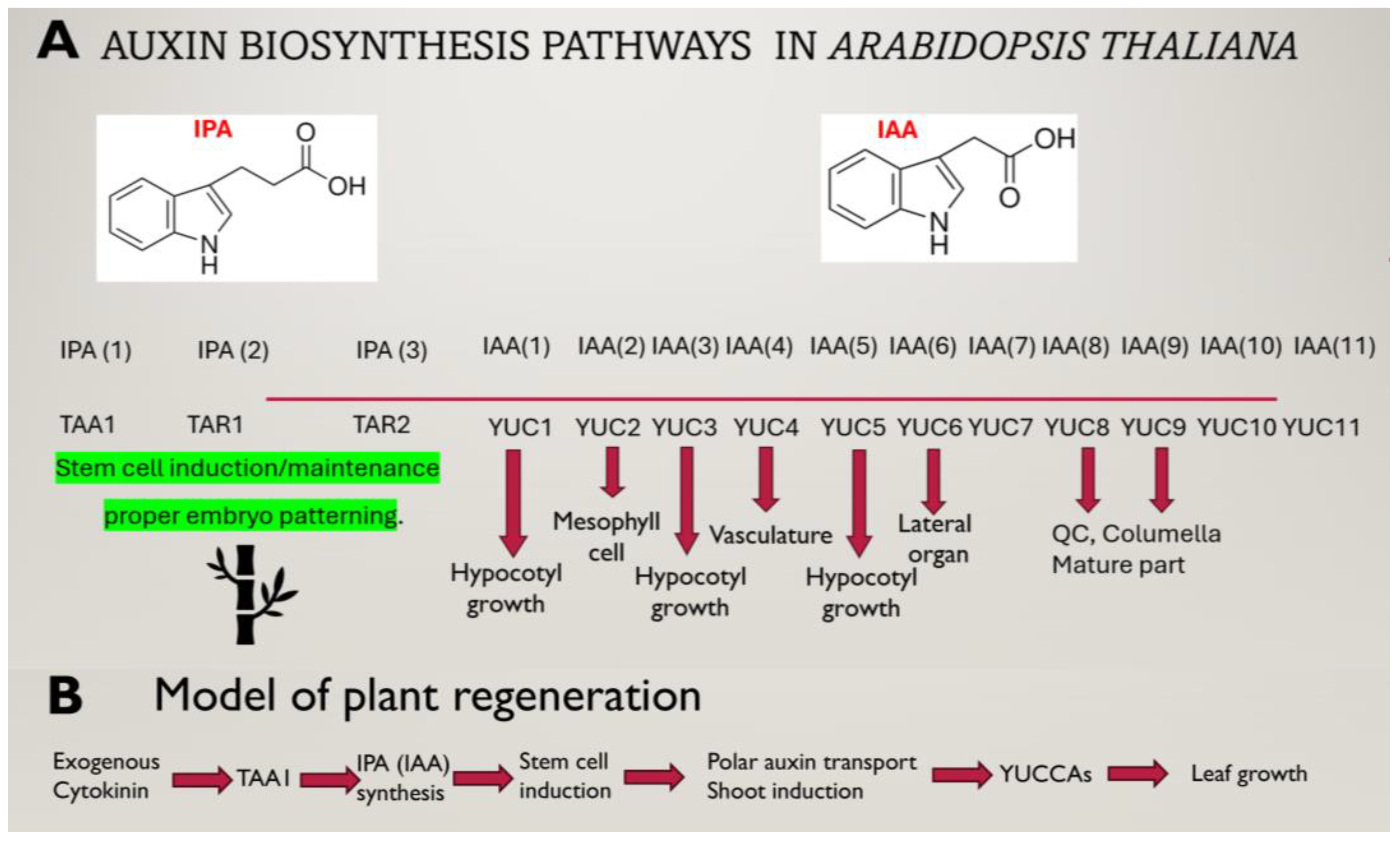
Figure 1. Model of auxin biosynthesis pathways and a possible model of de novo shoot formation in Arabidopsis thaliana. (A)—Auxin biosynthesis pathway in Arabidopsis thaliana. The localization and effects of different TAA1/YUCCA genes are shown under the arrows. (B)—Possible order of events during shoot formation. The key step in morphogenesis is the TAA1-mediated induction of IPA biosynthesis (in only a few cells) and the formation of auxin gradients.
Ijpb 16 00097 g001
Disclaimer/Publisher’s Note: The statements, opinions and data contained in all publications are solely those of the individual author(s) and contributor(s) and not of MDPI and/or the editor(s). MDPI and/or the editor(s) disclaim responsibility for any injury to people or property resulting from any ideas, methods, instructions or products referred to in the content.