Article

Impacts of Foliar Application of Se and TiO2 Nanoparticles on Growth, Development, and Flowering in Lilium Sunny Oriental

by
Nayla Tamara Sánchez-Granados
1,
Sergio Rubén Pérez-Ríos
1,*,
Yolanda González-García
2,
Fabian Fernández-Luqueño
3,
Eliazar Aquino-Torres
1,
Mariana Saucedo-García
1,
Ana Karen Zaldívar-Ortega
1,
Ma Isabel Reyes-Santamaria
1 and
Iridiam Hernández-Soto
1,*
1
Instituto de Ciencias Agropecuarias, Universidad Autónoma del Estado de Hidalgo, Av. Universidad Km 1 Rancho Universitario, Tulancingo de Bravo 43600, Hidalgo, Mexico
2
Centro de Investigación Regional Noreste, Campo Experimental Todos Santos, Instituto Nacional de Investigaciones Forestales, Agrícolas y Pecuarias, La Paz 23070, Baja California Sur, Mexico
3
Centro de Investigación y de Estudios Avanzados del IPN—Departamento de Biotecnología y Bioingeniería Gustavo A. Madero, Ciudad de México 07360, Mexico
*
Authors to whom correspondence should be addressed.
Int. J. Plant Biol. 2025, 16(3), 103; https://doi.org/10.3390/ijpb16030103
Submission received: 31 July 2025 / Revised: 26 August 2025 / Accepted: 2 September 2025 / Published: 5 September 2025
(This article belongs to the Section Plant Physiology)

Abstract

Lilium species produce some of the most commercially valuable ornamental flowers in the world, characterized by their attractiveness and high demand in cut flower markets. However, it is necessary to strengthen the competitiveness of this sector in the global market. Due to strong competition from international producers and an increasingly demanding market regarding quality, shelf life, and sustainability, alternatives are being sought to counteract the use of conventional agrochemicals. The use of nanoparticles has emerged as a promising strategy in ornamental horticulture due to their ability to enhance plant growth, improve stress tolerance, and stimulate physiological processes, ultimately contributing to higher quality and productivity. The hypothesis of this research is that the foliar application of selenium and titanium dioxide nanoparticles during the vegetative growth and flowering stages significantly enhances the growth, development, and flowering of Lilium plants when compared with untreated plants. Therefore, the physiological effects of SeNPs and TiO2NPs applied via foliar application in two concentrations (SeNPsD1, SeNPsD2, TiNPsD1, and TiNPsD2) were evaluated against absolute control. The treatments were applied in two phenological stages (vegetative and reproductive development), and their effects on vegetative and reproductive variables in Lilium plants were evaluated from 120 to 270 days after sowing. The surface of seeds obtained from SeNPsD1-treated plants was further analyzed via scanning electron microscopy (SEM) coupled with energy-dispersive X-ray spectroscopy (EDS). The results demonstrate that the application of SeNPs generated variable effects depending on the phenological stage. In the vegetative stage (46 DAS), SeNPsD2 increased the number of leaves by 118%, while SeNPsD1 increased the fresh weight of leaves by 110%. Regarding ovaries, the application of SeNPsD2 resulted in a 276% increase in fresh weight and a 230% increase in dry weight, while SeNPsD1 achieved an increase of 164% in fresh weight. Furthermore, at this stage, SeNPsD2 promoted a 223% increase in the number of bulbils, a 240% increase in fresh weight, and a 199% increase in dry weight. In the reproductive stage (69 DAS), SeNPsD1 increased the leaf fresh weight by 1% and yielded a 107% increase in the number of ovaries, in addition to 307% and 328% increases in their fresh and dry weights, respectively. In the same stage, SeNPsD2 increased the fresh ovary weight by 153%, compared with the control. Finally, capsule formation was observed only under the SeNPsD1 treatment. Meanwhile, TiO2NPs had an effect on the number of buds and the number of open buds: the number of buds increased by 115% with TiNPsD1 (69 DAS) and the number of open buds increased by 104% (46 DAS) with TiNPsD1; in the reproductive stage, the number increased by 115% with TiNPsD1 compared with the control. In the seed capsules of plants treated with selenium nanoparticles (SeNPsD1), although no surface selenium was detected via EDS, elements that had possibly been physiologically redistributed were identified, including iron (Fe), silicon (Si), and aluminum (Al). These findings confirm the hypothesis of this research, demonstrating that the foliar application of SeNPs and TiO2NPs to Lilium plants during the vegetative and reproductive stages significantly improves their vegetative growth, reproductive development, and floral quality under controlled conditions. This work presents the first comparative evidence regarding the effects of SeNPs and TiO2NPs on the vegetative and reproductive characteristics of Lilium Sunny Oriental, providing unprecedented information for the use of nanotechnology in ornamental horticulture. The findings confirm the potential use of nanoparticles as agents to optimize the productivity and commercial quality of ornamental flowers in highly competitive markets.

1. Introduction

Lilium spp. are some of the most valued ornamental flowers worldwide due to their beauty and diversity of colors, making them a highly sought-after product in international floriculture [1]. The cultivation of Lilium plants has been consolidated mainly in regions with advanced infrastructure for the production of cut flowers [2]. In 2022, the Netherlands led global exports with sales of approximately USD 150 million, followed by Costa Rica (USD 29 million) and Colombia (USD 20.5 million) [3]. Against the background of growing demand and technological development, the use of nanotechnology in the agricultural sector can be considered as a key tool to boost sustainable development [4]. Nevertheless, the application of nanoparticles in ornamental crops remains underexplored. This situation presents a strategic opportunity to investigate the effects of nanoparticles on Lilium production, especially regarding aspects such as growth, development, and flowering, thus contributing to the competitiveness of the ornamental sector in the global market.
SeNPs are among the most widely used nanoparticles in the agricultural sector, which have been reported to exert biostimulant effects by enhancing crop growth and development while modulating plant defense mechanisms. Several studies have shown that SeNPs can decrease the accumulation of hydrogen peroxide (H2O2) and superoxide anion (O2), indicating that selenium affects plant defense systems to control the production and accumulation of reactive oxygen species (ROS) [5,6]. This regulatory mechanism is crucial for maintaining cellular redox homeostasis, preventing oxidative damage, and ensuring optimal metabolic function. When applied to agricultural crops, SeNPs have been shown to improve their absorption and assimilation of nutrients, growth, antioxidant capacity, germination, nutritional value, and resistance to biotic and abiotic stresses [7,8,9].
Another nanomaterial of agricultural interest is titanium dioxide (TiO2), which has been recognized for its photocatalytic properties which can influence various physiological processes. TiO2NPs have been reported to stimulate cell division and elongation, enhance the absorption of light for photosynthesis, and increase chloroplast activity. Furthermore, they can protect plants against abiotic stresses by regulating stomatal aperture, thereby reducing excessive transpiration and improving water use efficiency [10]. These combined physiological effects make TiO2NPs promising candidates for sustainable agricultural production under adverse environmental conditions.
The above statements have been supported by various studies. For example, Treviño López et al. [11] evaluated the effects of Se nanoparticles on the production and productivity of cucumber plants (Cucumis sativus L.) and concluded that the agronomic performance was increased in plants treated with SeNPs; in addition, the NPs positively affected stem diameter and fresh weight of the plant. In another study, Reyes-Pérez et al. [9] concluded that the application of SeNPs to the foliage of chili pepper plants (Capsicum annuum L.) had positive effects on the morphological and reproductive development of this crop. Furthermore, Kolenčík et al. [12] applied TiO2NPs during the cultivation of sunflowers (Helianthus annuus L.) and showed that their application led to a physiological modification towards early ripening in sunflowers. For the above reasons, the hypothesis of this research is that foliar application of selenium and titanium dioxide nanoparticles to Lilium Sunny Oriental during the vegetative and flowering stages significantly improves growth, development, and flowering compared with untreated plants. To test this hypothesis, the physiological effects of SeNPs and TiO2NPs applied at two concentrations and different phenological stages were evaluated and compared, focusing on their influence on vegetative, reproductive, and floral quality variables. This research not only seeks to demonstrate the agronomic efficacy of nanoparticles but also to pave the way for innovative technologies that drive more sustainable and competitive floriculture. The controlled use of nanoparticles represents a strategic alternative to reduce dependence on conventional agrochemicals, mitigate negative environmental impacts, and optimize the use of available resources. Furthermore, it fosters the transition towards more resilient production models which are capable of addressing challenges such as climate change, resource scarcity, and increasing environmental regulations, ensuring the viability and long-term sustainability of the ornamental sector [13].

2. Materials and Methods

2.1. Crop Establishment and Management

The crop was established in a medium-technology greenhouse at the Institute of Agricultural Sciences of the Autonomous University of the State of Hidalgo (UAEH), located in the city of Tulancingo, Hidalgo, Mexico (20°3′39.93″ N, 98°23′0.41″ W). The region is situated at an altitude of 2157 m above sea level and is characterized by a temperate climate, with an average annual temperature of 16 °C [14]. The plant material used was Lilium Sunny Oriental Hybrid bulbs of caliber 18/20, which were planted in 10 L plastic bags containing a mixture of perlite and peat moss substrate in a 1:1 ratio (v/v). The pH and EC of the substrate were monitored at the beginning and during the development of the project, using a kit (LAQUAtwin 4M Kit®, HORIBA, Kyoto, Japan) and obtaining constant values of 5.5 pH and 1.2 ds m−2 for EC. Regarding crop nutrition, a universal Steiner solution adjusted to 25% was applied during the first four weeks after sowing and adjusted to 45% from the fifth week onwards, according to the methodology proposed by Gonzalez-Lemus et al. [15]. Fertilization was carried out once a week and watering was carried out three times a week (in drench), applying 200 mL per plant using a manual multipurpose pump.

2.2. Application of Treatments

The nanoparticles used in this study were acquired from Materials Nanostructures S.A. de C.V., Zapopan, Mexico, a company based in Mexico. The selenium nanoparticles (SeNPs) have a spherical morphology with a size less than 100 nm, gray color, density of 4.81 g cm−3, molecular weight of 78.96, melting point of 960.8 °C, boiling point of 222.12 °C, and possess weak ferromagnetic properties. Meanwhile, the TiO2NPs correspond to the anatase crystalline phase, with a hexagonal crystallographic system. They have a particle size less than 50 nm, white color, density of 4.23 g cm−3, molecular weight of 79.87, melting point of 1843 °C, and are also weakly ferromagnetic. Dispersions of both nanoparticles were made in two doses, 0.70 and 1.42 mL L−1, dissolved in 200 mL of deionized water. The volume of 200 mL was chosen due to the stability of the dispersion, with this volume allowing for homogeneous dispersion after stirring and maintaining the target concentration without agglomeration and minimizing sedimentation. The concentrations were selected based on previous studies demonstrating their effectiveness and absence of toxicity [16,17]. In particular, preliminary tests were carried out and, based on the results, two levels were selected: a minimum (0.70 mL L−1) and a maximum (1.42 mL L−1), both of which are within the safe and effective range. Furthermore, an adherent with surfactant properties (AGREX®, Agroenzymas, Tlalnepantla de Baz, Mexico) was applied at a dose of 1 mL L−1 of water to each of the solutions. Subsequently, the solutions were placed in a beaker and dispersed in an ultrasonic bath (32V118A®, LSS, Shanghai, China) with ultrasonic frequency of 40 kHz and ultrasonic power of 100 W at 25 °C for 35 min. Finally, after the ultrasonic bath, the solutions were placed in atomizers and used as foliar sprays, coating each plant with a volume of 3.2 mL. For comparison, we designed a control treatment that was prepared similarly to the other treatments but included no NPs (Figure 1). For practical purposes, these treatments are denoted as follows: SeNPsD1 (one dose of selenium), SeNPsD2 (two doses of selenium), TiNPsD1 (one dose of titanium dioxide), TiNPsD2 (two doses of titanium), and T0 (absolute control). The doses were applied at two time points: at 46 days and 69 days after sowing (see Figure 1).

2.3. Agronomic Variables

To evaluate the effects of the SeNPs and TiO2NPs on plant growth, development, and quality, the following parameters were assessed: Number of capsules per plant, total number of buds, number of open buds, number of malformed buds, average bud length, average flower diameter, average number of days the flower remained open, number of ovaries, fresh weight of ovaries, dry weight of ovaries, number of peduncles, fresh weight of peduncles, dry weight of peduncles, total plant height, stem height, stem diameter, fresh weight of stem, dry weight of stem, number of leaves, fresh weight of leaves, dry weight of leaves, chlorophyll (SPAD), fresh weight of bulb, dry weight of bulb, number of bulbils, fresh weight of bulbils, dry weight of bulbils, fresh weight of root, and dry weight of root. Specifically, plant height was evaluated using a flexometer (PRO-8-R®, Truper, CDMX, Mexico) from the stem base (neck) to the apex at 271 days after sowing (DAS), following the methodology proposed by Flores-Pérez et al. [18] Likewise, stem length was measured from the neck to the intersection of the first peduncle at 271 DAS [18]. For the stem diameter, a digital vernier (14388®, CALDI-6MP, Truper, Mexico) was used and the measurement was made at 271 DAS [18]. On this same day, chlorophyll measurement was performed using a digital chlorophyll meter (SPAD-502®, KONICA MINOLTA, Tokyo, Japan), taking five measurements on five random leaves of the same plant following the methodology proposed by Ontiveros-Capurata et al. [19]. After the emergence of the buds at 150 DAS, the total number of flower buds was quantified and the length of the buds was measured, using a tape measure (PRO-8-R®, Truper, CDMX, Jilotepec de Molina Enríquez, Mexico), from the base of the bud to the apex of the bud [20]. At the time of complete flower opening at 150 DAS, the flower diameter was measured using the same measuring tape (PRO-8-R®, Truper, CDMX, Mexico) between the apices of the petals [21], taking two measurements in the form of a cross from one end to the other of the petals. Constant monitoring was maintained during the flowering period, and the days that the buds remained open until the moment of senescence were counted [18]. For this analysis, the first two buds of each flower were used, within an interval ranging from 123 to 188 days after sowing; a record was kept of the days that the bud remained open, from the day that the tip first began to open until the last petals fell. At 271 DAS, the percentage of open buds (BFA) was calculated using the formula proposed by Soriano Melgar et al. [22]:
Where BA is the number of buttons open during the evaluation period and BT is the number of total buttons:
B F A   ( % ) = ( B A / B T ) 100
At 271 DAS, the capsules obtained from the crop were counted and collected; then, they were weighed fresh using an analytical balance (PW124®, Adam Equipment, Milton Keynes, UK) and their length and width were measured using a digital vernier caliper (14388®, CALDI-6MP, Truper, Mexico). At 271 days after sowing, the plants were removed from the bags and taken to the Soil Laboratory at the Institute of Agricultural Sciences (UAEH). Substrate residues were removed from the bulbs and roots by rinsing them under running water for 10 min. Each plant was sectioned into roots, bulbs, bulbils (the number of bulbils per plant was counted), stems, leaves, peduncles, and ovaries, and then weighed using an analytical balance (PW124®, Adam Equipment, UK) to determine the fresh weight. Once weighed, the plant parts were placed in paper bags and placed in a drying oven (LW-201C®, GRIEVE, Lake County, IL, USA) at a constant temperature of 60 °C for 72 h. After this time, they were removed and weighed again on an analytical balance (PW124®, Adam Equipment, UK) to determine the dry biomass [21].

2.4. Scanning Electron Microscopy and Energy Dispersive X-Ray Spectroscopy

Capsules containing Lilium seeds were obtained from plants treated with SeNPsD1. The objective of the analysis was to evaluate whether the nanoparticles had been translocated to the seeds and whether it was possible to detect their accumulation on the surface or inside using scanning electron microscopy (SEM) and X-ray energy dispersive spectroscopy (EDS). The seeds were incubated overnight in absolute ethanol (100%) to remove surface impurities and ensure complete dehydration. The dried seeds were mounted on aluminum supports with double-sided carbon tape, following the preparation protocol described by Bazzicalupo et al. [23]. The mounted samples were allowed to air dry at room temperature, then sputter-coated with a 10 nm layer of gold to improve surface conductivity and image resolution.
SEM observations were performed using a scanning electron microscope (JSM-6510LV®, JEOL, Tokyo, Japan) in high vacuum mode, with an accelerating voltage of 15.0 kV. For backscattered electron (BSE) imaging, a spot size of 6.8 was used at 500× magnification to visualize the overall morphology of the seed surface and detect high atomic number regions such as bright zones. For secondary electron (SE) imaging, a spot size of 4.1 was used at 2000× magnification to obtain detailed topographic information. To confirm the elemental composition of the bright regions observed in BSE mode, an EDS system (AURIGA-39-16®, AURIGA, Frankfurt am Main, Germany) was coupled to the SEM. EDS spectra were obtained directly from the selected areas, allowing for identification of the elements present on the seed surface. This combined SEM-EDS approach has been previously reported as a reliable method for investigating plant–nanomaterial interactions [24].

2.5. Statistical Analysis

A completely randomized design was carried out with four replicates per treatment, using one plant as the experimental unit. An analysis of variance was performed and, when significant differences were determined, Fisher’s LSD means test (p ≤ 0.05) was applied using the Infostat 2020 statistical software. The results of the agronomic variables are found as Supplementary Material Table S1.

3. Results

3.1. Vegetative Parameters

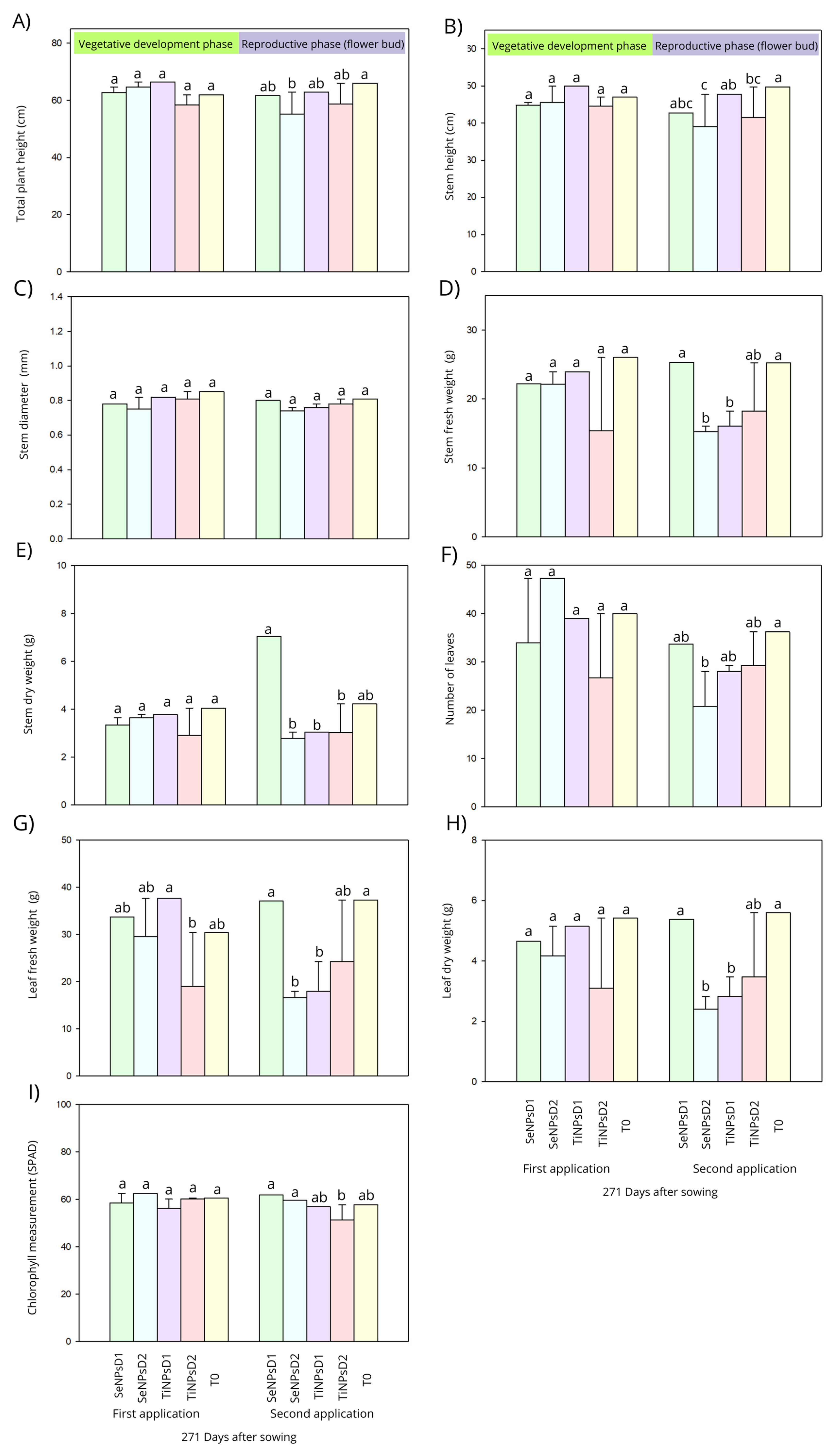
The results of the statistical analysis of variance and Fisher’s LSD mean test: p ≤ 0.05 indicates significant differences between the SeNPsD1, SeNPsD2, TiNPsD1, TiNPsD2, and T0 treatments for total plant height at the second application (69 DAS) (Figure 2A). At the first application (46 DAS), no statistical differences were detected, although all treatments slightly outperformed the control (T0) numerically: the SeNPsD2 group was 4% higher than T0, while TiNPsD1 showed a 7% increase compared with the control. The TiNPsD1 treatment was the most notable, outperforming TiNPsD2 by 113%. At the second application (69 DAS), the SeNPsD1 group was 2% higher than TiNPsD1 and 105% higher than TiNPsD2. The results of the statistical analysis indicate significant differences between the SeNPsD1, SeNPsD2, TiNPsD1, and TiNPsD2 treatments for the stem height (Figure 2B) at the second application (69 DAS). At the first application (46 DAS), the TiNPsD1 group increased 106% with respect to the control. The SeNPsD2 group was 9% higher than TiNPsD1. At the second application (69 DAS), the SeNPsD2 group was 19% higher than TiNPsD1. For the stem diameter variable (Figure 2C), the results of the statistical analysis demonstrated that there were no significant differences between the SeNPsD1, SeNPsD1, TiNPsD1, TiNPsD2, and T0 treatments for both applications (46 and 69 DAS). At the second application (69 DAS) SeNPsD1 was 2% higher than TiNPsD1 and 1% higher than TiNPsD2.
The results of the statistical analysis regarding the stem fresh weight (Figure 2D) demonstrated significant differences between the SeNPsD1, SeNPsD2, TiNPsD1, TiNPsD2, and T0 treatments at the second application (69 DAS). At the second application (69 DAS) SeNPsD1 was 37% and 27% higher than TiNPsD1 and TiNPsD2. Regarding the stem dry weight variable (Figure 2E), the results of the statistical analysis indicate significant differences for the SeNPsD1 treatment with respect to SeNPsD2, TiNPsD1, and TiNPsD2 at the second application (69 DAS). At the first application (46 DAS), TiNPsD1 was 14% higher than TiNPsD2. At the second application (69 DAS), SeNPsD1 showed a 167% increase over the control, as well as increases of 231% and 233% with respect to TiNPsD1 and TiNPsD2, respectively; furthermore, SeNPsD1 was 254% higher than SeNPsD2. Regarding the leaf number variable (Figure 2F), the results of the statistical analysis show significant differences: the SeNPsD2 treatment increased this variable by 118% with respect to T0 and 176% with respect to TiNPsD2 during the vegetative phase (46 DAS). At the reproductive phase (69 DAS) the SeNPsD1 group showed increases of 120% and 115% with respect to TiNPsD1 and TiNPsD2, respectively.
Regarding the fresh weight of the leaves (Figure 2G), the results of the statistical analysis demonstrated significant statistical differences between the TiNPsD1 and TiNPsD2 treatments at the first application (46 DAS), and SeNPsD1 and T0 with respect to SeNPsD2 and TiNPsD1 at the second application (69 DAS). At the first application (46 DAS), the TiNPsD2 treatment increased the fresh weight by 123% compared with T0, while the SeNPsD1 treatment increased the fresh weight by 110% with respect to T0. At the second application (69 DAS), SeNPsD1 was on average 176% superior to the TiO2NPs treatments. The results of the statistical analysis revealed significant differences for the SeNPsD1 and T0 treatments with respect to SeNPsD2 and TiNPsD1 for the leaf dry weight (Figure 2H) at the second application (69 DAS). At the first application (46 DAS), the SeNPsD1 group presented a 15% lower value than T0, and similarly for SeNPsD2 (24%), TiNPsD1 (5%), and TiNPsD2 (43%). At the second application (69 DAS), the SeNPsD1 group presented a reduction of 4% compared with the control, while TiNPsD1 and TiNPsD2 showed reductions of 58% and 38%, respectively. The results of the statistical analysis indicate significant differences for the SeNPsD1 and SeNPsD2 treatments with respect to TiNPsD2 for chlorophyll (SPAD) (Figure 2I) at the second application (69 DAS). Regarding this variable, the SeNPsD2 group presented a 103% higher value than T0 at the first application (46 DAS). The SeNPsD1 group was 103% higher than TiNPsD1, while SeNPsD2 outperformed TiNPsD1 by 110% and TiNPsD2 by 103%. At the second application (69 DAS), SeNPsD2 showed increases of 103% and 107% compared with the control and SeNPsD1, respectively.

3.2. Reproductive Parameters

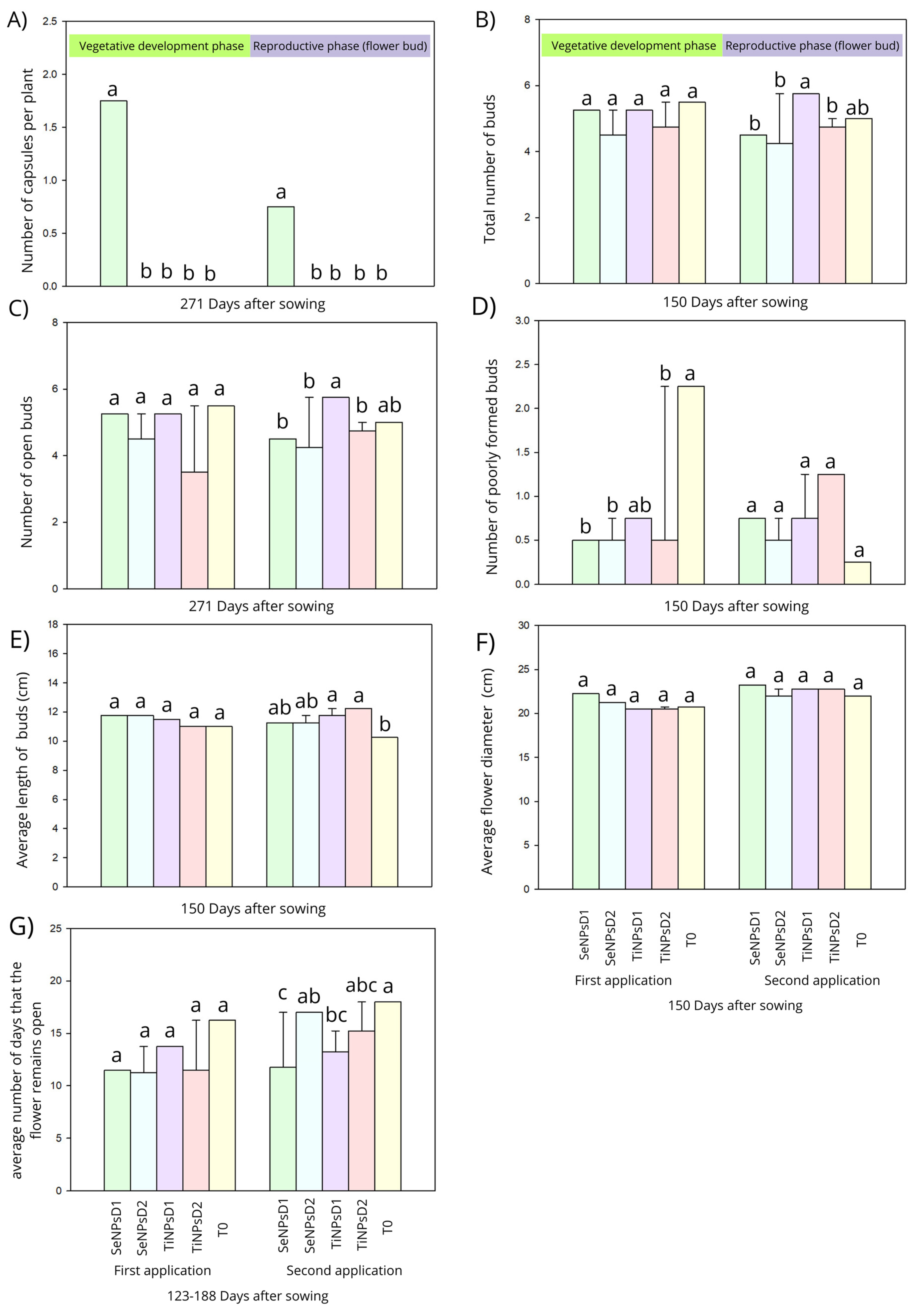
The results of the statistical analysis indicate significant differences between the SeNPsD1, SeNPsD2, TiNPsD1, TiNPsD2, and T0 treatments for the number of capsules per plant, number of days the buds were open, total number of buds, number of open buds, and number of malformed buds. Figure 3A shows the number of capsules per plant obtained from the Lilium plants at both applications (46 and 69 DAS). SeNPsD1 was the only treatment that promoted capsule formation at the first application (46 DAS), exceeding the control by 100%. Furthermore, at the second application (69 DAS), this effect remained exclusive to SeNPsD1. Regarding the total number of buds (Figure 3B), the results of the statistical analysis indicate significant differences at the second application (69 DAS) between the SeNPsD1, SeNPsD2, TiNPsD2, and T0 treatments. In this variable, the SeNPsD1 group was equal to TiNPsD1 and 110% higher than TiNPsD2 at the first application (46 DAS) At the second application (69 DAS), TiNPsD1 and TiNPsD2 showed smaller increases of 115% and a reduction of 5%, respectively, when compared with T0. The results of the statistical analysis indicate significant differences between the SeNPsD1, SeNPsD2, TiNPsD1, TiNPsD2, and T0 treatments at the second application (69 DAS) regarding the number of open buds (Figure 3C). At the first application (46 DAS), the SeNPsD1 and SeNPsD2 groups were 106% higher than the control (T0), while TiNPsD1 showed an increase of 104% compared with T0 and TiNPsD2 was similar to T0. The SeNPsD1 group was 102% higher than TiNPsD1 and 106% higher than TiNPsD2, and a similar trend was observed for SeNPsD2 with respect to TiNPsD1 and TiNPsD2. At the second application (69 DAS), TiNPsD1 and TiNPsD2 showed an increase of 115% with respect to the control (T0).
The results of the statistical analysis revealed significant differences between the SeNPsD1, SeNPsD2, TiNPsD1, TiNPsD2, and T0 treatments at the first application (46 DAS) for the number of malformed buds (Figure 3D). At the first application (46 DAS), the SeNPsD1 treatment was 34% more effective than TiNPsD1 and equivalent to TiNPsD2, while SeNPsD2 showed the same behavior with respect to TiNPsD1 and TiNPsD2. At the second application (69 DAS), SeNPsD1 and SeNPsD2 increased malformation by 300% and 100% compared with the control, while TiNPsD1 and TiNPsD2 increased malformation by 300% and 500%, respectively. Regarding the average length of the bud (Figure 3E), the results of the statistical analysis show that there were no significant differences at the first application (46 DAS); however, at the second application (69 DAS), there were significant differences between the treatments TiNPsD1 and TiNPsD2 with respect to T0. At the first application (46 DAS), the values for the SeNPsD1 group were 106% higher than the control (T0) and SeNPsD2, while TiNPsD1 showed an increase of 104% and TiNPsD2 did not differ from T0. The SeNPsD1 group was 102% higher than TiNPsD1 and 106% higher than TiNPsD2, and similar behavior was observed for SeNPsD2. At the second application (69 DAS), SeNPsD1 and SeNPsD2 maintained a 109% increase over the control, while TiNPsD1 and TiNPsD2 were 114% and 119% higher than T0, respectively. The statistical analysis revealed no significant differences among the SeNPsD1, SeNPsD2, TiNPsD1, TiNPsD2, and T0 treatments in the average flower diameter variable (Figure 3F) at either application. At the first application (46 DAS), treatments with SeNPs (SeNPsD1 and SeNPsD2) were 107% and 102% higher than the control (T0). The SeNPsD1 treatment was 108% superior to TiNPsD1 and TiNPsD2, while SeNPsD2 showed increases of 103% compared with TiNPsD1 and TiNPsD2. At the second application (69 DAS), SeNPsD1 maintained an increase of 105% compared with T0, while TiNPsD1 and TiNPsD2 were 103% superior to the control. The SeNPsD1 treatment was 102% superior to TiNPsD1 and TiNPsD2.
The results of the statistical analysis indicate significant differences between the SeNPsD1, SeNPsD2, TiNPsD1, TiNPsD2, and T0 treatments regarding the average number of days that the flower remained open in the reproductive stage (69 DAS) (Figure 3G). No statistical differences were observed at the first application (46 DAS); however, SeNPsD1 and SeNPsD2 showed reductions of 70% and 69% compared with the control (T0), while TiNPsD1 and TiNPsD2 were 16% and 30% lower. The SeNPsD1 group presented 17% lower values than TiNPsD1, while SeNPsD2 showed reductions of 19% and 3% with respect to TiNPsD1 and TiNPsD2, respectively. At the second application (69 DAS), the SeNPsD1 treatment reduced flowering by 35% compared with T0 and SeNPsD2 by 6%, while TiNPsD1 and TiNPsD2 reduced this variable by 27% and 16%, respectively. The SeNPsD1 treatment was 12% and 23% lower than TiNPsD1 and TiNPsD2, while SeNPsD2 was 26% and 11% higher in the same comparison.
The results of the statistical analysis indicate significant differences between the SeNPsD1, SeNPsD2, TiNPsD1, TiNPsD2, and T0 treatments regarding the number of ovaries, fresh weight of ovaries, dry weight of ovaries, number of peduncles, fresh weight of peduncles, and dry weight of peduncles. For the number of ovaries (Figure 4A), the results of the statistical analysis indicate significant statistical differences between the SeNPsD1, SeNPsD2, TiNPsD1, TiNPsD2, and T0 treatments at the first application (46 DAS). At this time, the SeNPsD1 treatment outperformed TiNPsD1 and TiNPsD2 by 109% and 50%, respectively, with SeNPsD2 similarly outperforming TiNPsD1 and TiNPsD2 by 154% and 283%. At the second application (69 DAS), SeNPsD1 yielded a value 107% higher than the control. The SeNPsD1 treatment was 187% and 171% higher than TiNPsD1 and TiNPsD2, while SeNPsD2 exceeded them by 150% and 42%, respectively. The fresh ovary weight is shown in Figure 4B; SeNPsD2 treatment at the first application (46 DAS) increased this variable by 276% compared with T0, followed by SeNPsD1 (with an increase of 164% compared with T0). At the second application (69 DAS), SeNPsD1 increased the fresh ovary weight by 307% compared with T0, while the SeNPsD2 treatment increased this variable by 153% compared with T0. The increase was 285% when directly comparing SeNPsD2 with TiNPsD2, while SeNPsD1 was 800% higher than TiNPsD2. Compared with TiNPsD1, SeNPsD2 was 285% higher and SeNPsD1 was 572% higher. The results of the statistical analysis revealed significant differences between the SeNPsD1, SeNPsD2, TiNPsD1, TiNPsD2, and T0 treatments for the dry ovary weight at the second application (69 DAS) (Figure 4C). At the first application (46 DAS), the SeNPsD2 treatment led to a 230% higher value than the control (T0), while TiNPsD1 and TiNPsD2 showed increases of 130% and 170% compared with T0. At the second application (69 DAS), SeNPsD1 was 328% higher than the control, while SeNPsD2 was 185% higher. The SeNPsD1 group was 460% and 328% higher than TiNPsD1 and TiNPsD2, while SeNPsD2 exceeded them by 260% and 185%, respectively. Regarding the number of peduncles (Figure 4D), the results of the statistical analysis show significant differences at the second application (69 DAS) between the SeNPsD1, SeNPsD2, TiNPsD1, TiNPsD2, and T0 treatments. At the first application (49 DAS), TiNPsD1 increased this variable by 105% compared with the control while, at the second application (69 DAS), TiNPsD1 was 121% greater than T0. For the fresh stem weight variable (Figure 4E), the results of the statistical analysis indicate significant differences between the SeNPsD1, SeNPsD2, TiNPsD1, TiNPsD2, and T0 treatments at the second application (69 DAS). At the first application (46 DAS), the SeNPsD1 and SeNPsD2 groups showed increases of 145% and 108% compared with T0, while an 112% increase was observed for TiNPsD1. The SeNPsD1 group was 128% and 221% higher than TiNPsD1 and TiNPsD2, while SeNPsD2 was 5% lower than TiNPsD1 and 164% higher than TiNPsD2. At the second application (69 DAS), TiNPsD1 and TiNPsD2 were 24% and 37% higher than T0. Again, SeNPsD1 was 122% and 134% higher than TiNPsD1 and TiNPsD2. The results of the statistical analysis demonstrated significant differences between the SeNPsD1, SeNPsD2, TiNPsD1, TiNPsD2, and T0 treatments regarding the peduncle dry weight at the second application (69 DAS) (Figure 4F). At the first application (49 DAS), TiNPsD2 increased this variable by 125% with respect to T0 while, at the second application (69 DAS), TiNPsD1 was 113% higher than T0.
The results of the statistical analysis indicate significant differences for the SeNPsD2 and TiNPsD1 treatments with respect to SeNPsD1 regarding the number of bulbils (Figure 5A) at the first application (46 DAS): at this time, the SeNPsD2 group presented a 223% higher value than T0, while TiNPsD1 and TiNPsD2 were 207% and 107% higher with respect to T0. Furthermore, SeNPsD2 was 193% higher than TiNPsD2. At the second application (69 DAS), the SeNPsD1 group showed an increase of 144% with respect to T0, while TiNPsD1 and TiNPsD2 were 137% and 182% higher with respect to T0. The results of the statistical analysis indicate significant differences between the SeNPsD1, SeNPsD2, TiNPsD1, and TiNPsD2 treatments at the two applications (46 DAS and 69 DAS) regarding the bulbil fresh weight (Figure 5B). At the first application (46 DAS), that in the SeNPsD2 group was increased by 240% compared with T0; TiNPsD1 was 124% higher than T0; and SeNPsD2 was 49% and 69% higher than TiNPsD1 and TiNPsD2. At the second application (69 DAS), the SeNPsD1 treatment yielded a 285% higher value than TiNPsD1; while TiNPsD1 was 85% higher than TiNPsD2. The results of the statistical analysis for the bulbil dry weight (Figure 5C) indicate significant differences between the SeNPsD1, SeNPsD2, TiNPsD1, TiNPsD2, and T0 treatments at the two applications (46 and 69 DAS): at the first application (46 DAS), SeNPsD2 was 199% higher than the control and TiNPsD1 was 116% higher than T0; at the second application (69 DAS), SeNPsD1 was 307% greater than TiNPsD1.
The results of the statistical analysis indicate significant differences for SeNPsD1 with respect to SeNPsD2 and TiNPsD1 for the fresh bulb weight (Figure 5D) at the second application (69 DAS). At the first application (46 DAS), the SeNPsD1 group was 114% higher than TiNPsD1 and 148% higher than TiNPsD2, while TiNPsD1 was 108% higher than TiNPsD2. At the second application (69 DAS), SeNPsD1 was 113% higher than T0 and SeNPsD2 was 104% higher than TiNPsD1. The results of the statistical analysis indicate significant differences in the bulb dry weight (Figure 5E) for the SeNPsD1 treatment with respect to SeNPsD2 and TiNPsD1 at the second application (69 DAS). At the first application (46 DAS), SeNPsD1 and SeNPsD2 were 5% and 30% lower than T0, while TiNPsD1 and TiNPsD2 were 22% and 94% lower than T0, respectively. At the second application (69 DAS), SeNPsD1 was 117%, 197%, and 167% higher than T0, TiNPsD1, and TiNPsD2. The results of the statistical analysis indicate significant differences for the SeNPsD1 and TiNPsD2 treatments with respect to T0 regarding the fresh root weight (Figure 5F) at the first application (46 DAS): at this time, TiNPsD1 was 16% higher than TiNPsD2. At the second application (69 DAS), SeNPsD1 was 142% and 144% higher than TiNPsD1 and TiNPsD2. Regarding the dry root weight (Figure 5G), the results of the statistical analysis significant differences for SeNPsD1 and TiNPsD2 with respect to T0 at the first application (46 DAS): at this time, SeNPsD2 was 15% and 47% higher than the TiO2NPs groups. At the second application (69 DAS), the SeNPsD1 group was 171% and 180% higher than TiNPsD1 and TiNPsD2, while SeNPsD2 was 117% and 123% higher than the same groups, respectively.

3.3. Scanning Electron Microscopy and Energy Dispersive X-Ray Spectroscopy

The sample of Lilium seeds from plants treated with selenium nanoparticles (SeNPsD1) were analyzed via scanning electron microscopy (SEM) at 500× with a BSE detector (Figure 6A). The seeds showed a relatively conserved plant epidermal structure and presented bright punctual regions, indicative of the accumulation of elements with high atomic number; one of these areas coincided with the site where the punctual analysis was performed via X-ray energy dispersive spectroscopy (EDS), which indicates iron (Fe) as the predominant element, accompanied by traces of carbon (C), nitrogen (N), aluminum (Al), and silicon (Si), with no signals attributable to selenium (Se). These results suggest that, in this specific region, there was no detectable accumulation of this element (Figure 6C). In the image at 2000× magnification obtained with an SE detector (Figure 6B), fine topographic details can be seen, with ovoid surface structures that could correspond to mineral aggregates or nanoparticle remains standing out, as well as cellular filaments associated with the seed’s morphology. The combination of both images with the EDS spectrum (Figure 6) allowed us to conclude that iron accumulated in a localized manner possibly as an environmental contaminant and that the presence of selenium should be evaluated through broader elemental mapping or additional analyses, as its absence at the considered point does not rule out a heterogeneous distribution in other regions.

4. Discussion

The results of the present study demonstrate that the application of SeNPs and TiO2NPs induces positive responses during the morphological and reproductive development of Lilium, confirming our initial hypothesis. SeNPs promoted significant increases in the vegetative and reproductive variables of Lilium plants, with variations observed according to the phenological stage (Figure 2, Figure 3, Figure 4 and Figure 5). These results could be explained by the ability of NPs to induce the activation of key genes involved in plant growth and flowering [24]. In particular, SeNPs have been associated with the overexpression of antioxidant genes such as GPX1, SOD1, CAT1, and APX1, which help to maintain cellular redox homeostasis under exposure to reactive oxygen species [25]. Such genetic modulation may explain the observed increases in the number of leaves, roots, and bulbs in the SeNP-treated plants (Figure 2F and Figure 5D–G), mediated by the promotion of cell division and protection against oxidative stress. Similarly, genes such as SOC1, LFY, and AP1—regulators of the onset of flowering—could have been activated in response to the modified nutritional and hormonal status induced by the nanoparticles [26]; this supports the greater number of buds observed in plants treated with TiO2 NPs (Figure 3B). The exposure of the Lilium plants to the nanoparticles possibly induced reconfiguration of the synthesis of primary and secondary metabolites [27]. Due to its chemical affinity with sulfur, selenium can be incorporated into amino acids (e.g., selenomethionine or selenocysteine), thus generating a reserve of sulfur compounds which are involved in stress tolerance and the regulation of cellular metabolism. These metabolites are also linked to the regulation of cell expansion and the induction of reproductive structures [28]. Similar results have been reported by AbdElgawad et al. [29], who observed that the application of SeNPs to broccoli sprouts (Brassica oleracea var. italica) improved root growth and the aerial biomass—effects that coincide with the increase in the number of bulbils recorded for Lilium plants after treatment with selenium nanoparticles (Figure 5A).
On the other hand, TiO2NPs increased the number of buds (by 115% with TiNPsD1 at 69 DAS) and the number of open buds (by 104% with TiNPsD1 at 46 DAS); furthermore, at the reproductive stage, the number was increased by 115% with TiNPsD1 when compared with the control (Figure 3). This could be explained by the ability of titanium dioxide to act as a photocatalyst, thus modifying carbon metabolism through increased photosynthetic activity and the production of sugars, organic acids, and phenolic compounds [30], which may be related to the higher number of peduncles (Figure 4A) and ovaries observed under the TiO2 treatments in this study (Figure 4D). At the root level, this modified metabolomic profile may promote greater secretion of exudates, thus stimulating the development of lateral roots [31], justifying the observed increase in the number of bulbils (Figure 5A). Azmat et al. [32] showed that the application of TiO2NPs significantly increased photosynthesis, chlorophyll concentration, and biomass in spinach (Spinacia oleracea L.). Likewise, Verma et al. [33] reported improvements in root and stem growth, leaf number, and chlorophyll content in bean (Phaseolus vulgaris L.) plants treated with TiO2NPs. Finally, Kamali et al. [34] applied TiO2NPs to petunia (Petunia hybrida). This treatment produced an increase in floral diameter, number of flowers, and ornamental quality, consistent with the better reproductive performance of Lilium plants recorded in this study (Figure 3C,F,G).
From a mechanistic perspective, these effects can be further explained by considering the initial interaction between the surface charges of nanoparticles and cell surfaces [35]. Plant membranes and cell walls possess a net negative charge at their interface with the apoplast, generating an electrical double layer (EDL) composed of a Stern layer (adsorbed ions) and a diffuse layer. Due to their high surface-to-volume ratio and surface charge density, nanoparticles interact with these surfaces through electrostatic attraction or molecular recognition, altering the surface potential (ψ0) and the transmembrane potential [36]. These changes can modify the activities of receptors, ion channels, and transporter proteins, triggering signals that affect gene expression and cellular metabolism [37]. A key factor is the formation of a protein corona on the surface of nanoparticles upon contact with biological fluids. This corona, which has positive or negative charges depending on the pH and ionic strength of the medium, acts as a mediator in the interaction with integral and peripheral membrane proteins [38]. In the case of SeNPs and TiO2NPs, such an interaction could explain the induction of pathways associated with cell division and elongation, increased photosynthetic efficiency, activation of the antioxidant system, and the redistribution of photoassimilates towards reproductive structures [39]. This justifies the increase in leaf biomass and its subsequent mobilization towards reproductive organs, reflected in the higher fresh and dry weights of ovaries and bulbils reported in this research.
Meanwhile, the absence of SeNPs in SEM micrographs and spot analysis using energy dispersive X-ray spectroscopy (EDS) (Figure 6) could be explained by the possible systematic redistribution of Se to non-superficial tissues or intracellular chemical transformations [40]. The presence of Fe, Si, and Al, despite not having been applied as nanoparticles, can be attributed to their endogenous origin, as well as to passive adsorption processes on the tissue surface during crop development [41]. In particular, as an essential micronutrient, iron can be actively mobilized towards reproductive structures, while silicon and aluminum are usually found as natural contaminants in agricultural matrices and can be associated with the external surface through interaction with mucilage or cuticular residues [42,43]. As no selenium was identified superficially, the physiological effects observed in the treated plants suggest that SeNPsD1 was absorbed and translocated—probably accumulating in subcellular compartments or integrating into internal metabolic pathways not reflected on the seed surface [44]. These results are consistent with those reported by Wang et al. [44], who showed how SeNPs are absorbed by rice (Oryza sativa L.) roots, being translocated and biotransformed into organic compounds at the intracellular level, although they were not detected on the surface by SEM-EDS. In another investigation, Diehl et al. [45] described that seed mucilage contains natural traces of cations such as Fe and Al, which are retained in external layers due to the adhesive properties of polysaccharides. Finally, Haughn & Western [46] have highlighted that seed mucilage is rich in pectins and cellulose, which are capable of interacting with external ions and retaining them on the surface.
Although nanoparticles have demonstrated positive effects in the agricultural sector, enhancing crop growth, development, and productivity, their constant application poses significant challenges that must be considered in the context of sustainability [47]. The repeated use of nanoparticles could alter the dynamics of the root and leaf microenvironment, modify the composition and functionality of the associated microbiota, and favor their accumulation in plant tissues or in the soil [48]. Determining these potential accumulations requires rigorous assessments of persistence, mobility, and long-term sublethal effects, both in plants and non-target organisms. Furthermore, crop responses between production cycles could vary due to factors such as residual accumulation, initial nutritional status, and environmental conditions during application, underscoring the need to develop tailored management strategies that maximize the benefits of nanoparticles while minimizing the risks associated with their continued use [49].

5. Conclusions

The foliar application of selenium and titanium dioxide nanoparticles on Lilium plants generated positive physiological responses, improving their biomass, reproductive performance, and floral quality. These results highlight the potential of these nanoparticles as tools for the physiological management of ornamental crops, with the aim of increasing their productivity and commercial value. However, additional studies are needed to clarify their long-term effects, environmental fate, and the physiological mechanisms involved, particularly regarding the redistribution of elements and potential impacts on subsequent growth cycles.

Supplementary Materials

The following supporting information can be downloaded at: https://www.mdpi.com/article/10.3390/ijpb16030103/s1, Table S1: The results of the agronomic variables.

Author Contributions

Conceptualization, S.R.P.-R. and I.H.-S. Data curation, N.T.S.-G., A.K.Z.-O., and I.H.-S., Formal analysis, A.K.Z.-O. and I.H.-S. Investigation, Y.G.-G., M.S.-G., S.R.P.-R., E.A.-T., M.I.R.-S. and I.H.-S.; Methodology, N.T.S.-G., F.F.-L., E.A.-T., Y.G.-G. and I.H.-S.; Resources, S.R.P.-R., M.I.R.-S., M.S.-G. and F.F.-L.; Software, A.K.Z.-O. and I.H.-S.; Supervision, S.R.P.-R. and I.H.-S.; Validation, S.R.P.-R. and I.H.-S.; Visualization, N.T.S.-G. and A.K.Z.-O.; Writing—original draft, N.T.S.-G. and I.H.-S.; Writing—review and editing, N.T.S.-G. and I.H.-S. All authors have read and agreed to the published version of the manuscript.

Funding

This research received no external funding.

Data Availability Statement

The original contributions presented in this study are included in the article/Supplementary Material. Further inquiries can be directed to the corresponding author(s).

Acknowledgments

The authors would like to acknowledge the Universidad Autónoma del Estado de Hidalgo (UAEH) for allowing us to conduct the study in their facilities.

Conflicts of Interest

The authors declare no conflicts of interest.

References

  1. Yan, Y.; Zhu, X.; Yu, Y.; Li, C.; Zhang, Z.; Wang, F. Nanotechnology Strategies for Plant Genetic Engineering. Adv. Mater. 2021, 34, 2106945. [Google Scholar] [CrossRef]
  2. Wei, L.; Wei, S.; Hu, D.; Feng, L.; Liu, Y.; Liu, H.; Liao, W. Comprehensive Flavor Analysis of Volatile Components During the Vase Period of Cut Lily (Lilium spp. ‘Manissa’) Flowers by HS-SPME/GC–MS Combined With E-Nose Technology. Front. Plant Sci. 2022, 13, 822956. [Google Scholar] [CrossRef]
  3. Jiang, F.; Zhou, L.; Zhou, W.; Zhong, Z.; Yu, K.; Xu, J.; Zou, L.; Liu, W. Effect of modified atmosphere packaging combined with plant essential oils on preservation of fresh-cut lily bulbs. LWT 2022, 162, 113513. [Google Scholar] [CrossRef]
  4. González-García, Y.; Juárez-Maldonado, A. Nanomaterials on Plant Growth and Stress Adaptation. Plants 2025, 14, 1651. [Google Scholar] [CrossRef]
  5. Zhu, Z.; Chen, Y.; Shi, G.; Zhang, X. Selenium delays tomato fruit ripening by inhibiting ethylene biosynthesis and enhancing the antioxidant defense system. Food Chem. 2017, 219, 179–184. [Google Scholar] [CrossRef]
  6. González-García, Y.; Juárez-Maldonado, A.; Benavides-Mendoza, A.; Ojeda-Barrios, D.L.; Tortella Fuentes, G.; Seabra, A.B. Improving Nutritional Quality of Food Crops Using Nanomaterials and Nanostimulants. In Plant Biostimulation with Nanomaterials; Smart Nanomaterials Technology; Springer: Singapore, 2025; pp. 187–221. [Google Scholar] [CrossRef]
  7. Song, J.; Yu, S.; Yang, R.; Xiao, J.; Liu, J. Opportunities for the use of selenium nanoparticles in agriculture. NanoImpact 2023, 31, 100478. [Google Scholar] [CrossRef]
  8. Zhang, T.; Qi, M.; Wu, Q.; Xiang, P.; Tang, D.; Li, Q. Recent research progress on the synthesis and biological effects of selenium nanoparticles. Front. Nutr. 2023, 10, 1183487. [Google Scholar] [CrossRef]
  9. Reyes-Pérez, J.J.; Llerena-Ramos, L.T.; Torres-Rodríguez, J.A.; Rodríguez-Rodríguez, S.; Sariñana-Navarrete, M.A. Respuesta Morfológica y Fenológica de Plantas de Chile Pimiento a la Suplementación de Nanopartículas de Selenio. Terra Latinoam. 2025, 43, 1–12. [Google Scholar] [CrossRef]
  10. Gamage, A.; Fernando, N.; Mani, S.; Manamperi, A.; Madhujith, T. Biodegradable Bionanocomposites in Agriculture Applications. In Nanoformulations for Agricultural Applications; Radhakrishnan, E.K., Aswani, R., Visakh, P.M., Eds.; Scrivener Publishing LLC.: Beverly, MA, USA, 2025; pp. 203–230. [Google Scholar] [CrossRef]
  11. Treviño López, E.A.; Sandoval-Rangel, A.; Benavides Mendoza, A.; Benavides Mendoza, A.; Ortega Ortiz, H.; Cadenas Pliego, G.; Cabrera de la Fuente, M. Nanopartículas de selenio absorbidas en hidrogeles de quitosán-polivinil alcohol en la producción de pepino injertado. Rev. Mex. Cienc. Agríc. 2021, 26, 159–169. [Google Scholar] [CrossRef]
  12. Kolenčík, M.; Ernst, D.; Urík, M.; Ďurišová, Ľ.; Bujdoš, M.; Šebesta, M.; Kratošová, G. Foliar application of low concentrations of titanium dioxide and zinc oxide nanoparticles to the common sunflower under field conditions. Nanomaterials 2020, 10, 1619. [Google Scholar] [CrossRef]
  13. Ur Rahim, H.; Qaswar, M.; Uddin, M.; Giannini, C.; Herrera, M.L.; Rea, G. Nano-enable materials promoting sustainability and resilience in modern agriculture. Nanomaterials 2021, 11, 2068. [Google Scholar] [CrossRef]
  14. INEGI (Instituto Nacional de Estadística, Geografía e Informática). Anuario Estadístico y Geográfico del Estado de Hidalgo, México; Gobierno del Estado de Hidalgo: Higalgo, Mexico, 2017; Volume 1, p. 1674. Available online: https://www.inegi.org.mx/contenido/productos/prod_serv/contenidos/espanol/bvinegi/productos/nueva_estruc/anuarios_2017/702825095093.pdf (accessed on 1 March 2025).
  15. González-Lemus, U.; Tapia-Zayago, F.A.; Pérez-Ríos, S.R.; Zaldívar-Ortega, A.K.; Rueda-Puente, E.O.; Hernández-Pérez, A.; Hernández-Soto, I. Lentil Biorooting Agents: An Ecological Alternative to Improve the Growth and Development of Italian Zucchini in Sustainable Production Systems. Horticulturae 2025, 11, 332. [Google Scholar] [CrossRef]
  16. Larue, C.; Veronesi, G.; Flank, A.M.; Surble, S.; Herlin-Boime, N.; Carrière, M. Comparative uptake and impact of TiO2 nanoparticles in wheat and rapeseed. J. Toxicol. Environ. Health A 2012, 75, 722–734. [Google Scholar] [CrossRef] [PubMed]
  17. Satti, S.H.; Raja, N.I.; Javed, B.; Akram, A.; Mashwani, Z.U.; Ahmad, M.S.; Ikram, M. Titanium dioxide nanoparticles elicited agro-morphological and physicochemical modifications in wheat plants to control Bipolaris sorokiniana. PLoS ONE 2021, 11, e0246880. [Google Scholar] [CrossRef]
  18. Flores-Pérez, S.; Castillo-González, A.M.; Valdez-Aguilar, L.A.; Avítia-García, E. Uso de diferentes proporciones de led rojos y azules para mejorar el crecimiento de Lilium spp. Rev. Mex. Cienc. Agric. 2021, 12, 5. [Google Scholar] [CrossRef]
  19. Ontiveros-Capurata, R.E.; Juárez-López, P.; Mendoza-Tafolla, R.O.; Alia-Tejacal, I.; Villegas-Torres, O.G.; Guillén-Sánchez, D.; Cartmill, A.D. Relationship between chlorophyll and nitrogen concentration, and fresh matter production in basil ‘Nufar’ (Ocimum basilicum) with three handheld chlorophyll meter readings: SPAD, atLEAF and MC-100. Rev. Chapingo Ser. Hortic. 2022, 28, 189–202. [Google Scholar] [CrossRef]
  20. Velasco-Lara, D.; De La Cruz-Guzmán, G.H.; Mandujano-Piña, M.; Arriaga-Frías, A.; Ramírez-Santiago, D. Dosis de paclobutrazol para modificar el aspecto visual de Lilium cv. Litouwen. Rev. Mex. Cienc. Agric. 2024, 15, 1–10. [Google Scholar] [CrossRef]
  21. Rios-Florida, L.G.; De La Cruz-Guzmán, G.H.; Arriaga-Frías, A.; Mandujano-Piña, M. Effect of paclobutrazol and Glomus intraradices on the crop of Lilium cv. Armandale and Tresor. Siembra 2022, 9, 1–9. [Google Scholar] [CrossRef]
  22. Soriano Melgar, L.A.A.; López-Guerrero, A.; Cortéz-Mazatan, G.; Mendoza-Mendoza, E.; Peralta-Rodríguez, R.D. Zinc Oxide and Zinc Oxide/Graphene Nanoparticles Used in Vase Solutions on Lisianthus (Eustoma grandiflorum) Postharvest Life. Agroproductividad 2018, 11, 137–144. [Google Scholar] [CrossRef]
  23. Bazzicalupo, M.; Calevo, J.; Adamo, M.; Giovannini, A.; Copetta, A.; Cornara, L. Seed Micromorphology, In Vitro Germination, and Early-Stage Seedling Morphological Traits of Cattleya purpurata (Lindl. & Paxton) Van den Berg. Horticulturae 2021, 7, 480. [Google Scholar] [CrossRef]
  24. Yang, C.; Wang, C.; Khan, Z.; Duan, S.; Li, Z.; Shen, H. Algal polysaccharides–Selenium nanoparticles regulate the uptake and distribution of selenium in rice plants. Front. Plant Sci. 2023, 14, 1135080. [Google Scholar] [CrossRef]
  25. Juárez-Maldonado, A.; Benavides-Mendoza, A.; Ojeda-Barrios, D.L.; Fuentes, G.T.; Seabra, A.B. Plant Biostimulation with Nanomaterials; Springer Nature: Berlin/Heidelberg, Germany, 2025; pp. 21–43. [Google Scholar] [CrossRef]
  26. Nogales, F.; Pajuelo, E.; Romero-Herrera, I.; Carreras, O.; Merchán, F.; Carrasco López, J.A.; Ojeda, M.L. Uncovering the Role of Selenite and Selenium Nanoparticles (SeNPs) in Adolescent Rat Adipose Tissue beyond Oxidative Balance: Transcriptomic Analysis. Antioxidants 2024, 13, 750. [Google Scholar] [CrossRef]
  27. Samynathan, R.; Venkidasamy, B.; Ramya, K.; Muthuramalingam, P.; Shin, H.; Kumari, P.S.; Sivanesan, I. A recent update on the impact of nano-selenium on plant growth, metabolism, and stress tolerance. Plants 2023, 12, 853. [Google Scholar] [CrossRef]
  28. Sánchez-Navarro, J.F.; González-García, Y.; Benavides-Mendoza, A.; Morales-Díaz, A.B.; González-Morales, S.; Cadenas-Pliego, G.; Juárez-Maldonado, A. Silicon nanoparticles improve the shelf life and antioxidant status of lilium. Plants 2021, 10, 2338. [Google Scholar] [CrossRef]
  29. Gong, H.Z.; Li, S.; Wang, F.Y.; Zhu, Y.; Jiang, Q.L.; Zhu, X.L.; Jiang, J. Titanium dioxide nanoparticles Disrupt ultrastructure and function of Rat thyroid tissue via oxidative stress. Heliyon 2024, 10, e34722. [Google Scholar] [CrossRef]
  30. Hossain, A.; Skalicky, M.; Brestic, M.; Maitra, S.; Sarkar, S.; Ahmad, Z.; Laing, A.M. Selenium biofortification: Roles, mechanisms, responses and prospects. Molecules 2021, 26, 881. [Google Scholar] [CrossRef] [PubMed]
  31. Li, C.C.; Jhou, S.M.; Li, Y.C.; Ciou, J.W.; Lin, Y.Y.; Hung, S.C.; Chang, H.H. Exposure to low levels of photocatalytic TiO2 nanoparticles enhances seed germination and seedling growth of amaranth and cruciferous vegetables. Sci. Rep. 2022, 12, 18228. [Google Scholar] [CrossRef] [PubMed]
  32. Azmat, R.; Altaf, I.; Moin, S. The reflection of the photocatalytic properties of TiO2 nanoparticles on photosynthetic activity of Spinacia oleracea plants. Pakistan J. Bot. 2020, 52, 1229–1234. [Google Scholar] [CrossRef] [PubMed]
  33. Verma, D.K.; Patel, S.; Kushwah, K.S. Synthesis of titanium dioxide (TiO2) nanoparticles and impact on morphological changes, seed yield and phytotoxicity of Phaseolus vulgaris L. Trop. Plant Res. 2020, 7, 158–170. [Google Scholar] [CrossRef]
  34. Kamali, M.; Shoor, M.; Feizi, H. Impacts of Nanosized and Bulk Titanium Dioxide on Flowering and Morpho-physiological Traits of Petunia (Petunia hybrida) under Salinity Stress. J. Hortic. Sci. 2018, 32, 199–212. [Google Scholar] [CrossRef]
  35. Juárez-Maldonado, A.; Ortega-Ortíz, H.; Morales-Díaz, A.B.; González-Morales, S.; Morelos-Moreno, Á.; Cabrera-De la Fuente, M.; Sandoval-Rangel, A.; Cadenas-Pliego, G.; Benavides-Mendoza, A. Nanoparticles and Nanomaterials as Plant Biostimulants. Int. J. Mol. Sci. 2019, 20, 162. [Google Scholar] [CrossRef]
  36. Kinraide, T.B.; Wang, P. The surface charge density of plant cell membranes (sigma): An attempt to resolve conflicting values for intrinsic sigma. J. Exp. Bot. 2010, 61, 2507–2518. [Google Scholar] [CrossRef] [PubMed]
  37. Wiley, D.; Weihs, G.F. Surface Charge Density. In Encyclopedia of Membranes; Springer: Berlin/Heidelberg, Germany, 2016; pp. 1864–1866. [Google Scholar] [CrossRef]
  38. Canellas, L.P.; Olivares, F.L.; Aguiar, N.O.; Jones, D.L.; Nebbioso, A.; Mazzei, P.; Piccolo, A. Humic and fulvic acids as biostimulants in horticulture. Sci. Hortic. 2015, 196, 15–27. [Google Scholar] [CrossRef]
  39. Almutairi, K.F.; Górnik, K.; Awad, R.M.; Ayoub, A.; Abada, H.S.; Mosa, W.F.A. Influence of Selenium, Titanium, and Silicon Nanoparticles on the Growth, Yield, and Fruit Quality of Mango under Drought Conditions. Horticulturae 2023, 9, 1231. [Google Scholar] [CrossRef]
  40. Djanaguiraman, M.; Anbazhagan, V.; Dhankher, O.P.; Prasad, P.V. Uptake, Translocation, Toxicity, and Impact of Nanoparticles on Plant Physiological Processes. Plants 2024, 13, 3137. [Google Scholar] [CrossRef]
  41. Bhat, M.A.; Mishra, A.K.; Shah, S.N.; Bhat, M.A.; Jan, S.; Rahman, S.; Jan, A.T. Soil and mineral nutrients in plant health: A prospective study of iron and phosphorus in the growth and development of plants. Curr. Issues Mol. Biol. 2024, 46, 5194–5222. [Google Scholar] [CrossRef]
  42. Sperotto, R.A.; Ricachenevsky, F.K.; Williams, L.E.; Vasconcelos, M.W.; Menguer, P.K. From soil to seed: Micronutrient movement into and within the plant. Front. Plant Sci. 2014, 5, 438. [Google Scholar] [CrossRef]
  43. Grillet, L.; Mari, S.; Schmidt, W. Iron in seeds–loading pathways and subcellular localization. Front. Plant Sci. 2014, 4, 535. [Google Scholar] [CrossRef] [PubMed]
  44. Wang, K.; Wang, Y.; Li, K.; Wan, Y.; Wang, Q.; Zhuang, Z.; Li, H. Uptake, translocation and biotransformation of selenium nanoparticles in rice seedlings (Oryza sativa L.). J. Nanobiotechnol. 2020, 18, 103. [Google Scholar] [CrossRef]
  45. Diehl, D.; Knott, M.; Schaumann, G.E. Purification effects show seed and root mucilage’s ability to respond to changing rhizosphere conditions. Biopolymers 2023, 114, e23561. [Google Scholar] [CrossRef] [PubMed]
  46. Haughn, G.W.; Western, T.L. Arabidopsis seed coat mucilage is a specialized cell wall that can be used as a model for genetic analysis of plant cell wall structure and function. Front. Plant Sci. 2012, 3, 64. [Google Scholar] [CrossRef] [PubMed]
  47. Sigala-Aguilar, N.A.; Juárez-Altamirano, R.; Flores-Rentería, D.; Torres-Gómez, A.; Fernández-Luqueño, F. Physiological and Morphological Responses of Crops When Applying Nanostimulant. In Plant Biostimulation with Nanomaterial; Springer: Singapore, 2025; Volume 1, pp. 1–22. [Google Scholar] [CrossRef]
  48. Hernandez-Perez, H.; Fabián, F.L.; Juárez-Maldonado, A. Assessment of Iron Oxide Engineered Nanoparticles in the Accumulation in Endogeic Earthworms, and Ferns Under Natural Forest Soil Conditions. Water Air Soil Pollut. 2025, 236, 517. [Google Scholar] [CrossRef]
  49. Sigala-Aguilar, N.A.; Torres-Gómez, A.P.; Pérez-Hernández, H.; Fernández-Luqueño, F. Beneficial and toxicological impact of nanotechnology in agriculture as the basis for a regulatory framework. In Nano-Bioinoculants; Academic Press: Cambridge, MA, USA, 2025; Volume 1, pp. 375–390. [Google Scholar] [CrossRef]
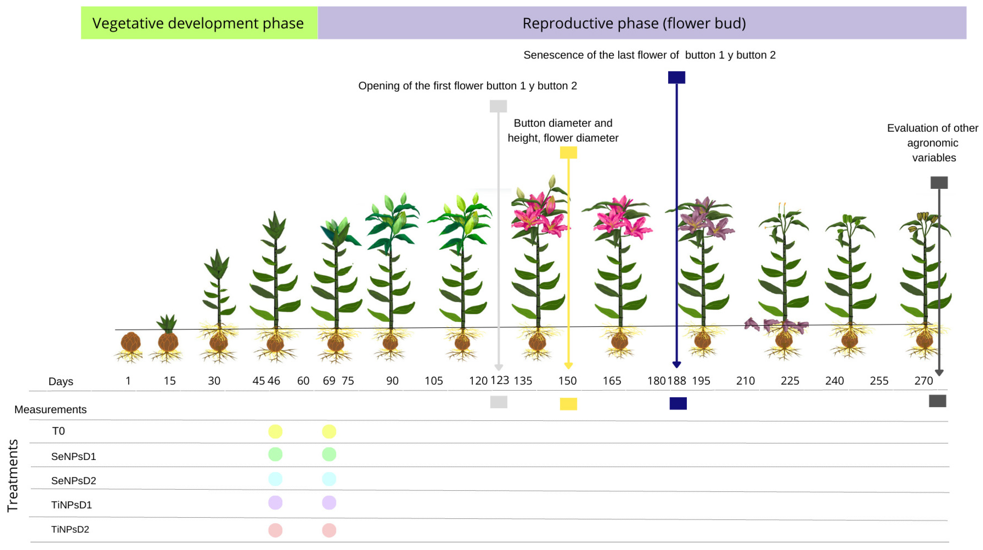
Figure 1. Application of treatments and data collection in Lilium cultivation.
Ijpb 16 00103 g001
Figure 2. (A) Total plant height; (B) stem length; (C) stem diameter; (D) stem fresh weight; (E) stem dry weight; (F) number of leaves; (G) leaf fresh weight; (H) leaf dry weight; (I) chlorophyll (SPAD) under the effects of five treatments: SeNPsD1 and SeNPsD2 (selenium nanoparticle doses), TiNPsD1 and TiNPsD2 (titanium dioxide nanoparticle doses), and T0 (absolute control). Different letters on bars indicate significant differences according to Fisher’s least significant difference test (p ≤ 0.05); the standard error is shown with the mean of four replicates per treatment. The labels at the top of the figures indicate the phenological stage of the crop: green, vegetative stage; purple, reproductive stage.
Ijpb 16 00103 g002
Figure 3. (A) Number of capsules per plant; (B) total number of buds; (C) number of open buds; (D) number of malformed buds; (E) average bud length; (F) average flower diameter; (G) average number of days the flower remained open under the effects of five treatments: SeNPsD1 and SeNPsD2 (selenium nanoparticle doses), TiNPsD1 and TiNPsD2 (titanium dioxide nanoparticle doses), and T0 (absolute control). Different letters on bars indicate significant differences according to Fisher’s least significant difference test (p ≤ 0.05); the standard error is shown with the mean of four replicates per treatment. The labels at the top of the figures indicate the phenological stage of the crop: green, vegetative stage; purple, reproductive stage.
Ijpb 16 00103 g003
Figure 4. (A) Number of ovaries; (B) fresh weight of ovaries; (C) dry weight of ovaries; (D) number of peduncles; (E) fresh weight of peduncles; (F) dry weight of peduncles under the effects of five treatments: SeNPsD1 and SeNPsD2 (selenium nanoparticle doses), TiNPsD1 and TiNPsD2 (titanium dioxide nanoparticle doses), and T0 (absolute control). Different letters on bars indicate significant differences according to Fisher’s least significant difference test (p ≤ 0.05); the standard error is shown with the mean of four replicates per treatment. The labels at the top of the figures indicate the phenological stage of the crop: green, vegetative stage; purple, reproductive stage.
Ijpb 16 00103 g004
Figure 5. (A) Number of bulbils; (B) fresh weight of bulbils; (C) dry weight of bulbils; (D) fresh weight of bulb; (E) dry weight of bulb; (F) fresh weight of root; (G) dry weight of root under the effects of five treatments: SeNPsD1 and SeNPsD2 (selenium nanoparticle doses), TiNPsD1 and TiNPsD2 (titanium dioxide nanoparticle doses), and T0 (absolute control). Different letters on bars indicate significant differences according to Fisher’s least significant difference test (p ≤ 0.05); the standard error is shown with the mean of four replicates per treatment. The labels at the top of the figures indicate the phenological stage of the crop: green, vegetative stage; purple, reproductive stage.
Ijpb 16 00103 g005
Figure 6. (A) SEM (500×); (B) SEM (2000×); (C) elemental analysis via EDS. In Lilium seeds exposed to SeNPsD1, elemental analysis demonstrates the presence of Fe, Al, Si, N, and C in the plant samples.
Ijpb 16 00103 g006
Disclaimer/Publisher’s Note: The statements, opinions and data contained in all publications are solely those of the individual author(s) and contributor(s) and not of MDPI and/or the editor(s). MDPI and/or the editor(s) disclaim responsibility for any injury to people or property resulting from any ideas, methods, instructions or products referred to in the content.