Article

lncRNA-mRNA-miRNA Networks in Arabidopsis thaliana Exposed to Micro-Nanoplastics

1
Department of Chemical, Biological, Pharmaceutical and Environmental Sciences, University of Messina, 98122 Messina, Italy
2
Department of Mathematics, University of Pavia, 27100 Pavia, Italy
3
Department of Biology, University of Florence, Sesto Fiorentino, 50019 Florence, Italy
4
Genomics for Climate Change Research Center (GCCRC), University of Campinas (UNICAMP), Campinas 13083-862, SP, Brazil
5
Council for Agricultural Research and Economics (CREA), Research Centre for Plant Protection and Certification (CREA-DC), 90128 Palermo, Italy
*
Author to whom correspondence should be addressed.
These authors contributed equally to this work.
Int. J. Plant Biol. 2025, 16(2), 70; https://doi.org/10.3390/ijpb16020070
Submission received: 6 May 2025 / Revised: 6 June 2025 / Accepted: 12 June 2025 / Published: 18 June 2025
(This article belongs to the Section Plant Response to Stresses)

Abstract

Long non-coding RNAs (lncRNAs) are key regulators of genetic networks in numerous biological processes. Micro-nanoplastics represent a novel abiotic stress, having a direct xenobiotic impact on plant cells, while the regulation of lncRNAs in Arabidopsis thaliana under this kind of abiotic stress remains largely unclear. We explored RNA-seq data sets of A. thaliana roots treated with two types of micro-nanoplastics: transparent polyethylene terephthalate (Tr-PET) and blue polyethylene terephthalate (Bl-PET) to reveal known and new unannotated lncRNAs. Our findings showed that the Tr-PET changed the expression of 104 lncRNAs, while the Bl-PET changed the expression of just 19. We speculate on the possible significance of the differential expressions for plant tolerance and resistance to micro-nanoplastic stress. A key finding of this work is that the studied lncRNAs tend to regulate their neighboring protein-coding genes. Consistent with this regulatory role, their promoters were found to contain cis-acting regulatory elements responsive to abscisic acid, light, MeJA, MYC/MYB, and other stress-related signals. Furthermore, some of the miRNAs that participate in plant development and defense were also predicted to be sponged by the differentially expressed lncRNAs. In summary, this study adds to our knowledge of A. thaliana lncRNAs through the discovery of new transcripts, describing their expression under micro-nanoplastic stress, and revealing their possible roles in post-transcriptional gene regulation.

1. Introduction

Long non-coding RNAs (lncRNAs) are non-coding protein RNAs longer than 200 nucleotides found in plants, insects, humans, microorganisms, and animals [1,2,3,4,5,6]. Recently, research efforts have been spent on the characterization of lncRNAs in several regulatory mechanisms in plants underlying the control of development and environmental responses. It has been elucidated that lncRNA–chromatin complexes are key leaders in the activation and repression of downstream responsive genes [7,8]. For many years, only protein-coding genes have been the major objectives for crop improvement while very little attention has been given to the regulatory non-coding RNAs. LncRNAs, especially, have been almost excluded as targets for biotechnological approaches, as well as for molecular marker-assisted selection. The understanding of their post-transcriptional role in modulating key protein-coding genes involved in plant stress tolerance/resistance is essential to promote their use in molecular breeding to develop future climate-smart crops. In this regard, the involvement of lncRNAs in the mechanism of RNA-directed DNA methylation during stress response requires future attention. With respect to protein-coding genes, long non-coding RNAs (lncRNAs) are created by RNA polymerases I to V from several distinct types of DNA regions. These locations include the DNA stretches found between two different protein-coding genes, which give rise to lncRNAs specifically called long intergenic non-coding RNAs (lincRNAs). LncRNAs can also originate from the antisense strands, meaning the DNA strand opposite to the one used for a protein-coding gene [2,4,9]. Most plant lncRNAs have transcription promoter regions and splicing signals, while their mature forms may have 5′-cap and 3′-polyadenylation signals [10]. In Arabidopsis thaliana, thousands of lncRNAs have been predicted, the tissue-specific or stress-induced expression profiles have been determined for some of them, and their molecular functions have been speculated upon [4,11,12,13,14,15]. However, although A. thaliana is a good model for studying lncRNAs, very little is known about most of these RNAs in this plant species.
It is widely accepted by the scientific community that they might play pivotal roles in the regulation of coding-protein genes through a wide range of mechanisms contributing to plant adaptation to adverse environmental conditions at both the transcriptional and post-transcriptional levels, such as RNA-mediated DNA methylation (RdDM) and inactivating and activating RNAs or enhancing mRNA translation [16,17,18,19,20,21]. The interest in lncRNAs is very high since preliminary results suggest that the biotechnological engineering of these has the potential to improve crop resilience to environmental stresses linked to anthropogenic activities and climate change [9,22]. Furthermore, it has been demonstrated that the expression profile of lncRNAs is modulated by both abiotic and biotic stresses and has been hypothesized to act to improve plant resilience [23,24,25,26]. Most of the studies have been focused on their role in typical abiotic stresses, such as drought, salinity, heat, trace metals, and nutrient starvation [25,27,28,29,30,31,32]. In tomatoes under drought stress, transcriptome responses in anther tissues revealed a large number of differentially expressed lncRNAs that target protein-coding genes related to phytohormone metabolism with essential roles in pollen development [33]. In addition, some lncRNAs are key players in responses to cold stress, such as COOLAIR, COLDWRAP, and COLDAIR, which modulate flowering in A. thaliana [34,35]. In addition, a lncRNA inhibits the flavodiiron protein (FLV) gene in A. thaliana, allowing for vernalization and controlling the flowering time epigenetically [36,37]. LncRNAs are important also for the survival of plants kept under high salinity via the control of photosynthesis, the regulation of growth, hormone signaling, reduction in the uptake of toxic ions, and the mitigation of reactive oxygen species [38,39,40]. Finally, lncRNAs are important regulators of cellular responses aimed at reducing the detrimental effects of xenobiotics, such as trace metals [41]. However, scarce knowledge is present on the identification of lncRNAs modulated by micro-nanoplastics in plants.
For knowledge, microplastics (<5 mm in size) and nanoplastics (<100 nm in size) originate from plastic bottles, bags, films, textiles, and degrading packaging [5,42]. Polyethylene and polyethylene terephthalate (PET) are two of the most common constituent polymers of micro-nanoplastics [43]. PET has negatively charged oxygen atoms and can promote stronger interactions with other molecules, while polyethylene has negative carbon atoms and can reduce other molecules [43,44]. In consequence, micro-nanoplastics have gained great relevance due to their xenobiotic potential for multiple organisms, including humans [45,46,47], aquatic animals [48,49], land animals [50], soil microbiomes [51], aquatic plants [52,53,54], and land plants [55,56,57,58]. Updated data have confirmed the negative impacts caused by micro-nanoplastics on the ecosystem in general [51,59,60,61,62,63]. These data also reinforce that risk assessments and remediation strategies are strongly needed [64,65]. The contamination of soil and water with these polymers has resulted in plant stress and negatively impacted agronomic performance [66,67]. The uptake and accumulation of micro-nanoplastics in plants have been shown to delay seed germination, cause penalties in root and plant growth, and reduce fruit/seed yields [56,68,69,70,71,72,73]. In addition, they have also been shown to alter morphological, physiological, biochemical, genetic, and epigenetic properties [54,60,74,75,76,77]. Significant changes in gene expression [58,78,79,80,81,82,83] and metabolomes [84,85,86] have been observed in different plant species treated with micro-nanoplastics. Previous findings in Cucurbita pepo showed significant differences during plant stress caused by both transparent PET (Tr-PET) and blue PET (Bl-PET), such as in plant growth and photosynthesis [72]. In particular, transcriptional changes in protein-coding genes mediated by micro-nanoplastic stress were recently studied in A. thaliana [58,78,87]. Both types of plastic material induced a root length reduction, while only Bl-PET reduced the rosette area [58]. While Tr-PET induced genes encoding proteins involved in xenobiotic signaling, Bl-PET has only a few effects on the transcriptomic profile, upregulating only a few genes involved in abiotic stresses. In addition, only Tr-PET has profound effects on genes involved in hormone biosynthesis and signaling, such as brassinosteroids and abscisic acid (ABA). Furthermore, Tr-PET and Bl-PET have received the most attention among xenobiotic compounds because they represent the majority of micro-nanoplastics found discarded and decomposing in nature [72]. Furthermore, it has also been suggested that transcriptional changes in lncRNAs triggered by micro-nanoplastics occur in A. thaliana [58]. Despite the growing interest in the impact of micro-nanoplastics on plant health, our understanding of how lncRNAs modulate plant defense responses and contribute to the adaptation of A. thaliana in such contexts remains limited.
In our study, based on RNA-seq datasets generated from A. thaliana plants exposed to treatment with micro-nanoplastics in a hydroponic system, we investigated the transcriptomic response to abiotic stressors through a first step of whole-genome annotation to identify novel unannotated lncRNAs. Subsequently, the expression profiles of all the novel unannotated and previously identified lncRNAs were determined from A. thaliana exposed to Tr-PET and Bl-PET. Finally, we delved into the biological implications of these differential expression patterns of lncRNAs, and the relationship with plant resilience to this kind of stress was discussed, along with the biotechnological potential of lncRNAs to improve plant tolerance to micro-nanoplastic, opening avenues for future research in sustainable agriculture and environmental health.

2. Materials and Methods

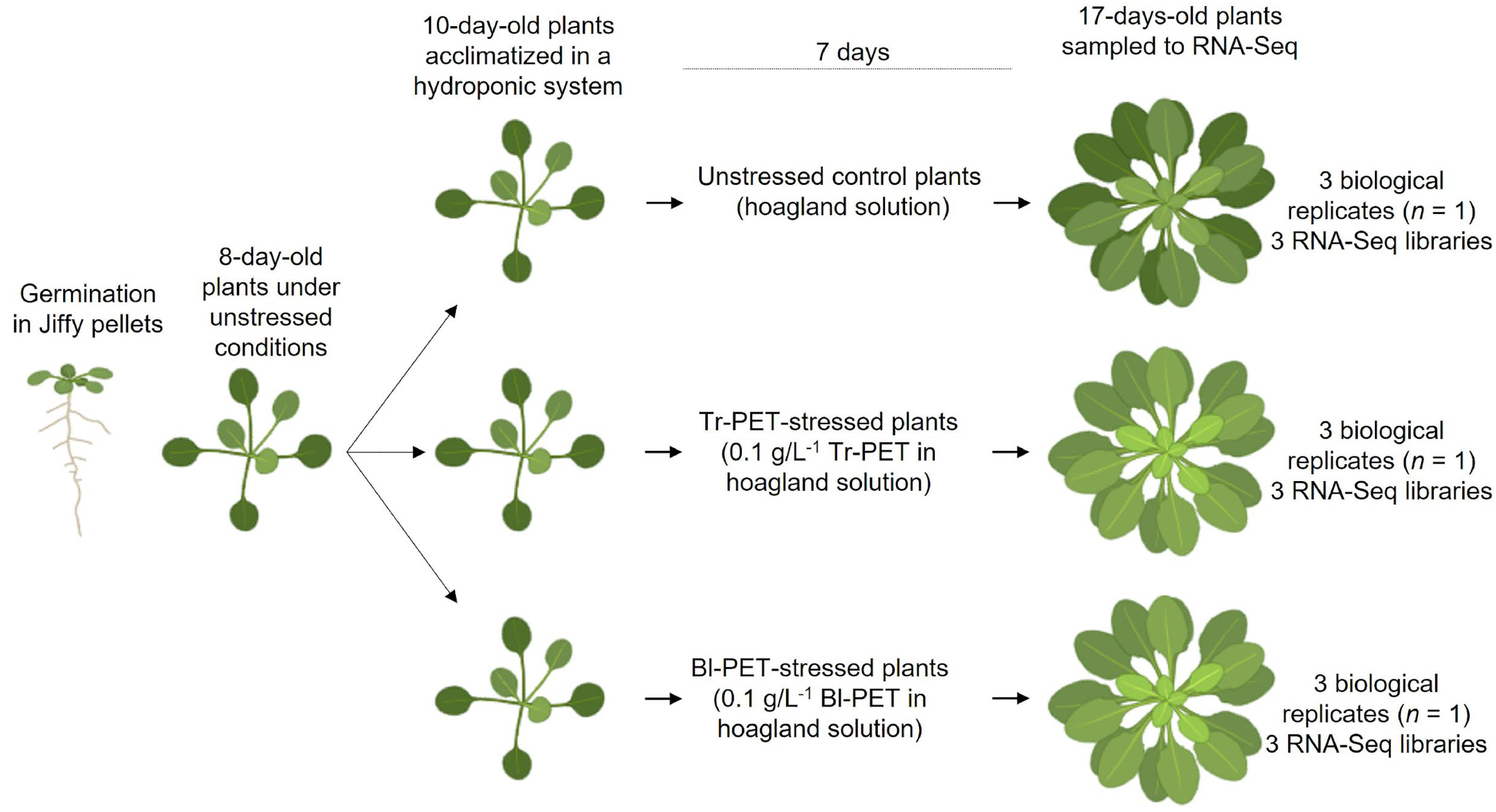
2.1. Plant Material and Stress Treatment

A. thaliana Col-0 seeds were stratified for 24 h in the dark at room temperature and germinated in Jiffy pellets (Jiffy Group International, Zwijndrecht, The Netherlands), and seedlings were transferred to a hydroponic growth system as described by Conn et al. [88] and Dainelli et al. [58]. After acclimation, ten-day-old plants were divided into three treatments consisting of (i) an unstressed control (only half-strength Hoagland solution), (ii) Tr-PET treatment (half-strength Hoagland solution supplemented with 0.1 g/L−1 transparent micro-nanoplastics), and (iii) Bl-PET treatment (half-strength Hoagland solution supplemented with 0.1 g/L−1 blue micro-nanoplastics). For that, micro-nanoplastics were produced from commercial blue and transparent bottles previously cleaned following the procedure described by Ekvall et al. [89] and Dainelli et al. [58]. The concentrations of Tr-PET and Bl-PET micro-nanoplastics used were considered equivalent to those in water from contaminated streams [60]. Plants were placed inside externally dark-coated cylindric glass vessels (11 cm height, 4 cm base diameter), and 8 vessels per treatment were prepared (n = 3 treatments × 8 plants each). These plants under the hydroponic system were kept in a growth room for 17 days (16 h photoperiod, 24–16 °C day/night temperature, 300 μmol m−2s−1 light, and 60–65% humidity). The Hoagland solutions were changed every eight days, and the containers were shaken every two days to homogenize the micro-nanoplastics with the Hoagland solution. Seventeen days after the onset of treatments, roots were harvested from stressed plants with micro-nanoplastics and unstressed control plants, frozen in liquid nitrogen, and stored at −80 °C. Three biological replicates were used for each of the three treatments, and each biological replicate was composed of one plant (Figure 1).

2.2. RNA Isolation, Library Sequencing, and Transcript Expression

Total RNA purification was performed using the RNeasy Plant Mini kit (Qiagen, Hilden, Germany) from 100 mg of roots ground with a pestle and mortar in liquid nitrogen. The RNA quality was determined using the RNA 6000 nano kit on the Agilent 2100 Bioanalyzer System (Bio-Rad, Hercules, CA, USA). Nine cDNA libraries (three libraries per treatment) were prepared from both polyadenylated and non-polyadenylated transcripts following the procedure of TruSeq Stranded mRNA Library Prep (Illumina, San Diego, CA, USA) using TruSeq RNA Single Indexes. The cDNA concentration of each library was determined using the dsDNA High Sensitivity Kit on the Qubit 4 Fluorometer (Invitrogen, Waltham, MA, USA). The sequencing of the RNA-seq libraries was performed using the NovaSeq 6000 S1 Reagent Kit (100 cycles, 2 × 100 + 10 + 10 bp parameters) on the Illumina NovaSeq 6000 sequencing platform (Illumina, Inc., San Diego, California, USA). All the libraries were run in a single-flow cell following Illumina’s standard procedure in XP mode. The RNA-seq raw read data in .fastq format were obtained from BCL files using the bcl2fastq2 v2.20 tool (Illumina). After assessing the quality of the sequenced RNA-seq libraries with FastQC v0.11.9 [90], adaptor sequences and low-quality bases were trimmed using Trimmomatic v0.39 [91] with the following parameters: ILLUMINACLIP:adapters.fa:2:30:10 HEADCROP:1 LEADING:3 TRAILING:3 SLIDINGWINDOW:4:18 MINLEN:40. Libraries with raw reads have been deposited in the ArrayExpress database at EMBL-EBI (www.ebi.ac.uk/arrayexpress) under accession number E-MTAB-13532.

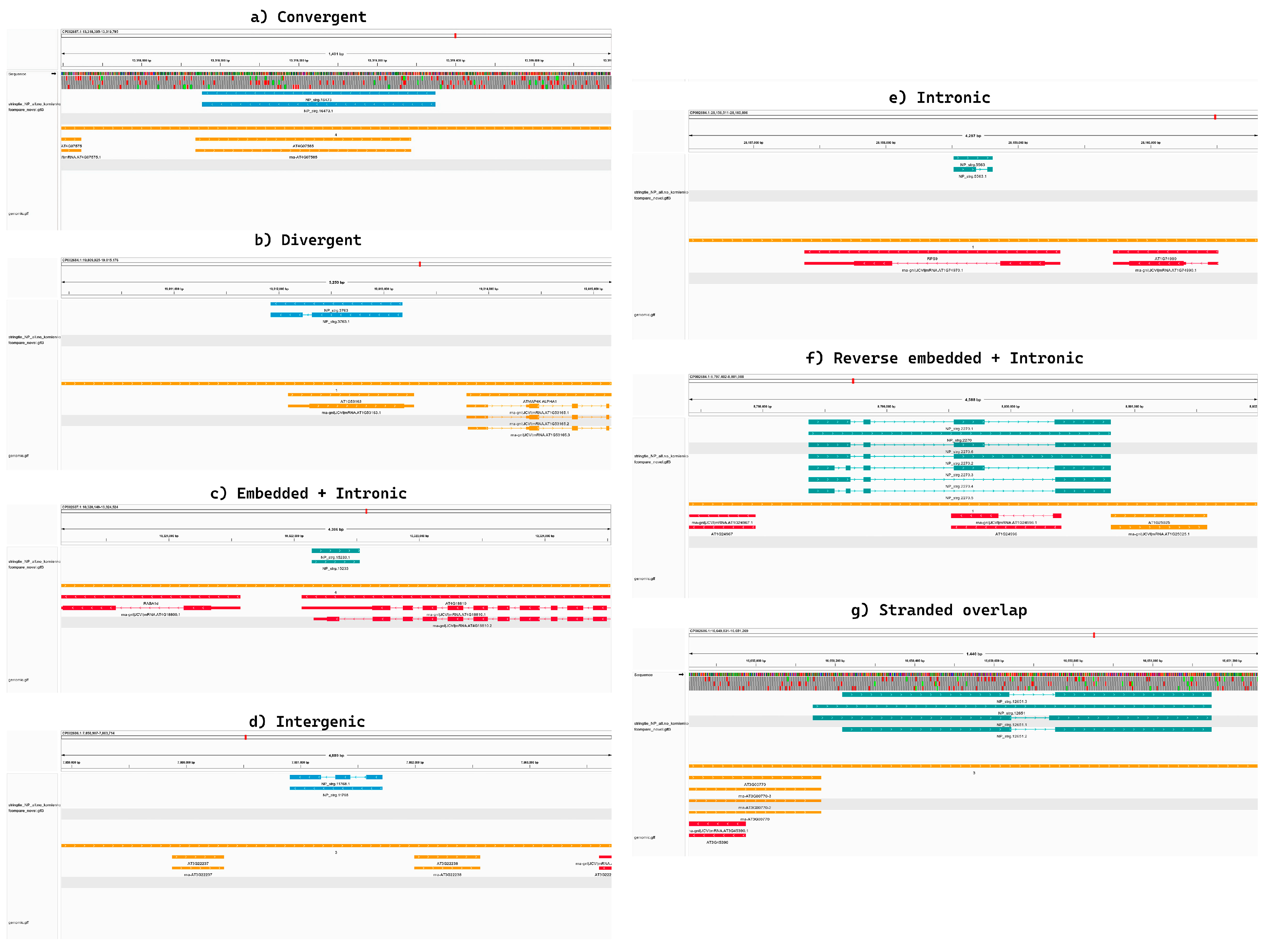
2.3. Identification of Novel Unannotated lncRNAs, Read Mapping, and Expression Profiling

To discover the novel transcripts, the official Araport 11 genome annotation was merged with the recently published one from Kornienko et al. [4] using AGAT utility v1.4.0 [92]. This annotation was used as an input file (−G flag) for guiding the de novo transcriptome assembly computed by StringTie v.2.2.1 (https://ccb.jhu.edu/software/stringtie/), specifying the strandedness of the library using the --rf parameter. All transcripts that did not fall, with the same directionality, into known genes/loci were then filtered based on their normalized transcription level (≥0.1 transcripts per million) and considered novel. The resulting transcripts were inspected using CPC2 software [93] to predict their potential coding/non-coding placement. The high-quality reads were aligned to the A. thaliana TAIR 10 assembly [94] using HiSat2 v2.2.1 [35] with default parameters, integrating our novel gene models with those of Araport11 and Kornienko et al. [4]. Read counts at the gene level were generated from alignment files using featureCounts v2.0.3 [95] with default parameters according to the “exon” feature and “transcript_id” meta-feature of the annotation. Differential expression analyses were carried out using Bioconductor EdgeR v3.28.1 [96]. The EdgeR package was used to filter out the not-expressed or poorly expressed transcripts (a transcript was considered “active” if the normalized read per million mapping to that gene was >1 in at least two libraries) to normalize the RNA libraries, and to perform the differential expression analyses with the Likelihood-Ratio Test (LRT). Protein-coding and non-coding transcripts with a log2(fold change) ≥ |0.5| and false discovery rate (FDR) < 0.05 were considered differentially expressed. PCA plots of the lncRNAs differentially regulated in the three pairwise comparisons were conducted (Additional File S14). To evaluate the differential expression profile at the isoform level, we first estimated the transcript abundances using StringTie v.2.2.1 with the following parameters: (−e) to estimate the transcript abundance, (−G) to set as the genome annotation the one combining Araport11, Kornienko et al. [4], and our novel annotation, and (−B) to produce the output in the format required from the Ballgown R package v2.40.0 (https://www.bioconductor.org/packages/release/bioc/html/ballgown.html) for the subsequent statistical tests conducted in the R environment thanks to the stattest module from the Ballgown R package. Principal Component Analysis (PCA) and Pearson correlation were computed with the prcomp and cor functions, respectively, from the R stats package v.4.4.1, while the corresponding graph, alongside the MA and Volcano plots, was generated through the ggplot2 and pheatmap R package. Finally, the Pearson correlation analysis was conducted using the corr module of the stats package v.4.4.1.

2.4. In Silico Prediction of Regulatory Function of Novel lncRNAs

To determine all the putative lncRNA/mRNA interactions, we first created the pRIblast database [97] relative to the protein-coding transcripts, extracted with AGAT tool v1.4.0 [92], and then we computed the interactions using all the known interactions predicted by Kornienko et al. [4] and our novel lncRNAs as queries. pRIblast uses a seed-and-extension algorithm to compute hybridization energies rather than statistical p-values; therefore, no multiple-testing correction step is required in the algorithm. To reduce potential false-positive calls, the resulting interactions were then intersected with the differentially expressed gene list to retain only the best interaction (resulting in the lowest interaction energy) between each lncRNA and its mRNA target. We reported the corresponding mRNA and its gene expression level for lncRNAs differentially expressed in at least a single comparison between the experimental conditions. Moreover, thanks to the use of the ViennaRNA Package and its submodule RNAcofold web server [98], figures relative to the secondary structure of the interaction model between RNAs were generated. The same approach was also applied to identify lncRNA/miRNA interactions by building the miRNA database by extracting miRNA sequences from the Araport11 genome annotation, before computing the interactions.

2.5. Gene Set Enrichment Analysis

To understand which biological process could be affected by the differentially expressed lncRNAs, we selected the protein-coding best hit of each lncRNA and used it as the queries for the enrichment analysis performed with the gProfiler webtool [99]. A. thaliana was used as the “organism”, while “only annotated genes” were used as the “statistical domain scope”, with a ≤0.05 threshold and by inspecting all the “Gene Ontology” categories alongside “KEGG, Reactome, and WikiPathways” as the “data sources”.

2.6. Network Predictive Protein–Protein Interaction Analysis

The protein–protein interaction and gene co-expression networks among protein-coding genes that are targets of the lncRNAs differentially modulated by Tr-PET and Bl-PET were provided by the STRING database [100] using the TAIR dataset as a reference and an FDR < 0.05.

2.7. Chromosomal Localization and Cis-Acting Regulatory Elements in Promoter Sequences

The chromosomal localization of the differentially expressed lncRNAs and their target protein-coding genes on the A. thaliana genome was generated using the MapGene2Chrom v2 web server [101]. Promoter sequences of 1500 nucleotides upstream of the start codon (ATG) from the differentially expressed lncRNAs were retrieved from the Phytozome v.13 database and submitted to the PlantCARE program [102] to predict the cis-acting regulatory elements.

3. Results

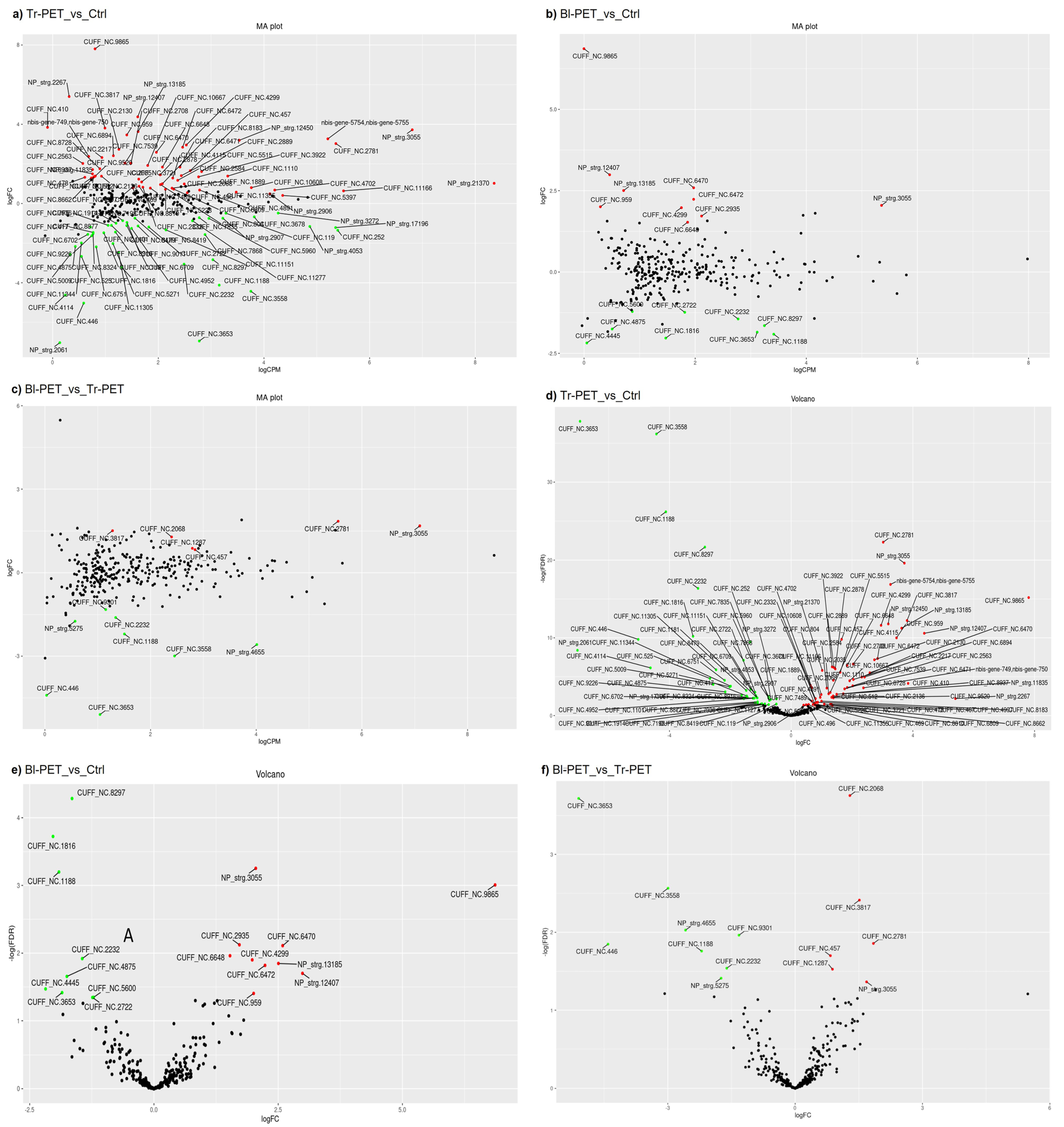
3.1. Global Characterization and Identification of lncRNAs and Their Target Protein-Coding Genes in A. thaliana Response to Micro-Nanoplastics

The expression levels of each lncRNA and their potential protein-coding targets were determined in each of the three pairwise comparisons. In total, 6,619,086–10,988,896 high-quality reads were assigned to at least a single gene among the replicates from the three conditions (the unstressed control and Tr-PET and Bl-PET treatments) (Additional File S1). These data resulted in 96.99 to 98.50% of the total reads used for the subsequent analysis. A total of 194,794 to 345,971 reads were unmapped and with ambiguity (104,307 to 867,131) and unassigned with multimapping (299,316 to 559,605), with a total spanning from 7,237,824 to 12,220,672 among the nine samples/libraries. The Pearson coefficients computed showed a very high correlation between biological replicates (overall correlation: rmin = 0.972; rmax = 0.995), with a correlation between 0.991 and 0.994 in the Tr-PET-treated tissues, between 0.988 and 0.994 in the Bl-PET-treated tissues, and between 0.993 and 0.995 for the unstressed control-treated tissues (Additional File S2). A total of 281 novel lncRNAs, producing a comprehensive set of 324 transcripts, were finally determined in this study (Additional File S3). Among the novel lncRNAs, 96 were intergenic, 80 were embedded, 72 were intronic, 44 were divergent, 40 were reverse-embedded, 31 were convergent, 4 were reverse-intronic (whether a protein-coding gene is present in the lncRNA intron), and 1 was in the same strand overlapping at 5′UTR (Additional File S4). An illustrative scheme of how the novel lncRNAs were classified based on their genomic location is shown in Figure 2a–g.
A differential expression analysis of the three pairwise comparisons (Tr-PET versus unstressed control, Bl-PET versus unstressed control, and Bl-PET versus Tr-PET) was conducted for the DEGs (differentially expressed protein-coding genes), lncRNAs previously identified by Kornienko et al. [4], and novel lncRNAs identified in this study (Table 1). In response to Tr-PET, 1152 DEGs were upregulated and 950 DEGs were repressed, while 104 lncRNAs were modulated, including 11 novel lncRNAs in addition to those previously characterized by Kornienko et al. [4]. In response to Bl-PET, only 427 DEGs were identified, 266 upregulated and 161 downregulated (Table 1). The Bl-PET treatment modulated only 19 lncRNAs, 10 upregulated and 9 downregulated, including 3 upregulated novel ones in addition to those identified by Kornienko et al. [4].
The total lists of the up- and downregulated lncRNAs in each of the three comparisons are presented in Additional File S5. The lncRNA position in the A. thaliana genome and other features are also presented in Additional File S3. The MA plots (Figure 3a–c) show other lncRNAs with high log2FC values in the Tr-PET versus unstressed control, such as NP_strg.2267 (upregulated) and NP_strg.2061 (repressed). In turn, the Volcano plots (Figure 3d,e) show similar results to those of the MA plots: the Tr-PET treatment modulated a much higher number of lncRNAs than the Bl-PET treatment, as shown by the higher number of differentially expressed lncRNAs. Among them, it is worth mentioning that some highly modulated lncRNAs, such as CUFF_NC.3653, CUFF_NC.3558, and CUFF_NC1188, were repressed, and CUFF_NC2781, NP-strg.3055, and CUFF_NC.9865 were upregulated. Among the lncRNAs modulated by the Tr-PET treatment, it is worth mentioning that CUFF_NC8297, CUFF_NC1816, and CUFF_NC1188 were repressed and CUFF_NC9865 was upregulated.

3.2. Functional Analysis of Target Protein-Coding Genes and Splice Variants

The pairwise comparisons conducted at the isoform level showed a high degree of deregulation and underscored significant differences in the stress-response pathway activated by the Tr-PET and Bl-PET treatments. In detail, looking at the Tr-PET versus unstressed control, a total of 2328 and 1994 total isoforms were up- and downregulated, respectively, with 10 upregulated and 8 downregulated novel transcripts (Table 1). In addition, in the Bl-PET versus unstressed control, a total of 1419 isoforms were modulated, 821 upregulated and 598 downregulated (Table 1). Only seven new lncRNAs were significantly modulated, three upregulated and four downregulated. Finally, in the Tr-PET versus Bl-PET, we observed 1597 differentially expressed isoforms, 784 upregulated and 813 downregulated, with just 3 out of the 324 novel lncRNA transcripts significantly affected. The best-predicted interacting protein-coding gene for each lncRNA significantly modulated in each of the three pairwise comparisons is presented in Additional Files S5 and S6. The Gene Set Enrichment Analysis (GSEA) of the target protein-coding genes of lncRNAs regulated by Tr-PET and Bl-PET is presented in Additional File S7. The GSEA results highlighted that lncRNAs may modulate protein-coding genes involved in key metabolic and biosynthetic pathways. Notably, the analysis revealed the potential regulation of genes associated with alpha-linolenic acid metabolism and zeatin biosynthesis, both of which showed significant enrichment (adjusted p-value = 0.05792). In particular, zeatin biosynthesis was enriched in the Tr-PET versus unstressed control, with a fold enrichment of 19,186, implicating genes such as UGT85A1 and IPT5. The involvement of these pathways suggests the role of lncRNAs in modulating hormone biosynthesis and fatty acid metabolism in response to environmental cues. Furthermore, the expression analysis uncovered a distinctive pattern of co-expression between the lncRNAs and their target mRNAs across different conditions. While most pairs displayed low expression levels for both RNA molecules, a subset exhibited an inverse relationship, where the high expression of lncRNAs corresponded with low mRNA levels, and vice versa (Additional File S8). This pattern implies condition-specific regulation, where lncRNAs may act as activators or repressors of their target mRNAs, potentially playing a crucial role in the fine-tuning of the gene expression within the plant’s regulatory network.

3.2.1. Identification of mRNAs Interacting with PET-Modulated lncRNAs

The predicted interactions between lncRNAs regulated by Tr-PET and Bl-PET and mRNAs were determined based on the sequence complementarity and interaction energy, as described in Section 2.4. For this, the best-target-predicted mRNA for each novel lncRNA that exhibited differential regulation in each of the three pairwise comparisons (Additional File S5) was identified. A statistical summary of the data revealed a wide range of interaction profiles among these lncRNAs. The total number of mRNA interactions per novel lncRNA varied significantly, ranging from 6224 to 4,454,490 interactions. Meanwhile, the differentially expressed lncRNAs previously identified by Kornienko et al. [4] showed a range varying from 39,490 to 4,089,918 interactions (Additional File S9). This large variability is evident in the novel lncRNAs, with some lncRNAs exhibiting an extensive network of interactions, while others showed relatively fewer associations. In the novel lncRNA dataset, the mean number of total mRNA interactions per lncRNA was approximately 362,717, while the median was 180,730. Similarly, the number of distinct mRNAs that interacted with each lncRNA also displayed variability, with an average of around 21,883 and a median of 23,653 distinct mRNAs.
In the differentially expressed lncRNA datasets identified by Kornienko et al. [4], the mean number of total mRNA interactions was 622,979, while the median was 500,339. Similarly, the number of distinct mRNAs that interacted with each lncRNA also displayed variability, with an average of around 44,531 and a median of 46,133 distinct mRNAs. These results, with a skewed distribution in both datasets, suggest that while some lncRNAs may play roles in more extensive regulatory networks, the majority have fewer interactions. Focusing on the top interactors, the novel lncRNAs NP_strg.12450.6, NP_strg.12450.5, and NP_strg.12450.4 exhibited the highest numbers of total mRNA interactions. Each of these lncRNAs interacted with an exceptionally high number of distinct mRNAs, 27,458 in the case of NP_strg.12450.6 and NP_strg.12450.5 and 27,393 for NP_strg.12450.4. This suggests that these particular lncRNAs could act as central regulatory hubs, influencing a wide array of biological processes through extensive mRNA regulation. At the lower end of the spectrum, lncRNAs such as NP_strg.6480.1, NP_strg.1264.1, and NP_strg.13972.1 displayed the “fewest” interactions, with only 6224, 13,224, and 14,296 total interactions, respectively, and interacted with as few as 3450 to 6563 distinct mRNAs (Additional File S10). This suggests that these lncRNAs might have more specialized or targeted regulatory functions, potentially affecting only a narrow set of biological pathways. Of the top interactors from the differentially expressed lncRNAs retrieved from Kornienko’s dataset were found CUFF_NC.804, CUFF_NC.8965, and CUFF_NC.8810, interacting with total numbers of 4,089,918, 2,856,096, and 2,120,172 mRNAs, respectively, with them resulting in totals of 48,165, 47,745, and 48,092 distinct interacting mRNAs. On the contrary, Kornienko’s lowest-interacting differentially expressed lncRNAs resulted in CUFF_NC.6471, CUFF_NC.7936, and CUFF_NC.6709, which displayed 39,490, 62,219, and 68,913 total interactions with mRNAs, representing different lncRNA/mRNA configurations of interactions, relative to 23,019, 28,558, and 30,447 distinct mRNAs. These findings reinforce the notion that lncRNAs in A. thaliana under Bl-PET and Tr-PET treatments exhibit diverse interaction profiles, with some functioning as key regulatory hubs while others engage in more specialized roles (Additional File S9).

3.2.2. Tr-PET Versus Unstressed Control

The number of the best-predicted target protein-coding genes of significantly modulated lncRNAs in the Tr-PET versus unstressed control was 104 (Additional File S5). In the Tr-PET versus unstressed control, several protein-coding genes were differentially regulated, some of which share regulatory patterns with genes affected by the Bl-PET versus unstressed control (Additional File S5). Among these, the At2g33830 gene stands out as a key player. This gene encodes a negative regulator of local and systemic acquired resistance (SAR) and serves as a target for the FLD-mediated activation of SAR. SAR is a critical plant defense mechanism against a broad spectrum of pathogens, and the upregulation of At2g33830 (log2FC 2.67) in the Tr-PET versus unstressed control indicates a robust activation of defense pathways, potentially in response to the stress imposed by the treatment condition. Notably, this gene was not differentially expressed in the Bl-PET versus unstressed control, highlighting the specificity of its response to the Tr-PET treatment. The lncRNA CUFF_NC.4299 was identified as interacting with At2g33830, suggesting a possible regulatory role of this lncRNA in modulating the SAR-related gene expression under the Tr-PET treatment. Another gene of interest, At4g25130, which encodes a chloroplast-localized methionine sulfoxide reductase, was repressed. The gene At3g49790, which encodes a carbohydrate-binding protein of unknown function, exhibited a slight upregulation (log2FC 1.10) in the Tr-PET versus unstressed control. Finally, At3g51325, a RING/U-box superfamily protein with an as-yet-unidentified function, also showed a modest upregulation (log2FC 1.12) in the Tr-PET versus unstressed control.

3.2.3. Bl-PET Versus Unstressed Control

In the Bl-PET versus unstressed control, the upregulation of genes was less pronounced, with no target protein-coding genes exhibiting a log2FC greater than 2 (Additional File S5). However, several genes displayed more subtle upregulation, with Log2FC values around 1. One such gene is At3g51325, the same RING/U-box superfamily protein mentioned earlier in the Tr-PET versus unstressed control. In this context, it showed a minor upregulation (log2FC 1.02), again suggesting potential involvement in ubiquitin-mediated protein degradation. The interaction with the lncRNA NP_strg.13185 remained consistent across treatments, indicating that this lncRNA may have a broader regulatory role that is not limited to a specific treatment condition. Interestingly, no target protein-coding genes were downregulated with log2FC values below −2 in the Bl-PET versus unstressed control, suggesting that the response to Bl-PET may not involve the strong repression of gene expression. However, At3g20020, which encodes protein arginine methyltransferase 6, did exhibit slight downregulation (log2FC −0.2). This gene is known to regulate photoperiodic flowering by interacting with nuclear factor Y, a key component of the flowering time regulatory network. The associated lncRNA CUFF_NC.5600 may play a role in fine-tuning the expression of flowering-related genes in response to environmental cues.

3.3. Identification of miRNAs Interacting with PET-Modulated lncRNAs

The predicted interactions between lncRNAs regulated by Tr-PET and Bl-PET and miRNAs were computationally determined similarly to the protein-coding genes, as described in Section 2.4. The best-target-predicted miRNA for each lncRNA that showed differential regulation in each of the three pairwise comparisons are presented in Additional Files S11 and S12. A statistical summary of the data across both our novel lncRNAs and the differentially expressed lncRNAs identified by Kornienko et al. [4] revealed a wide range of interaction profiles between the abovementioned non-coding RNAs. The total number of miRNA interactions per lncRNA varied from 2 to 2550, with the novel lncRNAs ranging from 2 to 1203 interactions, and Kornienko’s one showing an even broader spectrum, with interactions ranging from 1 to 2550 miRNAs. This highlights the high variability in miRNA interaction profiles and points toward a common trend: a subset of lncRNAs have disproportionately high interactions, while most lncRNAs interact with far fewer miRNAs (Additional File S12). In the novel lncRNA dataset, the average number of miRNA interactions per lncRNA was approximately 45, with a median of 36, reinforcing the idea that the distribution of interactions is heavily skewed. Similarly, Kornienko’s dataset displayed a median number of 56 distinct miRNA interactions, with an average of around 61 interactors. Both descriptive statistics suggested that while there is a moderate variability in the number of miRNAs that each lncRNA interacts with, some lncRNAs might have evolved to engage with a broader spectrum of miRNAs, potentially indicating more diverse regulatory roles. In both datasets, the top 10% of lncRNAs showed disproportionately high numbers of interactions: in Kornienko’s dataset, CUFF_NC.5397 stood out with the highest number of interactions (2550) and 223 distinct miRNAs, followed by CUFF_NC.804 (1771 total interactions and 198 distinct miRNAs) and CUFF_NC.5630 (1674 total interactions and 187 distinct miRNAs). Meanwhile, the novel lncRNA NP_strg.12450.4 exhibited the highest number of total miRNA interactions (1203) and 173 distinct miRNAs, followed closely by NP_strg.12450.6 and NP_strg.12450.5, which both displayed around 780 interactions and engaged with 145 and 146 distinct miRNAs, respectively. This confirms the presence of lncRNAs that could act as central regulatory hubs, potentially influencing a wide range of biological processes through extensive miRNA regulation. At the lower end of the interaction spectrum, both datasets feature lncRNAs with few interactions, suggesting specialized or limited regulatory functions. In fact, in both datasets were found lncRNAs interacting with a very limited number of miRNAs, as for the CUFF_NC.9083 and CUFF_NC.1386 non-coding RNAs interacting with a single miRNA, and for the NP_strg.12729.1 and NP_strg.13972.1 transcripts exhibiting only two interactions with two miRNAs, reflecting a possible role in more targeted miRNA regulation, likely affecting only a narrow set of biological pathways.

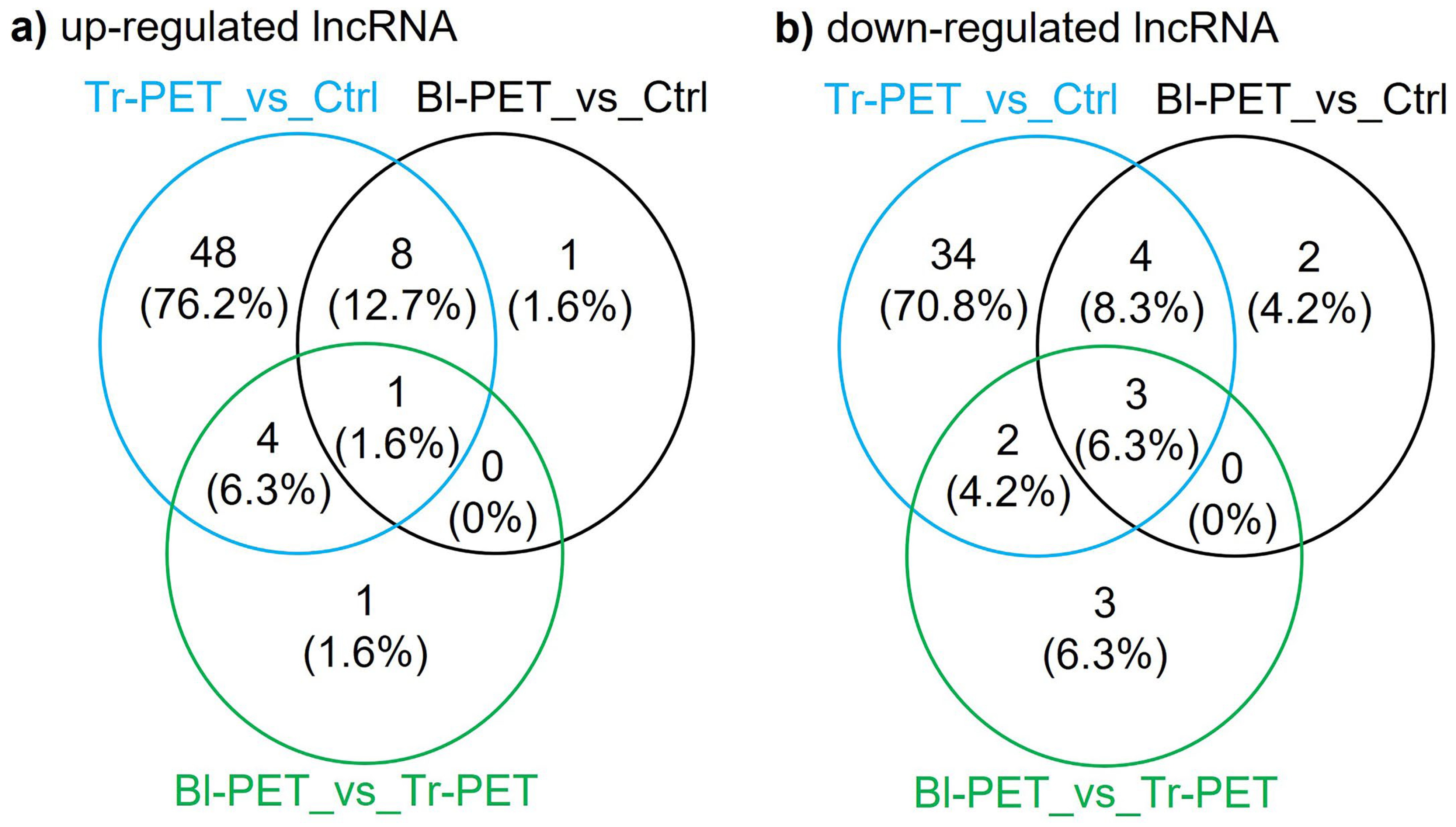
3.4. LncRNAs Up- or Down-Regulated by Micro-Nanoplastics

The RNA-seq data revealed 104 differentially modulated lncRNAs, of which 61 were up-regulated and 43 were downregulated, by the Tr-PET versus unstressed control, while another 19 lncRNAs were considered differentially modulated, 10 up-regulated and 9 downregulated, by the Bl-PET versus unstressed control (Figure 4). In addition, 14 lncRNAs were also considered differentially modulated, 6 up-regulated and 8 downregulated, by the Bl- PET versus Tr-PET (Figure 4).

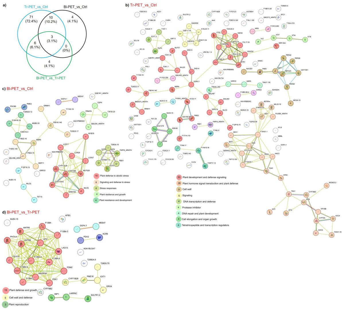
3.5. Functional Analysis of Protein-Coding Genes Targeted by lncRNAs

The RNA-seq data revealed 104 protein-coding genes targeted by the lncRNAs differentially modulated by the Tr-PET versus unstressed control, while another 19 protein-coding genes were identified in the Bl-PET versus unstressed control (Figure 5a). In addition, we identified 14 protein-coding genes targeted by the lncRNAs differentially modulated by the Bl-PET versus Tr-PET (Figure 5a). Protein–protein interaction network data revealed that coding-protein genes identified from the Tr-PET versus unstressed control grouped into nine main clusters ranging from 4 to 33 interactive proteins each (Figure 5b). Interestingly, the gene clusters were represented by the functions of plant development and defense signaling, plant hormone signal transduction and plant defense, the cell wall, signaling, DNA transcription and defense, protease inhibition, DNA repair and plant development, cell elongation and organ growth, and tetratricopeptide and transcription regulators. Protein-coding genes identified from the Bl-PET versus unstressed control were grouped into five main clusters ranging between 4 and 10 genes. The main cluster was composed of genes involved in the plant defense against abiotic stress and signaling and the defense against stress, stress responses, plant resilience and growth, and plant resistance and development (Figure 5c). Furthermore, protein-coding genes identified from the Bl-PET versus Tr-PET grouped into three main clusters of 3 to 15 genes each, involved in plant defense and growth, the cell wall and defense, and plant reproduction (Figure 5d).

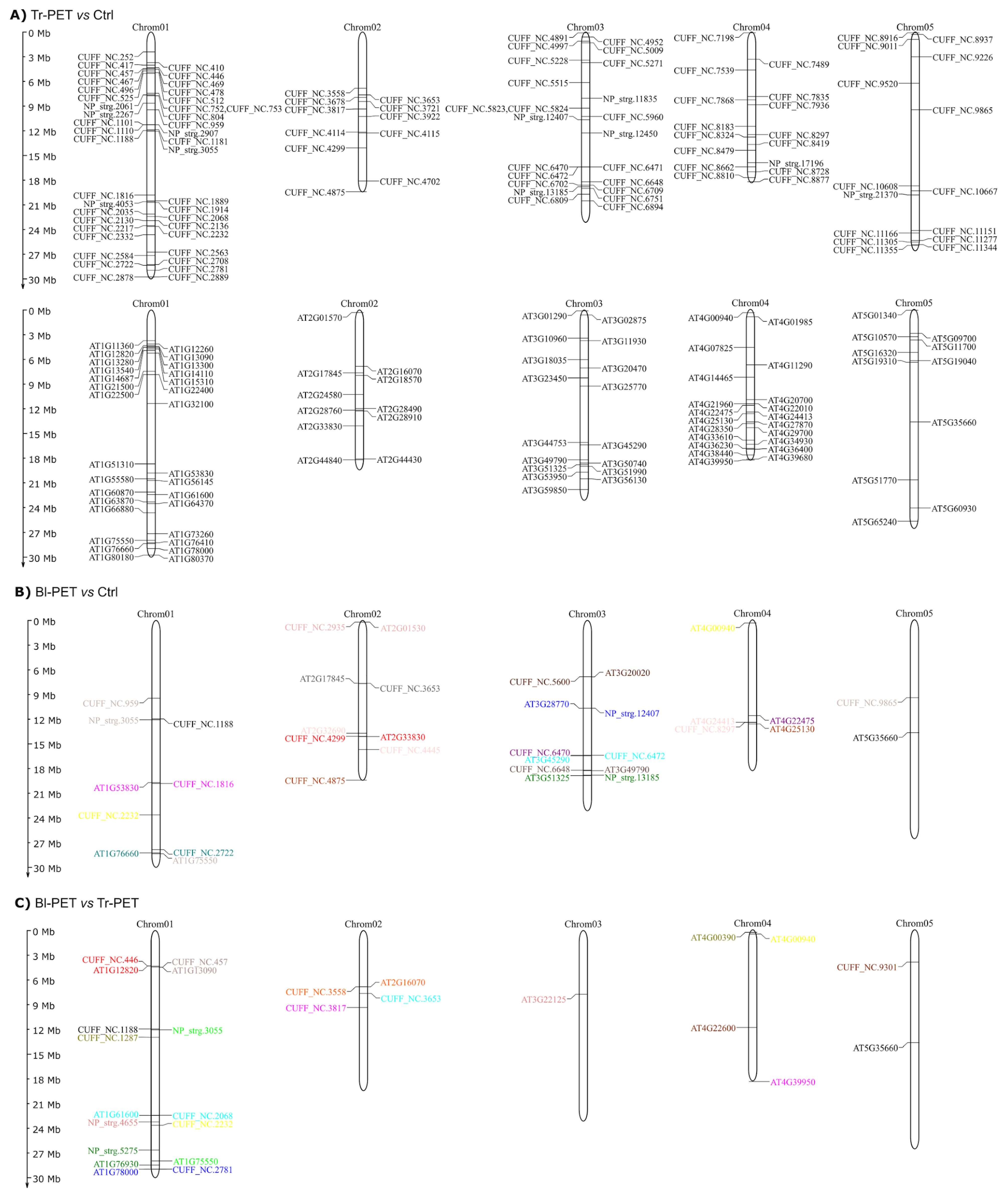
3.6. Chromosomal Localization of lncRNAs and Cis-Acting Regulatory Elements in Their Promoter Sequences

The mapping of the differentially expressed lncRNAs on A. thaliana chromosomes revealed that some of these genes are clustered, while most are widely distributed across the five chromosomes. Moreover, the protein-coding genes that are targeted by these lncRNAs were mostly mapped along the five chromosomes. Most lncRNAs were mapped distantly or on another chromosome relative to their target protein-coding gene (Figure 6). The cis-acting regulatory elements in the promoter sequences of the differentially expressed lncRNAs were identified and overrepresented 16 top elements, including hormone-responsive, stress-induced, tissue-specific, plant development or senescence, light-responsive, MYC/MYB-related, and anaerobic induction elements (Additional File S13). The ABA-responsive (0 to 15), light-responsive (1 to 20), anaerobic-induction (0 to 7), MeJA-responsive (0 to 16), MYC/MYB-related (0 to 24), and stress-responsive (0 to 13) elements were the cis-acting regulatory elements more represented in these promoter sequences of lncRNAs identified from the Tr-PET versus unstressed control, Bl-PET versus unstressed control, and Bl-PET versus Tr-PET. Some promoters also showed cis-acting regulatory elements responsive to gibberellin, auxin, salicylic acid, and ethylene, or for expression in the tissue- or development-stage-specific manner (meristem, endosperm, root, and senescence). Therefore, it was verified that in both Tr-PET and Bl-PET contrasts, the promoters of differentially expressed lncRNAs can respond to different stimuli, with some specificities among them, but highlighting the responses to abiotic stress and plant development.

4. Discussion

It is well-known that plants can learn, store, memorize, and respond to environmental stresses using epigenetic mechanisms that include the regulation of the expression of transcripts not encoding proteins such as lncRNAs [23,24]. This mechanism of stress memory has been considered as a sort of buffer zone in a generation. When the stress response overcomes the physiological buffer zone, it can be manifested at the genetic and epigenetic levels involving a trans-generational response [25]. The epigenetic mechanism acts as a link between the environment and genetic basis to silence loci [103] and activate others, allowing a stable, strong stress memory that prepares plants for subsequent environmental events via the modification of chromatin’s small RNAs in the reproductive parts of plants [104,105,106]. A finely tuned crosstalk of different types of RNAs (protein-coding and non-coding, lncRNAs and miRNAs) occurs in eukaryotic cells, including plants modulating signaling mechanisms in response to development and environmental factors and processes [19,20]. The role of lncRNA-mediated regulation in plants in response to environmental stresses has been increasingly appreciated [14,15]. In this study, 281 novel lncRNAs were identified in addition to the ones deposited in the TAIR database and recently identified by Kornienko et al. [4], and those regulated by Tr-PET and Bl-PET were identified. The Tr-PET treatment was shown to more significantly affect the abundance of lncRNAs than the Bl-PET treatment. Likewise, the Tr-PET treatment affected the expression of much more protein-coding genes than the Bl-PET treatment. The reason why the Tr-PET treatment seems to have a higher impact on all types of transcripts is unknown and should be further studied. A total of 11 novel lncRNAs were modulated by Tr-PET, and 3 additional lncRNAs were modulated by Bl-PET in addition to those reported by Kornienko et al. [4]. These lncRNAs represent a valuable addition to the recently performed TAIR lncRNA collection to gain insight into the role of lncRNAs in the complex mechanisms of the post-transcriptional modulation of plant stress responses to environmental stresses, especially xenobiotic molecules.

4.1. lncRNA Roles in Micro-Nanoplastics Stress

4.1.1. Tr-PET Versus Unstressed Control

The Tr-PET treatment modulated a much higher number of lncRNAs than the Bl-PET treatment. In total, 36 target protein-coding genes of lncRNAs modulated by Tr-PET were significantly regulated in the same treatment. Among them, several play a key role in the abiotic stress responses in plants. Methionine sulfoxide reductases are crucial for protecting chloroplasts from oxidative damage by reducing methionine sulfoxide back to methionine, thereby maintaining the functionality of chloroplast proteins under oxidative stress. The downregulation of At4g25130 (log2FC −0.98) in the Tr-PET versus unstressed control suggests a compromised ability to mitigate oxidative stress, which could have implications for the chloroplast function and overall plant health. The associated lncRNA CUFF_NC.4875 may play a regulatory role, potentially influencing the expression of stress-responsive genes in chloroplasts. The carbohydrate-binding protein (At3g49790) was slightly upregulated. Although the precise role of this protein remains unclear, its carbohydrate-binding capacity hints at a possible involvement in cell wall remodeling or pathogen recognition, processes that are often triggered by environmental stress. The lncRNA CUFF_NC.6648 was found to interact with this gene, which could provide insights into the regulation of carbohydrate metabolism in plants under stress conditions. Likewise, the RING/U-box superfamily genes (At3g51325 and At1g76410) were upregulated. Among the transcription factors targeted by lncRNAs modulated by Tr-PET treatment, it is worth noting the SOP1 (Suppressor of PAS2) encoding a zinc-finger protein co-localizing with the exosome-associated RNA helicase HEN2. This gene works as a co-factor of nuclear RNA quality control via the nucleoplasmic exosome [107]. Another gene encoding a putative C3HC4-type RING zinc-finger factor (At1g22500) was also upregulated and targeted by CUFF_NC.2563. This gene has been reported to be induced in response to light and ascorbate stimuli [108]. Another interesting finding was the upregulation of the negative regulator of local and systemic acquired resistance (At2g33830) targeted by the upregulated lncRNA CUFF_NC.4299. This may imply a possible effect of micro-nanoplastics on biotic stress responses against biotroph pathogens. This is also corroborated by the upregulation of a gene encoding a trypsin inhibitor (At1g73260) involved in modulating programmed cell death in plant–pathogen interactions.

4.1.2. Bl-PET Versus Unstressed Control

The Bl-PET treatment modulated only 19 lncRNAs, so it can be speculated that Bl-PET should not have a significant impact on the post-transcriptional regulation of protein-coding genes. Overall, the Bl-PET versus unstressed control results suggest that the Bl-PET induced less pronounced transcriptional changes compared to the Tr-PET treatment, with the limited downregulation of the target protein-coding genes. The interaction of specific lncRNAs with differentially expressed genes in both treatment comparisons highlights the potential regulatory roles of lncRNAs in the plant’s response to diverse environmental challenges. The functional enrichment of RNA-seq data in response to Bl-PET highlighted the lncRNA CUFF_NC.6472, targeting genes encoding enzymes with transferase activities, such as UDP-glycosyl- and hexosyl-transferases (UGT85A2). This category of genes is well-known to be involved in stress-related responses in plants, and some of them have been targeted with biotechnological approaches to confirm their ability to provide abiotic stress tolerance. Indeed, the glycosyltransferase family represents a large family of genes in plants, generally involved in the modification of small molecules responsible for important aspects not only related to development but also stress responses. For example, a paralog of UGT85A2 was found to significantly confer tolerance to salinity, osmotic, and drought stresses. Indeed, the overexpression of the UGT87A2 gene enhanced the germination and chlorophyll content, providing a higher survival rate and promoting the root length against abiotic stresses [109]. Furthermore, the transgenic overexpression of this gene promoted a reduction in the water loss rate due to smaller stomatal apertures. The transferase activities of these genes have been demonstrated towards several important biological molecules, including plant hormones, such as auxin [110], brassinosteroids [111], strigolactones [112], and ABA [113], as well as several secondary metabolites, including flavonoids [114], lignin precursors, and xenobiotics [115]. Therefore, based on these results, it was speculated that the effects of Bl-PET on the post-transcriptional regulation of protein-coding genes through the action of lncRNAs should be very small. A gene encoding a RING/U-box protein was upregulated and targeted by the upregulated lncRNA NP_strg.13185. The RING/U-box proteins are typically involved in ubiquitin-mediated protein degradation, a process essential for the regulation of various cellular activities, including stress responses. The interaction of this gene with the lncRNA NP_strg.13185 may be crucial for understanding the post-transcriptional regulation of protein turnover in response to micro-nanoplastic stress.

5. Conclusions

In conclusion, we have expanded the available resource of lncRNAs in A. thaliana involved in the plant response to stresses caused by xenobiotic compounds to strengthen future efforts to functionally characterize those lncRNAs that might play a key role in plant biology. In this work, we identified 281 novel lncRNA genes. Amongst these, 104 lncRNAs were modulated by Tr-PET and 19 were modulated by Bl-PET. In addition, their target protein-coding genes were identified, and their role in the plant response to stress caused by Tr-PET and Bl-PET was discussed. Our analysis highlighted that the function of the majority of these non-coding transcripts is to regulate their neighboring protein-coding genes, and that their promoter sequences contain cis-acting regulatory elements that are responsive to ABA, light, jasmonic acid, MYC/MYB transcription factors, and other stress-related signals. Highly interactive lncRNAs in both datasets may serve as key regulatory hubs, orchestrating miRNA-mediated gene expression in response to environmental challenges, such as micro-nanoplastics. The wide range of interactions indicates that lncRNAs participate in complex regulatory networks, highlighting their conserved functions within these and suggesting the possibility of functional redundancy. Their role in epigenetic mechanisms underlying plant stress memories should be further investigated to create crops primed to environmental stresses linked to climate change. Furthermore, the functional characterization of lncRNAs using CRISPR/Cas9 technology is at the forefront of crop genetic improvement.

Supplementary Materials

The following supporting information can be downloaded at https://www.mdpi.com/article/10.3390/ijpb16020070/s1, File S1. Assigned and unassigned reads based on their features in the nine root samples/libraries. File S2. The Pearson coefficients of the nine analyzed samples (*** corresponds to p-values ≤ 0.001). File S3. GTF file relative to the novel lncRNAs identified in this study. File S4. Types of lncRNAs identified in this present research study. File S5. List of differentially expressed novel or Kornienko’s lncRNAs alongside their best-interacting protein-coding genes and miRNAs found in the Tr-PET versus unstressed control, Bl-PET versus unstressed control, and Bl-PET versus Tr-PET. File S6. List of lncRNAs and their best target protein-coding genes retrieved in this study and differentially expressed lncRNAs from Kornienko’s dataset. LncRNA transcripts and their best-interacting mRNAs, hybridization, interaction energy, and base pairs found through RIblast analysis. File S7. GSEA data of best target protein-coding genes of lncRNAs regulated by micro-nanoplastics. File S8. Scatterplot representing pairs of lncRNAs/mRNAs best hit with their normalized (FPKM) expression levels in all three experimental conditions of this study. Ctrl: unstressed control. File S9. List of computed novel lncRNA versus mRNA interactions and Kornienko’s lncRNA versus mRNA interactions. File S10. Each image, generated through ViennaRNA, shows the concatenated sequences of a long non-coding RNA (lncRNA) and its best-interacting protein-coding transcript. The first listed sequence is the lncRNA (green), and the second is the corresponding protein-coding gene (red). File S11. Each image, generated through ViennaRNA, displays the concatenated sequences of an lncRNA (in green) and its best-interacting miRNA (in red). The first listed sequence is the lncRNA, and the second is the corresponding miRNA. File S12. List of computed novel lncRNA versus miRNA interactions and Kornienko’s lncRNA versus miRNA interactions. File S13. Cis-acting regulatory elements in promoter sequences (1500 nucleotides upstream of supposed start codon, ATG) of the differentially expressed lncRNAs. Promoter sequences of lncRNAs from contrasts: (a) plants exposed to transparent micro-nanoplastic versus plants maintained under unstressed control (Ctrl) condition (Tr-PET versus Ctrl); (b) plants exposed to blue micro-nanoplastic versus plants exposed to transparent micro-nanoplastic (Bl-PET versus Tr-PET). Number of each cis-acting regulatory element is shown within the heatmap. ABA: abscisic acid; MeJA: methyl jasmonate; MYB: MYB transcription factor binding domain; MYC: MYC transcription factor binding domain; SA: salicylic acid. NP_strg.2267 (1) to (4) and NP_strg.12450 (1) to (3) are promoter sequences of different isoforms for the NP_strg.2267 and NP_strg.12450 lncRNAs. File S14. Principal Component Analysis of the biological replicates included in this study. Red dots represent the unstressed control condition, green dots refer to the Bl-PET treatment, and blue dots refer to Tr-PET.

Author Contributions

Conceptualization, F.M. and L.G.; data curation, D.G. and L.G.; formal analysis, D.G. and A.B.; funding acquisition, F.M.; investigation, A.B., M.N., C.V., I.C., C.G. and M.D.; methodology, R.G.; resources, A.G.; supervision, F.M.; writing—original draft, R.G., D.G., A.B., M.F.B., A.G. and L.G.; writing—review and editing, R.G. and G.G. All authors have read and agreed to the published version of the manuscript.

Funding

This research received no external funding.

Data Availability Statement

The raw sequences of the nine RNA libraries were deposited in the EMBL-EBI ArrayExpress database under the accession number E-MTAB-13532.

Acknowledgments

This research was made possible through publicly available resources and tools, as well as through the broader scientific literature that provided a foundation for this work.

Conflicts of Interest

The authors declare that the research was conducted in the absence of any commercial or financial relationships that could be construed as potential conflicts of interest.

References

  1. Hovhannisyan, H.; Gabaldón, T. The Long Non-Coding RNA Landscape of Candida Yeast Pathogens. Nat. Commun. 2021, 12, 7317. [Google Scholar] [CrossRef] [PubMed]
  2. Mattick, J.S.; Amaral, P.P.; Carninci, P.; Carpenter, S.; Chang, H.Y.; Chen, L.-L.; Chen, R.; Dean, C.; Dinger, M.E.; Fitzgerald, K.A.; et al. Long Non-Coding RNAs: Definitions, Functions, Challenges and Recommendations. Nat. Rev. Mol. Cell Biol. 2023, 24, 430–447. [Google Scholar] [CrossRef] [PubMed]
  3. Jardim Poli, P.; Fischer-Carvalho, A.; Tahira, A.C.; Chan, J.D.; Verjovski-Almeida, S.; Murilo Sena, A. Long Non-Coding RNA Levels Are Modulated in Schistosoma Mansoni Following in Vivo Praziquantel Exposure. Non-Coding RNA 2024, 10, 27. [Google Scholar] [CrossRef] [PubMed]
  4. Kornienko, A.E.; Nizhynska, V.; Morales, A.M.; Pisupati, R.; Nordborg, M. Population-level annotation of lncRNAs in Arabidopsis reveals extensive expression variation associated with transposable element-like silencing. Plant Cell 2023, 36, 85–111. [Google Scholar] [CrossRef]
  5. Rahman, M.-M.; Omoto, C.; Kim, J. Genome-Wide Exploration of Long Non-Coding RNAs of Helicoverpa armigera in Response to Pyrethroid Insecticide Resistance. Insects 2024, 15, 146. [Google Scholar] [CrossRef]
  6. Zhang, Y.; Liu, H.; Niu, M.; Wang, Y.; Xu, R.; Guo, Y.; Zhang, C. Roles of Long Noncoding RNAs in Human Inflammatory Diseases. Cell Death Discov. 2024, 10, 235. [Google Scholar] [CrossRef]
  7. Gendrel, A.-V.; Heard, E. Noncoding RNAs and Epigenetic Mechanisms during X-Chromosome Inactivation. Annu. Rev. Cell Dev. Biol. 2014, 30, 561–580. [Google Scholar] [CrossRef]
  8. Meller, V.H.; Joshi, S.S.; Deshpande, N. Modulation of Chromatin by Noncoding RNA. Annu. Rev. Genet. 2015, 49, 673–695. [Google Scholar] [CrossRef]
  9. Zhao, X.; Li, F.; Ali, M.; Li, X.; Fu, X.; Zhang, X. Emerging Roles and Mechanisms of LncRNAs in Fruit and Vegetables. Hortic. Res. 2024, 11, uhae046. [Google Scholar] [CrossRef]
  10. Gonzales, L.R.; Blom, S.; Henriques, R.; Bachem, C.W.B.; Immink, R.G.H. LncRNAs: The Art of Being Influential without Protein. Trends Plant Sci. 2024, 29, 770–785. [Google Scholar] [CrossRef]
  11. Zhao, X.; Li, J.; Lian, B.; Gu, H.; Li, Y.; Qi, Y. Global Identification of Arabidopsis LncRNAs Reveals the Regulation of MAF4 by a Natural Antisense RNA. Nat. Commun. 2018, 9, 5056. [Google Scholar] [CrossRef] [PubMed]
  12. Palos, K.; Nelson, A.C.; Yu, L.; Brock, J.R.; Railey, C.E.; Wu, H.-Y.L.; Sokołowska, E.; Skirycz, A.; Hsu, P.Y.; Gregory, B.D.; et al. Identification and Functional Annotation of Long Intergenic Non-Coding RNAs in Brassicaceae. Plant Cell 2022, 34, 3233–3260. [Google Scholar] [CrossRef] [PubMed]
  13. Chen, W.; Zhu, T.; Shi, Y.; Chen, Y.; Li, W.J.; Chan, R.J.; Chen, D.; Zhang, W.; Yuan, Y.A.; Xu, J.; et al. An Antisense Intragenic LncRNA SEAIRa Mediates Transcriptional and Epigenetic Repression of SERRATE in Arabidopsis. Proc. Natl. Acad. Sci. USA 2023, 120, e2216062120. [Google Scholar] [CrossRef] [PubMed]
  14. He, Z.; Lan, Y.; Zhou, X.; Yu, B.; Zhu, T.; Yang, F.; Fu, L.-Y.; Chao, H.; Wang, J.; Feng, R.-X.; et al. Single-Cell Transcriptome Analysis Dissects LncRNA-Associated Gene Networks in Arabidopsis. Plant Commun. 2023, 5, 100717. [Google Scholar] [CrossRef]
  15. Liu, J.; Zhong, X. Epiallelic Variation of Non-Coding RNA Genes and Their Phenotypic Consequences. Nat. Commun. 2024, 15, 1375. [Google Scholar] [CrossRef]
  16. Wang, Y.; Folimonova, S.Y. Long Noncoding RNAs in Plant–Pathogen Interactions. Phytopathology 2023, 113, 1380–1386. [Google Scholar] [CrossRef]
  17. Cui, C.; Wan, H.; Li, Z.; Ai, N.; Zhou, B. Long noncoding RNA TRABA suppresses β-glucosidase-encoding BGLU24 to promote salt tolerance in cotton. Plant Physiol. 2024, 194, 1120–1138. [Google Scholar] [CrossRef]
  18. Fahad, M.; Tariq, L.; Muhammad, S.; Wu, L. Underground Communication: Long Non-Coding RNA Signaling in the Plant Rhizosphere. Plant Commun. 2024, 5, 100927. [Google Scholar] [CrossRef]
  19. Jin, X.; Wang, Z.; Li, X.; Ai, Q.; Wong, D.C.J.; Zhang, F.; Yang, J.; Zhang, N.; Si, H. Current Perspectives of LncRNAs in Abiotic and Biotic Stress Tolerance in Plants. Front. Plant Sci. 2023, 14, 1334620. [Google Scholar] [CrossRef]
  20. Numan, M.; Sun, Y.; Li, G. Exploring the Emerging Role of Long Non-Coding RNAs (LncRNAs) in Plant Biology: Functions, Mechanisms of Action, and Future Directions. Plant Physiol. Biochem. 2024, 212, 108797. [Google Scholar] [CrossRef]
  21. Yadav, A.; Mathan, J.; Dubey, A.K.; Singh, A. The Emerging Role of Non-Coding RNAs (NcRNAs) in Plant Growth, Development, and Stress Response Signaling. Non-Coding RNA 2024, 10, 13. [Google Scholar] [CrossRef] [PubMed]
  22. Jia, Y.; Zhao, H.; Niu, Y.; Wang, Y. Long Noncoding RNA from Betula platyphylla, BplncSIR1, Confers Salt Tolerance by Regulating BpNAC2 to Mediate Reactive Oxygen Species Scavenging and Stomatal Movement. Plant Biotechnol. J. 2023, 22, 48–65. [Google Scholar] [CrossRef] [PubMed]
  23. Saeid, B.; Bhalla, P.L.; Singh, M.B. Identifying Long Non-Coding RNAs Involved in Heat Stress Response during Wheat Pollen Development. Front. Plant Sci. 2024, 15, 1344928. [Google Scholar] [CrossRef]
  24. Bai, Y.; He, J.; Yao, Y.; An, L.; Cui, Y.; Li, X.; Yao, X.; Xiao, S.; Wu, K. Identification and Functional Analysis of Long Non-Coding RNA (LncRNA) and Metabolites Response to Mowing in Hulless Barley (Hordeum vulgare L. Var. Nudum Hook. F.). BMC Plant Biol. 2024, 24, 666. [Google Scholar] [CrossRef]
  25. Magar, N.D.; Shah, P.; Barbadikar, K.M.; Bosamia, T.C.; Madhav, M.S.; Mangrauthia, S.K.; Pandey, M.K.; Sharma, S.; Shanker, A.K.; Neeraja, C.N.; et al. Long Non-Coding RNA-Mediated Epigenetic Response for Abiotic Stress Tolerance in Plants. Plant Physiol. Biochem. 2024, 206, 108165. [Google Scholar] [CrossRef]
  26. Weng, M.; Zhang, D.; Wang, H.; Yang, C.; Lin, H.; Pan, Y.; Lin, Y. Long Non-Coding RNAs and Their Potential Function in Response to Postharvest Senescence of Sparassis latifolia during Cold Storage. Sci. Rep. 2024, 14, 747. [Google Scholar] [CrossRef]
  27. Wang, X.; Fan, H.; Wang, B.; Yuan, F. Research Progress on the Roles of LncRNAs in Plant Development and Stress Responses. Front. Plant Sci. 2023, 14, 1138901. [Google Scholar] [CrossRef]
  28. Ackah, M.; Jin, X.; Zhang, Q.; Amoako, F.K.; Wang, L.; Attaribo, T.; Zhao, M.; Yuan, F.; Herman, R.A.; Qiu, C.; et al. Long Noncoding RNA Transcriptome Analysis Reveals Novel LncRNAs in Morus Alba “Yu-711” Response to Drought Stress. Plant Genome 2022, 17, e2027. [Google Scholar] [CrossRef]
  29. Cai, J.; Zhang, Y.; He, R.; Jiang, L.; Qu, Z.; Gu, J.; Yang, J.; Legascue, M.F.; Wang, Z.-Y.; Ariel, F.; et al. LncRNA DANA1 Promotes Drought Tolerance and Histone Deacetylation of Drought Responsive Genes in Arabidopsis. EMBO Rep. 2024, 25, 796–812. [Google Scholar] [CrossRef]
  30. Cao, W.; Yang, L.; Zhuang, M.; Lv, H.; Wang, Y.; Zhang, Y.; Ji, J. Plant Non-Coding RNAs: The New Frontier for the Regulation of Plant Development and Adaptation to Stress. Plant Physiol. Biochem. PPB 2024, 208, 108435. [Google Scholar] [CrossRef]
  31. Imaduwage, I.; Hewadikaram, M. Predicted Roles of Long Non-Coding RNAs in Abiotic Stress Tolerance Responses of Plants. Mol. Hortic. 2024, 4, 20. [Google Scholar] [CrossRef] [PubMed]
  32. Traubenik, S.; Charon, C.; Blein, T. From Environmental Responses to Adaptation: The Roles of Plant LncRNAs. Plant Physiol. 2024, 195, 232–244. [Google Scholar] [CrossRef] [PubMed]
  33. Lamin-Samu, A.T.; Zhuo, S.; Ali, M.; Lu, G. Long Non-Coding RNA Transcriptome Landscape of Anthers at Different Developmental Stages in Response to Drought Stress in Tomato. Genomics 2022, 114, 110383. [Google Scholar] [CrossRef] [PubMed]
  34. Heo, J.B.; Sung, S. Vernalization-Mediated Epigenetic Silencing by a Long Intronic Noncoding RNA. Science 2010, 331, 76–79. [Google Scholar] [CrossRef]
  35. Kim, D.; Paggi, J.M.; Park, C.; Bennett, C.; Salzberg, S.L. Graph-Based Genome Alignment and Genotyping with HISAT2 and HISAT-Genotype. Nat. Biotechnol. 2019, 37, 907–915. [Google Scholar] [CrossRef]
  36. Sun, Q.; Csorba, T.; Skourti-Stathaki, K.; Proudfoot, N.J.; Dean, C. R-Loop Stabilization Represses Antisense Transcription at the Arabidopsis FLC Locus. Science 2013, 340, 619–621. [Google Scholar] [CrossRef]
  37. Whittaker, C.; Dean, C. The FLC Locus: A Platform for Discoveries in Epigenetics and Adaptation. Annu. Rev. Cell Dev. Biol. 2017, 33, 555–575. [Google Scholar] [CrossRef]
  38. Wang, T.-Z.; Liu, M.; Zhao, M.-G.; Chen, R.; Zhang, W.-H. Identification and Characterization of Long Non-Coding RNAs Involved in Osmotic and Salt Stress in Medicago truncatula Using Genome-Wide High-Throughput Sequencing. BMC Plant Biol. 2015, 15, 131. [Google Scholar] [CrossRef]
  39. Sun, Z.; Wang, Y.; Mou, F.; Tian, Y.; Chen, L.; Zhang, S.; Qiong, J.; Li, X. Genome-Wide Small RNA Analysis of Soybean Reveals Auxin-Responsive MicroRNAs That Are Differentially Expressed in Response to Salt Stress in Root Apex. Front. Plant Sci. 2016, 6, 1273. [Google Scholar] [CrossRef]
  40. Khandal, H.; Parween, S.; Roy, R.; Meena, M.K.; Chattopadhyay, D. MicroRNA Profiling Provides Insights into Post-Transcriptional Regulation of Gene Expression in Chickpea Root Apex under Salinity and Water Deficiency. Sci. Rep. 2017, 7, 4632. [Google Scholar] [CrossRef]
  41. Unver, T.; Tombuloglu, H. Barley Long Non-Coding RNAs (LncRNA) Responsive to Excess Boron. Genomics 2019, 112, 1947–1955. [Google Scholar] [CrossRef] [PubMed]
  42. Qian, N.; Gao, X.; Lang, X.; Deng, H.; Bratu, T.M.; Chen, Q.; Stapleton, P.; Yan, B.; Min, W. Rapid Single-Particle Chemical Imaging of Nanoplastics by SRS Microscopy. Proc. Natl. Acad. Sci. USA 2024, 121, e2300582121. [Google Scholar] [CrossRef] [PubMed]
  43. Martínez, A.; Barbosa, A. Chemical Reactivity Theory to Analyze Possible Toxicity of Microplastics: Polyethylene and Polyester as Examples. PLoS ONE 2024, 19, e0285515. [Google Scholar] [CrossRef] [PubMed]
  44. Ortega, D.E.; Cortés-Arriagada, D. Atmospheric Microplastics and Nanoplastics as Vectors of Primary Air Pollutants—A Theoretical Study on the Polyethylene Terephthalate (PET) Case. Environ. Pollut. 2023, 318, 120860. [Google Scholar] [CrossRef]
  45. Patil, P.B.; Maity, S.; Sarkar, A. Potential Human Health Risk Assessment of Microplastic Exposure: Current Scenario and Future Perspectives. Environ. Monit. Assess. 2022, 194, 898. [Google Scholar] [CrossRef]
  46. Dal Yöntem, F.; Aydoğan Ahbab, M. Mitochondria as a Target of Micro- and Nanoplastic Toxicity. Camb. Prism. Plast. 2024, 2, e6. [Google Scholar] [CrossRef]
  47. Djouina, M.; Loison, S.; Body-Malapel, M. Recent Progress in Intestinal Toxicity of Microplastics and Nanoplastics: Systematic Review of Preclinical Evidence. Microplastics 2024, 3, 217–233. [Google Scholar] [CrossRef]
  48. Eom, H.-J.; Nam, S.-E.; Rhee, J.-S. Polystyrene Microplastics Induce Mortality through Acute Cell Stress and Inhibition of Cholinergic Activity in a Brine Shrimp. Mol. Cell. Toxicol. 2020, 16, 233–243. [Google Scholar] [CrossRef]
  49. Naz, S.; Manan, A.; Khan, N.A.; Ullah, Q.; Zaman, F.; Qadeer, A.; Khan, I.B.; Danabas, D.; Kiran, A.; Skalickova, S.; et al. Unraveling the Ecotoxicological Effects of Micro and Nano-Plastics on Aquatic Organisms and Human Health. Front. Environ. Sci. 2024, 12, 1390510. [Google Scholar] [CrossRef]
  50. Aardema, H.; Vethaak, A.D.; Kamstra, J.H.; Legler, J. Farm Animals as a Critical Link between Environmental and Human Health Impacts of Micro-and Nanoplastics. Microplastics Nanoplastics 2024, 4, 5. [Google Scholar] [CrossRef]
  51. Zhou, Y.; Wang, J.; Zou, M.; Jia, Z.; Zhou, S.; Li, Y. Microplastics in Soils: A Review of Methods, Occurrence, Fate, Transport, Ecological and Environmental Risks. Sci. Total Environ. 2020, 748, 141368. [Google Scholar] [CrossRef] [PubMed]
  52. Jiang, X.; Chen, H.; Liao, Y.; Ye, Z.; Li, M.; Klobučar, G. Ecotoxicity and Genotoxicity of Polystyrene Microplastics on Higher Plant Vicia faba. Environ. Pollut. 2019, 250, 831–838. [Google Scholar] [CrossRef] [PubMed]
  53. Karalija, E.; Carbó, M.; Coppi, A.; Colzi, I.; Dainelli, M.; Gašparović, M.; Grebenc, T.; Gonnelli, C.; Papadakis, V.; Pilić, S.; et al. Interplay of Plastic Pollution with Algae and Plants: Hidden Danger or a Blessing? J. Hazard. Mater. 2022, 438, 129450. [Google Scholar] [CrossRef] [PubMed]
  54. Dainelli, M.; Castellani, M.B.; Pignattelli, S.; Falsini, S.; Ristori, S.; Papini, A.; Colzi, I.; Coppi, A.; Gonnelli, C. Growth, Physiological Parameters and DNA Methylation in Spirodela polyrhiza (L.) Schleid Exposed to PET Micro-Nanoplastic Contaminated Waters. Plant Physiol. Biochem. 2024, 207, 108403. [Google Scholar] [CrossRef]
  55. Falsini, S.; Colzi, I.; Chelazzi, D.; Dainelli, M.; Schiff, S.; Papini, A.; Coppi, A.; Gonnelli, C.; Ristori, S. Plastic Is in the Air: Impact of Micro-Nanoplastics from Airborne Pollution on Tillandsia usneoides (L.) L. (Bromeliaceae) as a Possible Green Sensor. J. Hazard. Mater. 2022, 437, 129314. [Google Scholar] [CrossRef]
  56. Dainelli, M.; Pignattelli, S.; Bazihizina, N.; Falsini, S.; Papini, A.; Baccelli, I.; Mancuso, S.; Coppi, A.; Castellani, M.B.; Colzi, I.; et al. Can Microplastics Threaten Plant Productivity and Fruit Quality? Insights from Micro-Tom and Micro-PET/PVC. Sci. Total Environ. 2023, 895, 165119. [Google Scholar] [CrossRef]
  57. Roy, T.; Dey, T.K.; Jamal, M. Microplastic/Nanoplastic Toxicity in Plants: An Imminent Concern. Environ. Monit. Assess. 2022, 195, 27. [Google Scholar] [CrossRef]
  58. Dainelli, M.; Colzi, I.; Giosa, D.; Gargiulo, G.; Passo, C.L.; Pernice, I.; Falsini, S.; Ristori, S.; Pignattelli, S.; Miniati, A.; et al. Coding and Non-Coding Transcripts Modulated by Transparent and Blue PET Micro-Nanoplastics in Arabidopsis thaliana. Plant Physiol. Biochem. 2025, 220, 109409. [Google Scholar] [CrossRef]
  59. Ciriminna, R.; Pagliaro, M. Biodegradable and Compostable Plastics: A Critical Perspective on the Dawn of Their Global Adoption. ChemistryOpen 2019, 9, 8–13. [Google Scholar] [CrossRef]
  60. Li, S.; Wang, T.; Guo, J.; Dong, Y.; Wang, Z.; Gong, L.; Li, X. Polystyrene Microplastics Disturb the Redox Homeostasis, Carbohydrate Metabolism and Phytohormone Regulatory Network in Barley. J. Hazard. Mater. 2021, 415, 125614. [Google Scholar] [CrossRef]
  61. Luo, H.; Liu, C.; He, D.; Sun, J.; Li, J.; Pan, X. Effects of Aging on Environmental Behavior of Plastic Additives: Migration, Leaching, and Ecotoxicity. Sci. Total Environ. 2022, 849, 157951. [Google Scholar] [CrossRef] [PubMed]
  62. Gupta, D.K.; Choudhary, D.; Vishwakarma, A.; Mudgal, M.; Srivastava, A.K.; Singh, A. Microplastics in Freshwater Environment: Occurrence, Analysis, Impact, Control Measures and Challenges. Int. J. Environ. Sci. Technol. 2022, 20, 6865–6896. [Google Scholar] [CrossRef]
  63. Ahmed, A.S.S.; Billah, M.M.; Ali, M.M.; Bhuiyan, M.K.A.; Guo, L.; Mohinuzzaman, M.; Hossain, M.B.; Rahman, M.S.; Islam, M.S.; Yan, M.; et al. Microplastics in Aquatic Environments: A Comprehensive Review of Toxicity, Removal, and Remediation Strategies. Sci. Total Environ. 2023, 876, 162414. [Google Scholar] [CrossRef] [PubMed]
  64. Tao, S.; Li, T.; Li, M.; Yang, S.; Shen, M.; Liu, H. Research Advances on the Toxicity of Biodegradable Plastics Derived Micro/Nanoplastics in the Environment: A Review. Sci. Total Environ. 2024, 916, 170299. [Google Scholar] [CrossRef]
  65. Tsochatzis, E.D.; Gika, H.; Theodoridis, G.; Maragou, N.; Thomaidis, N.; Corredig, M. Microplastics and Nanoplastics: Exposure and Toxicological Effects Require Important Analysis Considerations. Heliyon 2024, 10, e32261. [Google Scholar] [CrossRef]
  66. Chen, Y.; Li, Y.; Liang, X.; Lu, S.; Ren, J.; Zhang, Y.; Han, Z.; Gao, B.; Sun, K. Effects of Microplastics on Soil Carbon Pool and Terrestrial Plant Performance. Carbon Res. 2024, 3, 37. [Google Scholar] [CrossRef]
  67. Iqbal, S.; Xu, J.; Gui, H.; Bu, D.; Alharbi, S.A.; Khan, S.; Nadir, S. Interactive Effects of Microplastics and Typical Pollutants on the Soil-Plant System: A Mini-Review. Circ. Agric. Syst. 2024, 4, e007. [Google Scholar] [CrossRef]
  68. Bosker, T.; Bouwman, L.J.; Brun, N.R.; Behrens, P.; Vijver, M.G. Microplastics Accumulate on Pores in Seed Capsule and Delay Germination and Root Growth of the Terrestrial Vascular Plant Lepidium sativum. Chemosphere 2019, 226, 774–781. [Google Scholar] [CrossRef]
  69. Lian, J.; Wu, J.; Xiong, H.; Zeb, A.; Yang, T.; Su, X.; Su, L.; Liu, W. Impact of Polystyrene Nanoplastics (PSNPs) on Seed Germination and Seedling Growth of Wheat (Triticum aestivum L.). J. Hazard. Mater. 2020, 385, 121620. [Google Scholar] [CrossRef]
  70. Giorgetti, L.; Spanò, C.; Muccifora, S.; Bottega, S.; Barbieri, F.; Bellani, L.; Ruffini Castiglione, M. Exploring the Interaction between Polystyrene Nanoplastics and Allium cepa during Germination: Internalization in Root Cells, Induction of Toxicity and Oxidative Stress. Plant Physiol. Biochem. 2020, 149, 170–177. [Google Scholar] [CrossRef]
  71. De Silva, Y.; Rajagopalan, U.; Kadono, H. Microplastics on the Growth of Plants and Seed Germination in Aquatic and Terrestrial Ecosystems. Glob. J. Environ. Sci. Manag. 2021, 7, 347–368. [Google Scholar] [CrossRef]
  72. Colzi, I.; Renna, L.; Bianchi, E.; Castellani, M.B.; Coppi, A.; Pignattelli, S.; Loppi, S.; Gonnelli, C. Impact of Microplastics on Growth, Photosynthesis and Essential Elements in Cucurbita pepo L. J. Hazard. Mater. 2022, 423, 127238. [Google Scholar] [CrossRef] [PubMed]
  73. Lei, C.; Engeseth, N.J. Comparison of Growth and Quality between Hydroponically Grown and Soil-Grown Lettuce under the Stress of Microplastics. ACS EST Water 2022, 2, 1182–1194. [Google Scholar] [CrossRef]
  74. Rillig, M.C.; Lehmann, A.; Souza Machado, A.A.; Yang, G. Microplastic Effects on Plants. New Phytol. 2019, 223, 1066–1070. [Google Scholar] [CrossRef]
  75. Azeem, I.; Adeel, M.; Ahmad, M.A.; Shakoor, N.; Jiangcuo, G.D.; Azeem, K.; Ishfaq, M.; Shakoor, A.; Ayaz, M.; Xu, M.; et al. Uptake and Accumulation of Nano/Microplastics in Plants: A Critical Review. Nanomaterials 2021, 11, 2935. [Google Scholar] [CrossRef]
  76. Ekner-Grzyb, A.; Duka, A.; Grzyb, T.; Lopes, I.; Chmielowska-Bąk, J. Plants Oxidative Response to Nanoplastic. Front. Plant Sci. 2022, 13, 1027608. [Google Scholar] [CrossRef]
  77. Ma, J.; Aqeel, M.; Khalid, N.; Nazir, A.; Alzuaibr, F.M.; Al-Mushhin, A.A.M.; Hakami, O.; Iqbal, M.F.; Chen, F.; Alamri, S.; et al. Effects of Microplastics on Growth and Metabolism of Rice (Oryza sativa L.). Chemosphere 2022, 307, 135749. [Google Scholar] [CrossRef]
  78. Sun, X.-D.; Yuan, X.-Z.; Jia, Y.; Feng, L.-J.; Zhu, F.-P.; Dong, S.-S.; Liu, J.; Kong, X.; Tian, H.; Duan, J.-L.; et al. Differentially Charged Nanoplastics Demonstrate Distinct Accumulation in Arabidopsis thaliana. Nat. Nanotechnol. 2020, 15, 755–760. [Google Scholar] [CrossRef]
  79. Lian, J.; Liu, W.; Sun, Y.; Men, S.; Wu, J.; Zeb, A.; Yang, T.; Ma, L.Q.; Zhou, Q. Nanotoxicological Effects and Transcriptome Mechanisms of Wheat (Triticum aestivum L.) under Stress of Polystyrene Nanoplastics. J. Hazard. Mater. 2021, 423, 127241. [Google Scholar] [CrossRef]
  80. Teng, L.; Zhu, Y.; Li, H.; Song, X.; Shi, L. The Phytotoxicity of Microplastics to the Photosynthetic Performance and Transcriptome Profiling of Nicotiana tabacum Seedlings. Ecotoxicol. Environ. Saf. 2022, 231, 113155. [Google Scholar] [CrossRef]
  81. Pehlivan, N.; Gedik, K. Coping with the Un-Natural: Tracking Transcriptional Activation and Macromolecular Profiles in Arabidopsis under Microplastic Exposure. Environ. Exp. Bot. 2022, 199, 104870. [Google Scholar] [CrossRef]
  82. Martín, C.; Pirredda, M.; Fajardo, C.; Costa, G.; Sánchez-Fortún, S.; Nande, M.; Mengs, G.; Martín, M. Transcriptomic and Physiological Effects of Polyethylene Microplastics on Zea mays Seedlings and Their Role as a Vector for Organic Pollutants. Chemosphere 2023, 322, 138167. [Google Scholar] [CrossRef] [PubMed]
  83. Qiu, G.; Han, Z.; Wang, Q.; Wang, T.; Sun, Z.; Yu, Y.; Han, X.; Yu, H. Toxicity Effects of Nanoplastics on Soybean (Glycine max L.): Mechanisms and Transcriptomic Analysis. Chemosphere 2023, 313, 137571. [Google Scholar] [CrossRef] [PubMed]
  84. Wu, X.; Liu, Y.; Yin, S.; Xiao, K.; Xiong, Q.; Bian, S.; Liang, S.; Hou, H.; Hu, J.; Yang, J. Metabolomics Revealing the Response of Rice (Oryza sativa L.) Exposed to Polystyrene Microplastics. Environ. Pollut. 2020, 266, 115159. [Google Scholar] [CrossRef]
  85. Wang, Y.; Lv, X.; Wang, F.; Wang, Z.; Bian, Y.; Gu, C.; Wen, X.; Kengara, F.O.; Schäffer, A.; Jiang, X.; et al. Positively Charged Microplastics Induce Strong Lettuce Stress Responses from Physiological, Transcriptomic, and Metabolomic Perspectives. Environ. Sci. Technol. 2022, 56, 16907–16918. [Google Scholar] [CrossRef]
  86. Wang, Y.; Xiang, L.; Wang, F.; Redmile-Gordon, M.; Bian, Y.; Wang, Z.; Gu, C.; Jiang, X.; Schäffer, A.; Xing, B. Transcriptomic and Metabolomic Changes in Lettuce Triggered by Microplastics-Stress. Environ. Pollution 2023, 320, 121081. [Google Scholar] [CrossRef]
  87. Yu, Z.; Xu, X.; Guo, L.; Yuzuak, S.; Lu, Y. Physiological and Biochemical Effects of Polystyrene Micro/Nano Plastics on Arabidopsis thaliana. J. Hazard. Mater. 2024, 469, 133861. [Google Scholar] [CrossRef]
  88. Conn, S.J.; Hocking, B.; Dayod, M.; Xu, B.; Athman, A.; Henderson, S.; Aukett, L.; Conn, V.; Shearer, M.K.; Fuentes, S.; et al. Protocol: Optimising Hydroponic Growth Systems for Nutritional and Physiological Analysis of Arabidopsis thaliana and Other Plants. Plant Methods 2013, 9, 4. [Google Scholar] [CrossRef]
  89. Ekvall, M.T.; Lundqvist, M.; Kelpsiene, E.; Šileikis, E.; Gunnarsson, S.B.; Cedervall, T. Nanoplastics Formed during the Mechanical Breakdown of Daily-Use Polystyrene Products. Nanoscale Adv. 2019, 1, 1055–1061. [Google Scholar] [CrossRef]
  90. Babraham Bioinformatics—FastQC A Quality Control Tool for High Throughput Sequence Data. Available online: www.bioinformatics.babraham.ac.uk (accessed on 1 September 2024).
  91. Bolger, A.M.; Lohse, M.; Usadel, B. Trimmomatic: A Flexible Trimmer for Illumina Sequence Data. Bioinformatics 2014, 30, 2114–2120. [Google Scholar] [CrossRef]
  92. Dainat, J. AGAT: Another Gff Analysis Toolkit to Handle Annotations in Any GTF/GFF Format (Version v1.4.0) Zenodo. 2024. Available online: https://www.doi.org/10.5281/zenodo.3552717 (accessed on 1 September 2024).
  93. Kang, Y.-J.; Yang, D.-C.; Kong, L.; Hou, M.; Meng, Y.-Q.; Wei, L.; Gao, G. CPC2: A Fast and Accurate Coding Potential Calculator Based on Sequence Intrinsic Features. Nucleic Acids Res. 2017, 45, W12–W16. [Google Scholar] [CrossRef] [PubMed]
  94. Swarbreck, D.; Wilks, C.; Lamesch, P.; Berardini, T.Z.; Garcia-Hernandez, M.; Foerster, H.; Li, D.; Meyer, T.; Muller, R.; Ploetz, L.; et al. The Arabidopsis Information Resource (TAIR): Gene Structure and Function Annotation. Nucleic Acids Res. 2007, 36, D1009–D1014. [Google Scholar] [CrossRef] [PubMed]
  95. Liao, Y.; Smyth, G.K.; Shi, W. FeatureCounts: An Efficient General Purpose Program for Assigning Sequence Reads to Genomic Features. Bioinformatics 2013, 30, 923–930. [Google Scholar] [CrossRef] [PubMed]
  96. Robinson, M.D.; McCarthy, D.J.; Smyth, G.K. EdgeR: A Bioconductor Package for Differential Expression Analysis of Digital Gene Expression Data. Bioinformatics 2009, 26, 139–140. [Google Scholar] [CrossRef]
  97. Amatria-Barral, I.; González-Domínguez, J.; Touriño, J. PRIblast: A Highly Efficient Parallel Application for Comprehensive LncRNA–RNA Interaction Prediction. Future Gener. Comput. Syst. 2022, 138, 270–279. [Google Scholar] [CrossRef]
  98. Lorenz, R.; Bernhart, S.H.; Höner zu Siederdissen, C.; Tafer, H.; Flamm, C.; Stadler, P.F.; Hofacker, I.L. ViennaRNA Package 2.0. Algorithms Mol. Biol. 2011, 6, 46. [Google Scholar] [CrossRef]
  99. Kolberg, L.; Raudvere, U.; Kuzmin, I.; Adler, P.; Vilo, J.; Peterson, H. G:Profiler—Interoperable Web Service for Functional Enrichment Analysis and Gene Identifier Mapping (2023 Update). Nucleic Acids Res. 2023, 51, gkad347. [Google Scholar] [CrossRef]
  100. Szklarczyk, D.; Gable, A.L.; Lyon, D.; Junge, A.; Wyder, S.; Huerta-Cepas, J.; Simonovic, M.; Doncheva, N.T.; Morris, J.H.; Bork, P.; et al. STRING V11: Protein–Protein Association Networks with Increased Coverage, Supporting Functional Discovery in Genome-Wide Experimental Datasets. Nucleic Acids Res. 2019, 47, D607–D613. [Google Scholar] [CrossRef]
  101. Chao, J.; Kong, Y.; Wang, Q.; Sun, Y.; Gong, D.; Lv, J.; Liu, G. MapGene2Chrom, a Tool to Draw Gene Physical Map Based on Perl and SVG Languages. PubMed 2015, 37, 91–97. [Google Scholar] [CrossRef]
  102. Lescot, M. PlantCARE, a Database of Plant Cis-Acting Regulatory Elements and a Portal to Tools for in Silico Analysis of Promoter Sequences. Nucleic Acids Res. 2002, 30, 325–327. [Google Scholar] [CrossRef]
  103. Cavalli, G.; Heard, E. Advances in Epigenetics Link Genetics to the Environment and Disease. Nature 2019, 571, 489–499. [Google Scholar] [CrossRef] [PubMed]
  104. Shanker, A.K.; Bhanu, D.; Maheswari, M. Epigenetics and Transgenerational Memory in Plants under Heat Stress. Plant Physiol. Rep. 2020, 25, 583–593. [Google Scholar] [CrossRef]
  105. Mladenov, V.; Fotopoulos, V.; Kaiserli, E.; Karalija, E.; Maury, S.; Baranek, M.; Segal, N.; Testillano, P.S.; Vassileva, V.; Pinto, G.; et al. Deciphering the Epigenetic Alphabet Involved in Transgenerational Stress Memory in Crops. Int. J. Mol. Sci. 2021, 22, 7118. [Google Scholar] [CrossRef] [PubMed]
  106. Nair, A.; Bhukya, D.P.N.; Sunkar, R.; Chavali, S.; Allu, A.D. Molecular Basis of Priming-Induced Acquired Tolerance to Multiple Abiotic Stresses in Plants. J. Exp. Bot. 2022, 73, 3355–3371. [Google Scholar] [CrossRef]
  107. Gao, S.; Wang, J.; Jiang, N.; Zhang, S.; Wang, Y.; Zhang, J.; Li, N.; Fang, Y.; Yang, L.; Chen, S.; et al. Hyponastic Leaves 1 Protects Pri-MiRNAs from Nuclear Exosome Attack. Proc. Natl. Acad. Sci. USA 2020, 117, 17429–17437. [Google Scholar] [CrossRef]
  108. Gao, F.; Chen, J.; Ma, T.; Li, H.; Wang, N.; Li, Z.; Zhang, Z.; Zhou, Y. The Glutathione Peroxidase Gene Family in Thellungiella salsuginea: Genome-Wide Identification, Classification, and Gene and Protein Expression Analysis under Stress Conditions. Int. J. Mol. Sci. 2014, 15, 3319–3335. [Google Scholar] [CrossRef]
  109. Li, P.; Li, Y.; Wang, B.; Hou, Y.; Qin, L.; Hou, B.-K. The ArabidopsisUGT87A2, a Stress-Inducible Family 1 Glycosyltransferase, Is Involved in the Plant Adaptation to Abiotic Stresses. Physiol. Plant. 2016, 159, 416–432. [Google Scholar] [CrossRef]
  110. Tognetti, V.B.; Mühlenbock, P.; Van Breusegem, F. Stress Homeostasis—The Redox and Auxin Perspective. Plant Cell Environ. 2011, 35, 321–333. [Google Scholar] [CrossRef]
  111. Husar, S.; Berthiller, F.; Fujioka, S.; Rozhon, W.; Khan, M.; Kalaivanan, F.; Elias, L.; Higgins, G.; Li, Y.; Schuhmacher, R.; et al. Overexpression of the UGT73C6 Alters Brassinosteroid Glucoside Formation in Arabidopsis thaliana. BMC Plant Biol. 2011, 11, 51. [Google Scholar] [CrossRef]
  112. Islam, S.; Griffiths, C.A.; Blomstedt, C.K.; Le, T.-N.; Gaff, D.F.; Hamill, J.D.; Neale, A.D. Increased Biomass, Seed Yield and Stress Tolerance Is Conferred in Arabidopsis by a Novel Enzyme from the Resurrection Grass Sporobolus stapfianus That Glycosylates the Strigolactone Analogue GR24. PLoS ONE 2013, 8, e80035. [Google Scholar] [CrossRef]
  113. Priest, D.M.; Ambrose, S.J.; Vaistij, F.E.; Elias, L.; Higgins, G.; Ross, A.R.S.; Bowles, D.J. Use of the Glucosyltransferase UGT71B6 to Disturb Abscisic Acid Homeostasis in Arabidopsis thaliana. Plant J. 2006, 46, 492–502. [Google Scholar] [CrossRef] [PubMed]
  114. Ren, W.; Qiao, Z.; Wang, H.; Zhu, L.; Zhang, L. Flavonoids: Promising Anticancer Agents. Med. Res. Rev. 2003, 23, 519–534. [Google Scholar] [CrossRef] [PubMed]
  115. Meßner, B.; Thulke, O.; Schäffner, A.R. Arabidopsis Glucosyltransferases with Activities toward Both Endogenous and Xenobiotic Substrates. Planta 2003, 217, 138–146. [Google Scholar] [CrossRef] [PubMed]
Figure 1. Overview of treatments, experimental design, and sampling of roots for RNA-seq. Tr-PET: transparent polyethylene terephthalate; Bl-PET: blue polyethylene terephthalate.
Ijpb 16 00070 g001
Figure 2. Configurations of the novel lncRNAs identified in this study. (a) Convergent, (b) divergent, (c) embedded + intronic, (d) intergenic, (e) intronic, (f) reverse-embedded + -intronic, and (g) stranded overlap. Gene model color legend: novel lncRNAs from the plus strand (cyan); novel lncRNAs from the minus strand (blue); mRNAs from the plus strand (orange); mRNAs from the minus strand (red).
Ijpb 16 00070 g002
Figure 3. MA plots of novel and previously identified lncRNAs in the following pairwise comparisons: (a) plants exposed to transparent micro-nanoplastic versus plants kept under unstressed control (Ctrl) condition (Tr-PET versus Ctrl); (b) plants exposed to blue micro-nanoplastic versus plants maintained under unstressed control condition (Bl-PET versus Ctrl); (c) plants exposed to blue micro-nanoplastic versus plants exposed to transparent micro-nanoplastic. Red circles correspond to upregulated lncRNAs and green circles correspond to downregulated lncRNAs (log2FC ≥ +0.5 or ≤−0.5 and FDR < 0.05; Additional File S5). Volcano plots of novel and previously identified lncRNAs in the following pairwise comparisons: (d) Bl-PET versus unstressed control; (e) Tr-PET versus Bl-PET; (f) Tr-PET versus unstressed control.
Ijpb 16 00070 g003
Figure 4. Number of lncRNAs identified as differentially modulated by micro-nanoplastics in A. thaliana. Venn diagrams represent the number of lncRNAs in each of the three pairwise comparisons (log2FC ≥ +0.5 or ≤−0.5 and FDR < 0.05; Additional File S5): (a) upregulated, (b) downregulated. The studied conditions were: plants exposed to transparent micro-nanoplastic versus plants kept under unstressed control (Ctrl) condition (Tr-PET versus Ctrl), plants exposed to blue micro-nanoplastic versus plants maintained under unstressed control condition (Bl-PET versus Ctrl), and plants exposed to blue micro-nanoplastic versus plants exposed to transparent micro-nanoplastic (Bl-PET versus Tr-PET). LncRNAs with different trends of expression in the three pairwise comparisons were not considered.
Ijpb 16 00070 g004
Figure 5. Protein–protein interaction and gene co-expression networks among protein-coding genes targeted by the lncRNAs identified as differentially modulated (log2FC ≥ +0.5 or ≤−0.5 and FDR < 0.05; Additional Files S5–S7) by micro-nanoplastics in A. thaliana. (a) Venn diagrams representing the numbers of protein-coding genes targeted by the lncRNAs. Protein–protein interaction and gene co-expression networks among genes targeted by the lncRNAs differentially modulated in (b) plants exposed to transparent micro-nanoplastic versus plants maintained under unstressed control (Ctrl) condition (Tr-PET versus Ctrl), (c) plants exposed to blue micro-nanoplastic versus plants maintained under unstressed control condition (Bl-PET versus Ctrl), and (d) plants exposed to blue micro-nanoplastic versus plant protein clusters using the k-means clustering option provided by the STRING database. In addition to the protein-coding genes used as input, more nodes were added to the current networks. The interaction networks provided by the STRING database representing the known interactions are shown in light-blue lines when from curated databases and in pink lines when experimentally determined; predicted interactions are shown in dark-green lines for gene neighborhoods, red lines for gene fusions, and dark-yellow lines for gene co-occurrences; other protein–protein associations are shown in light-green lines for text mining, black lines for co-expression, and light-blue lines for protein homology. Colored nodes indicate query proteins and the first shell of interactors.
Ijpb 16 00070 g005
Figure 6. Chromosomal localization of differentially expressed lncRNAs and their respective target protein-coding genes. LncRNAs and target genes from contrasts: (A) plants exposed to transparent micro-nanoplastic versus plants maintained under unstressed control (Ctrl) condition (Tr-PET versus Ctrl); (B) plants exposed to blue micro-nanoplastic versus plants maintained under unstressed control condition (Bl-PET versus Ctrl); (C) plants exposed to blue micro-nanoplastic versus plants exposed to transparent micro-nanoplastic (Bl-PET versus Tr-PET). Legend of gene names: ATG: protein-coding genes regulated by lncRNAs; CUFF and NP: differentially expressed lncRNAs. Gene names in the same color: differentially expressed lncRNAs and their respective target protein-coding genes.
Ijpb 16 00070 g006
Table 1. Differentially expressed lncRNA genes (top) and isoforms (bottom) for each pairwise comparison between experimental conditions. For both genes and isoforms, the cut-off values were log2FC ≥ |0.5| and FDR ≤ 0.05. The term “Kornienko” refers to the comprehensive lncRNA annotation for A. thaliana established by Kornienko et al. [4]; “novel lncRNA” refers to those differentially expressed lncRNAs found in this study. Tr-PET: transparent polyethylene terephthalate; Bl-PET: blue polyethylene terephthalate.
Differentially Expressed GenesTr-PET vs. CtrlBl-PET vs. CtrlBl-PET vs. Tr-PET
UpDownTotalUpDownTotalUpDownTotal
Total11529502102266161427219334553
Kornienko plus novel identified lncRNAs6143104109196814
Novel lncRNAs7411303123
Differentially Expressed IsoformsTr-PET vs. CtrlBl-PET vs. CtrlBl-PET vs. Tr-PET
UpDownTotalUpDownTotalUpDownTotal
Total23281994432282159814197848131597
Kornienko plus novel identified lncRNAs1561142705043935454108
Novel lncRNAs10818347123
Disclaimer/Publisher’s Note: The statements, opinions and data contained in all publications are solely those of the individual author(s) and contributor(s) and not of MDPI and/or the editor(s). MDPI and/or the editor(s) disclaim responsibility for any injury to people or property resulting from any ideas, methods, instructions or products referred to in the content.

