Article

Antibiotics Resistance and PGPR Traits of Endophytic Bacteria Isolated in Arid Region of Morocco

1
Laboratory of Plant Biotechnology, Department of Biology, Faculty of Sciences, Ibn Zohr University (UIZ), Agadir 80000, Morocco
2
Laboratory of Microbial Biotechnology, Agrosciences and Environment (BioMAgE), Labeled Research Unit-CNRST N°4, Faculty of Sciences Semlalia, Cadi Ayyad University, Marrakech 40000, Morocco
3
Laboratory of Biology, Ecology and Health, Faculty of Sciences of Tetouan, Abdelmalek Essaadi University, Tétouan 93000, Morocco
4
Département de Biologie, Faculté des Sciences, Université Chouaïb Doukali El Jadida, El Jadida 24000, Morocco
5
Laboratory of Organic and Physical Chemistry, Faculty of Science, Ibn Zohr University, Agadir 80000, Morocco
*
Authors to whom correspondence should be addressed.
Int. J. Plant Biol. 2024, 15(4), 1063-1076; https://doi.org/10.3390/ijpb15040075
Submission received: 7 August 2024 / Revised: 4 October 2024 / Accepted: 14 October 2024 / Published: 18 October 2024
(This article belongs to the Section Plant–Microorganisms Interactions)

Abstract

:
This study aimed to characterize endophytic bacteria isolated from legume nodules and roots in the rhizosphere soils of Acacia trees in Morocco’s arid regions. The focus was on identifying bacterial strains with plant growth-promoting rhizobacteria (PGPR) traits and antibiotic resistance, which could enhance legume productivity under various abiotic stresses. Autochthonous legumes were used to harbor the endophytic bacteria, including chickpea (Cicer arietinum), faba bean (Vicia faba), lentil (Lens culinaris), and common bean (Phaseolus vulgaris). In a previous study, seventy-two isolates were obtained, and molecular characterization grouped them into twenty-two bacterial isolates. These twenty-two bacterial isolates were then further analyzed for their antibiotic resistance and key PGPR traits, such as phosphate solubilization, indole-3-acetic acid (IAA) production, and siderophore production. The results revealed that 86.36% of the isolates were resistant to erythromycin, 45.45% to ciprofloxacin, 22.73% to ampicillin-sulbactam, and 9.09% to tetracycline, with ciprofloxacin and tetracycline being the most effective. All isolates produced IAA, with HN51 and PN105 exhibiting the highest production at 6 µg of IAA per mg of protein. The other isolates showed varying levels of IAA production, ranging from moderate to low. Siderophore production, assessed using CAS medium, indicated that the strains PN121, LR142, LNR146, and HR26 exhibited high production, while the rest demonstrated moderate to low capacities. Additionally, 18.2% of the isolates demonstrated phosphate solubilization on YED-P medium, with PR135 and LNR135 being the most efficient, achieving solubilization indices of 2.14 and 2.13 cm, respectively. LR142 and LNR146 showed a moderate solubilization efficiency. Overall, these findings indicate that these isolated endophytic bacteria possess significant potential as biofertilizers, owing to their antibiotic resistance, IAA production, siderophore production, and phosphate solubilization abilities. These characteristics position them as promising candidates for enhancing legume growth under abiotic stress and contributing to sustainable agriculture in arid regions.

1. Introduction

The rhizosphere, a narrow zone of soil directly influenced by plant roots, is characterized by high microbiological and biochemical activity due to the release of organic and mineral nutrients via root exudation during photosynthesis. As a result, the rhizosphere is typically richer in microflora compared to the bulk soil [1,2,3]. Numerous studies have unveiled that the rhizosphere of spontaneous legumes in arid and semi-arid regions, such as Acacia, harbors bacteria well-adapted to various abiotic stresses, including salinity, drought, soil acidity, and high temperatures [4,5,6,7,8]. In fact, these stress-tolerant bacteria have been successfully utilized to enhance the productivity of legumes under challenging conditions such as salinity and phosphorus deficiency [9,10,11].
Endophytic bacteria within the root nodules of legumes have garnered significant research emphasis due to their pivotal role in improving plant growth and yield under both abiotic and biotic stresses [9,10,11,12,13]. Many of these endophytic bacteria, including both nodulating microsymbionts and non-nodulating strains, function as plant growth-promoting rhizobacteria (PGPR). PGPR naturally occur in soil and colonize various plant tissues, such as roots, leaves, stems, flowers, and seeds, enhancing plant stress tolerance through the production of metabolites that promote growth, induce systemic resistance against pathogens, and improve nutrient uptake [14,15].
Moreover, PGPR stimulate plant growth through numerous mechanisms, ranging from nitrogen fixation, phosphate solubilization, and phytohormone production (such as indole acetic acid [IAA]) to siderophore, exopolysaccharide production, and acting as biocontrol agents against pathogens [8,12,16,17,18,19,20,21,22,23]. In addition to improving the soil fertility [24], employing PGPR in agriculture reduces the need for chemical fertilizers and enhances plant mineral nutrition. To further explore these benefits, experiments have been conducted to elucidate the specific mechanisms of interaction between endophytic bacteria and plant hosts, focusing on the production of signaling molecules, modulation of plant hormone levels, and induction of plant defense responses [25,26]. This research has enhanced our understanding of how these bacteria promote plant growth and improve stress tolerance. Additionally, it has examined their impact on various physiological and biochemical processes, including nutrient uptake, root development, and overall plant health, with the goal of optimizing their use as biofertilizers [27,28]. Moreover, identifying the key bacterial factors involved in improving plant tolerance to abiotic stresses such as drought, salinity, and nutrient deficiencies will contribute to developing strategies that increase crop resilience under adverse conditions [29,30], thereby advancing our understanding of their potential to support sustainable agriculture.
In addition, legumes are vital to human nutrition, as they provide a higher protein content compared to many other crops [31]. Additionally, legumes play a key role in sustainable agriculture due to their ability to fix atmospheric nitrogen in symbiosis with rhizobia, thereby enhancing soil fertility [9,10,12,13,31,32]. However, legume productivity is often limited by various biotic and abiotic stresses [9,10,12,13].
Antibiotic resistance is an important trait of PGPR that warrants further investigation for several reasons [33]. First, antibiotic-resistant PGPR can help to protect plants from pathogenic microbes, thus improving plant health and productivity [34]. This protective effect is especially useful in environments where antibiotics, whether naturally occurring or applied by farmers, are present, as resistant PGPR can continue to function effectively [35]. Thus, understanding the antibiotic resistance profiles of PGPR is also crucial for evaluating potential risks, such as the transfer of resistance traits to pathogenic bacteria, which could impact human and animal health [36]. Therefore, identifying antibiotic resistance traits in PGPR that colonize legume root nodules is essential due to their agricultural and environmental significance [37].
By addressing these gaps in knowledge, this study aims to advance our understanding of the unique endophytic bacterial communities associated with legumes in arid regions. Specifically, it seeks to identify and characterize strains with promising traits for use as biofertilizers, thereby contributing to sustainable agricultural practices and enhancing legume productivity under adverse conditions.

2. Materials and Methods

2.1. Collection of Rhizospheric Soils and Plant Materials

Rhizospheric soil samples from Acacia plants were collected across nine sites in the Tata-Akka region. The legume seeds used in this study, including faba beans, common beans, lentils, and chickpeas, were sourced from local markets and are commonly cultivated by farmers in the region.
The seeds were surface disinfected for ten minutes using a 1:5 solution of sodium hypochlorite (NaClO), followed by thorough rinsing with sterile distilled water. To confirm the effectiveness of the sterilization process, one milliliter of the final rinse water was inoculated onto YEM medium as a positive control. Three seeds were then planted per pot in small containers (20 cm in diameter and 30 cm in height) filled with rhizospheric soil from nine different locations. The seeds were germinated in these soils and the plants were grown in a temperature-controlled glasshouse at 28 °C, with regular irrigation using a nutrient solution. The plants were harvested at their following respective flowering stages: chickpea (90 days after germination), faba bean (70 days), lentil (60 days), and common bean (40 days).

2.2. Isolation of Endophytic Bacteria

The plant roots were thoroughly washed under tap water to remove soil particles, and selected roots and nodules were cut into several fragments. Under aseptic conditions, the surfaces of the roots and nodules were disinfected with a 1:3 dilution of NaClO12° chlorometric (38 g/l of active chlorine) for 5 min, followed by 3 to 5 rinses with sterilized distilled water.
After disinfection, the roots and nodules from each plant were individually crushed and homogenized in a sterilized mortar and pestle with sterile water. The resulting suspensions were streaked onto Petri dishes containing yeast extract–mannitol (YEM) medium, which is routinely used for the isolation, purification, and culture of rhizobia [38]. All chemicals used for the growth media, as well as those related to the studied parameters in this work, were purchased from Sigma-Aldrich, (St. Louis, MO, USA), distributed locally in Morocco through Interlab SARL.
The Petri dishes were incubated at 28 °C for 24 h, and colonies were selected after this period. Repeated picking and re-picking yielded a total of 72 isolates, which were preserved in 40% (v/v) glycerol at −20 °C. In a previous study by Taoufiq et al. [4], the molecular characterization of these 72 isolates showed that they could be categorized into 22 clusters, indicating significant genotypic diversity among the strains. Specifically, Ensifermeliloti strains were isolated from the nodules of chickpea and common bean, while Rhizobium sp. strains were isolated from the nodules of common bean and lentil. Additionally, Enterobacter sp. strains were found in the roots of faba bean, common bean, chickpea, and lentil.

2.3. Antibiotics Resistance

The antibiotic resistance of the bacterial isolates was evaluated using the Becton Dickinson BBL disc diffusion method on YEM agar medium. Initially, each bacterial strain was suspended in sterile water to prepare a bacterial concentration of 1 × 10⁸ CFUs (c (St. Louis, MO, USA), distributed locally in Morocco through Interlab SARL. olony-forming units). This concentration was achieved by combining liquid culture growth, optical density (OD600) measurement, and colony counting. Specifically, the OD600 of the bacterial culture was measured at 600 nm using a UV visible JENWAY-6705 spectrophotometer, with a reading of 0.1 OD600 typically corresponding to 1 × 10⁸ CFU/mL for many bacterial species. Serial dilutions followed by plate counting were conducted to verify the accuracy of this concentration.
Once the bacterial suspension was prepared, it was evenly spread over YEM agar plates. Antibiotic susceptibility was tested using discs from Thermo Scientific™ Oxoid™ containing erythromycin (15 µg), ciprofloxacin (5 µg), ampicillin–sulbactam (20 µg), and tetracycline (30 µg) (Table 1). These discs were placed on the inoculated plates under aseptic conditions. The plates were incubated at 28 ± 2 °C for 48 h, after which the zones of inhibition around each disc were measured to determine the sensitivity of the isolates to the respective antibiotics. All experiments were performed in triplicate.
These antibiotics are widely used in biological studies, particularly in microbiological research, where they serve specific purposes depending on the study’s goals. They can inhibit key bacterial processes such as protein synthesis, DNA replication, and cell wall formation. For example, some antibiotics inhibit bacterial ribosomes or enzymes like DNA gyrase and topoisomerase IV, while others, such as beta-lactamase inhibitors, prevent bacterial enzymes from breaking down antibiotics like ampicillin, restoring their effectiveness against resistant strains. In addition, these antibiotics are often used to target Gram-positive bacteria and play a crucial role in selecting for or against strains based on their sensitivity or resistance to certain drug classes, such as macrolides.
In our study, the use of these antibiotics helped to assess the antibiotic resistance profiles of bacteria isolated from leguminous plants under abiotic stress conditions, such as drought or salinity, providing valuable insights into their resistance mechanisms.

2.4. Production of Indole 3-Acetic Acid

The ability of the endophytic bacteria to produce indole-3-acetic acid (IAA) was determined using the method of Wohler [39]. Indeed, tubes containing liquid YEM medium were inoculated with bacterial cultures and incubated at 28 °C with shaking at 120 rpm for 48 h. After incubation, the cultures were centrifuged at 3500 rpm for 15 min. The resulting pellet was resuspended in 3 mL of phosphate buffer solution (0.01 M, pH 7.1) containing glucose (1 g/100 mL) and 2 mL of L-tryptophan solution (1 g/100 mL H₂O) [39]. The suspension was then incubated at 28 °C for 24 h.
Following incubation, 2 mL of trichloroacetic acid (5%) and 1 mL of calcium chloride (0.5 M) were added to the mixture, which was subsequently centrifuged at 3500 rpm for 5 min. Three milliliters of the supernatant were then mixed vigorously with 2 mL of Salkowski reagent (prepared by combining 150 mL of perchloric acid, 7.5 mL of FeCl₃·6H₂O (0.5 M), and 250 mL of distilled water) and incubated in the dark at room temperature (24–26 °C) for 30 min.
The optical density at 535 nm (OD535) was measured using a UV visible JENWAY-6705 spectrophotometer, and the quantity of IAA produced by the different bacterial strains was estimated using a standard curve established under the same conditions, with known IAA concentrations (0, 1, 2, 3, 4, 5, 7, 10, 20, 30, 40, and 50 μg/mL). To prepare the calibration curve, 3 mL of each IAA solution at the specified concentrations was mixed with 2 mL of Salkowski reagent, agitated, and incubated in the dark at room temperature (24–26 °C) for 30 min. The OD535 values were plotted against the IAA concentration on the calibration curve (A535 = f (IAA)). The IAA concentration of each strain was then calculated based on the initial protein content, estimated using the Bradford method.
For protein quantification, an aliquot of the bacterial pellet used for IAA production was mixed with an equal volume of 1N NaOH, vortexed, and boiled for 20 min. After boiling, the mixture was centrifuged at 13,000 rpm for 5 min to remove any remaining cells. Then, 50 µL of each supernatant was diluted with distilled water to a final volume of 500 µL. From this dilution, 100 µL was transferred to a new tube and further diluted with distilled water to 800 µL. Finally, 200 µL of Bradford reagent was added, and the mixture was vortexed and incubated at room temperature (24–26 °C) for 1 h.
Moreover, the protein concentrations were determined by interpolation on a calibration curve prepared from a series of dilutions of a standard stock solution of bovine serum albumin (BSA) at 1 mg/mL. The standard solutions were prepared at the following concentrations: 0, 5, 10, 15, 20, 25, and 30 µg/mL. Absorbance was measured at 595 nm using a UV visible JENWAY-6705 spectrophotometer, and the resulting values were plotted on the calibration curve (A595 = f (BSA)). Each treatment was performed in triplicate to ensure accuracy.

2.5. Production of Siderophores

Siderophore production by the bacterial strains was detected using Chromo Azurol-S (CAS) medium, following the method described by Schwyn and Neilands [40] and Shahnaz et al. [41]. In CAS medium, iron is provided in complex with the dye chrome azurol. When siderophores are produced by an isolate, iron dissociates from the dye, causing a color change in the medium from blue to orange.
To prepare the solid CAS medium, 5 g of PIPES (1,4-piperazinediethanesulfonic acid), 5 g of tryptone (casein digest), 2.5 g of yeast extract, 2 g of NaCl, and 7.5 g of agar were mixed with 450 mL of distilled water. The pH was adjusted to 6.8 using NaOH pellets, and the medium was autoclaved at 121 °C for 20 min. After cooling the medium to 50 °C, 50 mL of the staining solution was added with continuous stirring.
The staining solution was prepared by mixing 25 mL of Solution 1 (25.25 g of chrome azurol S dissolved in 25 mL of distilled water), 5 mL of the iron solution (27 mg of FeCl₃·6H₂O dissolved in 99 mL of distilled water and 1 mL of 1 M HCl), and 20 mL of Solution 2 (36.45 g of HDTMA [C19H24BrN] in 20 mL of distilled water). The staining solution was sterilized by autoclaving at 121 °C for 20 min.
Each bacterial strain was inoculated by spotting 10 µL onto CAS agar plates, which were incubated at 28 °C for several days. Siderophore production was indicated by the formation of yellow/orange halos around the colonies, and the diameter of the halos was used as a measure of the quantity of siderophores secreted.

2.6. Solubilization of the Insoluble Inorganic Phosphate

The ability to solubilize the insoluble inorganic tricalcium phosphate of different strains was tested in Petri dishes containing YED medium (0.5% yeast extract, 1% glucose, and 2% agar) supplemented with 0.2% of tricalcium phosphate, as follows: Ca3 (PO4)2 (YED-P) [19]. The pH was adjusted to pH 6.8 and the medium was autoclaved at 121 °C for 20 min. A suspension of each strain was prepared by mixing a fresh culture of the isolate’s strains with sterile distilled water, then the Petri dishes containing YED-P medium were inoculated with 10 μL of the bacterial suspension. The Petri dishes were incubated at 28 °C for 7 days. All experiments were performed in triplicate. Phosphate-solubilizing strains were detected by the formation of clear halos around their colonies. The phosphate solubilization index was calculated by measuring the colony diameter and the halo zone diameter, using the following formula: Phosphate Solubilization index (PSI) = (colony diameter + halo zone diameter)/colony diameter.

3. Statistical Analysis

The experimental layout employed a randomized block design, with all measurements being performed in triplicate. Data for the box plots illustrating the halo zones indicative of siderophore production were generated using the Data Analysis ToolPak in Microsoft Office Excel 2007. Antibiotic resistance data were analyzed using a two-way analysis of variance (ANOVA), while data for the other parameters were subjected to a one-way ANOVA. For mean comparisons, a Least Significant Difference (LSD) test was applied, with analysis being performed using Costat software 6.4.

4. Results

4.1. Isolation and Identification of the Strains

The results of endophytic bacterial isolation and identification were shown in a previous work by Taoufiq et al. [4]

4.2. Resistance to Antibiotics

The endophytic isolates displayed resistance to all tested antibiotics. Specifically, 86.36% of the isolates were resistant to erythromycin (15 µg), 45.45% to ciprofloxacin (5 µg), 22.73% to ampicillin–sulbactam (20 µg), and 9.09% to tetracycline (30 µg). The diameters of the inhibition zones around the tetracycline and ciprofloxacin discs ranged from 1.4 to 3 cm, while the inhibition zones for erythromycin ranged from 1.2 to 1.4 cm, and for ampicillin–sulbactam, from 1.4 to 2.3 cm. These results indicate that tetracycline (30 µg) and ciprofloxacin (5 µg) were more effective against these endophytic bacteria compared to the other antibiotics tested (Table 2).

4.3. Production of Indole-3-Acetic Acid (IAA)

All isolates produced significant amounts of IAA, as shown in Figure 1. The isolates HN51 and PN105 demonstrated exceptional IAA production, with levels reaching 6 µg of IAA per mg of protein. Several other isolates, including HR33, HR46, and HR48, were moderate producers of IAA. In contrast, some isolates, such as LR142 and HN64, exhibited lower levels of IAA production.

4.4. Production of Siderophores

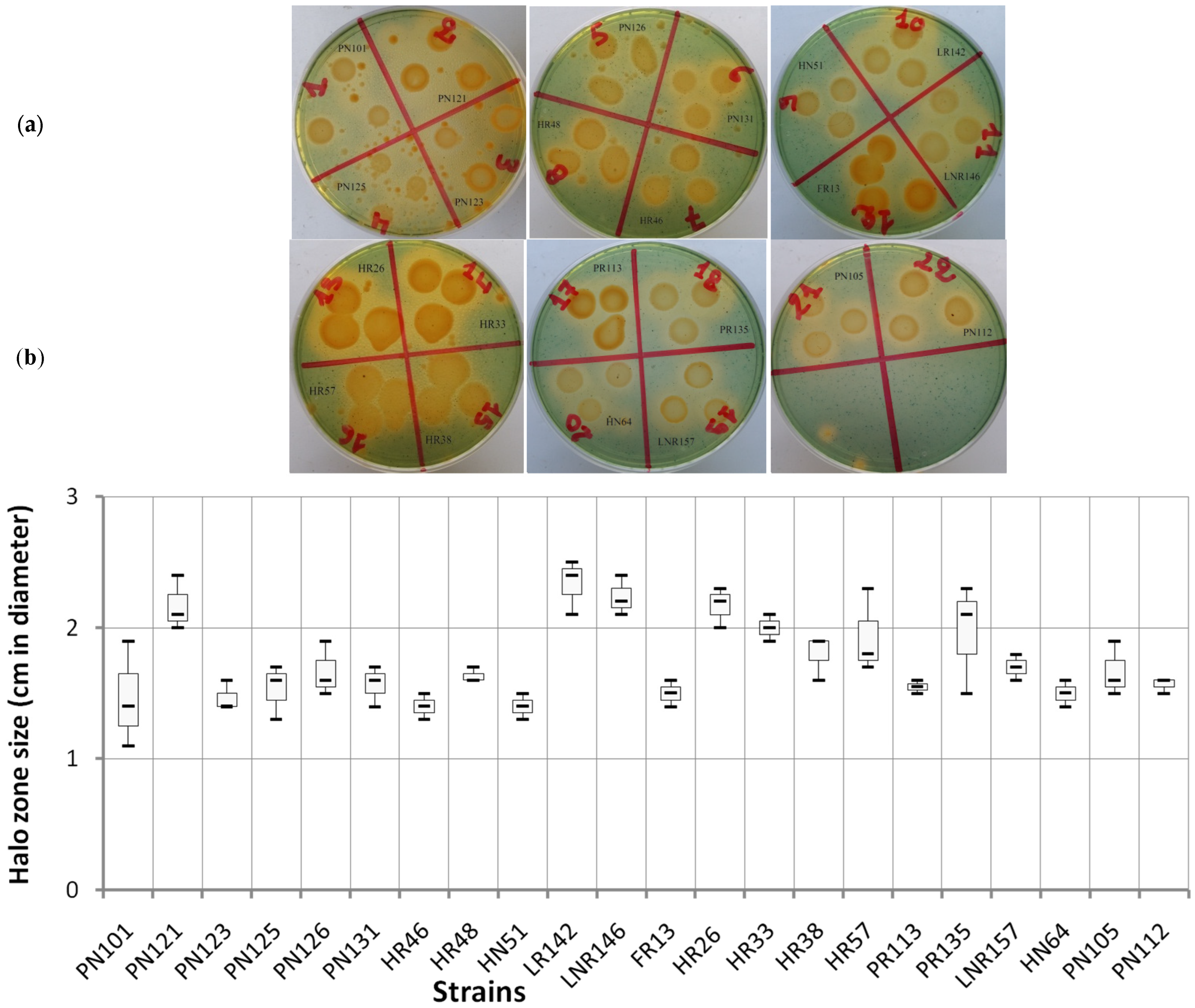
The images illustrate the halo zone sizes produced by different endophytic bacterial strains on CAS (Chrome Azurol S) medium, which reflect their siderophore production capabilities (Figure 2a). The box plots represent the lower and upper quartiles of the halo zone measurements for each strain (Figure 2b), with each value being the average of three observations to ensure data reliability. The strains PN121, LR142, LNR146, and HR26 exhibited larger halo zones, with medians of around or above 2 cm, indicating a high siderophore production capacity. These strains also showed outliers, particularly LR142, with data points significantly higher or lower than the rest, potentially reflecting experimental anomalies or natural variations in siderophore production. In contrast, the strains HR33, HR57, and PR135 had medians close to 2 cm and moderate interquartile ranges, indicating a reliable but not exceptional siderophore production capacity. Meanwhile, the strains PN101, PN123, PN125, PN126, PN131, HR46, HR48, HN51, FR13, HR38, FR113, LNR157, HN64, PN105, and PN112 displayed medians below 2 cm with smaller and less variable halo zones, suggesting a low siderophore production capacity.

4.5. Solubilization of Phosphate

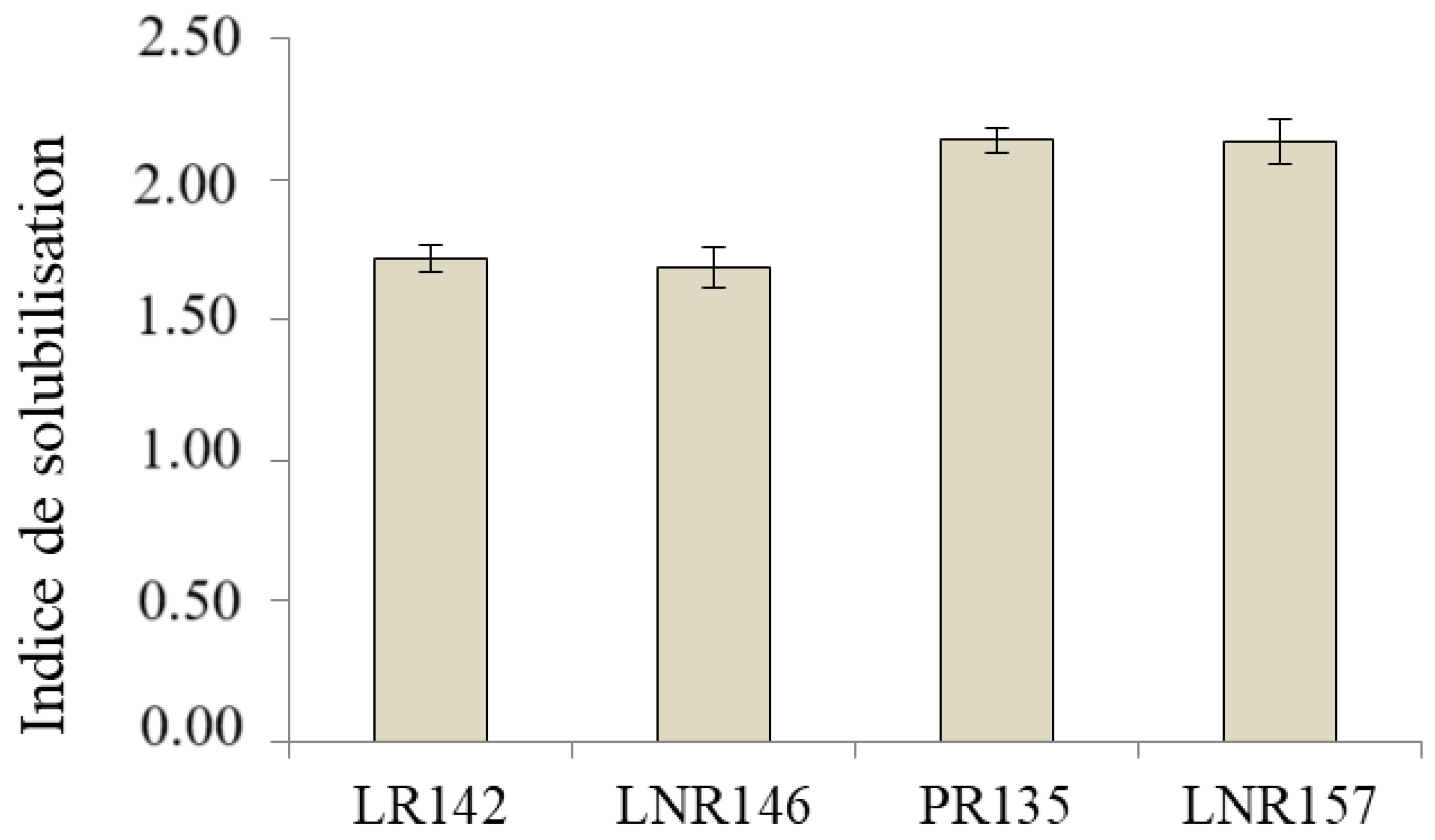
Only 18.2% of the endophytic isolates (4 out of 22) revealed phosphate solubilization, as evidenced by the clearing zones around bacterial colonies on the YED-P medium after 7 days of incubation (Figure 3). The strains PR135 and LNR135 were the most effective phosphate solubilizers, with solubilization indices (PSI) of 2.14 ± 0.04 and 2.13 ± 0.08, respectively. However, the strains LR142 and LNR146 exhibited a moderate phosphate solubilization efficiency, with solubilization indices of 1.72 ± 0.05 and 1.69 ± 0.07, respectively (Figure 4).

5. Discussion

Our results demonstrate that the studied endophytic bacteria exhibit resistance to erythromycin (86.36%), followed by ciprofloxacin (45.45%), ampicillin–sulbactam (22.73%), and tetracycline (9.09%). Additionally, these bacteria possess plant growth-promoting rhizobacteria (PGPR) traits, evidenced by their ability to produce indole-3-acetic acid (IAA). Most isolates also produce siderophores, while only the strains LR142, LNR146, PR135, and LNR135 have the capability to solubilize insoluble phosphate.
The observed antibiotic resistance in these soils, despite the absence of an anthropogenic influence, can be attributed to the natural antibiotic production by soil microorganisms as a biocontrol mechanism against pathogens [33,34,35,36,37,42,43]. Soil microorganisms such as Streptomyces erythreus, Streptomyces rimosus, Penicillium, and Bacillus polymyxa produce antibiotics like erythromycin, tetracycline, penicillin, and polymyxin, respectively [37]. Antibiotic resistance, which may arise through lateral gene transfer, is a significant mechanism by which resistance develops among soil bacteria [36,44,45]. Studies have documented antibiotic resistance in various species, including Rhizobium trifolii (MTCC905), Sinorhizobium morelense sp. nov., and Enterobacter cloacae [41,46,47,48,49]. Antibiotic resistance has also been utilized to study the diversity of endophytic bacteria in legumes such as Phaseolus coccineus [50], Glycine max L. Merr. [51], and Arachis hypogaea [52].
Indole-3-acetic acid (IAA) is a key phytohormone synthesized primarily from L-tryptophan. It plays a crucial role in stimulating plant growth through various mechanisms, including root elongation, nodule formation, and seed germination. Consequently, bacteria that produce IAA can serve as effective biofertilizers to enhance plant growth [18,20,53,54]. The production of IAA is widespread among both soil rhizobacteria and plants. Notable strains capable of IAA production include Rhizobium, Bacillus amyloliquefaciens FZB42, Streptomyces CMU-H009, and Streptomyces viridis NBRC 13373T [18,20,53].
Iron deficiency is a major cause of chlorosis in plants, leading to significant losses in field crops. Utilizing bacteria that produce siderophores offers an effective alternative for preventing or correcting iron deficiency.
Siderophores are low-molecular-weight secondary metabolites produced by many bacteria to solubilize and mobilize iron from their environment, making this essential element accessible to the cells. They have a high affinity for ferric iron (Fe3⁺), and the siderophore-Fe3⁺ complex is subsequently taken up by these bacteria through specific membrane transporters. In addition to iron, siderophores can bind various other metals, playing a significant role in the bioremediation of soils contaminated with heavy metals. Siderophores come in several types, including catechol (phenolate), hydroxamate, and carboxylic acid [55,56,57]. These compounds enhance plant growth by increasing iron availability or indirectly through their biocontrol properties. Scher and Baker [16] highlighted the antifungal action of siderophores, noting that iron chelation is crucial for the germ tube elongation of Fusarium oxysporum.
Furthermore, siderophore production has been shown in several genera and species of bacteria such as Azotobacter, Pseudomonas, Rhizobium meliloti, Bacillus niabensis PT-32-1, Bacillus subtilis SWI16, HPC21, and HPC24, and Bacillus mojavensis JCEN3 [21,23,41,57,58,59].
Phosphate-solubilizing abilities vary significantly between the strains of the same species. Phosphate solubilization has been described in several genera and species such as Rhizobium, Bacillus, Enterobacter, Pantoea, Klebsiella, Rahnella, Burkholderia, Pseudomonas rhizosphaerae, Pseudomonas putida SP21 and SP22, and Sinorhizobium meliloti [17,49,60,61,62,63,64,65]. The results showed that PR135 and LNR135 presented high phosphate-solubilizing indexes of 2.14 ± 0.04 and 2.13 ± 0.08, respectively, compared to LR142 and LNR146 with phosphate-solubilizing indexes of 1.72 ± 0.05 and 1.69 ± 0.07, respectively. These results are comparable with those obtained by Marraet al. [66] who found indexes of solubilization of 2.12 and 1.78 in Bradyrhizobium sp. UFLA 03–84 and Bradyrhizobium japonicum BR3267, respectively. Also, Sridevi et al. [67] noted that Rhizobium species isolated from the nodules of Crotalaria retusa and Crotalaria verrucosa had a high solubilization capacity for tricalcic phosphate, ranging from 2.40 to 2.70.
An inverse relationship between phosphate solubilization capacity and the pH of the medium has been observed. In particular, 2-ketogluconic acid, produced by Rhizobiummeliloti SU 47, has been identified as a key factor responsible for dissolving inorganic phosphate [60]. Several studies have shown that inoculating crops with plant growth-promoting rhizobacteria (PGPR) yields numerous agronomic benefits, enhancing plant growth while reducing the need for chemical fertilizers. For instance, the symbiotic relationship between Rhizobium species and legumes serves as a vital nitrogen source, boosting soil fertility and improving crop yields [5,67,68,69].
Zou et al. [70] demonstrated that inoculation with Rhizobium significantly enhanced the symbiotic nitrogen fixation of Acacia ampliceps under saline conditions. Additionally, inoculating soil with phosphate-solubilizing and nitrogen-fixing strains of Mesorhizobium mediterraneum, isolated from chickpea nodules, led to a substantial increase in chickpea dry biomass and elevated the contents of essential minerals such as phosphorus (125%), nitrogen, calcium, potassium, and magnesium [19]. Furthermore, Sadki et al. [71] showed that inoculating Phaseolus vulgaris with bacteria isolated from arid regions positively impacted its growth and antioxidant responses under saline conditions (25 mm).

6. Conclusions

The endophytic bacteria isolated in this study not only demonstrated notable antibiotic resistance, but also revealed essential traits of plant growth-promoting rhizobacteria (PGPR), such as phosphate solubilization, indole-3-acetic acid (IAA) production, and siderophore production. Their significant resistance to antibiotics, particularly erythromycin, ciprofloxacin, ampicillin–sulbactam, and tetracycline, highlights their potential resilience in challenging environments. These findings suggest that these bacteria have considerable potential as biofertilizers to enhance legume growth under abiotic stresses such as drought and salinity. However, before recommending these bacteria as biofertilizers for sustainable agriculture, it is essential to proceed with caution. Future studies must address the potential risks related to environmental and human health, particularly the spread of antibiotic resistance genes. Moreover, investigating safer alternatives will be crucial to ensuring both environmental and public health safety.
Strains like LR142, LNR146, PR135, and LNR135 showed particularly promising results in phosphate solubilization, emphasizing their usefulness in improving legume productivity in arid regions. Therefore, these selected endophytic bacteria present a viable solution for enhancing legume cultivation in areas impacted by both biotic and abiotic factors, contributing to sustainable agricultural practices in Morocco’s arid environments. Further research is recommended to evaluate the impact of inoculating these bacterial strains on legume growth under both controlled and field conditions. Furthermore, while this study focused on leguminous plants in the Tata-Akka region, future research should expand to include a broader range of plant species and investigate other regions with different environmental conditions. This broader approach will be crucial for providing a more comprehensive understanding of the applicability and benefits of these endophytic bacteria across diverse ecosystems and will be the subject of our future investigations.

Author Contributions

K.T., L.A., O.A. and M.F. developed the experimental design, and were responsible for the preparation of the manuscript. K.O. and K.E.B. established the molecular characterization of the strains. A.T., R.F. and H.O. contributed in preparation and revision of the manuscript. All authors have read and agreed to the published version of the manuscript.

Funding

This work was supported by the ministerial fellowship (Morocco).

Institutional Review Board Statement

Not applicable.

Informed Consent Statement

Not applicable.

Data Availability Statement

Data are contained within the article.

Conflicts of Interest

The authors declare that they have no conflicts of interest whatsoever.

References

  1. Herman, R.P.; Provencio, K.R.; Herrera-Matos, J.; Torrez, R.J. Resource islands predict the distribution of heterotrophic bacteria in Chihuahuan Desert soils. Appl. Environ. Microbiol. 1995, 61, 1816–1821. [Google Scholar] [CrossRef]
  2. Grego, S.; Moscatelli, M.C.; Di Mattia, E.; Marinari, S.; Cacciari, I. Activité biochimique de la rhizosphère d’Acacia raddiana au nord et du sud du Sahara. In Un Arbre au Désert: Acacia raddiana; Grouzis, M., Le Floc’h, E., Eds.; IRD: Paris, France, 2003; pp. 231–247. ISBN 2-7099-1522-7. [Google Scholar]
  3. Lal, B.; Khanna, S. Selection of salt-tolerant Rhizobium isolates of Acacia nilotica. World J. Microbiol. Biotechnol. 1994, 10, 637–639. [Google Scholar] [CrossRef]
  4. Taoufiq, K.; Faghire, M.; Tahrouch, S.; Nejemeddine, M.; Oufdou, K.; Hatimi, A. Isolation and identification of endophytic bacteria isolated from legumes nodules and roots cultivated in acacia rhizosphere soils collected from Tata-Akka region of Morocco. Indian J. Nat. Sci. 2018. [Google Scholar]
  5. Zahran, H.H. Rhizobia from wild legumes: Diversity, taxonomy, ecology, nitrogen fixation and biotechnology. J. Biotechnol. 2001, 91, 143–153. [Google Scholar] [CrossRef]
  6. Zahran, H.H. Enhancement of rhizobia–legumes symbioses and nitrogen fixation for crops productivity improvement. In Microbial Strategies for Crop Improvement; Khan, M.S., Zaidi, A., Musarrat, J., Eds.; Springer: Berlin/Heidelberg, Germany, 2009; pp. 227–254. [Google Scholar]
  7. Essendoubi, M.; Brhada, F.; Eljamali, J.E.; Filali-Maltouf, A.; Bonnassie, S.; Georgeault, S.; Blanco, C.; Jebbar, M. Osmoadaptative responses in the rhizobia nodulating Acacia isolated from south-eastern Moroccan Sahara. Environ. Microbiol. 2007, 9, 603–611. [Google Scholar] [CrossRef]
  8. Naseem, H.; Bano, A. Role of plant growth-promoting rhizobacteria and their exopolysaccharide in drought tolerance of maize. J. Plant Interact. 2014, 9, 689–701. [Google Scholar] [CrossRef]
  9. Faghire, M.; Mandri, B.; Oufdou, K.; Bargaz, A.; Ghoulam, C.; Ramírez-Bahena, M.H.; Velázquez, E.; Peix, A. Identification at the species and symbiovar levels of strains nodulating Phaseolus vulgaris in saline soils of the Marrakech region (Morocco) and analysis of the otsA gene putatively involved in osmotolerance. Syst. Appl. Microbiol. 2012, 35, 156–164. [Google Scholar] [CrossRef]
  10. Faghire, M.; Farissi, M.; Taoufiq, K.; Fghire, R.; Bargaz, A.; Mandri, B.; Oufdou, K.; Laury, A.; Drevon, J.J.; Ghoulam, C. Genotypic variation of nodules’ enzymatic activities in symbiotic nitrogen fixation among common bean (Phaseolus vulgaris L.) genotypes grown under salinity constraint. Symbiosis 2013, 60, 115–122. [Google Scholar] [CrossRef]
  11. Bargaz, A.; Faghire, M.; Farissi, M.; Drevon, J.J.; Ghoulam, C. Oxidative stress in the root nodules of Phaseolus vulgaris is induced under conditions of phosphorus deficiency. Acta Physiol. Plant. 2013, 35, 1633–1644. [Google Scholar] [CrossRef]
  12. Faghire, M.; Bargaz, A.; Farissi, M.; Palma, F.; Mandri, B.; Lluch, C.; TejeraGarcía, N.A.; Herrera-Cervera, J.A.; Oufdou, K.; Ghoulam, C. Effect of salinity on nodulation, nitrogen fixation and growth of common bean (Phaseolus vulgaris) inoculated with rhizobial strains isolated from the Haouz region of Morocco. Symbiosis 2011, 55, 69–75. [Google Scholar] [CrossRef]
  13. Benidire, L.; Lahrouni, M.; El Khalloufi, F.; Gottfert, M.; Oufdou, K. Effects of Rhizobium leguminosarum inoculation on growth, nitrogen uptake and mineral assimilation in Viciafaba plants under salinity stress. J. Agric. Sci. Technol. 2017, 889–901. [Google Scholar]
  14. Benbrahim, K.F.; Berrada, H.; El Ghachtouli, N.; Ismaili, M. Les acacias: Des plantes fixatrices d’azote prometteuses pour le développement durable des zones arides et semi-arides [Acacia: PromisingNitrogen fixing trees for sustainabledevelopment in arid and semi-arid areas]. Int. J. Innov. Appl. Stud. 2014, 8, 46. [Google Scholar]
  15. Kandel, S.L.; Joubert, P.M.; Doty, S.L. Bacterial endophyte colonization and distribution within plants. Microorganisms 2017, 5, 77. [Google Scholar] [CrossRef]
  16. Scher, F.M.; Baker, R. Effect of Pseudomonas putida and a synthetic iron chelator on induction of soil suppressiveness to Fusarium wilt pathogens. Phytopathology 1982, 72, 1567–1573. [Google Scholar] [CrossRef]
  17. Halder, A.K.; Mishra, A.K.; Bhattacharyya, P.; Chakrabartty, P.K. Solubilization of rock phosphate by Rhizobium and Bradyrhizobium. J. Gen. Appl. Microbiol. 1990, 36, 81–92. [Google Scholar] [CrossRef]
  18. Datta, C.; Basu, P.S. Indole acetic acid production by a Rhizobium species from root nodules of a leguminous shrub, Cajanuscajan. Microbiol. Res. 2000, 155, 123–127. [Google Scholar] [CrossRef]
  19. Peix, A.; Rivas-Boyero, A.A.; Mateos, P.F.; Rodriguez-Barrueco, C.; Martinez-Molina, E.; Velazquez, E. Growth promotion of chickpea and barley by a phosphate solubilizing strain of Mesorhizobiummediterraneum under growth chamber conditions. Soil Biol. Biochem. 2001, 33, 103–110. [Google Scholar] [CrossRef]
  20. Idris, E.E.; Iglesias, D.J.; Talon, M.; Borriss, R. Tryptophan-dependent production of indole-3-acetic acid (IAA) affects level of plant growth promotion by Bacillus amyloliquefaciens FZB42. Mol. Plant-Microbe Interact. 2007, 20, 619–626. [Google Scholar] [CrossRef]
  21. Ahmad, F.; Ahmad, I.; Khan, M.S. Screening of free-living rhizospheric bacteria for their multiple plant growth promoting activities. Microbiol. Res. 2008, 163, 173–181. [Google Scholar] [CrossRef]
  22. Kyungseok, P.; Kloepper, J.W.; Ryu, C.M. Rhizobacterial exopolysaccharides elicit induced resistance on cucumber. J. Microbiol. Biotechnol. 2008, 18, 1095–1100. [Google Scholar]
  23. Karadayi, M.; Gulluce, M.; Dogan, S.; Gulluce, E. Isolation and Molecular Characterization of Bacteria with Plant Growth Promoting Characteristics from Magnesite Mining Fields in Kütahya-Turkey. J. Appl. Biol. Sci. 2016, 10, 27–32. [Google Scholar]
  24. Adesemoye, A.O.; Torbert, H.A.; Kloepper, J.W. Plant growth-promoting rhizobacteria allow reduced application rates of chemical fertilizers. Microb. Ecol. 2009, 58, 921–929. [Google Scholar] [CrossRef]
  25. Glick, B.R. Plant Growth-Promoting Bacteria: Mechanisms and Applications. Scientifica 2012, 2012, 1–15. [Google Scholar] [CrossRef]
  26. Compant, S.; Duffy, B.; Nowak, J.; Cleves, P.; Barka, E.A. Use of plant growth-promoting bacteria for sustainable agriculture. J. Plant Nutr. Soil Sci. 2019, 182, 563–579. [Google Scholar]
  27. Rosenblueth, M.; Martínez-Romero, E. Bacterial endophytes are more than just endosymbionts. Funct. Plant Biol. 2006, 33, 701–712. [Google Scholar]
  28. de Carvalho, C.A.A.; Teixeira SM, R.; Figueira, A.V. Plant growth-promoting bacteria in agriculture: Benefits and applications. Microorganisms 2021, 9, 1478. [Google Scholar]
  29. Mitter, B.; Pfaffenbichler, N.; Riedel, T. Endophytes: The Plant Microbiome and Its Impact on Plant Health and Agriculture. J. Appl. Microbiol. 2017, 123, 938–949. [Google Scholar]
  30. Kumar, A.; Pandey, S.S.; Kumar, V. Endophytic bacteria: A novel approach for the enhancement of plant growth and stress tolerance. Environ. Exp. Bot. 2020, 176, 104041. [Google Scholar]
  31. Broughton, W.J.; Hernandez, G.; Blair, M.; Beebe, S.; Gepts, P.; Vanderleyden, J. Beans (Phaseolus spp.) model food legumes. Plant Soil 2003, 252, 55–128. [Google Scholar] [CrossRef]
  32. Manchanda, G.; Garg, N. Salinity and its effects on the functional biology of legumes. Acta Physiol. Plant. 2008, 30, 595–618. [Google Scholar] [CrossRef]
  33. Naamala, J.; Jaiswal, S.K.; Dakora, F.D. Antibiotics resistance in Rhizobium: Type, process, mechanism and benefit for agriculture. Curr. Microbiol. 2016, 72, 804–816. [Google Scholar] [CrossRef]
  34. Howie, W.J.; Suslow, T.V. Role of antibiotic biosynthesis in the inhibition of Pythium ultimum in the cotton spermosphere and rhizosphere by Pseudomonas fluorescens. Mol. Plant-Microbe Interact. 1991, 4, 393–399. [Google Scholar] [CrossRef]
  35. Yokoyama, T.; Ando, S.; Tsuchiya, K. Serological properties and intrinsic antibiotic resistance of soybean bradyrhizobia isolated from Thailand. Soil Sci. Plant Nutr. 1999, 45, 505–515. [Google Scholar] [CrossRef]
  36. Ghosh, S.; LaPara, T.M. The effects of subtherapeutic antibiotic use in farm animals on the proliferation and persistence of antibiotic resistance among soil bacteria. Int. Soc. Microb. Ecol. J. 2007, 1, 191. [Google Scholar] [CrossRef]
  37. Nwosu, V.C. Antibiotic resistance with particular reference to soil microorganisms. Res. Microbiol. 2001, 152, 421–430. [Google Scholar] [CrossRef]
  38. Vincent, J.M. The cultivation, isolation and maintenance of rhizobia. In A Manual for the Practical Study of Root-Nodule; Vincent, J.M., Ed.; Blackwell Scientific Publications: Oxford, UK, 1970; pp. 1–13. [Google Scholar]
  39. Wöhler, I. Auxin-indole derivatives in soils determined by a colorimetric method and by high performance liquid chromatography. Microbiol. Res. 1997, 152, 399–405. [Google Scholar] [CrossRef]
  40. Schwyn, B.; Neilands, J.B. Universal chemical assay for the detection and determination of siderophores. Anal. Biochem. 1987, 160, 47–56. [Google Scholar] [CrossRef]
  41. Sultana, S.; Alam, S.; Karim, M.M. Screening of siderophore-producing salt-tolerant rhizobacteria suitable for supporting plant growth in saline soils with iron limitation. J. Agric. Food Res. 2021, 4, 100–150. [Google Scholar] [CrossRef]
  42. Fernandes Júnior, P.I.; Lima, A.A.D.; Passos, S.R.; Gava, C.A.T.; Oliveira, P.J.D.; Rumjanek, N.G.; Xavier, G.R. Phenotypic diversity and amylolytic activity of fast-growing rhizobia from pigeonpea [Cajanuscajan (L.) Millsp. Braz. J. Microbiol. 2012, 43, 1604–1612. [Google Scholar] [CrossRef]
  43. Singh, A.K.; Bhatt, R.P.; Pant, S.; Bedi, M.K.; Naglot, A. Characterization of Rhizobium isolated from root nodules of Trifolium alexandrinum. J. Agric. Technol. 2011, 7, 1705–1723. [Google Scholar]
  44. Kimenju, J.W.; Karanja, N.K.; Macharia, I. Plant parasitic nematodes associated with common bean in Kenya and the effect of Meloidogyne infection on bean nodulation. Afr. Crop Sci. J. 1999, 7, 503–510. [Google Scholar] [CrossRef]
  45. Melakeberhan, H.; Brooke, R.C.; Webster, J.M. Relationship between physiological response of French beans of different age to Meloidogyne incognita and subsequent yield loss. Plant Pathol. 1987, 35, 203–213. [Google Scholar] [CrossRef]
  46. Wang, E.T.; Tan, Z.Y.; Willems, A.; Fernández-López, M.; Reinhold-Hurek, B.; Martínez-Romero, E. Sinorhizobiummorelense sp. nov., a Leucaena leucocephala-associated bacterium that is highly resistant to multiple antibiotics. Int. J. Syst. Evol. Microbiol. 2002, 52, 1687–1693. [Google Scholar] [CrossRef]
  47. Amarger, N Rhizobia in the field. Adv. Agron. 2001, 73, 109–168.
  48. Ramesh, A.; Sharma, S.K.; Sharma, M.P.; Yadav, N.; Joshi, O.P. Plant growth-promoting traits in Enterobacter cloacae subsp. dissolvens MDSR9 isolated from soybean rhizosphere and its impact on growth and nutrition of soybean and wheat upon inoculation. Agric. Res. 2014, 3, 53–66. [Google Scholar] [CrossRef]
  49. Khalifa, A.Y.; Alsyeeh, A.M.; Almalki, M.A.; Saleh, F.A. Characterization of the plant growth promoting bacterium, Enterobacter cloacae MSR1, isolated from roots of non-nodulating Medicago sativa. Saudi J. Biol. Sci. 2016, 23, 79–86. [Google Scholar] [CrossRef]
  50. Arredondo-Peter, R.; Escamilla, E. Sodium dodecyl sulfate-polyacrylamide gel electrophoresis protein banding patterns among Rhizobium leguminosarumbiovarphaseoli strains isolated from the Mexican bean Phaseoluscoccineus. Appl. Environ. Microbioology 1993, 59, 3960–3962. [Google Scholar] [CrossRef]
  51. Ramirez, M.E.; Israel, D.W.; Wollum, A.G., II. Phenotypic and genotypic diversity of similar serotypes of soybean bradyrhizobia from two soil populations. Soil Biol. Biochem. 1997, 29, 1539–1545. [Google Scholar] [CrossRef]
  52. El-Akhal, M.R.; Rincon, A.; Mourabit, N.E.; Pueyo, J.J.; Barrijal, S. Phenotypic and genotypic characterizations of rhizobia isolated from root nodules of peanut (Arachishypogaea L.) grown in Moroccan soils. J. Basic Microbiol. 2009, 49, 415–425. [Google Scholar] [CrossRef]
  53. Khamna, S.; Yokota, A.; Peberdy, J.F.; Lumyong, S. Indole-3-acetic acid production by Streptomyces sp. isolated from some Thai medicinal plant rhizosphere soils. Eur. Asian J. BioSci. 2010, 4. [Google Scholar] [CrossRef]
  54. Mohite, B. Isolation and characterization of indole acetic acid (IAA) producing bacteria from rhizospheric soil and its effect on plant growth. J. Soil Sci. Plant Nutr. 2013, 13, 638–649. [Google Scholar] [CrossRef]
  55. Haas, H. Molecular genetics of fungal siderophore biosynthesis and uptake: The role of siderophores in iron uptake and storage. Appl. Microbiol. Biotechnol. 2003, 62, 316–330. [Google Scholar] [CrossRef] [PubMed]
  56. Azmat, R. The impact of siderophore secretion by Pseudomonas stutzeri to chelating cu metal in solution culture. Pak. J. Bot. 2014, 46, 383–387. [Google Scholar]
  57. Kesaulya, H.; Hasinu, J.V.; Tuhumury, G.N. Potential of Bacillus spp. produces siderophoresinsuppressing the wilt disease of banana plants. In IOP Conference Series: Earth and Environmental Science; IOP Publishing: Bristol, England, 2018; Volume 102, p. 012016. [Google Scholar]
  58. Arora, N.K.; Kang, S.C.; Maheshwari, D.K. Isolation of siderophore-producing strains of Rhizobium meliloti and their biocontrol potential against Macrophominaphaseolina that causes charcoal rot of groundnut. Curr. Sci. 2001, 81, 673–677. [Google Scholar]
  59. Kesaulya, H.; Zakaria, B.; Syaiful, S.A. Isolation and physiological characterization of PGPR from potato plant rhizosphere in medium land of Buru Island. Procedia Food Sci. 2015, 3, 190–199. [Google Scholar] [CrossRef]
  60. Halder, A.K.; Chakrabartty, P.K. Solubilization of inorganic phosphate by Rhizobium. Folia Microbiol. 1993, 38, 325–330. [Google Scholar] [CrossRef]
  61. Rodíguez, H.; Fraga, R. Phosphate solubilizing bacteria and their role in plant growth promotion. Biotechnol. Adv. 1999, 17, 319–339. [Google Scholar] [CrossRef]
  62. Peix, A.; Rivas, R.; Mateos, P.F.; Martínez-Molina, E.; Rodríguez-Barrueco, C.; Velazquez, E. Pseudomonasrhizosphaerae sp. nov., a novel speciesthatactively solubilizes phosphate in vitro. Int. J. Syst. Evol. Microbiol. 2003, 53, 2067–2072. [Google Scholar] [CrossRef]
  63. Rosas, S.B.; Andrés, J.A.; Rovera, M.; Correa, N.S. Phosphate-solubilizing Pseudomonasputida can influence the rhizobia–legume symbiosis. Soil Biol. Biochem. 2006, 38, 3502–3505. [Google Scholar] [CrossRef]
  64. Rfaki, A.; Nassiri, L.; Ibijbijen, J. Phosphate-solubilizing bacteria in the rhizosphere of some cultivated legumes from Meknes region, Morocco. Br. Biotechnol. J. 2014, 4, 946–956. [Google Scholar] [CrossRef]
  65. Rfaki, A.; Nassiri, L.; Ibijbijen, J. Isolation and Characterization of phosphate solubilizing bacteria from the rhizosphere of faba bean (Viciafaba L.) in Meknes Region, Morocco. Br. Microbiol. Res. J. 2015, 6, 247–254. [Google Scholar] [CrossRef]
  66. Marra, L.M.; Oliveira, S.M.D.; Soares, C.R.F.S.; Moreira, F.M.D.S. Solubilisation of inorganic phosphates by inoculant strains from tropical legumes. Sci. Agric. 2011, 68, 603–609. [Google Scholar] [CrossRef]
  67. Sridevi, M.; Mallaiah, K.V.; Yadav, N.C.S. Phosphate solubilization by Rhizobium isolates from Crotalaria species. J. Plant Sci. 2007, 2, 635–639. [Google Scholar] [CrossRef]
  68. Vessey, J.K. Plant growth promoting rhizobacteria as biofertilizers. Plant Soil 2003, 255, 571–586. [Google Scholar] [CrossRef]
  69. Richardson, A.E.; Barea, J.M.; McNeill, A.M.; Prigent-Combaret, C. Acquisition of phosphorus and nitrogen in the rhizosphere and plant growth promotion by microorganisms. Plant Soil 2009, 321, 305–339. [Google Scholar] [CrossRef]
  70. Zou, N.; Dart, P.J.; Marcar, N.E. Interaction of salinity and rhizobial strain on growth and N2-fixation by Acacia ampliceps. Soil Biol. Biochem. 1995, 27, 409–413. [Google Scholar] [CrossRef]
  71. Sadki, I.; Taoufiq, K.; Aberchane, L.; El Biari, K.; Tahrouch, S.; Oufdou, K.; Faghire, M.; Hatimi, A. Effect of inoculation by indigenous endophytic bacteria from the arid region in Morocco (Tata-Akka) on the antioxidative responses of Phaseolus vulgaris L. J. Mater. Environ. Sci. 2021, 12, 205–216. [Google Scholar]
Figure 1. Concentration (µg of IAA/ mg of proteins) of indole-3-acetic acid produced by the endophytic bacteria using the reagent of Salkowski. Results averaged from numbers of measurements (n = 3) of each strain. Means followed by the same letter within a column are not significantly different (p < 0.05) using the LSD test.
Ijpb 15 00075 g001
Figure 2. Detection of siderophore-producing abilities by the endophytic bacteria strains using chrome azurol agar (CAS) medium S assay method. (a) Orange halo on CAS agar indicating the positive response of siderophore production by the endophytic bacteria strains PN101 (1), PN121 (2), PN123 (3), PN125 (4), PN126 (5), PN131 (6), HR46 (7), HR48 (8), HN51 (9), LR142 (10), LNR146 (11) FR13 (12), HR26 (13), HR33 (14), HR38 (15), HR57 (16), PR113, (17), PR135 (18) LNR157 (19), HN64 (20), PN105 (21), and PN112, (b) Box plot demonstrating halo zone measurements in diameter (cm) averaged from numbers of observations (n = 3) of each strain (the boxes represent the lower and upper quartiles, the horizontal line within the boxes is the median, the bars are the minimum and maximum data points).
Ijpb 15 00075 g002
Figure 3. Cleared zones on test plates indicating tricalcium phosphatesolubilization by the endophytic bacteria on YED-P medium with Ca3 (PO4)2as sole phosphorus source.
Ijpb 15 00075 g003
Figure 4. Solubilization index of inorganic phosphate by the endophytic bacteria on YED-P medium with Ca3 (PO4)2as sole phosphorus source. All measurements were performed in triplicate. Means followed by the same letter within a column are not significantly different (p < 0.05) using the LSD test.
Ijpb 15 00075 g004
Table 1. Antibiotics used for resistance test.
AntibioticQuantity of Antibiotic/Disc µg
E15Erythromycin15
CIP5Ciprofloxacin5
SAM20Ampicillin–sulbactam20
TET30Tetracycline30
Table 2. Resistance to antibiotics of the endophytic bacteria using disc diffusion test on YEM medium and inhibitory zone diameter (I.Z.D) measured. E15: Erythromycin; CIP5: Ciprofloxacin; SAM20: Ampicillin–sulbactam; and TET30: Tetracycline. Inhibitory zone diameter (I.Z.D.) measurements in diameter (cm) averaged from numbers of observations (n = 3) of each strain. Means followed by the same letter within a column are not significantly different (p < 0.05) using the LSD test.
E15CIP5SAM20TET30
GrowthI.Z.D.GrowthI.Z.D.GrowthI.Z.D.GrowthI.Z.D.
PN101+0 o3 b2.3 e3 b
PN1211.4 l+0 o2.1 fg1.9 hi
PN123+0 o2.1 fg1.8 ij3 b
PN125+0 o1.7 jk1.6 k1.4 l
PN126+0+02.21.9
PN131+0 o3.2 a1.7 jk3.3 a
HR461.3 lm+0 o1.2 m2.1 fg
HR481.2 m1.4 l1.8 ij1.7 jk
HN51+0 o+0 o1.8 ij2.6 d
LR142+0 o+0 o+0 o1.9 hi
LNR146+0 o1.3 lm+0 o2.9 bc
FR13+0 o2.8 c1.8 ij2.9 bc
HR26+0 o+0 o2.1fg3 b
HR33+0 o+0 o+0 o+0 o
HR38+0 o+0 o1.8 ij2.9 bc
HR57+0 o+0 o2 gh+0 o
PR113+0 o+0 o1.6 k1.6 k
PR135+0 o3 b1.8 ij1.9 hi
LNR157+0 o1.4 l+0 o2.1 fg
HN64+0 o2.5 d2.2 ef2.6 d
PN105+0 o2.1 fg1.4 l1.8 ij
PN112+0 o3 b+0 o1.9 hi
(+): Growth; (−): No growth; (I.Z.D): inhibitory zone diameter (cm).
Disclaimer/Publisher’s Note: The statements, opinions and data contained in all publications are solely those of the individual author(s) and contributor(s) and not of MDPI and/or the editor(s). MDPI and/or the editor(s) disclaim responsibility for any injury to people or property resulting from any ideas, methods, instructions or products referred to in the content.