Article

An Exploratory Study of Antibiotic Resistance and Virulence-Associated Markers in Enterococcus faecalis and Enterococcus faecium Isolates from Bulgarian Influent Wastewater

1
Department of General and Industrial Microbiology, Faculty of Biology, Sofia University “St. Kliment Ohridski”, 1164 Sofia, Bulgaria
2
Research Group: Microbiological Risks in the Environment, Sofia University “St. Kliment Ohridski”, 1164 Sofia, Bulgaria
*
Author to whom correspondence should be addressed.
Microbiol. Res. 2026, 17(7), 132; https://doi.org/10.3390/microbiolres17070132
Submission received: 29 May 2026 / Revised: 29 June 2026 / Accepted: 6 July 2026 / Published: 8 July 2026
(This article belongs to the Section Antimicrobials and Antimicrobial Resistance)

Abstract

Influent wastewater, characterized by a diverse bacterial composition of human and animal origin, is a suitable source for studying the spread of antibiotic resistance and virulence genes, particularly in Enterococcus faecalis and Enterococcus faecium. This study aimed to evaluate the potential of these two species as reservoirs and indicators of antibiotic resistance and virulence genes. Wastewater treatment plants near the three largest cities of Bulgaria (Sofia, Varna and Burgas) were selected for sampling. Influent wastewater samples were collected for a period of 3 months (February, March and May) and used in the analyses. Overall, 34 bacterial isolates (E. faecalis, n = 13 and E. faecium, n = 21) were isolated and identified by MALDI-ToF. Phenotypic and genotypic methods were used to evaluate their antibiotic resistance profiles (focusing on clinically relevant antibiotics) and virulence potential. High dissemination of phenotypic ampicillin and quinupristin-dalfoprisin resistance (97% of all tested strains and 81% of all E. faecium strains, respectively) was established. In March, the highest number of antibiotic-resistant profiles was observed, including the emergence of MDR strains, primarily among E. faecium strains. Molecular analyses revealed a dissemination of genes encoding resistance to aminoglycosides and β-lactam antibiotics. The most prevalent virulence genes were gelE and ace, found only in E. faecalis strains. All gelE-positive strains also exhibited phenotypic gelatinase activity. In this study, E. faecium shows greater antibiotic resistance potential, whereas E. faecalis exhibits increased virulence capacity. These exploratory findings support the usefulness of influent wastewater as a matrix for monitoring antibiotic-resistant and potentially virulent enterococcal isolates, while larger-scale and seasonally representative studies are needed to confirm temporal and geographic patterns.

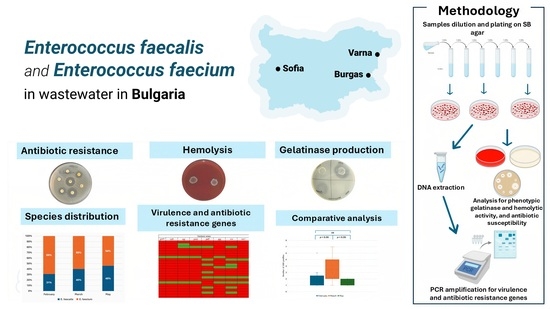
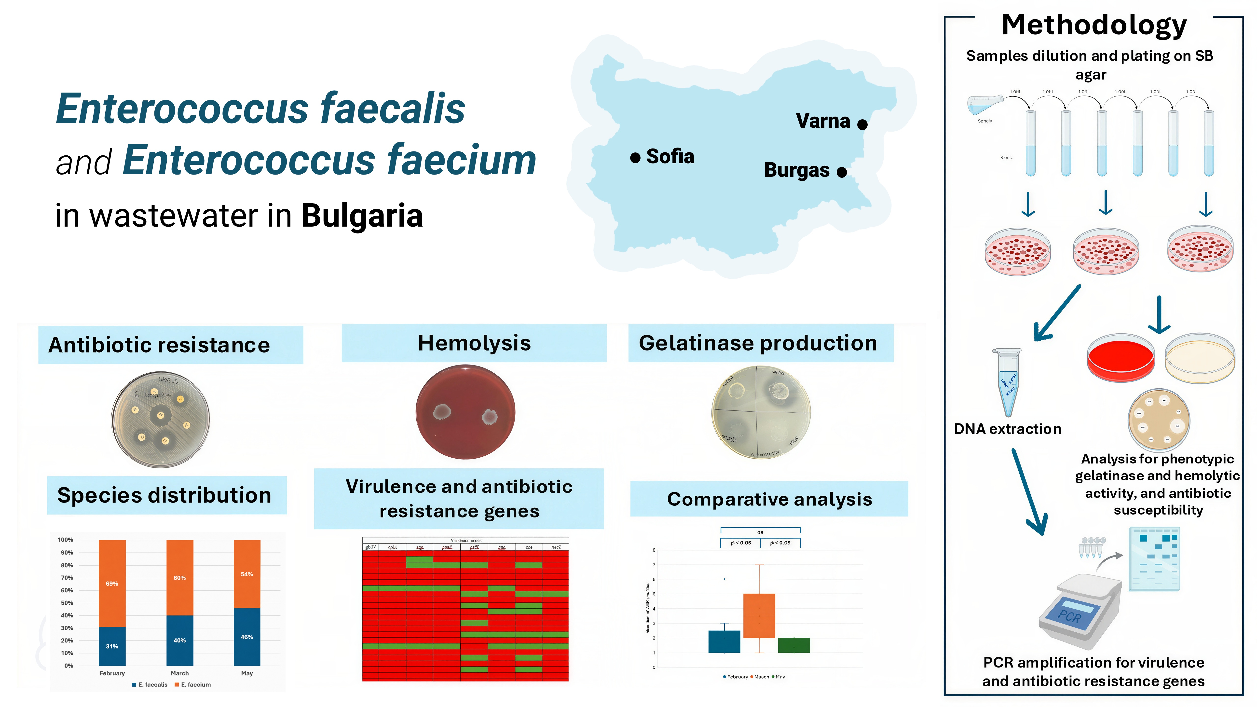
Graphical Abstract

1. Introduction

Influent wastewater represents a critical and highly informative matrix for wastewater-based epidemiology, enabling the detection and quantification of targeted microbial species as well as the genetic determinants associated with antimicrobial resistance and virulence. It is assumed that this environment provides favorable conditions for horizontal gene transfer, thereby promoting the spread of genes associated with antibiotic resistance and establishing it as a significant reservoir for the persistence and dissemination of resistance determinants [1]. In Europe, the recast Urban Wastewater Treatment Directive introduces, for the first time, a legal requirement to monitor antimicrobial resistance in wastewater from major urban agglomerations (≥100,000 person equivalents) [2].
Members of the genus Enterococcus are widely distributed across diverse ecological niches, with Enterococcus faecalis and Enterococcus faecium being particularly prevalent in wastewater and commonly regarded as indicator microorganisms of fecal contamination [3,4]. Both species are considered opportunistic pathogens, causing severe nosocomial infections in certain categories of people, mainly with compromised immune systems [4].
The clinical relevance of E. faecalis and E. faecium is associated with different but complementary characteristics. E. faecalis is generally characterized by a broader repertoire of virulence-associated factors, including adhesins, aggregation substance, collagen-binding proteins, extracellular enzymes such as gelatinase, cytolysin production, biofilm formation, and factors involved in host tissue colonization and persistence [5]. These determinants may contribute to adhesion, colonization, biofilm formation and persistence [5]. In contrast, E. faecium is often regarded as a species with lower virulence-associated gene content but greater adaptive and antimicrobial-resistance potential [6,7]. Important resistance mechanisms in enterococci include intrinsic or acquired reduced susceptibility to β-lactams, high-level aminoglycoside resistance mediated by aminoglycoside-modifying enzymes, glycopeptide resistance associated mainly with van operons, and resistance to macrolides and tetracyclines mediated by genes such as ermB, mefA, and tet determinants [6,8]. Therefore, the simultaneous evaluation of antibiotic-resistance profiles and selected virulence-associated markers is important for assessing the potential public health relevance of wastewater-derived enterococcal isolates.
These characteristics highlight the relevance of E. faecalis and E. faecium as reservoirs of antibiotic-resistance determinants and as species capable of harboring virulence-associated genes [6]. As a consequence, E. faecium is included in the ESKAPE group of pathogens, which are identified as critical multidrug-resistant bacteria for which effective therapies are of emerging importance [7]. According to the 2024 annual report of the European Centre for Disease Prevention and Control, healthcare-associated isolates in Bulgaria exhibited high levels of antimicrobial resistance. Specifically, resistance to gentamicin reached 80.9%, while resistance to ampicillin and, most notably, vancomycin was reported at 14.5% in 2024 [9].
Comprehensive monitoring of antibiotic resistance among enterococci in wastewater—covering both influent and effluent—should be considered critically important. This is due to the potential dissemination of antibiotic-resistant strains even after wastewater treatment, posing a tangible risk not only to the environment but also to public health [10]. However, studies of the antibiotic resistance of microbial communities in influent wastewaters in Bulgaria seem to be very limited. For example, in a survey conducted in 2025 by Tsvetanova and Bushnakov, antimicrobial resistance in influent and effluent wastewater was monitored, revealing that 10% (n = 26) of the total identified Enterococcus isolates (n = 259) were multidrug-resistant [11]. In contrast, most of the studies conducted in Bulgaria in recent years have primarily focused on assessing the dissemination of antibiotic resistance in major rivers such as the Yantra River and the Iskar River, which are identified as principal receiving water bodies for treated wastewater discharge, thus evaluating potential environmental risk factors [12,13]. For example, Donchev et al. introduced the possibility of antibiotic resistance genes spreading in the Iskar River by the Wastewater Treatment Plant (WWTP) in Samokov by reporting genes, relevant to macrolide and tetracycline resistance, as the most prevalent in the bacterial community in this habitat [13]. Similar dissemination of antibiotic resistance has been reported by other authors for different water habitats, Yantra River [12].
Given the limited research on influent wastewater as a reservoir of antibiotic resistance and virulence-associated markers in Bulgaria, this exploratory study aimed to provide new insights into the role of wastewater-derived E. faecalis and E. faecium isolates in this context. Influent wastewater samples were collected from WWTPs serving the cities of Sofia, Varna, and Burgas during three sampling months (February, March and May) in 2025. The primary analyses focused on the phenotypic and genotypic characterization of antibiotic resistance and selected virulence-associated markers among the obtained isolates. The results provide preliminary evidence supporting the relevance of influent wastewater as a useful matrix for monitoring antibiotic-resistant and potentially virulent enterococcal isolates in Bulgaria.

2. Materials and Methods

2.1. Sampling, Enterococcal Isolation and Enumeration

Wastewater samples were collected at the entrance of the WWTPs of the cities of Sofia, Varna and Burgas, Bulgaria, during three selected sampling months: February, March, and May 2025. Prior to isolation of targeted enterococci, all samples were processed to remove the larger debris. To determine the total number of fecal enterococci in these samples, the plate count method was employed using Slanetz and Bartley (SB) agar (HiMedia, Mumbai, India) as a selective medium. Only colonies exhibiting the characteristic red-brown color were enumerated, purified through three consecutive subcultures in de Man, Rogosa, and Sharpe (MRS) broth (HiMedia, Mumbai, India) and isolated as pure cultures. The enterococcal cultures were subsequently stored at 4 °C and used for further analyses.

2.2. Isolate Identification

The obtained pure cultures were further Gram-stained and identified to the species level by MALDI-TOF mass spectrometry (Autobio, Zhengzhou, China). The analyses were done in the microbiological laboratory of Vita Hospital, Sofia, Bulgaria. According to the manufacturer, scores between 9.5 and 10.0 indicate reliable subspecies-level identification, and scores between 9.0 and 9.5 are considered reliable at the species level. Scores ranging from 6.0 to 9.0 support identification at the genus level, whereas scores below 6.0 are considered unreliable.

2.3. Evaluation of Phenotypic Antibiotic Resistance

The newly isolated enterococci were screened for antibiotic susceptibility/resistance according to the European Committee on Antimicrobial Susceptibility Testing using the Kirby–Bauer disk diffusion method [14,15]. All E. faecalis isolates were tested to 12 antibiotics—ampicillin (AMP), 2 μg/disc; imipenem (IPM), 10 μg/disc; levofloxacin (LE), 5 μg/disc; norfloxacin (NX), 10 μg/disc; high-level gentamicin (HLG), 30 μg/disc; high-level streptomycin (HLS), 300 μg/disc; teicoplanin (TEI), 30 μg/disc; vancomycin (VA), 5 μg/disc; eravacycline (ERV), 20 μg/disc; tigecycline (TGC), 15 μg/disc; linezolid (LZ), 10 μg/disc; and nitrofurantoin (NIT), 100 μg/disc (HiMedia, Mumbai, India). All E. faecium isolates were tested to 11 antibiotics—ampicillin (AMP), 2 μg/disc; levofloxacin (LE), 5 μg/disc; norfloxacin (NX), 10 μg/disc; high-level gentamicin (HLG), 30 μg/disc; high-level streptomycin (HLS), 300 μg/disc; teicoplanin (TEI), 30 μg/disc; vancomycin (VA), 5 μg/disc; eravacycline (ERV), 20 μg/disc; tigecycline (TGC), 15 μg/disc; linezolid (LZ), 10 μg/disc; and pristinomycin (quinupristin/dalfopristin; RP), 15 μg/disc (HiMedia, Mumbai, India). Log bacterial cultures were obtained after cultivation of the strains on MRS agar (HiMedia Inc., Mumbai, India) at 37 °C for 24 h. Bacterial suspensions were prepared in sterile saline (108 CFU/mL, MacFarland 0.5) and plated on Mueller-Hinton agar (MHA, Merck KGaA, Darmstadt, Germany). The antibiotic paper disks were placed on the surface of Petri dishes and incubated for 24 h at 37 °C. The interpretation of the results was done according to EUCAST [14]. Isolates resistant to three or more classes of antibiotics were considered multidrug-resistant (MDR) [16].

2.4. Hemolytic Activity

The evaluation of hemolytic activity was performed according to the method described by Pandova et al. (2024) in both aerobic and anaerobic conditions [3]. Pure bacterial cultures were surface-spot inoculated onto Columbia agar plates supplemented with 5% sheep blood (GRASO Biotech, Starogard Gdański, Poland) and incubated at 37 °C for 48 h under anaerobic conditions (OXOID AnaeroGen™ sachets (Oxoid Ltd., Thermo Fisher Scientific, Basingstoke, UK) in sealed anaerobic containers, following the manufacturer’s instructions). The plates were examined for hemolysis, and the results were interpreted according to the following criteria: clear zones around the colonies—β-hemolysis (positive), lack of zone—γ-hemolysis (negative) and greenish zones—α-hemolysis. As a positive control for β-hemolysis, Bacillus cereus NBIMCC 1085 was used.

2.5. Gelatinase Activity

Phenotypic gelatinase activity was tested according to the procedure described by Pandova et al. (2024) [3]. Pure bacterial cultures were surface spot inoculated on agar plates containing 5 g/L peptone (Merck, Darmstadt, Germany), 30 g/L gelatin (Difco, Detroit, MI, USA), 3 g/L yeast extract (Gibco, Paisley, Scotland), and 15 g/L agar (Plant agar, Duchefa Biochemie, Haarlem, The Netherlands), with a pH of 7.0, and were incubated at 37 °C for 48 h. After the cultivation, the agar surface was flooded with a saturated solution of (NH3)2SO4 (55 g/100 mL dH2O). The presence of clear zones around the spots was interpreted as a positive result. For positive control, strains with known gelatinase activity from the laboratory collection were used (E. faecalis BM 1 and BM 2) [3].

2.6. DNase Activity

Phenotypic nuclease (DNase) activity was tested according to Batish et al. (1982) [17]. Pure bacterial cultures were surface-spot inoculated onto DNase agar plates (Difco Laboratories, Detroit, MI, USA) supplemented with methyl green (0.05 g/L) (Honeywell Riedel-de Haen AG, Wunstorfer Str. 40, D-30926 Seelze, Germany). The plates were incubated for 24 h at 37 °C, and the observation of decolorization zones around the spots indicated DNase activity of the tested isoaltes. As a positive control for DNase activity, Staphylococcus aureus ATCC 6538 was used.

2.7. Molecular Detection of Antibiotic Resistance and Virulence Associated Genes

All bacterial cultures were individually cultivated in 10 mL MRS broth at 37 °C for 24 h. The resulting biomass was harvested by centrifugation at 10,000× g and was washed twice with 500 µL 1M NaCl. Total DNA from all isolates was extracted using the GenElute Bacterial Genomic DNA Kit (Sigma-Aldrich, Merck KGaA, Darmstadt, Germany). To improve cell lysis, 2 µL of 1000 units/mg mutanolysin (Merck KGaA, Darmstadt, Germany) was added during the enzyme lysis step.
Polymerase Chain Reaction (PCR) was carried out for a selected panel of eight virulence (cylB, esp, gls24, nucl, psaA, agg, gelE, and ace) and nineteen antibiotic resistance-related genes (ermB, blaZ, blages, blaNDM1, blaSHV, blatem, blactx-m, blandm1, vanA, aphA, mefA, aac6′-aph2″, ant (6)-la, aadA (ant(3″)), strA (aph(3″)-Ib), strB (aph(6)-Id), tetM, tetS and tetL) commonly presented in clinical and environmental enterococci. All PCR amplifications were performed in a total reaction volume of 25 µL, containing 16.5 μL ultrapure H2O, 0.5 μL (5 pmol/μL) of each primer, 6.5 μL VWR Red Taq polymerase master Mix (VWR International bvba/sprl, Haasrode Researchpark Zone 3, Geldenaaksebaan 464 B-3001 Leuven, Haasrode, Belgium) and 1 μL of template DNA. The reaction conditions were as follows: initial denaturation at 95 °C for 5 min, followed by 25 cycles of denaturation at 94 °C for 45 s, annealing temperature according to primer specificity (Supplementary Table S1) [8,18,19,20,21,22,23,24,25,26,27,28,29] for 45 s, extension step at 72 °C for 45 s, and a final extension step at 72 °C for 7 min. The PCR products were analyzed by electrophoresis using a 1.5% agarose gel in 1X TBE buffer at 100 V for 30 min. For the size evaluation of the amplified products, a 100 bp DNA ladder (SERVA FastLoad 100 bp DNA ladder, SERVA Electrophoresis GmbH, Carl-Benz-Str. 7, Heidelberg, Germany) was used as a molecular size marker.

2.8. Statistical Analysis

For the analysis of enterococcal counts, CFU/mL values were log10-transformed prior to statistical evaluation. Normality of log10-transformed CFU/mL values was assessed using the Shapiro–Wilk test, and homogeneity of variance was evaluated using Levene’s test. Differences among sampling months and cities were analyzed using one-way ANOVA, followed by Tukey’s post-hoc test where applicable.
The number of antibiotic-resistance profiles and virulence-associated genes were analyzed per isolate. Normality of these datasets was also assessed using the Shapiro–Wilk test. Non-parametric statistical tests were also applied. Differences among the three groups, including comparisons across sampling months and cities, were evaluated using the Kruskal–Wallis test, followed by pairwise Mann–Whitney U tests with Bonferroni correction, where applicable. Differences between two groups, such as comparisons between E. faecalis and E. faecium, were assessed using the Mann–Whitney U test.
Statistical analyses were performed using Python version 3.13.13 with the SciPy package. A p-value below 0.05 was considered statistically significant.

3. Results

3.1. Enterococcal Counts and Species Distribution

This study analyzes the enterococcal counts and species distribution in influent wastewater samples collected during three selected months: February, March, and May 2025, from three of the largest cities in Bulgaria (Sofia, Varna and Burgas). The enterococcal counts are shown in Table 1.
Enterococcal counts differed among the selected sampling months. Lower enterococcal colony counts were detected in the winter month of February, whereas higher counts were observed in the spring months of March and May. Statistical analysis of log10-transformed CFU/mL values showed a significant difference among the sampling months (p = 0.0007), as determined by one-way ANOVA. Tukey’s post-hoc test showed significant differences between February and March (p = 0.003) and between February and May (p = 0.001), whereas no significant difference was observed between March and May (p = 0.880). No statistically significant difference was detected among the cities of Sofia, Varna, and Burgas (one-way ANOVA, p = 0.2255).
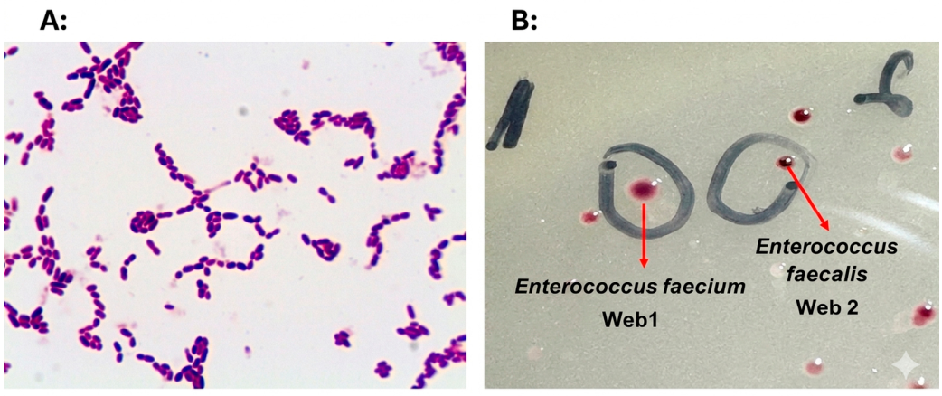
A total of 42 isolates were isolated based on the typical colony morphology of enterococci formed on SB media. Generally, E. faecalis and E. faecium exhibited species-specific differences in TTC (2,3,5-triphenyltetrazolium chloride) reduction, with E. faecalis showing a greater reducing capacity, resulting in the formation of colonies with a more intense red-brown coloration, whereas E. faecium typically produced colonies with less pronounced pigmentation (Figure 1). They showed typical micromorphology-Gram-positive ovoid cells.
All isolates (n = 42) were subjected to MALDI-TOF analysis. Of them, 34 isolates were successfully identified as members of the genus Enterococcus (n = 11 from Sofia, n = 9 from Burgas, n = 14 from Varna). However, the remaining eight isolates failed species identification and were excluded from the analysis. All Enterococcus isolates and their identification are shown in Table 2. As can be seen from the table, all isolates had an ID score above 9, except one isolate (Wev 7), showing an ID score of 8.383. Among the identified enterococcal isolates, E. faecium was the most prevalent species, representing 62% (n = 21, including Wev 7) of all enterococcal isolates, while E. faecalis accounted for 38% (n = 13) of the identified species.
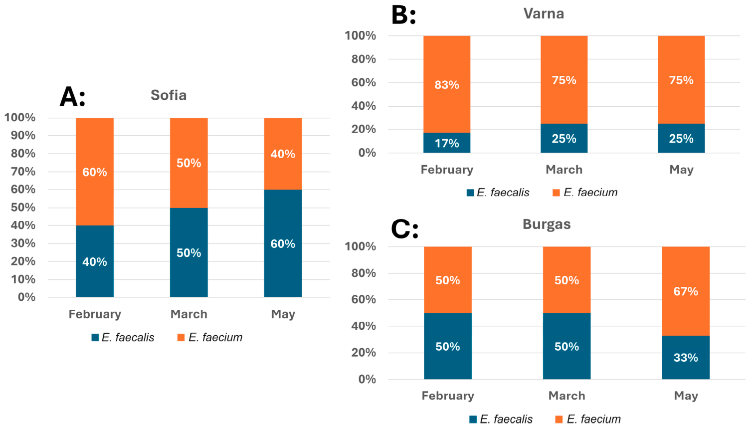
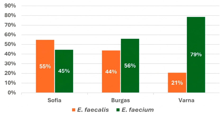
In the samples from Sofia and Burgas, E. faecalis and E. faecium were overall equally distributed (55%/45% and 44%/56%, respectively) (Figure 2). In Varna, however, E. faecium isolates predominated, accounting for 79% of all isolates, compared to 21% for E. faecalis.
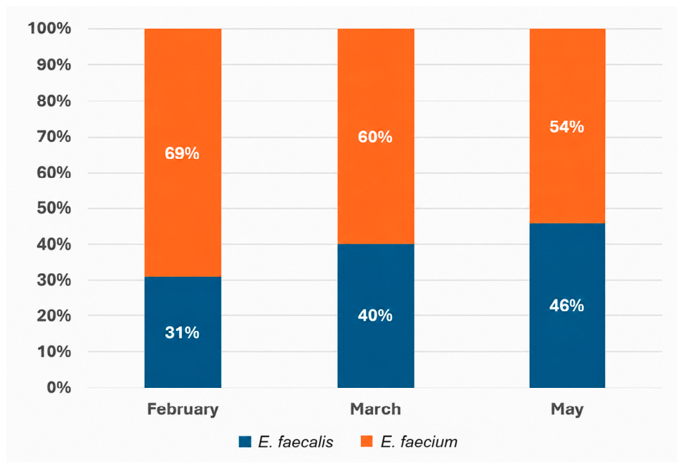
Interestingly, an increase in the percentage of E. faecalis isolates was observed among the strains collected during the selected sampling months (from 31% in February to 46% in May) (Figure 3).
The overall analysis of the species distribution in the influent wastewater across the selected cities showed a slight increase in the number of E. faecalis in the cities of Varna and Sofia. However, in Burgas, the percentage of identified E. faecalis and E. faecium is equal in February and March, whereas the percentage of E. faecalis isolates decreases in May (Figure 4).

3.2. Antibiotic Resistance

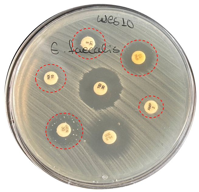
All 34 enterococcal isolates were subjected to phenotypic antibiotic susceptibility testing, using clinically relevant antibiotics. The obtained results were interpreted according to EUCAST (2025) criteria [14]. All isolates were susceptible to linezolid and teicoplanin, and the majority of them (97%, n = 33) were resistant to ampicillin (except E. faecium Wes1). All E. faecalis isolates were susceptible to nitrofurantoin. Resistance to quinupristin-dalfopristin was detected in 81% (n = 17) of all E. faecium strains. All E. faecalis isolates showed intermediate susceptibility to imipenem except one, E. faecalis Wes10, which was found to be resistant (Figure 5).
Resistance to the tested tetracyclines (eravacycline and tigecycline) was observed in 15% (n = 5) of all strains. Three of them (E. faecium Wes8, Wev6, and Wev10) exhibited resistance to both antibiotics, whereas the remaining two strains (E. faecium Wev3 and Wev9) were resistant only to eravacycline. Four isolates (12%) showed a suspected vancomycin-resistant phenotype based on fuzzy edges of inhibition zones in the disk diffusion assay. Resistance to fluoroquinolones (levofloxacin and norfloxacin) was found in 6% (n = 2; E. faecalis Wes10 and E. faecium Wev9) of all analyzed strains (Figure 5). High-level aminoglycoside resistance (HLAR) was detected in 27% (n = 9) of the strains. Resistance to both of the antibiotics (streptomycin and gentamicin) was determined only in strain E. faecium Wev9. Three strains showed resistance only to gentamycin, and five strains were resistant only to streptomycin.
Based on these results, 27% (n = 9) of the strains were considered multidrug-resistant (MDR) (Table 3). Interestingly, all the MDR strains were isolated during the period February—March. Approximately half of these strains (n = 5) are isolated from wastewater samples from Varna, three from Sofia, and one from Burgas. The majority of these strains (89%, n = 8) were identified as E. faecium and only one as E. faecalis (Wes10).
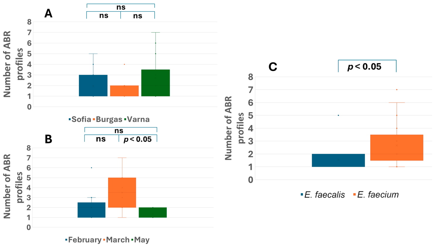
Statistical analysis of the number of antibiotic-resistant profiles between the isolates from different cities showed no significant differences (p > 0.05) (Figure 6A). The average antibiotic-resistant profiles varied from 2 to 3 in the cities (n = 2 in Sofia and Burgas, and n = 3 in Varna).
The comparison of antibiotic-resistance profiles across sampling periods revealed a statistically significant difference among the three sampling months, as determined by the Kruskal–Wallis test (p = 0.0318). In March, the highest number of antibiotic-resistance profiles per isolate was observed, whereas February and May exhibited lower averages. Pairwise Mann–Whitney U tests with Bonferroni correction showed a significant difference only between March and May (corrected p = 0.0365), while the differences between February and March (corrected p = 0.3444) and February and May (corrected p = 0.7351) were not statistically significant (Figure 6). The comparison of the antibiotic resistance profiles between E. faecalis and E. faecium isolates showed significant differences (p < 0.05), with an average of three antibiotic resistance profiles in E. faecium isolates and an average of 2 antibiotic resistance profiles in E. faecalis (Figure 6C).
The genetic determinants of the observed phenotypic antibiotic-resistance profiles were also investigated. The PCR screening revealed no amplification for the mefA (encodes macrolide efflux pump), vanA (encodes high-level vancomycin resistance), blaZ, blages, blaNDM1, and blaSHV (β-lactamases encoding penicillin resistance) genes in any of the analyzed strains. As mentioned above, phenotypic ampicillin resistance was detected in 97% of all analyzed strains. Genes encoding β-lactamases (blatem and blactx-m) were amplified in 38% (n = 13) of them. Positive amplifications for blatem were detected in 26% (n = 9) of all analyzed strains, whereas for blactx-m in 15% (n = 5). Interestingly, the blactx-m gene was detected only in E. faecalis strains (Wes 3, Wes 11, Wes 13, Wes 14, and Web 5). The blatem, however, was distributed both in E. faecalis (Wes 3 and Web 11) and E. faecium (Wes 2, Wes 8, Web 9, Wev 4, Wev 7, Wev 9 and Wev 14) isolates. Even though 81% of all E. faecium strains showed phenotypic quinupristin-dalfopristin resistance, ermB presence was determined in only 33% (n = 7) of all E. faecium isolates. The presence of the mefA gene was also not confirmed in them. More interestingly, some of the strains that amplified the ermB gene (E. faecium Wes 12 and Wev 13) showed phenotypic quinupristin-dalfopristin susceptibility. Overall, the ermB gene was detected in 38% (n = 13) of all analyzed strains. Several strains (n = 7; 19%) (E. faecalis Wes 3, Wes 10, Web 8, and E. faecium Wes 2, Wev 2, Wev 9 and Wev 14) gave amplification only for genes determining aminoglycoside resistance (aph(3′)-IIIa and/or aac(6′)–aph(2″)). All of them amplified the gene aph(3′)-IIIa (aphA-3), which encodes aminoglycoside 3′-phosphotransferase and is commonly present in strains resistant to kanamycin, neomycin, and amikacin. All but one (E. faecium Wev 8) HLGR enterococci (E. faecalis Wes 10, E. faecalis Web 8, and E. faecium Wev 9) amplified the aac(6′)–aph(2″) gene.
Additional sets of primers were used to further explain phenotypic antibiotic resistance to HLRS (Table 4) and eravacycline and tigecycline (tetracyclines) resistance (Table 5). These analyses were carried out only with the strains showing phenotypic antibiotic resistance to these antibiotics. None of the analyzed strains amplified the genes strA and strB. The genes ant (6)-la and aadA (ant(3″) were amplified in respectively 3 isolates (E. faecalis Wes 3 and E. faecium Wes 2, and Wev 9) and 3 E. faecium isolates (Wes 2, Wev 6, and Web 7) of the analyzed strains.
Phenotypes resistant to eravacycline and tigecycline (tetracyclines) were subjected to PCR with three additional sets of primers (for the genes tetM, tetL, and tetS). None of the strains showing phenotypic resistance to tetracyclines gave a positive amplification product for the tetL and tetS genes. Eighty percent (n = 4) of all strains resistant to tetracyclines amplified the tetM gene (Table 5).

3.3. Virulence-Associated Profiles

All enterococcal isolates were tested for phenotypic hemolytic activity on blood agar and for nuclease and gelatinase activities. None of them had DNAase activity, although four isolates (E. faecalis Wes 10, Web 5, Web 8 and Wev17) showed positive PCR amplification for the nuc1 gene. Phenotypic gelatinase activity was observed in 21% (n = 7) of all analyzed strains, identified as E. faecalis (Wes 13, Web 2, Web 5, Web 11, Wev 1, Wev 11, and Wev 17). All of them also amplified the gelE gene. On the other hand, two strains (Wes 3 and Wes 11) were found to possess the gelE gene (as determined by molecular analyses) but did not exhibit phenotypic gelatinase activity. None of the strains exhibited phenotypic β-hemolysis on blood agar (both aerobically and anaerobically), although 2 strains (E. faecalis Wes 10 and Web 8; 6%) amplified the cylB gene fragment. All strains exhibited phenotypic γ-hemolytic profiles.
Overall, 44% (n = 15) of all analyzed strains amplified at least one of the genes encoding virulence factors tested in this study (Table 6). Most of them (n = 12; 80%) were identified as E. faecalis. Five of these strains (38%) were found to possess between four and seven virulence-associated genes, suggesting their high virulence potential. Generally, only 20% (n = 3) of all strains, identified as E. faecium, generated positive amplification for single virulence-associated genes. Interestingly, 56% (n = 19) of all the strains did not amplify any of the analyzed genes for virulence factors. Simultaneously, the E. faecalis strain Web 8 was considered to have the greatest virulence potential, harboring seven of the eight tested virulence-associated genes. The most frequently amplified virulence gene was ace (encoding an adhesin for host tissues), found in 29% of all analyzed strains (n = 10). Next was the gene encoding gelatinase (27%, n = 9), followed by the genes esp and agg (15%, n = 5), psa and nuc1 (12%; n = 4), and the least amplified were the genes gls24 and cylB (6%; n = 2).
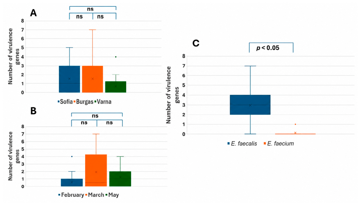
Statistical analysis of the number of virulence-associated genes per isolate showed no significant differences among the cities of Sofia, Burgas and Varna (Kruskal–Wallis test, p = 0.4927) or among the sampling months February, March and May (Kruskal–Wallis test, p = 0.2488). In contrast, a significant difference was detected between E. faecalis and E. faecium isolates (Mann–Whitney U test, p = 0.0093), confirming the higher number of virulence-associated genes in E. faecalis (Figure 7).

4. Discussion

This study aimed to contribute to the limited research specifically addressing influent wastewater as a key reservoir for the accumulation and dissemination of antibiotic resistance and virulence-associated genes in Bulgaria, focusing specifically on the participation of the enterococcal population in this process. The obtained results showed differences among the selected sampling months in the number of enterococci, with higher average values observed in March and May. According to some authors, a possible explanation for this increase in enterococcal numbers could be attributed to rising temperatures during spring (up to 10 °C) or to wastewater flow [30]. Despite continued temperature rises from March to May, the total enterococcal count remains relatively stable in our study. One possible reason for the leveling off in enterococcal count could be the increased UV index in May, although other factors may also play a role (seasonal changes, close monitoring of wastewater and treatment processes).
Our results indicate a significant variation in antibiotic-resistance profiles across the sampling months, with the highest number of profiles observed in March. However, pairwise post-hoc analysis showed a statistically significant difference only between March and May, possibly reflected by the complex influence of environmental and human factors. Similar observations are reported by other researchers, where the highest number of resistant enterococci was found also in March [30].
Isolates of E. faecalis and E. faecium, in the influent wastewater of Sofia and Burgas, were represented in equivalent amounts, while in Varna, E. faecium isolates constituted 79% of all identified species. Likewise, the increased antibiotic resistance of E. faecium strains may explain the high percentage of MDR strains isolated from samples from this city.
The higher antibiotic resistance patterns of the E. faecium species are also confirmed in this study, as the statistics show a significantly higher number of antibiotic resistance profiles in this species. Other authors also describe E. faecium as the most resistant enterococcal species in the genus Enterococcus [1,5]. Moreover, 89% of all MDR strains are identified as E. faecium.
The antibiotic resistance patterns of the enterococcal isolates in our study demonstrate resistance to ampicillin (2 µg/disc) in almost all enterococcal isolates (n = 33, 97%). Some enterococci possess intrinsic resistance to β-lactam antibiotics (e.g., ampicillin) due to low-affinity penicillin-binding proteins (PBPs) or β-lactamase production [31]. Although blaZ, blages, blaNDM1 and blaSHV genes (encoding β-lactamase) were not detected in the analyzed strains in our study, several strains (38%) were positive for the presence of blatem and blactx-m genes. According to the European Antimicrobial Resistance Surveillance Network (EARS-Net), all analyzed E. faecium isolates collected from blood and cerebrospinal fluid samples in Bulgaria in 2024 were resistant to ampicillin [9]. The same EARS-Net data showed that none of the analyzed E. faecalis isolates were resistant to ampicillin. Although our results do not correlate directly with the measured clinically relevant resistance in E. faecalis, some authors describe more than 60% resistant to ampicillin E. faecalis isolates from dairy cattle farms [32]. Same authors also describe that half (50%) of their tested isolates were positive for the presence of the blatem gene [32]. The same gene was also detected in enterococci from the Bulgarian river Yantra, although some did not exhibit the corresponding phenotypic resistance [33]. Sulaiman et al. (2023) also detected bla genes (blactx-m), although in E. faecalis isolates from children with bacteremia [34]. However, they link this genetic determinant to cefotaxime and ceftriaxone resistance. Overall, the broad ampicillin resistance in our study could be attributed to the previously described low-affinity PBPs or genes encoding β-lactamases. The difference between our findings and the EARS-Net 2024 report probably reflects the mixed nature of our isolates—from wastewaters (combining clinical, veterinary, and environmental flow). Prolonged exposure to sub-inhibitory concentrations of β-lactams and other selective agents in wastewater could potentially contribute to the persistence of strains carrying altered penicillin-binding proteins or, more rarely, β-lactamase production. Furthermore, the high bacterial abundance in wastewater environments may create conditions that could favor genetic exchange between different bacterial species and/or strains.
In our study, high-level resistance to aminoglycoside antibiotics (both gentamycin and streptomycin) was observed in 27% of all analyzed strains, with HLGR strains representing 9% of them. These results confirmed the enterococcal resistance pattern observed in Bulgaria, consistent with a 2025 study analyzing untreated and treated wastewater samples in the country, which reported that approximately 10% of all analyzed enterococcal strains were HLGR [11]. Most importantly, the authors determine a significant decrease in those strains after treatment of the wastewaters [11]. In our work, the presence of phenotypic HLGR profiles strongly correlates with the presence of the gene aac(6′)–aph(2″) (present in all but one HLGR strains). High prevalence of this gene in the wastewater enterococci was also described by Tsvetanova & Bushnakov (2025) [11]. Regarding the HLSR strains, all but one (E. faecalis Wev 11) amplified the ant (6)-la and/or aadA (ant(3″)) genes. These results correlate with those reported by other authors, analyzing hospital wastewaters, who detect these two genes in respectively 97% and 24% of all HLSR isolates [35].
Vancomycin-resistant enterococci (VRE) are considered a serious threat due to their association with high mortality rates and limited therapeutic options [36,37]. In our study, only 12% of all isolates showed susceptive phenotypic resistance patterns to vancomycin (fuzzy edges of the inhibition zones). However, none of these strains amplified the vanA gene. It should be noted that, besides vanA, eight additional van operons (vanB, vanC, vanD, vanE, vanG, vanL, vanM, and vanN) associated with vancomycin resistance have been reported in enterococci [38]. The transcription of these operons determines a variety of phenotypic resistance in enterococci. The vanA operon determines a high degree of resistance to both vancomycin and teicoplanin. On the other hand, vanB strains exhibit high-level resistance exclusively to vancomycin. The vanC resistance type confers low resistance only to vancomycin. Strains containing vanE and vanG demonstrate only intermediate-level resistance to vancomycin. The vanM is genetically and phenotypically similar to vanA, vanB, and vanD, whereas both vanL and vanN are similar to vanC [38,39]. The present study focused exclusively on the detection of vanA, while the occurrence of other van determinants remains a subject of ongoing and future investigations. While antibiotics like linezolid, daptomycin, tigecycline, and quinupristin/dalfopristin (Q/D) are available for vancomycin-resistant enterococcal infections, their effectiveness is often compromised by the emergence of resistance [40,41].
E. faecalis is considered to have intrinsic resistance to quinupristin-dapfopristin (a combination of streptogramin A and B) [42]. A large portion of the E. faecium strains (81%), however, were also resistant to quinupristin-dapfopristin in our study. In these strains mefA (encodes macrolide efflux pump) gene was not detected. However, some of the strains possessing the ermB gene (E. faecium Wes 12 and Wev 13) showed phenotypic quinupristin-dalfopristin susceptibility. These results indicate that possibly this gene is not the major determinant for quinupristin-dapfopristin resistance in the analyzed strains. A study from Greece, analyzing clinically relevant isolates of E. faecium for the period between 2005–2006, determined a reduced susceptibility to quinupristin-dalfopristin in 28.9% of all analyzed strains, without prior exposure to the agent [43]. Wastewaters, however, collect strains from diverse origins, not only clinically relevant strains. Virginiamycin, a combination of streptogramin A and B, has been used as a growth promoter in animal feed for many years. As a consequence, this selective pressure resulted in the emergence of virginiamycin-resistant strains of E. faecium, which are cross-resistant to quinupristin-dalfopristin [44,45]. Based on these observations, the use of virginiamycin has been banned in the European Union. The potential dissemination of enterococcal strains of animal origin in the wastewater could be a possible reason for the wide quinupristin-dalfopristin resistance observed in our study. Furthermore, Soltani et al. (2000) describe wide dissemination of vat(E) genes in their quinupristin-dalfopristin-resistant strains of E. faecium, isolated from hospital patients and nonhuman sources in European countries [44]. Moreover, they confirmed the location of this gene on a plasmid [44], which further confirms the possibility of its facilitated genetic transfer.
Fifteen percent of all analyzed strains were resistant to eravacycline and/or tigecycline. In almost all of them (except one—E. faecium Wes 8), the gene tetM was amplified, associated in the literature as encoding resistance to tetracyclines (in which group are also eravacycline and tigecycline) [46]. On the other hand, as described in previous work of our research team, this gene is widely distributed amongst the enterococcal populations in Bulgaria [47], and the phenotypic resistance to these two antibiotics could also be attributed to other genetic determinants.
All analyzed isolates in this study were found to be susceptible to linezolid and teicoplanin, indicating that these antibiotics remain effective therapeutic options. All E. faecalis isolates were also susceptible to nitrofurantoin. These results are consistent with the observations reported by Zaheer et al. (2020) [48], who identified little to no nitrofurantoin resistance among their E. faecalis isolates.
This work reports that 44% (n = 15) of the analyzed strains harbour multiple virulence genes, with significantly more virulence genes found in E. faecalis isolates. Although the percentage of detected strains with virulence genes were significantly lower compared to other authors, our results indicate more highly virulent strains of E. faecalis compared to the data reported by Adegoke et al. [1]. In our study, the most amplified genes were ace (29%, n = 10) and gelE (27%, n = 9). Adegoke et al. detected these genes in respectively 18% and 13.6% of all their analyzed strains [1]. The gene esp was also detected at a higher frequency across all analyzed strains (15%), whereas Adegoke et al. found it in 4.4% of their strains [1]. Moreover, they describe E. faecalis strains as strongly associated with the gelE gene, as observed in our results as well. We also detected the expression of phenotypic gelatinase activity in most of the strains where gelE was detected (seven out of 9 isolates PCR positive). The expression of the gelE gene is known to be controlled by the fsr operon and the population density of the enterococcal population [49]. However, the fsr operon appears to be easily lost or deleted [49]. Although we have no data on the presence of the full fsr genes in these two isolates at that time, we can hypothesize that the lack of phenotypic gelatinase activity in them could be due to deletions in the fsr operon. The production of cytolysin in enterococci is associated with induced septicemia and a fivefold increased risk of acutely terminal outcome in patients [50]. In this study, none of the isolates had β-hemolytic activity, although two of the strains amplified the cylB gene. Adegoke et al. (2025) also detected inconsistencies with the presence of this gene and the phenotypic expression of hemolysin [1]. These authors describe the poor distribution of cyl genes in their work (only one isolate had the gene), as was also seen in our results (only two strains amplified the correct fragment for the cyl gene).
Considering this, E. faecalis can be regarded as a pathogen of critical importance (as seen also in this work), and given its increasing resistance to multiple antibiotics, non-antibiotic therapeutic strategies, such as phage-based solutions, need to be developed in the future. This conclusion was further supported by statistical analysis, which showed a significantly higher number of virulence-associated genes in E. faecalis than in E. faecium isolates (Mann–Whitney U test, p = 0.0093). However, effective phage therapy against this pathogen depends on the isolation and characterization of diverse phages and the exploration of their therapeutic potential. For that reason, our research team successfully isolated and characterized several bacteriophages, targeting different E. faecalis strains [51]. Moreover, some of these phages are able to lyse three of the five highly virulent E. faecalis strains, described in this work (E. faecalis Wes 3, Wes 10 and Wev 17).
Sofia, Burgas and Varna are the three major cities in Bulgaria with big populations (with total population of 1,295,931 in Sofia, 388,919 in Burgas and 437,521 in Varna, reported by the National Statistical Institute of Bulgaria, 2024) [52]. These cities are not only big in population, but they also attract large tourist waves from around the world. The geographical location of the cities of Varna and Burgas further amplifies the importance of antimicrobial surveillance of wastewaters, as they are located on the Black Sea coast.

5. Conclusions

It can be concluded that wastewater inflow may be considered as a reservoir for antibiotic-resistant enterococci. The detection of resistant E. faecalis and E. faecium isolates in influent wastewater from the three largest Bulgarian cities supports the relevance of this matrix for monitoring of antibiotic-resistant enterococcal isolates. In addition, the high levels of antibiotic resistance observed in some isolates represent a potential risk to both the environment and public health in the event of wastewater treatment failure. This study also highlights species-specific patterns of antibiotic resistance and virulence potential, with E. faecium identified as the predominant antibiotic-resistant species, while E. faecalis was characterized by a greater number of virulence-associated genes. The predominance of multidrug-resistant profiles, mainly among E. faecium isolates, underscores the importance of this species as a potential indicator of resistance dissemination in wastewater, whereas the higher virulence-associated gene burden in E. faecalis suggests its relevance for assessing pathogenic potential. Consequently, regular surveillance is essential to enable timely and effective mitigation measures. Such monitoring could provide valuable information for public health authorities, wastewater management systems, and One Health strategies aimed at limiting the environmental spread of antibiotic-resistant and potentially pathogenic enterococci. However, larger-scale studies including more isolates, continuous sampling, and seasonally representative time points are needed to confirm long-term temporal and geographic patterns.

Supplementary Materials

The following supporting information can be downloaded at: https://www.mdpi.com/article/10.3390/microbiolres17070132/s1, Table S1: Primer pairs used for the detection of virulence and antibiotic resistance genes.

Author Contributions

Conceptualization: M.P., Y.K. and P.H.; Methodology: M.P., Y.K. and P.H.; Software: M.P.; Validation: Y.K. and P.H.; formal analysis: R.P. and M.P.; investigation: M.P. and R.P.; resources: Y.K., S.I. and P.H.; writing—original draft preparation: M.P.; writing—review and editing: Y.K. and P.H.; visualization: M.P.; supervision: P.H.; project administration: S.I.; funding acquisition: S.I. All authors have read and agreed to the published version of the manuscript.

Funding

This research was funded by the European Union-Next Generation EU, through the National Recovery and Resilience Plan of the Republic of Bulgaria, project No. BG-RRP-2.004-0008, scientific group 3.2.4.

Institutional Review Board Statement

Not applicable.

Informed Consent Statement

Not applicable.

Data Availability Statement

The original contributions presented in this study are included in the article/Supplementary Material. Further inquiries can be directed to the corresponding author.

Conflicts of Interest

The authors declare no conflicts of interest.

References

  1. Adegoke, A.A.; Madu, C.E.; Reddy, P.; Fatunla, O.K.; Stenström, T.A.; Okoh, A.I. Virulence and Antibiotic Resistance Genes in Enterococcus from Wastewater for Reuse and Their Health Impact. Microorganisms 2025, 13, 1045. [Google Scholar] [CrossRef] [PubMed]
  2. European Parliament; Council of the European Union. Directive (EU) 2024/3019 of the European Parliament and of the Council of 27 November 2024 concerning urban wastewater treatment (recast). Off. J. Eur. Union 2024, L series 2024/3019. Available online: https://eur-lex.europa.eu/eli/dir/2024/3019/oj (accessed on 4 March 2026).
  3. Pandova, M.; Kizheva, Y.; Tsenova, M.; Rusinova, M.; Borisova, T.; Hristova, P. Pathogenic Potential and Antibiotic Susibility: A Comprehensive Study of Enterococci from Different Ecological Settings. Pathogens 2024, 13, 36. [Google Scholar] [CrossRef] [PubMed]
  4. Iweriebor, B.C.; Obi, L.C.; Okoh, A.I. Virulence and antimicrobial resistance factors of Enterococcus spp. isolated from fecal samples from piggery farms in Eastern Cape, South Africa. BMC Microbiol. 2015, 15, 136. [Google Scholar] [CrossRef] [PubMed]
  5. Fisher, K.; Phillips, C. The ecology, epidemiology and virulence of Enterococcus. Microbiology 2009, 155, 1749–1757. [Google Scholar] [CrossRef] [PubMed]
  6. Hegstad, K.; Mikalsen, T.; Coque, T.; Werner, G.; Sundsfjord, A. Mobile genetic elements and their contribution to the emergence of antimicrobial resistant Enterococcus faecalis and Enterococcus faecium. Clin. Microbiol. Infect. 2010, 16, 541–554. [Google Scholar] [CrossRef] [PubMed]
  7. Miller, W.R.; Arias, C.A. ESKAPE pathogens: Antimicrobial resistance, epidemiology, clinical impact and therapeutics. Nat. Rev. Microbiol. 2024, 22, 598–616. [Google Scholar] [CrossRef] [PubMed]
  8. Conwell, M.; Daniels, V.; Naughton, P.J.; Dooley, J.S. Interspecies transfer of vancomycin, erythromycin and tetracycline resistance among Enterococcus species recovered from agrarian sources. BMC Microbiol. 2017, 17, 19. [Google Scholar] [CrossRef] [PubMed]
  9. European Centre for Disease Prevention and Control. European Antimicrobial Resistance Surveillance Network (EARS-Net). Available online: https://www.ecdc.europa.eu/en/about-us/networks/disease-networks-and-laboratory-networks/ears-net-data (accessed on 15 July 2025).
  10. Gotkowska-Płachta, A. The Prevalence of Virulent and Multidrug-Resistant Enterococci in River Water and in Treated and Untreated Municipal and Hospital Wastewater. Int. J. Environ. Res. Public Health 2021, 18, 563. [Google Scholar] [CrossRef] [PubMed]
  11. Tsvetanova, Z.; Boshnakov, R. Antimicrobial Resistance of Waste Water Microbiome in an Urban Waste Water Treatment Plant. Water 2025, 17, 39. [Google Scholar] [CrossRef]
  12. Tsvetanova, Z.; Najdenski, H. Antimicrobial Resistance of Heterotrophic Bacteria and Enterobacteriaceae Inhabiting an Anthropogenic-Affected River Stretch in Bulgaria. Processes 2023, 11, 2792. [Google Scholar] [CrossRef]
  13. Donchev, D.; Ivanov, I.N.; Stoikov, I.; Ivanova, M. Metagenomic Investigation of the Short-Term Temporal and Spatial Dynamics of the Bacterial Microbiome and the Resistome Downstream of a Wastewater Treatment Plant in the Iskar River in Bulgaria. Microorganisms 2024, 12, 1250. [Google Scholar] [CrossRef] [PubMed]
  14. The European Committee on Antimicrobial Susceptibility Testing. Breakpoint Tables for Interpretation of MICs and Zone Diameters, Version 15.0; EUCAST: Växjö, Sweden, 2025; Available online: https://www.eucast.org (accessed on 15 July 2025).
  15. Bauer, A.W.; Kirby, W.M.M.; Sherris, J.C.; Turck, M. Antibiotic susceptibility testing by a standard single disk method. Am. J. Clin. Pathol. 1966, 45, 493–496. [Google Scholar] [CrossRef]
  16. Magiorakos, A.P.; Srinivasan, A.; Carey, R.B.; Carmeli, Y.; Falagas, M.E.; Giske, C.G.; Harbarthg, S.; Hindlerh, J.F.; Kahlmeteri, G.; Olsson-Liljequist, B.; et al. Multidrug-resistant, extensively drug-resistant and pandrug-resistant bacteria: An international expert proposal for interim standard definitions for acquired resistance. Clin. Microbiol. Infect. 2012, 18, 268–281. [Google Scholar] [CrossRef] [PubMed]
  17. Batish, V.K.; Chander, H.; Ranganathan, B. Characterization of deoxyribonuclease-positive enterococci isolated from milk and milk products. J. Food Prot. 1982, 45, 348–352. [Google Scholar] [CrossRef] [PubMed]
  18. McBride, S.M.; Fischetti, V.A.; LeBlanc, D.J.; Moellering, R.C.; Gilmore, M.S. Genetic diversity among Enterococcus faecalis. PLoS ONE 2007, 2, e582. [Google Scholar] [CrossRef] [PubMed]
  19. van Asselt, G.J.; Vliegenthart, J.S.; Petit, P.L.C.; van de Klundert, J.A.M.; Mouton, R.P. High-level aminoglycoside resistance among enterococci and group A streptococci. J. Antimicrob. Chemother. 1992, 30, 651–659. [Google Scholar] [CrossRef] [PubMed]
  20. Garofalo, C.; Vignaroli, C.; Zandri, G.; Aquilanti, L.; Bordoni, D.; Osimani, A.; Clementi, F.; Biavasco, F. Direct detection of antibiotic resistance genes in specimens of chicken and pork meat. Int. J. Food Microbiol. 2007, 113, 75–83. [Google Scholar] [CrossRef] [PubMed]
  21. Jia, W.; Li, G.; Wang, W. Prevalence and Antimicrobial Resistance of Enterococcus Species: A Hospital-Based Study in China. Int. J. Environ. Res. Public Health 2014, 11, 3424–3442. [Google Scholar] [CrossRef] [PubMed]
  22. Jensen, L.B.; Frimodt-Møller, N.; Aarestrup, F.M. Presence of erm gene classes in gram-positive bacteria of animal and human origin in Denmark. FEMS Microbiol. Lett. 1999, 170, 151–158. [Google Scholar] [CrossRef] [PubMed]
  23. Liu, C.; Zhang, Z.Y.; Dong, K.; Yuan, J.P.; Guo, X.K. Antibiotic resistance of probiotic strains of lactic acid bacteria isolated from marketed foods and drugs. Biomed. Environ. Sci. 2009, 22, 401–412. [Google Scholar] [CrossRef] [PubMed]
  24. Wang, Y.; Dong, J.; Wang, J.; Chi, W.; Zhou, W.; Tian, Q.; Hong, Y.; Zhou, X.; Ye, H.; Tian, X.; et al. Assessing the drug resistance profiles of oral probiotic lozenges. J. Oral Microbiol. 2022, 14, 2019992. [Google Scholar] [CrossRef] [PubMed]
  25. Eaton, T.J.; Gasson, M.J. Molecular Screening of Enterococcus virulence determinants and potential for genetic exchange between food and medical isolates. Appl. Environ. Microbiol. 2001, 67, 1628–1635. [Google Scholar] [CrossRef] [PubMed]
  26. Mannu, L.; Paba, A.; Daga, E.; Comunian, R.; Zanetti, S.; Duprè, I.; Sechi, L.A. Comparison of the incidence of virulence determinants and antibiotic resistance between Enterococcus faecium strains of dairy, animal and clinical origin. Int. J. Food Microbiol. 2003, 88, 291–304. [Google Scholar] [CrossRef] [PubMed]
  27. Mbyemeire, H.; Ssekatawa, K.; Kato, C.D.; Wampande, E.M. Molecular characterization and distribution of cephalosporin resistance determinants in Escherichia coli and Klebsiella pneumoniae isolated from patients attending Kampala International University Teaching Hospital in Bushenyi, Western Uganda. Alex. J. Med. 2021, 57, 205–214. [Google Scholar] [CrossRef]
  28. Lapierre, L.; Cornejo, J.; Zavala, S.; Galarce, N.; Sánchez, F.; Benavides, M.B.; Guzmán, M.; Sáenz, L. Phenotypic and genotypic characterization of virulence factors and susceptibility to antibiotics in Salmonella Infantis strains isolated from chicken meat: First findings in Chile. Animals 2020, 10, 1049. [Google Scholar] [CrossRef] [PubMed]
  29. Tiemtoré, R.Y.W.; Dabiré, A.M.; Ouermi, D.; Sougué, S.; Benao, S.; Simporé, J. Isolation and identification of Escherichia coli and Klebsiella pneumoniae strains resistant to the oxyimino-cephalosporins and the monobactam by production of GES type extended spectrum beta-lactamase (ESBL) at Saint Camille Hospital Center in Ouagadougou, Burkina Faso. Infect. Drug Resist. 2022, 15, 3191–3204. [Google Scholar] [CrossRef] [PubMed]
  30. Lépesová, K.; Olejníková, P.; Mackuľak, T.; Tichý, J.; Birošová, L. Annual changes in the occurrence of antibiotic-resistant coliform bacteria and enterococci in municipal wastewater. Environ. Sci. Pollut. Res. 2019, 26, 18470–18483. [Google Scholar] [CrossRef] [PubMed]
  31. Martin, J.F.; Alvarez-Alvarez, R.; Liras, P. Penicillin-binding proteins, β-lactamases, and β-lactamase inhibitors in β-lactam-producing actinobacteria: Self-resistance mechanisms. Int. J. Mol. Sci. 2022, 23, 5662. [Google Scholar] [CrossRef] [PubMed]
  32. Fahim, N.A.I.; Rana, M.L.; Hasan, M.A.E.; Salam, S.; Masud, R.I.; Huda, N.; Saha, S.; Rahman, M.T. Biofilms and antibiotic resistance profile of Enterococcus faecalis in selected dairy cattle farm environments in Bangladesh. PLoS ONE 2025, 20, e0323667. [Google Scholar] [CrossRef] [PubMed]
  33. Tsvetanova, Z.; Boshnakov, R.; Kim, T.C.; Najdenski, H. Prevalence of Antibiotic-Resistant Enterococci in an Anthropogenic-Affected River Section. Water 2026, 18, 1149. [Google Scholar] [CrossRef]
  34. García-Solache, M.; Rice, L.B. The Enterococcus: A model of adaptability to its environment. Clin. Microbiol. Rev. 2019, 32, 10–1128. [Google Scholar] [CrossRef] [PubMed]
  35. Jannati, E.; Khademi, F.; Manouchehrifar, M.; Maleki, D.; Amirmozaffari, N.; Sadat Nikbin, V.; Arzanlou, M. Antibiotic resistance and virulence potentials of E. faecalis and E. faecium in hospital wastewater: A case study in Ardabil, Iran. J. Water Health 2023, 21, 1277–1290. [Google Scholar] [CrossRef] [PubMed]
  36. Maleki, D.; Manouchehrifar, M.; Kheljan, M.N.; Mossavi, S.H.; Jannati, E.; Doghaheh, H.P.; Teimourpour, R.; Khademi, F.; Arzanlou, M. Vancomycin-resistant Enterococcus species: Antimicrobial resistance and virulence genes profile. Gene Rep. 2021, 25, 101338. [Google Scholar] [CrossRef]
  37. Eichel, V.M.; Last, K.; Brühwasser, C.; von Baum, H.; Dettenkofer, M.; Götting, T.; Grundmann, H.; Güldenhöven, H.; Liese, J.; Martin, M.; et al. Epidemiology and outcomes of vancomycin-resistant Enterococcus infections: A systematic review and meta-analysis. J. Hosp. Infect. 2023, 141, 119–128. [Google Scholar] [CrossRef] [PubMed]
  38. Mareković, I.; Markanović, M.; Lešin, J.; Ćorić, M. Vancomycin-Resistant Enterococci: Current understandings of resistance in relation to transmission and preventive strategies. Pathogens 2024, 13, 966. [Google Scholar] [CrossRef] [PubMed]
  39. Iqbal, F.; Alocious, A.; Joy, S.C.; Stanly, E.A.R.; Rajesh, C.; Unnikrishnan, M.K.; Steinke, D.; Chandra, P. Vancomycin-resistant enterococci: A rising challenge to global health. Clin. Epidemiol. Glob. Health 2024, 28, 101663. [Google Scholar] [CrossRef]
  40. Shariati, A.; Dadashi, M.; Chegini, Z.; van Belkum, A.; Mirzaii, M.; Khoramrooz, S.S.; Darban-Sarokhalil, D. The global prevalence of daptomycin, tigecycline, quinupristin/dalfopristin, and linezolid-resistant Staphylococcus aureus and coagulase-negative staphylococci strains: A systematic review and meta-analysis. Antimicrob. Resist. Infect. Control 2020, 9, 56. [Google Scholar] [CrossRef] [PubMed]
  41. So, M. Vancomycin-resistant Enterococcus in hematology-oncology patients: A review on colonization, screening, infections, resistance, and antimicrobial stewardship. Curr. Treat. Options Infect. Dis. 2020, 12, 285–295. [Google Scholar] [CrossRef]
  42. Carey, S.A.; Goldstein, R.E.R.; Gibbs, S.G.; Claye, E.; He, X.; Sapkota, A.R. Occurrence of vancomycin-resistant and -susceptible Enterococcus spp. in reclaimed water used for spray irrigation. Environ. Res. 2016, 147, 350–355. [Google Scholar] [CrossRef] [PubMed]
  43. Karanika, M.; Prati, A.; Kiritsi, M.; Spiliopoulou, I.; Neonakis, I.; Anifantaki, M.; Petinaki, E. Reduced susceptibility to quinupristin/dalfopristin in Enterococcus faecium in Greece without prior exposure to the agent. Int. J. Antimicrob. Agents 2008, 31, 55–57. [Google Scholar] [CrossRef] [PubMed]
  44. Soltani, M.; Beighton, D.; Philpott-Howard, J.; Woodford, N. Mechanisms of resistance to quinupristin-dalfopristin among isolates of Enterococcus faecium from animals, raw meat, and hospital patients in Western Europe. Antimicrob. Agents Chemother. 2000, 44, 433–436. [Google Scholar] [CrossRef] [PubMed]
  45. Werner, G.; Klare, I.; Witte, W. Association between quinupristin/dalfopristin resistance in glycopeptide-resistant Enterococcus faecium and the use of additives in animal feed. Eur. J. Clin. Microbiol. Infect. Dis. 1998, 17, 401–402. [Google Scholar] [CrossRef] [PubMed]
  46. Tsiklauri, R.; Kobakhidze, S.; Kotetishvili, M. Interactive networks of donors and recipients of the TetM gene and its evolutionary dynamics across the bacterial domain. Sci. Rep. 2025, 15, 35312. [Google Scholar] [CrossRef] [PubMed]
  47. Pandova, M.; Kizheva, Y.; Hristova, P. Relationship Between CRISPR–Cas Systems and Acquisition of Tetracycline Resistance in Non-Clinical Enterococcus Populations in Bulgaria. Antibiotics 2025, 14, 145. [Google Scholar] [CrossRef] [PubMed]
  48. Zaheer, R.; Cook, S.R.; Barbieri, R.; Goji, N.; Cameron, A.; Petkau, A.; Polo, R.O.; Tymensen, L.; Stamm, C.; Song, J.; et al. Surveillance of Enterococcus spp. reveals distinct species and antimicrobial resistance diversity across a One-Health continuum. Sci. Rep. Correction in Sci. Rep. 2020, 10, 13401. https://doi.org/10.1038/s41598-020-69044-5.. 2020, 10, 3937. [Google Scholar] [CrossRef] [PubMed]
  49. Lopes, M.; Simões, A.; Tenreiro, R.; Joaquim, J.; Marques, F.; Crespo, M. Activity and expression of a virulence factor, gelatinase, in dairy enterococci. Int. J. Food Microbiol. 2006, 112, 208–214. [Google Scholar] [CrossRef] [PubMed]
  50. Huycke, M.M.; Spiegel, C.A.; Gilmore, M.S. Bacteremia caused by hemolytic, high-level gentamicin-resistant Enterococcus faecalis. Antimicrob. Agents Chemother. 1991, 35, 1626–1634. [Google Scholar] [CrossRef] [PubMed]
  51. Kizheva, Y.K.; Dimova, T.; Pandova, M.; Gladicheva, Y.; Petrova, R.; Paunova-Krasteva, T.; Urshev, Z.; Ivanov, S.; Hristova, P. Characterization of wastewater-derived bacteriophages infecting Enterococcus faecalis in Bulgaria: Insights into the novel phage vB_SEF_8. Front. Microbiol. 2025, 16, 1674800. [Google Scholar] [CrossRef] [PubMed]
  52. National Statistical Institute (Bulgaria). Population by Districts, 2024; NSI: Sofia, Bulgaria, 2025; Available online: https://www.nsi.bg (accessed on 28 January 2026).
Figure 1. (A) Gram-staining of strain Enterococcus faecalis Wev11 and (B) colony morphology on SB media of strains Enterococcus faecium Web1 and Enterococcus faecalis Web2.
Microbiolres 17 00132 g001
Figure 2. Distribution of enterococcal species within the selected WWTP (sampling points).
Microbiolres 17 00132 g002
Figure 3. Distribution of enterococcal species by period of sampling.
Microbiolres 17 00132 g003
Figure 4. Enterococcal species distribution based on time sampling in (A) Sofia, (B) Varna and (C) Burgas.
Microbiolres 17 00132 g004
Figure 5. Phenotypic antibiotic resistance of the MDR strain E. faecalis Wes 10. The figure shows resistance to ampicillin (AMP), fluoroquinolones (levofloxacin (LE) and norfloxacin (NX)), high-level gentamycin (GEN) and imipenem (IPM), indicated by red circles.
Microbiolres 17 00132 g005
Figure 6. Boxplot of the distribution of the number of antibiotic resistance profiles between enterococcal isolates from different cities (A), different sampling months (B), and different enterococcal species (C). Significant differences are considered when p < 0.05; when p ≥ 0.05, non-significant differences (ns) are assigned.
Microbiolres 17 00132 g006
Figure 7. Boxplot of the distribution of virulence genes between enterococcal isolates from different cities (A), different sampling months (B), and different enterococcal species (C). Significant differences are considered when p < 0.05; when p ≥ 0.05 non-significant differences (ns) are assigned.
Microbiolres 17 00132 g007
Table 1. Total enterococcal count in wastewater samples, evaluated on SB media.
MonthSofia, CFU/mLBurgas, CFU/mLVarna, CFU/mLMean, CFU/mL
February3.6 ± 1.2 × 1031.1 ± 0.2 × 1043.0 ± 0.8 × 1035.9 ± 4.5 × 10³
March2.6 ± 0.9 × 1046.9 ± 1.5 × 1041.8 ± 1.3 × 1043.8 ± 2.8 × 10⁴
May1.3 ± 0.3 × 1045.3 ± 1.6 × 1047.8 ± 1.1 × 1044.8 ± 2.7 × 10⁴
Mean, CFU/mL1.4 ± 1.1 × 1044.4 ± 3.0 × 1043.3 ± 4.0 × 10⁴NA
NA—not applicable.
Table 2. Isolate Source, Sampling Time, and MALDI-TOF Identification Data.
Month of SamplingIsolateSpeciesCityID Score
1FebruaryWes 1Enterococcus faeciumSofia9.266
2FebruaryWes 2Enterococcus faeciumSofia9.528
3FebruaryWes 3Enterococcus faecalisSofia9.653
4FebruaryWes 4Enterococcus faecalisSofia9.528
5FebruaryWes 5Enterococcus faeciumSofia9.394
6MarchWes 8Enterococcus faeciumSofia9.535
7MarchWes10Enterococcus faecalisSofia9.648
8MayWes 11Enterococcus faecalisSofia9.508
9MayWes 12Enterococcus faeciumSofia9.085
10MayWes 13Enterococcus faecalisSofia9.058
11MayWes 14Enterococcus faecalisSofia9.339
12FebruaryWeb 1Enterococcus faeciumBurgas9.182
13FebruaryWeb 2Enterococcus faecalisBurgas9.457
14MarchWeb 4Enterococcus faeciumBurgas9.552
15MarchWeb 5Enterococcus faecalisBurgas9.67
16MarchWeb 7Enterococcus faeciumBurgas9.578
17MarchWeb 8Enterococcus faecalisBurgas9.692
18MayWeb 9Enterococcus faeciumBurgas9.067
19MayWeb 11Enterococcus faecalisBurgas9.232
20MayWeb 13Enterococcus faeciumBurgas9.138
21FebruaryWev 1Enterococcus faecalisVarna9.628
22FebruaryWev 2Enterococcus faeciumVarna9.398
23FebruaryWev 3Enterococcus faeciumVarna9.089
24FebruaryWev 4Enterococcus faeciumVarna9.247
25FebruaryWev 6Enterococcus faeciumVarna9.399
26FebruaryWev 7Enterococcus faeciumVarna8.383
27MarchWev 8Enterococcus faeciumVarna9.598
28MarchWev 9Enterococcus faeciumVarna9.571
29MarchWev 10Enterococcus faeciumVarna9.544
30MarchWev 11Enterococcus faecalisVarna9.646
31MayWev 13Enterococcus faeciumVarna9.563
32MayWev 14Enterococcus faeciumVarna9.206
33MayWev 15Enterococcus faeciumVarna9.551
34MayWev 17Enterococcus faecalisVarna9.543
Table 3. Multidrug-resistant phenotypes of the analyzed enterococcal strains.
MDR PhenotypeNumber of IsolatesSpecies IdentificationCityMonth
Three antibiotic groups
Amp, Hls, RpWes 2E. faeciumSofiaFebruary
Amp, Rp, Erv, TgcWes 8E. faeciumSofiaMarch
Amp, Rp, ErvWev 3E. faeciumVarnaFebruary
Amp, Gen, RpWev 8E. faeciumVarnaMarch
Four antibiotic groups
Amp, Imi, Lev, Nor, GenWes 10E. faecalisSofiaMarch
Amp, Va, Rp, Erv, TgcWev 10E. faeciumVarnaMarch
Amp, Hls, Va, RpWeb 7E. faeciumBurgasMarch
Five antibiotic groups
Amp, Hls, Va, Rp, Erv, TgcWev 6E. faeciumVarnaFebruary
Amp, Lev, Nor, Gen, Hls, Rp, ErvWev 9E. faeciumVarnaMarch
Table 4. Enterococcal strains with HLRS phenotypes and PCR results for their potential genetic determinants.
HLRS Phenotype
(Strains)
Streptomycin Resistance Genes
ant (6)-laaadA
(ant(3′′))
strA
(aph(3″)-Ib)
strB
(aph(6)-Id)
E. faecium Wes 2
E. faecalis Wes 3
E. faecium Wev 6
E. faecalis Wev 11
E. faecium Web 7
E. faecium Wev 9
Red color—negative result, no amplification product; Green color—positive result, specific amplification product.
Table 5. Enterococcal strains with eravacycline and/or tigecycline resistant phenotypes and PCR results for their potential genetic determinants.
Tetracyclines Resistance ProfilesStrainTetracycline Resistance Genes
tetMtetStetL
EravacyclineE. faecium Wev 3
E. faecium Wev 9
Eravacycline + TigecyclineE. faecium Wes 8
E. faecium Wev 6
E. faecium Wev 10
Red color—negative result, no amplification product; Green color—positive result, specific amplification product.
Table 6. Distribution of virulence-associated genes among the analyzed enterococcal strains.
StrainsVirulence Genes
gls24cylBesppsaAgelEaggacenuc1
E. faecium Wes 1
E. faecium Wes 2
E. faecalis Wes 3
E. faecalis Wes 4
E. faecium Wes 5
E. faecium Wes 8
E. faecalis Wes 10
E. faecalis Wes 11
E. faecium Wes 12
E. faecalis Wes 13
E. faecalis Wes 14
E. faecium Web 1
E. faecalis Web 2
E. faecium Web 4
E. faecalis Web 5
E. faecium Web 7
E. faecalis Web 8
E. faecium Web 9
E. faecalis Web 11
E. faecium Web 13
E. faecalis Wev 1
E. faecium Wev 2
E. faecium Wev 3
E. faecium Wev 4
E. faecium Wev 6
E. faecium Wev 7
E. faecium Wev 8
E. faecium Wev 9
E. faecium Wev 10
E. faecalis Wev 11
E. faecium Wev 13
E. faecium Wev 14
E. faecium Wev 15
E. faecalis Wev 17
Red color—negative result, no amplification product; Green color—positive result, specific amplification product.
Disclaimer/Publisher’s Note: The statements, opinions and data contained in all publications are solely those of the individual author(s) and contributor(s) and not of MDPI and/or the editor(s). MDPI and/or the editor(s) disclaim responsibility for any injury to people or property resulting from any ideas, methods, instructions or products referred to in the content.