Article

Semi-Aquatic Turtles as Potential Reservoirs for Resistant and Virulent Aeromonas spp.

1
CIISA—Centre for Interdisciplinary Research in Animal Health, Faculty of Veterinary Medicine, University of Lisbon, 1300-477 Lisbon, Portugal
2
AL4AnimalS—Associate Laboratory for Animal and Veterinary Sciences, 1300-477 Lisbon, Portugal
3
BioISI—Biosystems and Integrative Sciences Institute, Faculty of Sciences, University of Lisbon, 1741-016 Lisbon, Portugal
4
cE3c—Centre for Ecology, Evolution and Environmental Changes, CHANGE—Global Change and Sustainability Institute, Faculty of Sciences, University of Lisbon, 1749-016 Lisbon, Portugal
*
Authors to whom correspondence should be addressed.
These authors contributed equally to this work.
Microbiol. Res. 2026, 17(3), 56; https://doi.org/10.3390/microbiolres17030056
Submission received: 5 February 2026 / Revised: 27 February 2026 / Accepted: 3 March 2026 / Published: 5 March 2026

Abstract

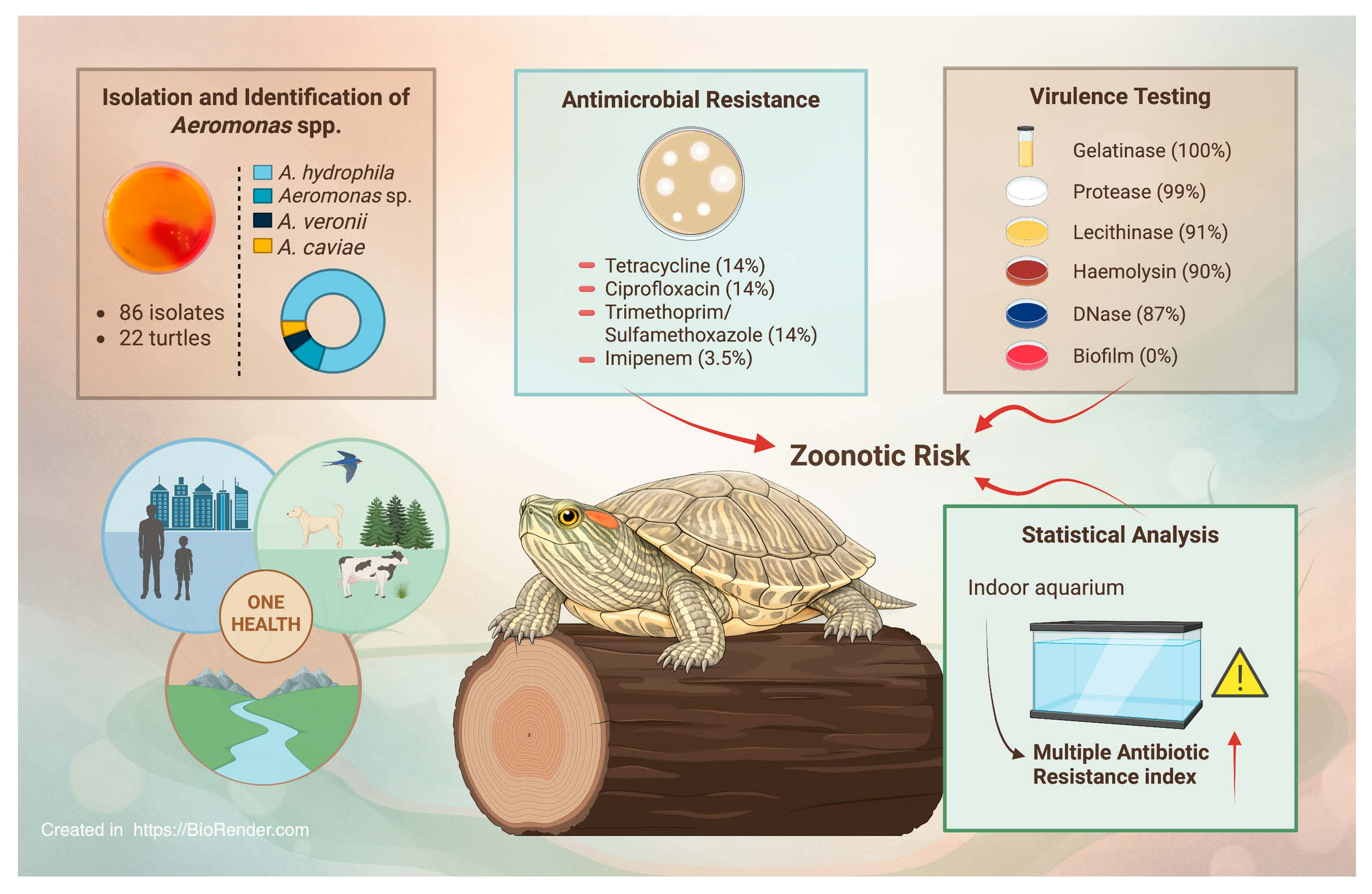
Semi-aquatic turtles are popular companion animals and represent an important One Health interface in Portugal due to their potential to harbour zoonotic pathogens. This study aimed to characterize the virulence and antimicrobial resistance (AMR) profiles of Aeromonas spp. isolated from captive semi-aquatic turtles in Portugal. Cloacal swabs (n = 31) were collected from turtles under human care, and Aeromonas spp. were isolated using selective media and identified by multiplex PCR. Antimicrobial susceptibility was assessed by disk diffusion using eleven antibiotics, while the phenotypic virulence profile was evaluated by determining isolates’ ability to express five hydrolytic enzymes and to form biofilm. A total of 86 Aeromonas isolates were recovered, with A. hydrophila being the most prevalent (77.9%). Most isolates displayed high pathogenic potential: over 87% produced DNase, haemolysin, and lecithinase, while nearly all produced protease and gelatinase. Also, 44.2% of the isolates were resistant to at least one antibiotic, and 12 (14.0%) were multidrug resistant. Higher Multiple Antibiotic Resistance (MAR) indices were significantly associated with turtles housed in indoor aquariums (p < 0.05). These findings indicate that captive semi-aquatic turtles may act as reservoirs of virulent and antimicrobial-resistant Aeromonas spp., highlighting potential zoonotic risks and supporting the need for a One Health approach in their management.

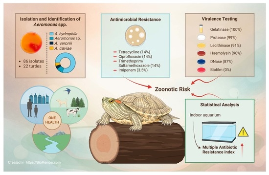
Graphical Abstract

1. Introduction

Semi-aquatic turtles, belonging to the order Testudines, represent a unique group of reptiles adapted to both aquatic and terrestrial environments [1,2,3]. These species rely on water for foraging, thermoregulation, and predator avoidance, while also depending on land for basking, nesting, and hibernation [2]. Their ecological role is crucial, as they contribute to nutrient cycling, and maintain the ecosystem balance by acting as both predators and prey [1,4]. In Portugal, only two native semi-aquatic species are recognized: the European pond turtle (Emys orbicularis) and the Mediterranean pond turtle (Mauremys leprosa), both of which inhabit diverse freshwater habitats and are indicators of wetland health [5,6,7]. However, the popularity of turtles as pets has led to the introduction of invasive species, such as the red-eared slider (Trachemys scripta elegans), which threatens native populations through competition for resources, habitat displacement, and potential crossbreeding [8,9,10].
The increasing popularity of turtles as exotic pets also carries significant public health implications. Semi-aquatic turtles can harbour a variety of zoonotic pathogens, most notably Salmonella spp., which can be asymptomatically shed through faces and contaminate both the animal and its environment [11,12]. Beyond Salmonella spp., turtles may carry opportunistic bacteria, including Aeromonas spp., Pseudomonas spp., Citrobacter spp., and Klebsiella spp., capable of causing gastrointestinal, wound, and systemic infections in humans [11,13]. Certain populational groups, such as children, the elderly, and immunocompromised individuals, are particularly vulnerable to these zoonotic infections [12]. The emergence of multidrug-resistant (MDR) strains among these bacteria heightens the risk, establishing turtles as potential reservoirs of clinically significant antimicrobial-resistant organisms [14].
Aquatic environments serve as critical reservoirs for antimicrobial resistance (AMR), gathering antibiotics, resistant bacteria, and mobile genetic elements from human, agricultural, and aquaculture sources [15,16,17]. Freshwater turtles are constantly exposed to these selective pressures, and their microbiota can reflect the resistance factors present in their habitats, making them valuable sentinels of environmental AMR [11]. Captive conditions, including artificial diets, water quality issues, crowding, and stress, can further disrupt the natural microbial balance of these animals, promoting the proliferation of opportunistic pathogens and the dissemination of resistant strains [18,19].
Among the opportunistic bacteria mentioned, the genus Aeromonas warrants particular attention. These Gram-negative, facultatively anaerobic bacteria are widely distributed in aquatic ecosystems and commonly colonize the gut of poikilothermic animals [20,21]. While often commensal, Aeromonas spp. can cause septicaemia, ulcerative stomatitis, and other severe infections in stressed or immunocompromised turtles [20,22]. Virulence factors, including haemolysins, aerolysin, proteases, and biofilm-forming capabilities, enable these bacteria to evade host defences and establish infections [20,23]. Many strains can also carry antimicrobial resistance genes, such as blaTEM, blaSHV, qnrS, and mutations in gyrA, conferring resistance to β-lactams, quinolones, and other clinically important antibiotics [13,24].
Previous studies have reported outbreaks of A. hydrophila in captive turtles, resulting in acute septicaemia and high mortality, highlighting their pathogenic potential [22,24]. It is important to note that A. hydrophila is often classified within a broader species complex, which can include genotypically diverse isolates with varied pathogenic potential [25]. Both wild and captive turtles frequently carry MDR strains with high multiple antibiotic resistance (MAR) indices, reflecting exposure to contaminated environments and amplifying their role as reservoirs of resistant bacteria [20,23].
Despite the recognized risks, data on the prevalence, virulence, and antimicrobial resistance profile of Aeromonas in captive semi-aquatic turtles in Portugal remain scarce. Understanding these dynamics is critical to evaluating zoonotic risks, informing veterinary care, and guiding management strategies under a One Health framework [17,26]. The present study aims to characterize Aeromonas spp. from captive turtles in Portugal, providing insights into their virulence profiles, antimicrobial resistance patterns, and potential implications for animal welfare and public health.

2. Materials and Methods

2.1. Study Population and Sample Collection

Over a 16-week period, a total of 31 cloacal swabs were obtained from turtles under human care, representing multiple species, at three veterinary facilities in Portugal: a private clinic in Lisbon, the Teaching Hospital of the Faculty of Veterinary Medicine, University of Lisbon, and a mobile veterinary service. Turtles selected for this study included all semi-aquatic turtles presented for consultation at these locations during the referred period. Samples encompassed turtles brought in for routine check-ups as well as individuals showing clinical signs. To ensure diversity, if the animals lived in large communal ponds or aquariums, a maximum of three turtles were randomly selected for sampling. Metadata recorded for each animal included species, sex, age, enclosure type (aquarium or outdoor pond), presence of other animals in the same enclosure, and history of antibiotic use in the six months preceding sampling.
Cloacal samples were collected using sterile AMIES swabs (VWR, Leuven, Belgium), gently inserted into the cloaca after cleaning the external cloacal region with 70% ethylic alcohol, avoiding contamination. All procedures were non-invasive and performed by experienced personnel during routine procedures, after owners’ consent. Swabs were stored at 4 °C until transportation to the Microbiology and Immunology Laboratory at FMV/ULisboa.

2.2. Characterisation of the Sampled Population

A total of 31 turtles were included in this study, comprising 51.6% females (n = 16) and 41.9% males (n = 13), while 6.5% (n = 2) were very young individuals for which the sex could not be accurately determined. The animals’ age ranged from 18 months to 37 years, with a median age of 10 years. The sampled population comprised seven species, with Trachemys scripta (32.3%; n = 10), Mauremys reevesii (19.4%; n = 6), and Pseudemys concinna (19.4%; n = 6) being the most represented (Table 1). Most samples originated from the Lisbon district (83.9%; n = 26), with the remaining samples collected at the Santarém district (16.1%; n = 5). Regarding housing conditions, 38.7% (n = 12) of the turtles lived in outdoor ponds, whereas 61.3% (n = 19) were kept in indoor aquariums, and 64.5% (n = 20) shared their enclosure with other animals. Only a small number of animals had received antibiotics in the six months prior to sampling (9.7%; n = 3). All data can be consulted in Table S1.

2.3. Bacterial Isolation and Identification

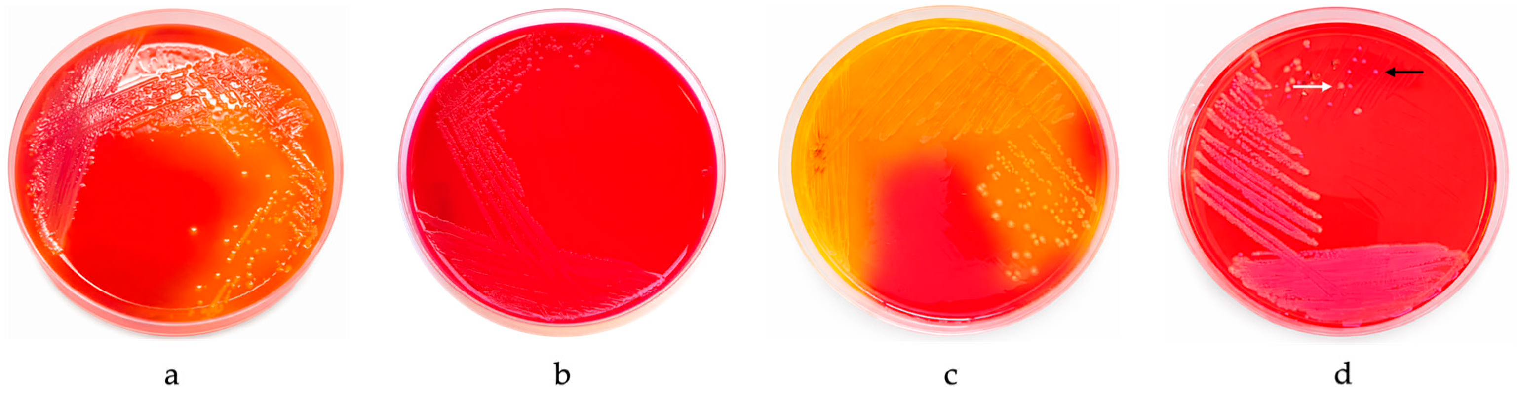
All cloacal swabs were processed individually, and no sample pooling was performed at any stage of bacterial isolation. Samples were inoculated in Brain Heart Infusion (BHI) broth (VWR, Leuven, Belgium) at 37 °C for 24 h. Subsequently, 10 µL of each enriched suspension was plated on Glutamate Starch Phenol Red (GSP) agar supplemented with sodium penicillin (100,000 IU/L) (Merck, Kenilworth, NJ, USA), and incubated at 37 °C for 12 h. Afterwards, up to four morphologically distinct colonies compatible with Aeromonas spp. (large, yellow colonies, surrounded by a yellow zone) were randomly selected per sample and re-isolated on the same medium, followed by pure culture on BHI agar at 37 °C for 24 h. Reference strains A. caviae ATCC 1976, A. hydrophila ATCC 7966, and A. media ATCC 33907 were used as positive controls, while Escherichia coli ATCC 25922 was used as a negative control (Figure 1).
Presumptive Aeromonas isolates were evaluated by Gram-staining and subjected to the oxidase test. Gram-negative and oxidase-positive isolates were further identified at the species level using multiplex-PCR.

2.4. Multiplex-PCR

DNA from each isolate was extracted using the boiling method [27]. Briefly, fresh colonies from overnight cultures were suspended in 150 µL PCR-grade water, heated at 100 °C for 10 min and centrifuged at 18,800× g for 10 min. The resulting supernatant was collected, and the concentration and purity of DNA were assessed using a NanoDrop™ Spectrophotometer (ThermoFisher Scientific, Waltham, MA, USA). Then, the supernatants with an absorbance A260/A280 ratio between 1.7 and 2 were diluted in PCR-grade water to 50 ng/µL for subsequent PCR analysis.
The multiplex-PCR protocol applied allowed differentiation between A. caviae, A. media, A. hydrophila, and A. veronii, and included an internal genus-specific control [27,28]. PCR reactions were prepared in a final volume of 20 µL, comprising 10 µL of Supreme NZYTaq 2 Green Master Mix (NZYTech, Lisbon, Portugal), 2.6 µL of PCR-grade water (Sigma-Aldrich, St. Louis, MO, USA), 0.05 µL of A-16S primers (0.05 µM), 0.5 µL of A-cav primers (0.5 µM), 0.2 µL of A-med primers (0.2 µM), 0.45 µL of A-hyd primers (0.45 µM), 0.15 µL of A-ver primers (0.15 µM) (STABVIDA, Caparica, Portugal) (Table 2), and 2.0 µL of template DNA. PCR amplification was performed using a thermocycler (VWR, Radnor, PA, USA) and the following conditions: an initial denaturation at 95 °C for 2 min, followed by 6 cycles of 94 °C for 40 s, 68 °C for 50 s, and 72 °C for 40 s; then 30 cycles of 94 °C for 40 s, 66 °C for 50 s, and 72 °C for 40 s [27,28].
Positive controls included DNA from reference strains mentioned in the previous section and from a clinical A. veronii isolate identified by Sanger sequencing of the 16S amplicon; negative controls included DNA from E. coli ATCC 25922, P. aeruginosa ATCC 27853, and water. PCR products were analyzed by gel electrophoresis, using agarose 2% (w/v) in 1x TBE buffer (NZYTech, Lisbon, Portugal). Gels were run for 1 h and 15 min at 90 V. A molecular weight marker, NZYDNALadder VI (NZYTech, Lisbon, Portugal), was used for reference, and the results were visualized using a UV transilluminator with imaging captured on a Bio-Rad ChemiDoc XRS image system (Bio-Rad Laboratories, Hercules, CA, USA).

2.5. Antimicrobial Susceptibility Profile

The antimicrobial susceptibility profiles of isolates were assessed using the Kirby–Bauer disk diffusion method, following CLSI (2025) guidelines. Eleven antimicrobials from seven classes were tested, including beta-lactams combined with beta-lactamase inhibitors (piperacillin-tazobactam, TZP, 110 µg), third-generation cephalosporins (ceftazidime, CAZ, 30 µg), fourth-generation cephalosporins (cefepime, FEP, 30 µg), cephamycins (cefoxitin, FOX, 30 µg), fluoroquinolones (ciprofloxacin, CIP, 5 µg), aminoglycosides (gentamicin, CN, 10 µg), monobactams (aztreonam, ATM, 30 µg), folate pathway inhibitors (sulfamethoxazole-trimethoprim, SXT, 25 µg), phenicols (chloramphenicol, C, 30 µg), tetracyclines (tetracycline, TE, 30 µg) and carbapenems (imipenem, IPM, 10 µg) (Oxoid, Basingstoke, Hampshire, UK). Antibiotics for susceptibility testing were chosen according to their relevance in veterinary and human medicine and the antimicrobial protocols commonly used for semi-aquatic turtles.
First, bacterial suspensions in saline were standardized to 0.5 McFarland (~108 CFU/mL) and inoculated onto Mueller-Hinton agar (Oxoid, Basingstoke, Hampshire, UK) plates, followed by antimicrobial disks application and incubation at 37 °C for 16–18 h. After the diameter of the inhibition halos was measured, the isolates were classified as susceptible (S), intermediate (I), or resistant (R) according to CLSI standards [29]. E. coli ATCC 25922 and P. aeruginosa ATCC 27853 were also tested as quality control, and 10% of isolates were retested to confirm reproducibility.
For defining MDR, intermediate results were considered as resistant. MDR was defined as non-susceptibility to ≥1 agent in ≥3 antimicrobial classes, according to [30].
The MAR index was additionally calculated for each isolate as the ratio between the number of antibiotics to which each isolate was resistant and the total number of antibiotics tested [23].

2.6. Virulence Profile

The phenotypic virulence profile of the isolates was assessed by evaluating their ability to produce biofilm and enzymes associated with bacterial pathogenicity, including protease, DNase, lecithinase, haemolysin, and gelatinase, according to Fernandes et al. [23]. Protease activity was determined using Skim Milk agar (VWR, Leuven, Belgium), with P. aeruginosa ATCC 27853 and S. aureus ATCC 29213 serving as positive and negative controls, respectively. DNase production was evaluated on DNase agar (Thermo Scientific Remel, Lenexa, KS, USA) supplemented with 0.01% toluidine blue (Merck, Darmstadt, Germany), using A. hydrophila ATCC 7966 and E. coli ATCC 25922 as positive and negative controls, respectively. Lecithinase activity was assessed on Tryptic Soy agar (VWR, Leuven, Belgium) supplemented with 10% egg yolk emulsion (VWR, Leuven, Belgium), with P. aeruginosa ATCC 27853 being tested as positive control and E. coli ATCC 25922 as negative control. Haemolysin production was evaluated on Columbia Agar supplemented with 5% sheep blood (bioMérieux, Marcy-l’Etoile, France), using S. aureus ATCC 25923 as the positive control and E. faecium CCUG 36804 as the negative control. Gelatinase production was assessed using Nutrient Gelatin agar (Oxoid, Basingstoke, Hampshire, UK), with P. aeruginosa ATCC 27853 and E. coli ATCC 25922 serving as positive and negative controls. Biofilm formation was evaluated on Congo Red Agar, composed of Brain Heart Infusion Broth at 3.7%, bacteriological agar (VWR, Leuven, Belgium) at 1.4%, sucrose (Sigma-Aldrich, Steinheim, Germany) at 5%, and Congo Red reagent (Sigma-Aldrich, Steinheim, Germany) at 0.08%, with E. faecium ATCC 35667 being tested as positive control and E. coli ATCC 25922 as negative control.
Results from the assays were assessed after incubation at 37 °C for 24 to 48 h, except for gelatinase activity (37 °C for 72 h). A 10% replica was evaluated in each test. Virulence indices were calculated for each isolate as the ratio between the number of virulence factors produced by the isolate and the total number of virulence determinants tested [23].

2.7. Statistical Analysis

Data were compiled in a spreadsheet (Excel, Microsoft Corporation, Redmond, WA, USA) and subsequent statistical analysis was performed using SAS 9.4 software (SAS Institute Inc., Cary, NC, USA). Age was summarized as median for animals with known age; individuals with indeterminate age were excluded from age-related analyses.
Multivariable logistic regression (PROC LOGISTIC) was used to assess associations between independent variables (enclosure type, presence of other animals, recent antimicrobial treatment, and PCR-identified species) and both virulence factor expression and antibiotic resistance ability. Cumulative logistic regression evaluated associations between enclosure type, presence of other animals, recent antimicrobial treatment, and PCR-identified species with virulence and MAR indices. A manual backward elimination was performed, using a p-value criterion of 0.157 and without prior univariable pre-filtering, to obtain the final models [25].
Spearman correlation assessed relationships between MAR and virulence indices, as well as resistance levels across antibiotics. Significance was defined at p ≤ 0.05, with trends considered at 0.05 < p ≤ 0.10.

3. Results

3.1. Isolates’ Isolation and Identification

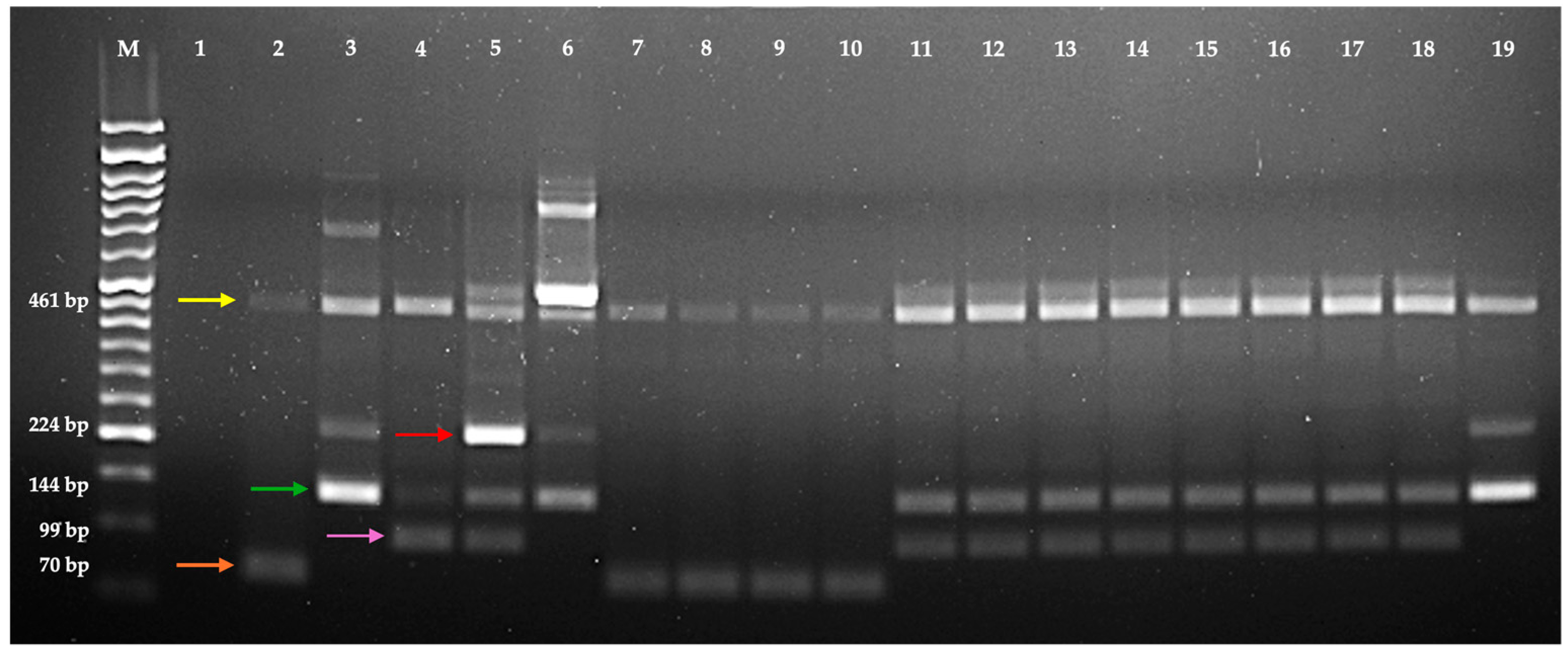
From the 31 cloacal samples collected, 22 (71.0%) produced colonies on GSP agar with morphology consistent with Aeromonas spp., allowing to obtain a total of 86 presumptive isolates. All isolates were Gram-negative and oxidase-positive. Multiplex-PCR allowed the identification of 67 isolates as A. hydrophila (77.9%), 6 as A. veronii (7.0%), and 4 as A. caviae (4.7%). Nine isolates (10.5%) were positive for the genus-specific internal control but could not be identified at the species level and were classified as Aeromonas spp. As expected, two specific amplicons were detected per isolate, corresponding to the genus- and species-specific targets. Although additional bands were also observed, these were also consistently detected in the reference strains, being absent in the negative controls, indicating reproducible non-specific amplification rather than random artefacts (Figure 2). As shown in Table 3, the distribution of Aeromonas species varied among turtle species.

3.2. Characterization of Isolates’ Antimicrobial Susceptibility Profile

Among the 86 isolates analysed, 44.2% (n = 38) exhibited resistance or intermediate susceptibility to at least one of the eleven antibiotics tested. Thirteen (15.1%) isolates were resistant to two or more antibiotics, and 12 (14.0%) fulfilled the criteria for MDR [30]. As shown in Table 3, MDR isolates were distributed across multiple turtle species. The highest resistance rates were observed for tetracycline (14.0%; n = 12), ciprofloxacin (14.0%; n = 12), and trimethoprim-sulfamethoxazole (14.0%; n = 12). Resistance to imipenem was observed in 3 (3.5%) isolates, and 11 (12.8%) isolates demonstrated intermediate susceptibility to this antibiotic. No resistance was detected against piperacillin-tazobactam, ceftazidime, cefepime, cefoxitin, gentamicin, aztreonam, and chloramphenicol (Table 4). A. caviae presented the highest mean MAR index (0.11) despite the low number of isolates (n = 4), followed by A. hydrophila (0.07), A. veronii (0.05), and Aeromonas spp. (0.03).

3.3. Characterization of Isolates’ Virulence Profile

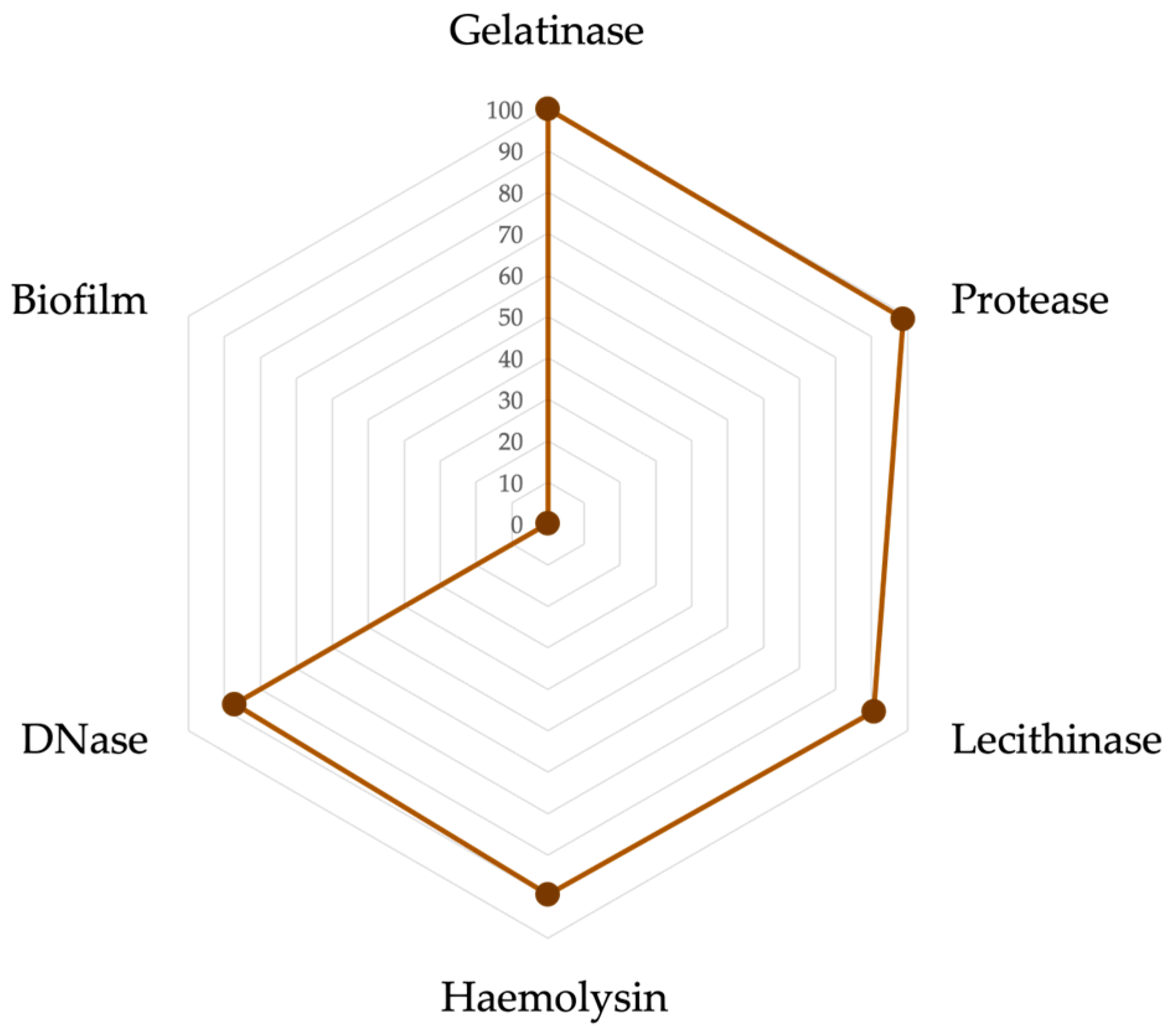
All isolates (100%; n = 86) exhibited gelatinase activity, and protease activity was detected in 98.8% (n = 85) of the isolates. Lecithinase, haemolysin, and DNase production were observed in 90.7% (n = 78), 89.5% (n = 77), and 87.2% (n = 75) of the isolates, respectively. None of the isolates produced biofilm (Figure 3). The highest virulence indices were recorded for A. hydrophila (0.83), A. caviae (0.79), and A. veronii (0.70), whereas the Aeromonas spp. group displayed the lowest index (0.41).

3.4. Statistical Analysis

No significant associations were found between the identified bacterial species and turtle species, enclosure type, or history of antibiotic use in the six months preceding sampling (p > 0.05).
Similarly, no significant differences in the MAR index were observed across Aeromonas species, turtle species, cohabitation with other animals, history of medication use in the six months preceding sampling, or age (p > 0.05). However, a statistically significant association was detected between the MAR index and enclosure type, with turtles housed in indoor aquariums exhibiting higher MAR indices than those maintained in outdoor ponds (p < 0.05).
No significant differences in virulence indices were observed among Aeromonas species, turtle species, enclosure type, cohabitation with other animals, medication history, or age (p > 0.05). Furthermore, no significant correlation was identified between virulence and MAR indices (p > 0.05).

4. Discussion

The present study highlights the role of semi-aquatic turtles as potential reservoirs of Aeromonas species, with implications for animal and public health. Among the 86 isolates obtained, A. hydrophila was the most prevalent species, followed by A. veronii and A. caviae, while 10.5% of the isolates could not be identified to a species level. These findings are consistent with previous reports identifying A. hydrophila as the dominant species in chelonians and other aquatic animals [20,22], whereas A. veronii and A. caviae are less frequently reported [25,31]. Although A. hydrophila was identified as the dominant species in this study, it is important to note that A. hydrophila is part of a broader species complex [25], which can include genotypically diverse isolates with varied pathogenic potential. Species assignments should therefore be interpreted within the limitations of the used multiplex PCR assay. The presence of unidentified Aeromonas isolates reflects the taxonomic complexity of the genus and may represent less common or novel species with currently unknown virulence and resistance potential. The high prevalence of A. hydrophila is notable given its role as a major opportunistic pathogen in chelonians and other aquatic animals, being associated with severe diseases such as ulcerative stomatitis, pneumonia, and haemorrhagic septicaemia, particularly in stressed or immunocompromised individuals [21,22]. The predominance of A. hydrophila aligns with its reported ubiquity in aquatic environments and its role as a primary pathogen in chelonian septicemia [22].
Antimicrobial resistance was observed in almost half of the 44.2% of isolates, with 13.9% being classified as MDR. The highest resistance rates (14.0%) were noted for tetracycline, ciprofloxacin, and sulfamethoxazole-trimethoprim, while no resistance was detected towards ceftazidime, cefepime, piperacillin-tazobactam, aztreonam, gentamicin, or chloramphenicol. Overall, these resistance patterns were reflected in a relatively low mean MAR index, with slight variations observed among Aeromonas species. These results align with previous studies on turtles reporting generally low AMR prevalence in populations with limited antibiotic exposure [32]. Some of these antimicrobial agents, such as ceftazidime, cefepime, piperacillin-tazobactam, and aztreonam, are frequently reserved for the treatment of complicated and hospital-acquired infections in humans, such as complicated intra-abdominal and urinary tract infections, and pneumonia, especially when resistant Gram-negative bacteria are involved [33]. Thus, the findings indicate that resistance to last-line or broad-spectrum agents is not yet widespread among the isolates under study.
Regarding virulence factors’ production, all isolates displayed gelatinase activity, and the majority expressed protease, DNase, haemolysin, and lecithinase, while biofilm production was not detected. These results suggest a high intrinsic virulence potential in the sampled turtles, particularly of A. hydrophila, consistent with its recognized role as a primary opportunistic pathogen in chelonians and other aquatic animals [21]. The virulence index values corroborate this pattern, with A. hydrophila isolates showing the highest scores. The near-ubiquitous production of gelatinase and protease is particularly relevant for turtle health, as these enzymes facilitate tissue invasion and are associated with ulcerative shell and skin lesions [34].
Overall, the disparity observed between high virulence potential and relatively low antimicrobial resistance rates may highlight the complex dynamics of bacterial adaptation, where virulence traits may be maintained in low-antibiotic-pressure environments independently of the expression of resistance mechanisms [35,36]. Semi-aquatic turtles thus serve as a relevant model for One Health studies, representing a direct interface between animal, human, and environmental health [26,37]. Also, the presence of highly virulent and, in some cases, MDR Aeromonas isolates underscores the importance of proper husbandry, surveillance, and public awareness to mitigate zoonotic and ecological risks [9,11,12].
The low reported antibiotic use prior to sampling suggests minimal direct selective pressure for maintaining AMR determinants, which often carry a fitness cost [35]. This contrasts with the high prevalence of virulence factors, which are likely maintained for ecological fitness within the host [25]. However, our data indicate that enclosure type can locally alter these dynamics. Turtles housed in indoor aquariums exhibited significantly higher MAR indices than those in outdoor ponds. A statistically significant association was observed between turtles housed in indoor aquariums and MAR indices. This finding was influenced by a group of 12 MDR isolates recovered from turtles sharing a single aquarium. Although clonal relatedness between isolates was not assessed, the presence of multiple MDR isolates in the same enclosed system may reflect localized accumulation of resistant bacteria. Indoor aquariums, unlike outdoor ponds, are closed systems in which bacterial populations can thrive, potentially facilitating persistence and horizontal gene transfer (HGT), a mechanism contributing to antimicrobial resistance dissemination [38]. These observations should be interpreted with caution, considering the limited sample size and lack of genetic confirmation, which prevent definitive conclusions regarding potential clonal spread.
The health of turtles in indoor aquariums depends heavily on husbandry practices, which are often suboptimal due to limited owner knowledge. Stressors such as overcrowding, inadequate filtration, improper diet, and insufficient UVB lighting can compromise physiological health, modify gut microbiota and increase susceptibility to opportunistic and potentially resistant pathogens [39]. Continuous exposure to microorganisms from handlers and tap water, which may contain antibiotic residues or resistant bacteria, further contributes to this risk [32]. Therefore, indoor aquariums may function as dynamic micro-ecosystems where environmental, animal, and human health factors converge, creating conditions that may favour the emergence and dissemination of antimicrobial resistance.
Importantly, the detection of potentially virulent Aeromonas isolates in the cloaca represents a continuous source of environmental contamination, as these bacteria are readily shed into aquarium water. This may facilitate exposure of turtle owners and household members, as routine tank maintenance involves direct contact with water, splashes, or contaminated equipment, creating a potential route for zoonotic transmission [12]. In humans, Aeromonas species are primarily associated with gastroenteritis, wound infections, and septicaemia [25], with infections more likely to occur following exposure to contaminated water or in individuals with underlying conditions. Collectively, these findings underscore that Aeromonas carriage by captive turtles may not be a confined animal health issue but represents a shared risk at the human–animal–environment interface.
Moreover, as turtles defecate directly into their aquatic environment, potentially pathogenic and virulent Aeromonas isolates are continuously shed into the tank water, creating a dynamic microbial reservoir to which humans can be exposed during routine maintenance. These findings highlight the relevance of a One Health perspective in domestic settings. Veterinarians should prioritize culture and susceptibility testing, educate owners on proper husbandry, and promote strict hygiene practices. Maintaining high water quality, practicing hand hygiene, and avoiding the release of unwanted pets are essential to reduce disease risks and environmental impact [9,11,12]. Finally, the presence of AMR in pet turtles reinforces the need for integrated surveillance programs and public education to address antimicrobial resistance within a One Health framework, in line with global recommendations [40]. Therefore, these animals can act as sentinels for environmental AMR and as potential sources of exposure for their handlers, underscoring the interconnectedness of animal, human, and environmental health in domestic settings.
A limitation of this study was the inability to identify 10.5% of the isolates to the species level using the multiplex PCR assay, relying instead on genus-level classification. It should also be noted that A. hydrophila belongs to a taxonomically complex group of closely related species (the A. hydrophila complex), and accurate discrimination among members may require more comprehensive genomic approaches. This limitation underscores the need for future research employing different techniques, such as 16S rRNA or whole-genome sequencing, to achieve complete taxonomic characterization of the Aeromonas community in turtles, as well as to evaluate the genetic relatedness among isolates. Additionally, in vitro competition assays between MDR and susceptible isolates obtained in this study could be performed to directly assess the fitness cost of resistance in the absence of antibiotics [35]. Such investigations would provide more definitive insights into the relationship between antimicrobial resistance and virulence within microbial communities of pet turtles.

5. Conclusions

This study provides an initial characterization of Aeromonas spp. colonizing captive semi-aquatic turtles in Portugal, highlighting their high intrinsic virulence potential and the occurrence of antimicrobial resistance, including MDR isolates. A. hydrophila was the predominant species identified in this study, consistent with previous reports in chelonians. The findings reveal a clear disparity between virulence and resistance traits, suggesting that virulence factors are maintained independently of antimicrobial exposure in low-pressure environments. Higher MAR indices in turtles housed in indoor aquariums may indicate that closed systems may act as localized reservoirs for resistant isolates. Overall, these results emphasize the role of semi-aquatic turtles as sentinel species at the interface of animal, human, and environmental health, underlining the importance of appropriate husbandry, veterinary surveillance, and public awareness to mitigate zoonotic and ecological risks. Future studies employing genomic approaches and broader environmental sampling are needed to fully characterize the Aeromonas community and its implications in a One Health perspective. These findings underscore the necessity of integrating companion reptile health into broader antimicrobial stewardship and surveillance programs under a One Health umbrella.

Supplementary Materials

The following supporting information can be downloaded at https://www.mdpi.com/article/10.3390/microbiolres17030056/s1, Table S1: Metadata of sampled animals.

Author Contributions

Conceptualization, G.M. and M.O.; methodology, G.M., R.A., E.C., L.C. and M.O.; software, G.M., R.A. and G.P.; validation, G.M., R.A., G.P., E.C., L.T. and M.O.; formal analysis, G.M., R.A., G.P. and M.O.; investigation, G.M., R.A. and M.O.; resources, L.T. and M.O.; data curation, G.M., R.A. and M.O.; writing—original draft preparation, G.M. and R.A.; writing—review and editing, G.M., R.A., G.P., E.C., L.C., L.T. and M.O.; visualization, G.M.; supervision, M.O.; project administration, M.O.; funding acquisition, L.T. and M.O. All authors have read and agreed to the published version of the manuscript.

Funding

This research was funded by the Portuguese Foundation for Science and Technology (FCT) under projects UID/276/2025 (CIISA) and LA/P/0059/2020 (AL4AnimalS).

Institutional Review Board Statement

All animals were cared for according to the rules given by the current EU (Directive 2010/63/EC) and Portuguese legislation (DL 113/2013), by the competent authority in Portugal (https://www.dgav.pt/animais, accessed on 11 January 2026). The samples collected for this study were gathered by trained veterinary surgeons during routine procedures, after owners’ consent. This study did not include animal experiments; only non-invasive samples were collected, and no ethics committee approval was needed.

Informed Consent Statement

Not applicable.

Data Availability Statement

The original contributions presented in this study are included in the article/Supplementary Material. Further inquiries can be directed to the corresponding authors.

Acknowledgments

The authors would like to acknowledge CIISA—the Centre for Interdisciplinary Research in Animal Health, Faculty of Veterinary Medicine, University of Lisbon—and AL4AnimalS—Associate Laboratory for Animal and Veterinary Sciences.

Conflicts of Interest

The authors declare no conflicts of interest.

References

  1. Lovich, J.E.; Ennen, J.R.; Agha, M.; Gibbons, J.W. Where Have All the Turtles Gone, and Why Does It Matter? Bioscience 2018, 68, 771–781. [Google Scholar] [CrossRef] [Scilit]
  2. Pearson, S.H.; Avery, H.W.; Spotila, J.R. Juvenile Invasive Red-Eared Slider Turtles Negatively Impact the Growth of Native Turtles: Implications for Global Freshwater Turtle Populations. Biol. Conserv. 2015, 186, 115–121. [Google Scholar] [CrossRef] [Scilit]
  3. Scheelings, T.F. BSAVA Manual of Reptiles, 3rd ed.; Girling, S.J., Raiti, P., Eds.; British Small Animal Veterinary Association: Toronto, ON, Canada, 2019. [Google Scholar]
  4. Stephens, P.R.; Wiens, J.J. Ecological Diversification and Phylogeny of Emydid Turtles. Biol. J. Linn. Soc. 2003, 79, 577–610. [Google Scholar] [CrossRef] [Scilit]
  5. Cadi, A.; Joly, P. Competition for Basking Places between the Endangered European Pond Turtle (Emys orbicularis galloitalica) and the Introduced Red-Eared Slider (Trachemys scripta elegans). Can. J. Zool. 2003, 81, 1392–1398. [Google Scholar] [CrossRef] [Scilit]
  6. Charcos com Vida Cágado Comum. Available online: https://charcoscomvida.pt/animals/cagado-comum/ (accessed on 29 December 2025).
  7. Charcos com Vida Cágado-de-Carapaça-Estriada. Available online: https://charcoscomvida.pt/animals/cagado-de-carapaca-estriada/ (accessed on 29 December 2025).
  8. Semenov, D.V. Slider Turtle, Trachemys scripta elegans, as Invasion Threat (Reptilia; Testudines). Russ. J. Biol. Invasions 2010, 1, 296–300. [Google Scholar] [CrossRef] [Scilit]
  9. Banha, F.; Gama, M.; Anastácio, P.M. The Effect of Reproductive Occurrences and Human Descriptors on Invasive Pet Distribution Modelling: Trachemys scripta elegans in the Iberian Peninsula. Ecol. Modell. 2017, 360, 45–52. [Google Scholar] [CrossRef] [Scilit]
  10. Polo-Cavia, N.; López, P.; Martín, J. Competitive Interactions during Basking between Native and Invasive Freshwater Turtle Species. Biol. Invasions 2010, 12, 2141–2152. [Google Scholar] [CrossRef] [Scilit]
  11. Hossain, S.; Heo, G.J. Pet-Turtles: A Potential Source of Human Pathogenic Bacteria. Arch. Microbiol. 2021, 203, 3785–3792. [Google Scholar] [CrossRef] [Scilit]
  12. Sodagari, H.R.; Habib, I.; Shahabi, M.P.; Dybing, N.A.; Wang, P.; Bruce, M. A Review of the Public Health Challenges of Salmonella and Turtles. Vet. Sci. 2020, 7, 56. [Google Scholar] [CrossRef] [Scilit] [PubMed]
  13. Wimalasena, S.H.M.P.; Shin, G.W.; Hossain, S.; Heo, G.J. Potential Enterotoxicity and Antimicrobial Resistance Pattern of Aeromonas Species Isolated from Pet Turtles and Their Environment. J. Vet. Med. Sci. 2017, 79, 921–926. [Google Scholar] [CrossRef] [Scilit]
  14. Wendt, M.; Silva, B.C.J.; Heo, G.-J. Virulence Factors and Antimicrobial Resistance of Pseudomonas aeruginosa Isolated from Pet Turtles. Asian J. Anim. Vet. Adv. 2017, 12, 205–211. [Google Scholar] [CrossRef] [Scilit]
  15. Berendonk, T.U.; Manaia, C.M.; Merlin, C.; Fatta-Kassinos, D.; Cytryn, E.; Walsh, F.; Bürgmann, H.; Sørum, H.; Norström, M.; Pons, M.N.; et al. Tackling Antibiotic Resistance: The Environmental Framework. Nat. Rev. Microbiol. 2015, 13, 310–317. [Google Scholar] [CrossRef] [Scilit]
  16. Roca, I.; Akova, M.; Baquero, F.; Carlet, J.; Cavaleri, M.; Coenen, S.; Cohen, J.; Findlay, D.; Gyssens, I.; Heure, O.E.; et al. The Global Threat of Antimicrobial Resistance: Science for Intervention. New Microbes New Infect. 2015, 6, 22–29, Erratum in New Microbes New Infect. 2015, 8, 175.. [Google Scholar] [CrossRef] [Scilit] [PubMed]
  17. White, A.; Hughes, J.M. Critical Importance of a One Health Approach to Antimicrobial Resistance. Ecohealth 2019, 16, 404–409. [Google Scholar] [CrossRef] [Scilit]
  18. Mathews, J. If You Must Touch a Turtle, Wash Your Hands. Oak Leaf 2022, 3, 28–36. [Google Scholar]
  19. Scheelings, T.F.; Van, T.T.H.; Moore, R.J.; Skerratt, L.F. Location Matters: Variations in Cloacal Microbiota Composition of Spatially Separated Freshwater Turtles. Microb. Ecol. 2024, 87, 140. [Google Scholar] [CrossRef] [Scilit] [PubMed]
  20. Guz, L.; Nowakiewicz, A.; Puk, K.; Zięba, P.; Gnat, S.; Matuszewski, Ł. Virulence and Antimicrobial Resistance Pattern of Aeromonas spp. Colonizing European Pond Turtles Emys orbicularis and Their Natural Environment. First Study from Poland. Animals 2021, 11, 772. [Google Scholar] [CrossRef] [Scilit]
  21. Harikrishnan, R.; Balasundaram, C. Modern Trends in Aeromonas hydrophila Disease Management with Fish. Rev. Fish. Sci. 2005, 13, 281–320. [Google Scholar] [CrossRef] [Scilit]
  22. Pasquale, V.; Baloda, S.B.; Dumontet, S.; Krovacek, K. An Outbreak of Aeromonas hydrophila Infection in Turtles (Pseudemis scrtipta). Appl. Environ. Microbiol. 1994, 60, 1678–1680. [Google Scholar] [CrossRef] [Scilit]
  23. Fernandes, M.; Grilo, M.; Carneiro, C.; Cunha, E.; Tavares, L.; Patino-Martinez, J.; Oliveira, M. Antibiotic Resistance and Virulence Profiles of Gram-Negative Bacteria Isolated from Loggerhead Sea Turtles (Caretta caretta) of the Island of Maio, Cape Verde. Antibiotics 2021, 10, 771. [Google Scholar] [CrossRef] [Scilit]
  24. Khafagy, A.R.; Kamel, A.M.; Abou El Ghait, E.-s.N.; Mohamed, N.M.; Eidaroos, N.H.; Hagag, F.A.; Fathi, M. Characterisation, Prevalence and Antibiogram of Aeromonas hydrophila in Turtles: Insights into Virulence Factors, Antimicrobial Resistance Genes and Innate Immunity. Afr. J. Herpetol. 2024, 73, 131–148. [Google Scholar] [CrossRef] [Scilit]
  25. Janda, J.M.; Abbott, S.L. The Genus Aeromonas: Taxonomy, Pathogenicity, and Infection. Clin. Microbiol. Rev. 2010, 23, 35–73. [Google Scholar] [CrossRef] [Scilit]
  26. Velazquez-Meza, M.E.; Galarde-López, M.; Carrillo-Quiróz, B.; Alpuche-Aranda, C.M. Antimicrobial Resistance: One Health Approach. Vet. World 2022, 15, 743–749. [Google Scholar] [CrossRef] [Scilit]
  27. Grilo, M.L.; Amaro, G.; Chambel, L.; Marques, C.S.; Marques, T.A.; Gil, F.; Sousa-Santos, C.; Robalo, J.I.; Oliveira, M. Aeromonas spp. Prevalence, Virulence, and Antimicrobial Resistance in an Ex Situ Program for Threatened Freshwater Fish—A Pilot Study with Protective Measures. Animals 2022, 12, 436. [Google Scholar] [CrossRef] [Scilit]
  28. Persson, S.; Al-Shuweli, S.; Yapici, S.; Jensen, J.N.; Olsen, K.E.P. Identification of Clinical Aeromonas Species by rpoB and gyrB Sequencing and Development of a Multiplex PCR Method for Detection of Aeromonas hydrophila, A. caviae, A. veronii, and A. media. J. Clin. Microbiol. 2015, 53, 653–656. [Google Scholar] [CrossRef] [Scilit]
  29. CLSI. Methods for Antimicrobial Dilution and Disk Susceptibility Testing of Infrequently Isolated or Fastidious Bacteria; Clinical and Laboratory Standards Institute: Wayne, PA, USA, 2015; Volume CLSI guideline M45. [Google Scholar]
  30. Magiorakos, A.P.; Srinivasan, A.; Carey, R.B.; Carmeli, Y.; Falagas, M.E.; Giske, C.G.; Harbarth, S.; Hindler, J.F.; Kahlmeter, G.; Olsson-Liljequist, B.; et al. Multidrug-Resistant, Extensively Drug-Resistant and Pandrug-Resistant Bacteria: An International Expert Proposal for Interim Standard Definitions for Acquired Resistance. Clin. Microbiol. Infect. 2012, 18, 268–281. [Google Scholar] [CrossRef] [Scilit] [PubMed]
  31. Rasmussen-Ivey, C.R.; Figueras, M.J.; McGarey, D.; Liles, M.R. Virulence Factors of Aeromonas hydrophila: In the Wake of Reclassification. Front. Microbiol. 2016, 7, 1337. [Google Scholar] [CrossRef] [Scilit] [PubMed]
  32. Xi, C.; Zhang, Y.; Marrs, C.F.; Ye, W.; Simon, C.; Foxman, B.; Nriagu, J. Prevalence of Antibiotic Resistance in Drinking Water Treatment and Distribution Systems. Appl. Environ. Microbiol. 2009, 75, 5714–5718. [Google Scholar] [CrossRef] [Scilit]
  33. Paul, M.; Carrara, E.; Retamar, P.; Tängdén, T.; Bitterman, R.; Bonomo, R.A.; de Waele, J.; Daikos, G.L.; Akova, M.; Harbarth, S.; et al. European Society of Clinical Microbiology and Infectious Diseases (ESCMID) Guidelines for the Treatment of Infections Caused by Multidrug-Resistant Gram-Negative Bacilli (Endorsed by European Society of Intensive Care Medicine). Clin. Microbiol. Infect. 2022, 28, 521–547. [Google Scholar] [CrossRef] [Scilit] [PubMed]
  34. Tomás, J.M. The Main Aeromonas Pathogenic Factors. Int. Sch. Res. Not. 2012, 2012, 256261. [Google Scholar] [CrossRef] [Scilit]
  35. Andersson, D.I.; Hughes, D. Antibiotic Resistance and Its Cost: Is It Possible to Reverse Resistance? Nat. Rev. Microbiol. 2010, 8, 260–271. [Google Scholar] [CrossRef] [Scilit] [PubMed]
  36. Beceiro, A.; Tomás, M.; Bou, G. Antimicrobial Resistance and Virulence: A Successful or Deleterious Association in the Bacterial World? Clin. Microbiol. Rev. 2013, 26, 185–230. [Google Scholar] [CrossRef] [Scilit] [PubMed]
  37. Mackenzie, J.S.; Jeggo, M. The One Health Approach-Why Is It so Important? Trop. Med. Infect. Dis. 2019, 4, 88. [Google Scholar] [CrossRef] [Scilit] [PubMed]
  38. Von Wintersdorff, C.J.H.; Penders, J.; Van Niekerk, J.M.; Mills, N.D.; Majumder, S.; Van Alphen, L.B.; Savelkoul, P.H.M.; Wolffs, P.F.G. Dissemination of Antimicrobial Resistance in Microbial Ecosystems through Horizontal Gene Transfer. Front. Microbiol. 2016, 7, 173. [Google Scholar] [CrossRef] [Scilit]
  39. Acierno, M.J.; Mitchell, M.A.; Roundtree, M.K.; Zachariah, T.T. Effects of Ultraviolet Radiation on 25-Hydroxyvitamin D3 Synthesis in Red-Eared Slider Turtles (Trachemys scripta elegans). Am. J. Vet. Res. 2006, 67, 2046–2049. [Google Scholar] [CrossRef] [Scilit]
  40. World Health Organization. Global Action Plan on Antimicrobial Resistance; WHO: Geneva, Switzerland, 2025. [Google Scholar]
Figure 1. Bacterial growth on GSP agar. (a) pure culture of A. hydrophila ATCC 7966, used as positive control; (b) pure culture of E. coli ATCC 25922, used as negative control; (c) pure culture resulting from the inoculation of an enriched BHI suspension (identified as A. caviae); (d) mixed culture resulting from the inoculation of an enriched BHI suspension, showing presumptive Aeromonas yellow colonies (white arrow) and pink colonies (non-Aeromonas) (black arrow).
Microbiolres 17 00056 g001
Figure 2. Multiplex-PCR amplification for identification of the Aeromonas isolates under study at the species level. M—molecular weight marker; 1—negative control (PCR-grade water); 2—A. caviae ATCC 1976; 3—A. hydrophila ATCC 7966; 4—A. media ATCC 33907; 5—A. veronii isolate identified by Sanger sequencing of the 16S amplicon; 6—isolate identified as A. veronii; 7–10—isolates identified as A. caviae; 11–18—isolates not identified as any of the four reference species and therefore classified as Aeromonas spp.; 19—isolate identified as A. hydrophila. Arrows indicate the specific amplification products of the internal genus-specific control (yellow), A. caviae (orange), A. hydrophila (green), A. media (pink), and A. veronii (red). Reproducible non-specific bands are visible but consistently present in the positive controls and absent in the negative control.
Microbiolres 17 00056 g002
Figure 3. Radar plot illustrating the virulence profiles of the isolates (n = 86). Axes represent the percentage of isolates positive for each virulence factor (0–100%).
Microbiolres 17 00056 g003
Table 1. Turtle species sampled in this study (n = 31).
Turtle SpeciesSamples, n (%)
Trachemys scripta10 (32.3)
Mauremys reevesii6 (19.4)
Pseudemys concinna6 (19.4)
Graptemys pseudogeographica4 (12.9)
Mauremys leprosa2 (6.4)
Sternotherus odoratus2 (6.4)
Sternotherus carinatus1 (3.2)
Table 2. Primers used in the multiplex-PCR for Aeromonas species identification [28].
PrimerTarget Organism (Gene)SequenceProduct (bp)
A-cav FA. caviae (gyrB)5′-TGCTGCTGACCATCCGC-3′70
A-cav R5′-GGTGCCTGCGGCTCG-3′
A-med FA. media (gyrB)5′-GGCCAAGCGTCTGCGT-3′99
A-med R5′-CGCCCTCGTAGCAGAAGTGA-3′
A-hyd FA. hydrophila (gyrB)5′-AGTCTGCCGCCAGTGGC-3′144
A-hyd R5′-CRCCCATCGCCTGTTCG-3′
A-ver FA. veronii (rpoB)5′-CGTGCCGGCTTTGAAGTC-3′224
A-ver R5′-GATCACGTACTTGCCTTCTTCAATA-3′
A-16S FAeromonas spp.
(16S rRNA gene)
5′-CGACGATCCCTAGCTGGTCT-3′461
A-16S R5′-GCCTTCGCCACCGGTAT-3′
Table 3. Distribution of Aeromonas species and MDR isolates according to turtle species. Data are expressed as the number of isolates (n) and percentage relative to the total number of isolates (n = 86).
Turtle SpeciesA. hydrophila
n (%)
A. caviae
n (%)
A. veronii
n (%)
Aeromonas spp.
n (%)
MDR Isolates
n (%)
Trachemys scripta20 (23.3) 8 (9.3)
Mauremys reevesii8 (9.3)4 (4.7)4 (4.7) 4 (4.7)
Pseudemys concinna15 (17.4) 1 (1.2)
Graptemys pseudogeographica8 (9.3) 2 (2.3)
Mauremys leprosa4 (4.7)
Sternotherus odoratus8 (9.3) 8 (9.3)
Sternotherus carinatus4 (4.7)
Total67 (77.9)4 (4.7)6 (7.0)9 (10.5)12 (14.0)
Table 4. Results of the antimicrobial susceptibility profiling of the Aeromonas spp. isolates under study. S—susceptible; I—intermediate; R—resistant.
Antimicrobial ClassAntimicrobial AgentBacterial Isolates, n (%)
SIR
Beta-lactam with beta-lactamase inhibitorPiperacillin-tazobactam 86 (100.0)0 (0.0)0 (0.0)
Third-generation cephalosporinsCeftazidime86 (100.0)0 (0.0)0 (0.0)
Cefepime86 (100.0)0 (0.0)0 (0.0)
CephamycinsCefoxitin77 (89.5)9 (10.5)0 (0.0)
FluoroquinolonesCiprofloxacin74 (86.0)0 (0.0)12 (14.0)
AminoglycosidesGentamicin86 (100.0)0 (0.0)0 (0.0)
MonobactamsAztreonam86 (100.0)0 (0.0)0 (0.0)
Folate pathway inhibitorsSulfamethoxazole-trimethoprim74 (86.0)0 (0.0)12 (14.0)
PhenicolsChloramphenicol86 (100.0)0 (0.0)0 (0.0)
TetracyclinesTetracycline69 (80.2)5 (5.8)12 (14.0)
CarbapenemsImipenem72 (83.7)11 (12.8)3 (3.5)
Disclaimer/Publisher’s Note: The statements, opinions and data contained in all publications are solely those of the individual author(s) and contributor(s) and not of MDPI and/or the editor(s). MDPI and/or the editor(s) disclaim responsibility for any injury to people or property resulting from any ideas, methods, instructions or products referred to in the content.