Communication

Herd-Level Prevalence of Hepatitis E Virus in Greek Pig Farms

by
Efthymia Stamelou
1,
Konstantinos Papageorgiou
1,*,
Aikaterini Stoikou
1,
Dimitrios Chatzopoulos
2,
Dimitrios Papadopoulos
1,
Ioannis A. Giantsis
3,
Charalambos Billinis
4,
Evanthia Petridou
1 and
Spyridon K. Kritas
1
1
Laboratory of Microbiology and Infectious Diseases, School of Veterinary Medicine, Faculty of Health Sciences, Aristotle University of Thessaloniki, 54124 Thessaloniki, Greece
2
Laboratory of One Health, Infectious Diseases and Zoonoses, Department of Public and One Health, University of Thessaly, 43100 Karditsa, Greece
3
Department of Animal Science, Faculty of Agricultural, Forestry and Natural Environment, Aristotle University of Thessaloniki, 54124 Thessaloniki, Greece
4
Laboratory of Microbiology and Parasitology, Faculty of Veterinary Medicine, University of Thessaly, 43100 Karditsa, Greece
*
Author to whom correspondence should be addressed.
Microbiol. Res. 2025, 16(9), 208; https://doi.org/10.3390/microbiolres16090208
Submission received: 15 July 2025 / Revised: 17 September 2025 / Accepted: 17 September 2025 / Published: 18 September 2025

Abstract

Hepatitis E virus (HEV) is an emerging zoonotic pathogen, with swine identified as a major reservoir. Despite the global significance of HEV, epidemiological data regarding its presence in Greek pig farms remain limited. This study investigated the presence of HEV RNA in swine populations across Greece. In 2019, a total of 280 fecal samples from finishing pigs were collected from 28 pig farms in diverse geographic regions. Pooled samples were analyzed by real-time RT-PCR targeting the conserved ORF3 region of the HEV genome (without genotyping). HEV RNA was detected in 42.9% (12/28) of farms, with positive farms identified in five of the six surveyed regions, suggesting widespread viral circulation. These findings confirm, for the first time, the presence of HEV in the Greek swine population, while the observed prevalence aligns with intermediate to high levels reported in other European countries. Considering the zoonotic potential of HEV, especially via occupational exposure or consumption of contaminated pork products, these results highlight the need for continued surveillance and further investigation into potential public health implications.

1. Introduction

Hepatitis E virus (HEV) is the leading cause of viral hepatitis in humans and is considered the most frequent enterically transmitted hepatitis worldwide. Each year, an estimated 20 million HEV infections occur globally, resulting in over 3 million symptomatic cases of acute hepatitis and more than 44,000 deaths [1,2]. Most hepatitis E cases occur in young adults (15–45 years old) [3]. HEV is a zoonotic virus that can be transmitted from animals to humans [4]. Its classification continues to evolve due to the identification of new strains in a wide range of animal species [5]. HEV is a non-enveloped, single-stranded, positive-sense RNA virus with a diameter of approximately 27–34 nm. It belongs to the Hepeviridae family, which consists of two subfamilies: Orthohepevirinae and Parahepevirinae. The Orthohepevirinae subfamily comprises the genera Paslahepevirus and Rocahepevirus, which infect both domestic and wild animals, humans, and bats, as well as Avihepevirus, which affects only birds [6,7,8]. The Paslahepevirus genus includes two species: P. balayani and P. alci. P. balayani (formerly known as Orthohepevirus A) includes eight distinct genotypes (HEV-1 to HEV-8), which infect humans and a wide range of animals [9,10]. HEV-1 and HEV-2 are restricted to humans and are responsible for large-scale waterborne epidemics in developing countries [11,12,13,14]. HEV-3 circulates in humans, deer, rabbits, mongooses, and swine. HEV-4 has been detected in humans, sheep, cows, goats, cattle, yaks, wild boars, and pigs. HEV-5 and HEV-6 are found in wild boars. HEV-7 has been identified in dromedary camels, and HEV-8 has recently been detected in Bactrian camels [10,15,16]. HEV-3 and HEV-4 are zoonotic, and HEV-7 is considered to have zoonotic potential, as demonstrated by a single human case—an immunosuppressed patient from the Middle East who consumed camel meat and milk [17,18,19]. Swine serve as reservoir hosts for HEV-3 and HEV-4, and HEV infection in pigs is asymptomatic [20,21,22,23,24]. Regarding geographical distribution, HEV-1 predominates in Asia and Africa, while HEV-2 is mainly found in Mexico [20]. HEV-3 and HEV-4 are common in industrialized countries; HEV-3 is globally distributed, whereas HEV-4 is primarily limited to East Asia (including Japan, China, and the Republic of Korea) and Europe [25,26]. HEV-5 and HEV-6 have been identified in wild boars in Japan, while HEV-7 and HEV-8 are mainly found in Middle Eastern countries and China, respectively [27]. In Europe, HEV-3 is the most frequently detected genotype in both humans and animals [17,22,28].
HEV was first identified in pigs in 1997 in the United States [29]. Since then, it has been detected in swine farms worldwide [30,31]. Pigs are usually infected early in life, around 8–12 weeks of age, when maternally derived antibodies have diminished. Virus shedding in feces peaks at about 3 months of age [4,32]. Transmission occurs through direct contact with feces or other body secretions from HEV-positive pigs [23,33]. Globally, it is estimated that about 60% of domestic pigs and 27% of wild boars have been exposed to HEV, based on seroprevalence data. At the same time, 12.71% of domestic and 9.5% of wild swine are HEV RNA-positive, indicating active infection. Additionally, approximately 10% of commercial pork products test positive for HEV RNA [34]. In European countries, HEV seroprevalence in pigs ranges from 8% to 93%, while the average seroprevalence in Dutch pig farms exceeds 70% [35]. HEV RNA prevalence in domestic swine in Europe ranges from 0% to 49.48%, with an overall prevalence of 17.19% [34].
In Greece, only one study on HEV in swine has been reported, published in 2009, in which 96 serum samples were tested for HEV antibodies. The HEV seroprevalence was 79.17% [36]. However, seroprevalence may underestimate the true prevalence of active infection, particularly in asymptomatic animals. In contrast, molecular techniques such as PCR enable direct detection of viral RNA, providing a more accurate assessment of current infections. To date, no study has been conducted in Greece to determine the prevalence of HEV RNA in swine. Consequently, an epidemiological investigation was considered necessary to determine the prevalence of HEV in Greek pig farms.

2. Materials and Methods

2.1. Collection of Samples

A total of 280 fecal samples from fattening pigs (15–17 weeks old) were collected in 2019. Due to COVID-19 pandemic, the processing of the samples was delayed, so the samples were analysed later, in 2023. The samples originated from 28 pig herds randomly selected across the entire Greek territory (8.0% of Greek farms), based on herd size (half hosting fewer and half hosting more than 300 sows), including four farms with fewer than 100 sows. The farms were located in the Regions of Central Macedonia, Epirus, Thessaly, Sterea Hellas, Peloponnese, and Crete (Figure 1). The selection of swine farms that were sampled was carried out after taking into consideration the total number of pig farms and the pig population in the different geographical regions of Greece, so that the results would be representative. Samples were collected directly from the rectum using swabs at a rate of ten samples from each farm. Rectal sampling was chosen because HEV is primarily transmitted through the fecal–oral route [20]. The samples were derived from 10 different pens to ensure that the total population of pigs on each farm was adequately represented. Since each pen contained roughly 25 fattening pigs, and each pen was considered as a single sampling unit, the 10 samples collected per farm provided a representative coverage of the total number of fattening pigs (15–17 weeks old), the target age group of the present study. Notably, a farm with 300 sows typically produces roughly 7000 fattening pigs annually. At the time of sampling, such a farm would have approximately 250–300 pigs aged 15–17 weeks, depending on factors such as the management of the farms, etc. After sampling, the swabs were put in sterile tubes and transferred to isothermal boxes with ice to the Laboratory of Microbiology and Infectious Diseases, School of Veterinary Medicine, Aristotle University of Thessaloniki. Each sample was re-suspended in 1 mL of phosphate-buffered saline (PBS) and vortexed for 5 min. The samples were then centrifuged at 12,000× g for 10 min, and 40 μL of each sample’s supernatant was pooled in groups of five (200 μL total volume) and stored at −80 °C until further processing. Two pools were created per farm, resulting in a total of 56 pools. The storage of samples at −80 °C preserved RNA integrity at levels sufficient for downstream analyses.

2.2. HEV RNA Real-Time PCR Detection

RNA was extracted from the pooled samples using the QIAamp cador Pathogen Mini Kit (Qiagen, Hilden, Germany), following the manufacturer’s instructions. The concentration and purity of the extracted RNA were assessed using an Eppendorf Biospectrophotometer (Eppendorf, Wien, Austria). Aliquots of the extracted RNA were stored at −80 °C until use. HEV RNA was amplified by real-time RT-PCR. The primers used were: 5′-GGTGGTTTCTGGGGTGAC-3′ and 5′-AGGGGTTGGTTGGATGAA-3′. These primers amplify a 69 bp region of the highly conserved ORF3 region of the HEV genome, as described by La Bella et al. [37]. For HEV detection, the KAPA SYBR® FAST qPCR Kit (Sigma-Aldrich, St. Louis, MO, USA) was used. Each 20 μL reaction mixture contained 10 μL of KAPA SYBR FAST qPCR Master Mix buffer (2×, 0.4 μL of each primer (final concentration 10 μM), 0.4 μL of High ROX, 2 μL of template RNA, and 6.8 μL of RNase-free water. Amplification was performed in a StepOne Real-Time PCR System (Sigma-Aldrich, St. Louis, MO, USA) under the following conditions: enzyme activation at 95 °C for 3 min, followed by 40 cycles of denaturation at 95 °C for 3 s and annealing/extension/data acquisition at 60 °C for 20 s. A melt curve analysis was performed afterward, from 55 °C to 95 °C, with signal acquisition every 5 s. Although genotyping, even at a partial sequencing level, would have added further value to the present work, this was beyond the scope and resources of the current study. We consider this a significant direction for future research, and we plan to address it in subsequent studies.

3. Results

Out of the 28 swine farms tested, 12 (42.9%) were positive for the presence of HEV RNA. Table 1 presents the prevalence of HEV across different Greek regions during the study period. The distribution of HEV-positive and -negative farms by region is illustrated in Figure 1.
In total, 15 out of 56 pooled samples tested positive for HEV RNA. In three of the 28 farms, both pooled samples were positive, while in the remaining 25 farms, only one of the two pooled samples tested positive. Notably, both pooled samples were positive in two farms located in the region of Macedonia and one farm in the region of Thessaly, confirming the presence of HEV RNA.
With regard to the correlation between the application of biosafety-biosecurity measures and the HEV status, distinct patterns were observed among the positive farms. Specifically, 4 out of the 12 HEV-positive farms had fewer than 100 sows and implemented minimal biosafety-biosecurity measures, limited only to the presence of a fence to isolate the pigs. Another 4 positive farms, with herd sizes of up to 300 sows, applied basic biosafety-biosecurity practices, including a well-maintained fence, quarantine for newly arriving animals, and regular disinfection programs. The remaining 4 positive farms, each with more than 300 sows, applied more advanced biosecurity measures, such as all-in/all-out management, quarantine of incoming animals for at least one month with laboratory testing, special areas for sick animals, a well-maintained fence, disinfection programs, and the submission of both environmental and animal samples for laboratory testing.
In contrast, the 16 negative farms—half with fewer than 300 sows and half with more than 300 sows—were characterized, in general, by well-established and consistently applied biosafety-biosecurity measures, including quarantine, fencing, disinfection programs, and all-in/all-out management.

4. Discussion

The aim of the present study was to assess the presence of HEV RNA at the pig farm level in Greece. Our findings confirm the detection of HEV in 42.9% of the farms tested, suggesting a notably high prevalence of HEV RNA in Greek swine production systems. According to a recent study [34], the global prevalence of HEV RNA in pigs is estimated at 12.71%, with the corresponding rate in Europe being 17.19%. Within Europe, the prevalence among domestic pigs varies significantly, ranging from 0% in Croatia to 49.48% in Denmark, while the highest recorded prevalence globally was in Nigeria at 76.67%. In other Mediterranean countries such as Italy, Spain and Portugal, the reported HEV prevalence in domestic pigs is 19.20%, 11.77% and 7.94%, respectively. In France, the overall HEV prevalence in domestic pigs is 15.6%, while in Africa it varies significantly from 5.88% to 76.67% in the various African countries, with a total prevalence of 12.29% [34]. The prevalence observed in our study is comparable to rates reported in countries such as Estonia (22.94%), Finland (22.39%), Lithuania (22.55%), Hungary (20.97%), Sweden (25.40%), and Canada (22.70%) [34]. Generally, the RNA-based prevalence is lower than the overall HEV seroprevalence, which is estimated at 57.46% in Europe and 59.33% globally [34]. This trend is also observed in Greece, where a previous study reported an HEV seroprevalence of 79.17% in pigs [36]. The higher seroprevalence of HEV compared to the presence of HEV RNA is mainly due to the fact that antibodies can persist long after infection, referring to either a recent or past exposure, whereas the detection of HEV RNA indicates an active infection. Therefore, it is more likely to detect antibodies against HEV in an animal than to identify the virus itself.
Unlike most studies, which focus on individual animals, our investigation targeted the detection of HEV at the farm level. This methodological distinction implies that the proportion of HEV-positive animals may differ from the proportion of positive farms. Regarding the sampled population, fattening pigs were selected as they represent a critical stage in the HEV transmission cycle. It has been shown that after pigs are transferred to fattening units, the proportion of HEV-shedding animals increases sharply [24]. Viremia generally occurs between 11 and 18 weeks of age [38,39,40], with peak viral shedding typically observed between 12 and 15 weeks [41,42]. Therefore, sampling fattening pigs was considered appropriate for detecting HEV RNA.
The widespread detection of HEV-positive farms across various regions of Greece suggests the virus is well-established within the national swine population. This is particularly significant given the zoonotic nature of HEV, which can be transmitted to humans through contact with infected animals or their products. For example, veterinarians working with swine in the U.S. show a 1.5-fold higher seroprevalence of HEV compared to general blood donors [43]. Similarly, individuals with occupational exposure to pigs—such as swine farmers, meat inspectors, and slaughterhouse workers—exhibit higher HEV seroprevalence rates than control populations in Germany and Spain [44,45]. Furthermore, in rural Taiwan, the risk of HEV infection among swine farmers was reported to be 3.5 times higher than that of the general population [46]. These findings support the hypothesis that occupational exposure to pigs significantly increases the risk of HEV infection. In Greece, an epidemiological study conducted in the northeastern part of the country found that 10.4% of hemodialysis patients tested positive for anti-HEV IgG, with significant associations between seropositivity and contact with animals such as pigs and deer. Another study in Central Greece reported an HEV seroprevalence of 4.8% among hemodialysis patients [47]. Although that study included individuals from Thessaly—a region known for its high livestock density—it did not establish a direct correlation between seropositivity and animal contact. Further research is needed to determine whether a link exists between HEV seropositivity in individuals from the regions included in our study and exposure to pigs or the consumption of pork products. Future studies should also focus on identifying the HEV genotypes circulating in Greek pig farms, in order to enable a more detailed epidemiological investigation and to potentially establish correlations between porcine and human HEV strains. These findings highlight the importance of establishing a One Health surveillance framework in Greece, particularly with regard to swine, pork products, and human health.
It is important to note that according to the results of the present study, in general, the HEV-negative farms implemented well-established biosafety-biosecurity practices, in contrast to the positive farms, where most were applying only minimal to basic biosafety-biosecurity measures. This finding highlights the potential role of the biosafety-biosecurity programs on the health status of the pig farms. However, further and more detailed investigation is required to confirm and better understand this correlation in future studies. To avoid over-interpretation, it should be emphasized that no statistical analysis was performed in the present study.
Of note, as in a previous Greek study [36], the samples analyzed in our research were collected from clinically healthy pigs. This observation is consistent with other reports indicating that HEV infections in swine, whether naturally occurring or experimentally induced, are typically asymptomatic [20,48]. This asymptomatic nature of infection underscores the risk faced by individuals in contact with pigs, as it is not possible to distinguish infected from non-infected animals based only on clinical signs.
Another point to highlight is the presence of negative pool samples in HEV-positive farms, as observed in the present study. Indeed, in 3 out of 12 positive farms, both pool samples tested positive, whereas in the remaining 9 farms, only 1 pool sample did. This is expected due to the fact that the samples were collected from different pens within each farm. Thus, some animals may have tested negative as they were not showing viremia at the time of sampling. Similarly, it is possible that some negative-tested farms contained positive animals that were not identified in the sampling, although the study was designed to adequately represent the total number of pigs from each farm. Additionally, it is important to clarify that the aim of the present study was to examine the presence of HEV at the farm level and not individually. Therefore, the HEV prevalence among individual pigs might be different compared to the farm-level prevalence presented here. Furthermore, because pooled samples were tested, a potential reduction in diagnostic sensitivity cannot be excluded. To minimize this limitation, RT-qPCR was used instead of conventional RT-PCR, as the former offers higher sensitivity and reduces the likelihood of such bias. Based on previous studies, pooling samples can lead to a Ct increase of 2–4 cycles, with associated drop in sensitivity compared to individual sample testing [49,50,51].
In conclusion, this study provides evidence for the circulation of HEV in pig farms across Greece. To the best of our knowledge, this is the first report confirming the presence of HEV RNA in swine in the country. Future studies could explore the HEV genotypes circulating in Greek pig farms, the potential associations between HEV presence in pigs and farm-level factors such as biosecurity measures and management practices, as well as the implications for public health.

Author Contributions

Conceptualization, E.S., K.P. and S.K.K.; methodology, K.P. and E.S.; validation, K.P.; formal analysis, E.S., A.S., D.C., D.P. and I.A.G.; investigation, C.B., I.A.G. and E.P.; writing—original draft preparation, E.S.; writing—review and editing, K.P. and S.K.K.; supervision, S.K.K. All authors have read and agreed to the published version of the manuscript.

Funding

This research received no external funding.

Institutional Review Board Statement

This study was conducted in accordance with all applicable European (EU Directive 2010/63/EU), national and institutional guidelines for the care and use of animals. Ethical review and approval were waived for this study, as the sampling procedure (collection of fecal material) was non-invasive, did not involve any experimental treatment or distress to the animals, and was performed as part of routine veterinary monitoring practices.

Informed Consent Statement

Not applicable.

Data Availability Statement

Dataset is available upon request from the authors.

Conflicts of Interest

The authors declare no conflicts of interest.

References

  1. Petrik, J.; Lozano, M.; Seed, C.R.; Faddy, H.M.; Keller, A.J.; Scuracchio, P.S.P.; Wendel, S.; Andonov, A.; Fearon, M.; Delage, G.; et al. Hepatitis E. Vox Sang. 2016, 110, 93–130. [Google Scholar] [CrossRef]
  2. Perez-Gracia, M.T.; García, M.; Suay, B.; Mateos-Lindemann, M.L. Current knowledge on hepatitis E. J. Clin. Transl. Hepatol. 2015, 3, 117–126. [Google Scholar] [CrossRef]
  3. Kumar, S.; Subhadra, S.; Singh, B.; Panda, B.K. Hepatitis E virus: The current scenario. Int. J. Infect. Dis. 2013, 17, 228–233. [Google Scholar] [CrossRef]
  4. Ahmad, T.; Jin, H.; Dhama, K.; Yatoo, M.I.; Tiwari, R.; Bilal, M.; Dhawan, M.; Emran, T.B.; Alestad, J.H.; Alhani, H.M.; et al. Hepatitis E virus in pigs and the environment: An updated review of public health concerns. Narra J. 2022, 2, 78. [Google Scholar] [CrossRef]
  5. Izopet, J.; Xia, N. Hepatitis E Vaccines. In Plotkin’s Vaccines, 8th ed.; Orenstein, W., Offit, P., Edwards, K.M., Plotkin, S., Eds.; Elsevier: Philadelphia, PA, USA, 2023; Chapter 29; ISBN 9780323790581. [Google Scholar]
  6. Purdy, M.A.; Drexler, J.F.; Meng, X.-J.; Norder, H.; Okamoto, H.; Van Der Poel, W.H.M.; Reuter, G.; De Souza, W.M.; Ulrich, R.G.; Smith, D.B. ICTV Virus Taxonomy Profile: Hepeviridae 2022: This Article Is Part of the ICTV Virus Taxonomy Profiles Collection. J. Gen. Virol. 2022, 103, 001778. [Google Scholar] [CrossRef]
  7. Prpić, J.; Baymakova, M. Hepatitis E Virus (HEV) Infection among Humans and Animals: Epidemiology, Clinical Characteristics, Treatment, and Prevention. Pathogens 2023, 12, 931. [Google Scholar] [CrossRef] [PubMed]
  8. Emerson, S.U.; Nguyen, H.; Torian, U.; Purcell, R.H. ORF3 Protein of Hepatitis E Virus Is Not Required for Replication, Virion Assembly, or Infection of Hepatoma Cells In Vitro. J. Virol. 2006, 80, 10457–10464. [Google Scholar] [CrossRef]
  9. Sooryanarain, H.; Meng, X.J. Swine hepatitis E virus: Cross-species infection, pork safety and chronic infection. Virus Res. 2020, 284, 197985. [Google Scholar] [CrossRef] [PubMed]
  10. Thakur, V.; Ratho, R.K.; Kumar, S.; Saxena, S.K.; Bora, I.; Thakur, P. Viral hepatitis E and chronicity: A growing public health concern. Front. Microbiol. 2020, 11, 577339. [Google Scholar] [CrossRef]
  11. Hakim, M.S.; Wang, W.; Bramer, W.M.; Geng, J.; Huang, F.; de Man, R.A.; Peppelenbosch, M.P.; Pan, Q. The global burden of hepatitis E outbreaks: A systematic review. Liver Int. 2017, 37, 19–31. [Google Scholar] [CrossRef] [PubMed]
  12. Carratalà, A.; Joost, S. Population density and water balance influence the global occurrence of hepatitis E epidemics. Sci. Rep. 2019, 9, 10042. [Google Scholar] [CrossRef] [PubMed]
  13. Kamani, L.; Padhani, Z.A.; Das, J.K. Hepatitis E: Genotypes, Strategies to Prevent and Manage, and the Existing Knowledge Gaps. JGH Open 2021, 5, 1127–1134. [Google Scholar] [CrossRef]
  14. Di Profio, F.; Sarchese, V.; Palombieri, A.; Fruci, P.; Lanave, G.; Robetto, S.; Martella, V.; Di Martino, B. Current Knowledge of Hepatitis E Virus (HEV) Epidemiology in Ruminants. Pathogens 2022, 11, 1124. [Google Scholar] [CrossRef] [PubMed]
  15. Sridhar, S.; Teng, J.L.L.; Chiu, T.H.; Lau, S.K.P.; Woo, P.C.Y. Hepatitis E virus genotypes and evolution: Emergence of camel hepatitis E variants. Int. J. Mol. Sci. 2017, 18, 869. [Google Scholar] [CrossRef] [PubMed]
  16. Primadharsini, P.P.; Nagashima, S.; Okamoto, H. Genetic variability and evolution of hepatitis E virus. Viruses 2019, 11, 456. [Google Scholar] [CrossRef]
  17. Monini, M.; Di Bartolo, I.; De Sabato, L.; Ianiro, G.; Agostinelli, F.; Ostanello, F. Hepatitis E Virus (HEV) in Heavy Pigs in Slaughterhouses of Northern Italy: Investigation of Seroprevalence, Viraemia, and Faecal Shedding. Animals 2023, 13, 2942. [Google Scholar] [CrossRef]
  18. Woo, P.C.Y.; Lau, S.K.P.; Teng, J.L.L.; Cao, K.-Y.; Wernery, U.; Schountz, T.; Chiu, T.H.; Tsang, A.K.L.; Wong, P.-C.; Wong, E.Y.M.; et al. New Hepatitis E Virus Genotype in Bactrian Camels, Xinjiang, China, 2013. Emerg. Infect. Dis. 2016, 22, 2219–2221. [Google Scholar] [CrossRef]
  19. Woo, P.C.Y.; Lau, S.K.P.; Teng, J.L.L.; Tsang, A.K.L.; Joseph, M.; Wong, E.Y.M.; Tang, Y.; Sivakumar, S.; Xie, J.; Bai, R.; et al. New Hepatitis E Virus Genotype in Camels, the Middle East. Emerg. Infect. Dis. 2014, 20, 1044–1048. [Google Scholar] [CrossRef]
  20. Proietto, S.; Leedom Larson, K.R. Hepatitis E Virus; Swine Health Information Center and Center for Food Security and Public Health: Ames, IA, USA, 2016; Available online: http://www.cfsph.iastate.edu/pdf/shic-factsheet-hepatitise-virus (accessed on 28 June 2025).
  21. Sridhar, S.; Lau, S.K.; Woo, P.C. Hepatitis E: A disease of reemerging importance. J. Formos. Med. Assoc. 2015, 114, 681–690. [Google Scholar] [CrossRef]
  22. Ricci, A.; Allende, A.; Bolton, D.; Chemaly, M.; Davies, R.; Fernandez Escamez, P.S.; Herman, L.; Koutsoumanis, K.; Lindqvist, R.; Norrung, B.; et al. Public health risks associated with hepatitis E virus (HEV) as a food-borne pathogen. EFSA J. 2017, 15, e04886. [Google Scholar] [CrossRef]
  23. Khuroo, M.S.; Khuroo, N.S. Transmission of Hepatitis E Virus in Developing Countries. Viruses 2016, 8, 253. [Google Scholar] [CrossRef] [PubMed]
  24. Meester, M.; Tobias, T.J.; Bouwknegt, M.; Kusters, N.E.; Stegeman, J.A.; van der Poel, W.H.M. Infection dynamics and persistence of hepatitis E virus on pig farms—A review. Porc. Health Manag. 2021, 7, 16. [Google Scholar] [CrossRef]
  25. González, M.M.; Sanabria, L.P.; Castaño-Osorio, J.C. Hepatitis E Virus: A review of the current status and perspectives. Infectio 2022, 26, 181–188. [Google Scholar] [CrossRef]
  26. Hakze-van der Honing, R.W.; van Coillie, E.; Antonis, A.F.; van der Poel, W.H. First isolation of hepatitis E virus genotype 4 in Europe through swine surveillance in The Netherlands and Belgium. PLoS ONE 2011, 6, e22673. [Google Scholar] [CrossRef] [PubMed]
  27. Songtanin, B.; Molehin, A.J.; Brittan, K.; Manatsathit, W.; Nugent, K. Hepatitis E Virus Infections: Epidemiology, Genetic Diversity, and Clinical Considerations. Viruses 2023, 15, 1389. [Google Scholar] [CrossRef] [PubMed]
  28. Dalton, H.R.; Izopet, J. Transmission and Epidemiology of Hepatitis E Virus Genotype 3 and 4 Infections. Cold Spring Harb. Perspect. Med. 2018, 8, a032144. [Google Scholar] [CrossRef]
  29. Meng, X.J.; Purcell, R.H.; Halbur, P.G.; Lehman, J.R.; Webb, D.M.; Tsareva, T.S.; Haynes, J.S.; Thacker, B.J.; Emerson, S.U. A novel virus in swine is closely related to the human hepatitis E virus. Proc. Natl. Acad. Sci. USA 1997, 94, 9860–9865. [Google Scholar] [CrossRef]
  30. Meng, X.J. Emerging and re-emerging swine viruses. Transbound Emerg. Dis. 2012, 59, 85–102. [Google Scholar] [CrossRef]
  31. Ahmed, R.; Nasheri, N. Animal reservoirs for hepatitis E virus within the Paslahepevirus genus. Vet. Microbiol. 2023, 278, 109618. [Google Scholar] [CrossRef]
  32. Salines, M.; Andraud, M.; Rose, N. From the epidemiology of hepatitis E virus (HEV) within the swine reservoir to public health risk mitigation strategies: A comprehensive review. Vet. Res. 2017, 48, 31. [Google Scholar] [CrossRef]
  33. Baumann-Popczyk, A.; Popczyk, B.; Golab, E.; Rozej-Bielicka, W.; Sadkowska-Todys, M. A cross-sectional study among Polish hunters: Seroprevalence of hepatitis E and analysis of factors contributing to HEV infections. Med. Microbiol. Immunol. 2017, 206, 367–378. [Google Scholar] [CrossRef]
  34. Li, P.; Ji, Y.; Li, Y.; Ma, Z.; Pan, Q. Estimating the global prevalence of hepatitis E virus in swine and pork products. One Health 2022, 14, 100362. [Google Scholar] [CrossRef] [PubMed]
  35. Meester, M.; Bouwknegt, M.; Hakze-van der Honing, R.; Vernooij, H.; Houben, M.; van Oort, S.; van der Poel, W.H.M.; Stegeman, A.; Tobias, T. Repeated cross-sectional sampling of pigs at slaughter indicates varying age of hepatitis E virus infection within and between pig farms. Vet. Res. 2022, 53, 50. [Google Scholar] [CrossRef]
  36. Asimoula, S.; Tzika, E.; Alexopoulos, C.; Kyriakis, S.C.; Froesner, G. First report of serological evidence of hepatitis E virus infection in swine in northern Greece. Acta Vet. 2009, 59, 205–211. [Google Scholar] [CrossRef][Green Version]
  37. La Bella, G.; Basanisi, M.G.; Nobili, G.; Coppola, R.; Damato, A.M.; Donatiello, A.; Occhiochiuso, G.; Romano, A.C.; Toce, M.; Palazzo, L.; et al. Evidence of Circulation and Phylogenetic Analysis of Hepatitis E Virus (HEV) in Wild Boar in South-East Italy. Viruses 2023, 15, 2021. [Google Scholar] [CrossRef] [PubMed]
  38. Leblanc, D.; Ward, P.; Gagné, M.-J.; Poitras, E.; Müller, P.; Trottier, Y.-L.; Simard, C.; Houde, A. Presence of hepatitis E virus in a naturally infected swine herd from nursery to slaughter. Int. J. Food Microbiol. 2007, 117, 160–166. [Google Scholar] [CrossRef]
  39. De Deus, N.; Casas, M.; Peralta, B.; Nofrarías, M.; Pina, S.; Martín, M.; Segalés, J. Hepatitis E virus infection dynamics and organic distribution in naturally infected pigs in a farrow-to-finish farm. Vet. Microbiol. 2008, 132, 19–28. [Google Scholar] [CrossRef]
  40. Feng, R.; Zhao, C.; Li, M.; Harrison, T.J.; Qiao, Z.; Feng, Y.; Ma, Z.; Wang, Y. Infection dynamics of hepatitis E virus in naturally infected pigs in a Chinese farrow-to-finish farm. Infect. Genet. Evol. 2011, 11, 1727–1731. [Google Scholar] [CrossRef]
  41. Krog, J.S.; Larsen, L.E.; Breum, S.O. Tracing hepatitis E virus in pigs from birth to slaughter. Front. Vet. Sci. 2019, 6, 50. [Google Scholar] [CrossRef] [PubMed]
  42. Motoya, T.; Umezawa, M.; Goto, K.; Doi, I.; Nagata, N.; Ikeda, Y.; Sakuta, A.; Sasaki, N.; Ishii, K. High prevalence of hepatitis E virus infection among domestic pigs in Ibaraki prefecture, Japan. BMC Vet. Res. 2019, 15, 87. [Google Scholar] [CrossRef]
  43. Meng, X.J.; Wiseman, B.; Elvinger, F.; Guenette, D.K.; Toth, T.E.; Engle, R.E.; Emerson, S.U.; Purcell, R.H. Prevalence of antibodies to hepatitis E virus in veterinarians working with swine and in normal blood donors in the United States and other countries. J. Clin. Microbiol. 2002, 40, 117–122. [Google Scholar] [CrossRef] [PubMed]
  44. Galiana, C.; Fernández-Barredo, S.; García, A.; Gómez, M.T.; Pérez-Gracia, M.T. Occupational exposure to hepatitis E virus (HEV) in swine workers. Am. J. Trop. Med. Hyg. 2008, 78, 1012–1015. [Google Scholar] [CrossRef] [PubMed]
  45. Krumbholz, A.; Mohn, U.; Lange, J.; Motz, M.; Wenzel, J.J.; Jilg, W.; Walther, M.; Straube, E.; Wutzler, P.; Zell, R. Prevalence of hepatitis E virus-specific antibodies in humans with occupational exposure to swine in Portugal. Med. Microbiol. Immunol. 2012, 201, 239–244. [Google Scholar] [CrossRef]
  46. Lee, J.T.; Shao, P.L.; Chang, L.Y.; Xia, N.S.; Chen, P.J.; Lu, C.Y.; Huang, L.M. Seroprevalence of hepatitis E virus infection among swine farmers and the general population in rural Taiwan. PLoS ONE 2013, 8, e67180. [Google Scholar] [CrossRef]
  47. Kogias, D.; Skeva, A.; Smyrlis, A.; Mourvati, E.; Kantartzi, K.; Romanidou, G.; Kalientzidou, M.; Rekari, V.; Konstantinidou, E.; Kiorteve, P.; et al. Hepatitis E Virus (HEV) Infection in Hemodialysis Patients: A Multicenter Epidemiological Cohort Study in North-Eastern Greece. Pathogens 2023, 12, 667. [Google Scholar] [CrossRef]
  48. Meng, X.J.; Baldwin, C.A.; Elvinger, F.; Halbur, P.; Wilson, C.A. Swine Hepatitis E Virus. In Diseases of Swine, 9th ed.; Straw, B.E., D’Allaire, S., Taylor, D.J., Eds.; Blackwell Publishing: Oxford, UK, 2006; pp. 537–545. [Google Scholar]
  49. Bautista, D.; Villar, L.A.; Reyes, R.; Cleves, M.A.; Gelves, M.; Lozano-Para, A.; Bueno-Ariza, N.; Orostegui, M.; Martinez-Vega, R.A.; Diaz-Galvis, M. Sensitivity and efficiency of RNA sample pooling for real-time quantitative polymerase chain reaction testing for SARS-CoV-2. J. Public Health Emerg. 2022, 6, 25. [Google Scholar] [CrossRef]
  50. Handous, I.; Hannachi, N.; Marzouk, M.; Hazgui, O.; Ben-Alaya, N.B.E.B.; Bougadida, J. Pooling nasopharyngeal swab specimens to increase testing capacity for SARS-CoV-2 by real-time RT-PCR. Biol. Proced. Online 2021, 23, 15. [Google Scholar] [CrossRef]
  51. Abdelrazik, A.M.; El-Said, M.N.; Abdelaziz, H.M. Evaluation of pooling strategy of SARS-CoV-2 RT-PCR in limited resources setting in Egypt at low prevalence. Trop. Anim. Health Prod. 2023, 55, 416. [Google Scholar] [CrossRef] [PubMed]
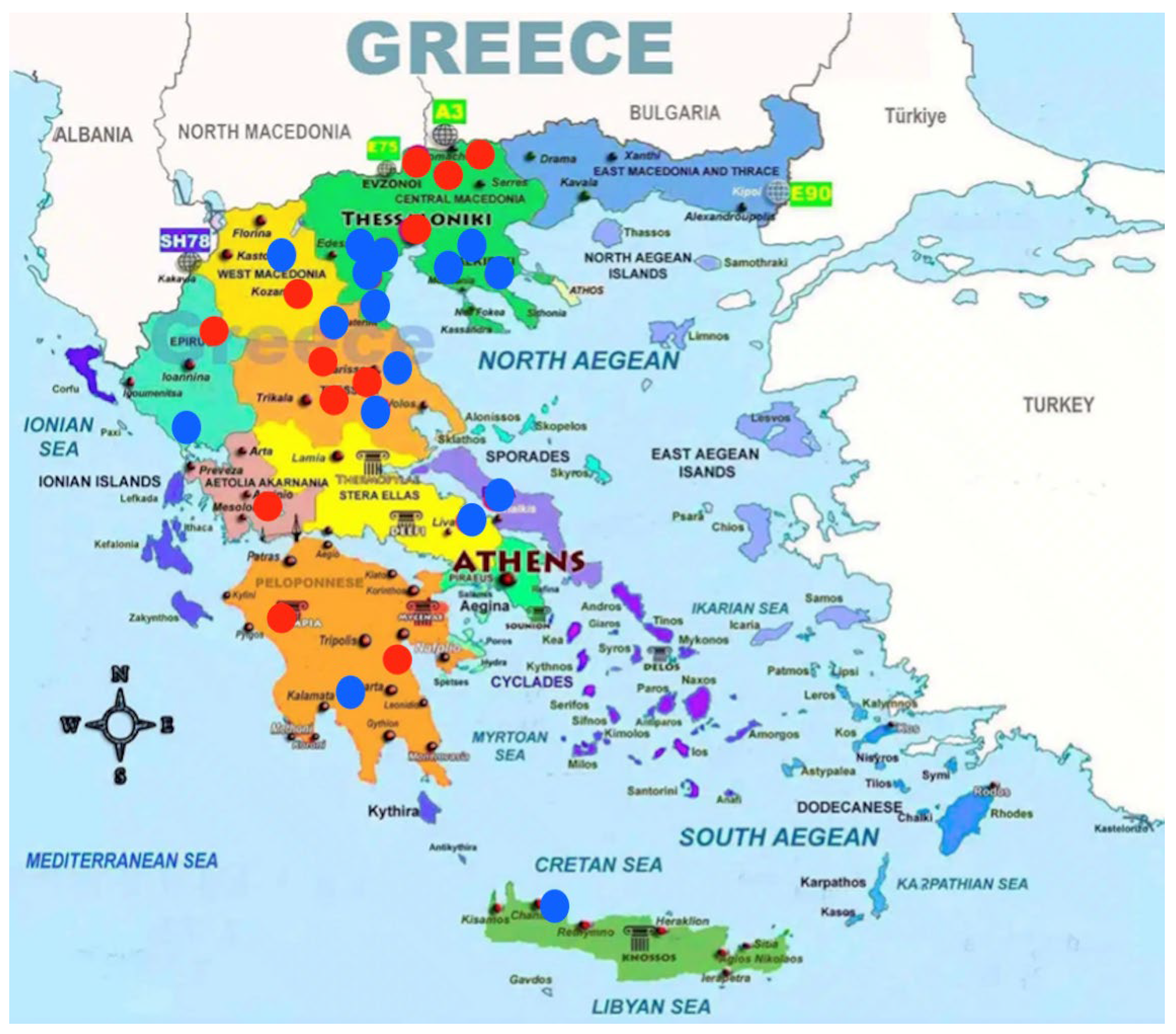
Figure 1. Map of Greece showing the locations of the sampled swine farms, marked with dots. Red dots indicate farms where HEV was detected, and blue dots indicate HEV-negative farms.
Microbiolres 16 00208 g001
Table 1. Distribution of the tested swine farms across the different regions of Greece.
RegionNumber of Sampled FarmsNumber of Sampled PigsHEV Positive FarmsHEV
Negative Farms
% Percentage of HEV Positive Farms
Macedonia131305838.5
Epirus2201150
Thessaly6603350
Sterea Hellas3301233.3
Peloponnese3302166.6
Crete110010
TOTAL28280121642.9
Disclaimer/Publisher’s Note: The statements, opinions and data contained in all publications are solely those of the individual author(s) and contributor(s) and not of MDPI and/or the editor(s). MDPI and/or the editor(s) disclaim responsibility for any injury to people or property resulting from any ideas, methods, instructions or products referred to in the content.