Article

Inhibitory Effects of Selected Chemical Substances on the Growth of Filamentous Fungi Occurring in Cellar Management

Department of Viticulture and Oenology, Faculty of Horticulture, Mendel University in Brno, Valtická 337, 69144 Lednice, Czech Republic
*
Author to whom correspondence should be addressed.
Microbiol. Res. 2025, 16(8), 182; https://doi.org/10.3390/microbiolres16080182
Submission received: 17 June 2025 / Revised: 28 July 2025 / Accepted: 2 August 2025 / Published: 4 August 2025

Abstract

This study evaluated the inhibitory efficacy of sulphur dioxide, hydrogen peroxide, copper sulphate pentahydrate, chlorine-based formulations, a chlorine-free formulation, ethanol, and acetic acid against Cladosporium cladosporioides, Aspergillus niger, and Penicillium expansum. An in vitro inhibition test was employed to investigate the inhibitory properties. The results demonstrated different sensitivities of filamentous fungi to the inhibitors. All tested substances displayed fungicidal properties. Sulphur dioxide (40% NH4HSO3 solution) inhibited growth at a 4% v/v concentration. No minimum effective concentration was established for H2O2; only a 30% w/v solution inhibited P. expansum. CuSO4·5H2O completely inhibited fungal growth at 5% w/v solution, with 2.5% w/v also proving effective. For the chlorine-based product, 40% w/v solution (48 g∙L−1 active chlorine) had the most substantial effect, though it only slowed growth, and NaClO solution completely inhibited growth at 2.35 g NaClO per 100 g of product (50% w/v solution). FungiSAN demonstrated fungicidal effects; however, the recommended dose was insufficient for complete inhibition. Ethanol exhibited the lowest efficacy, while the inhibitory threshold for CH3COOH was found to be a 5% v/v solution. The findings of this study may serve as a basis for informed decision-making when selecting the most suitable product, depending on specific application conditions.

Graphical Abstract

1. Introduction

The cellar environment hosts a diverse community of microorganisms found on material surfaces, as well as in the air [1,2,3]. Among these microorganisms are yeasts, bacteria, and filamentous fungi, including genera such as Penicillium, Aspergillus, and Cladosporium [1]. The total number of identified genera of filamentous fungi is remarkably high—up to 294 in a new winery before the first harvest, decreasing to 132 after two years of use [4]. Contamination can occur at any stage of production, from grapes to the final steps of wine packaging [5]. Filamentous fungi are present on surfaces such as floors, walls, vats, winemaking equipment, pumps, and pipes, affecting both used and new equipment [4,6]. Filamentous fungi of the Aspergillus spp. and Penicillium spp. genera can proliferate in barrels if proper sanitation is not maintained [7].
Some filamentous fungi can occasionally affect workers’ health by causing respiratory diseases such as allergies, asthma, non-specific respiratory symptoms, and irritation [8,9,10]. In the global winemaking sector, there is growing concern, particularly about filamentous fungi of the Aspergillus and Penicillium genera, as they are the primary producers of ochratoxin A [5,11,12] and contributors to cork taint in wine [13,14].
Sanitation can be performed using chemical treatments with substances such as sulphur dioxide (liquid/gas), ozone, peracetic acid (PAA), sodium percarbonate, citric acid, sodium hydroxide, and halogen compounds [7,15]. Physical treatments can also be applied, including hot water and high-pressure cleaning, steam, high-power ultrasound, and ultraviolet radiation [7]. Sulphur dioxide is one of the oldest antimicrobial and antioxidant agents used, not only to stop fermentation and prevent oxidation but also to inhibit filamentous fungal growth in cellar management [16,17].
Traditionally, sanitation agents in winemaking have been based on chlorine, hot water, and steam. However, chlorinated preparations promote the formation of TCA (2,4,6-trichloroanisole), which is why non-chlorinated chemicals are now preferred [18]. Aqueous solutions are the most commonly used; their effectiveness is enhanced by temperature and pressure. Water steam can also be employed for disinfection and cleaning, depending on the temperature and duration of exposure [19,20]. The chemicals and sanitation agents used eventually end up in wastewater, increasing environmental risks and driving the development of more eco-friendly alternatives. These include preparations based on PAA and hydrogen peroxide, often combined with surfactants and chelating agents [15,21].
Ozone is also utilized for effective sanitation, as it destroys microorganisms through oxidation. However, its toxicity poses a health risk; it can also corrode metals such as brass and copper [7,22,23].
The aim of this study was to experimentally verify the effectiveness of selected chemical substances and preparations in inhibiting the growth of Aspergillus niger, Penicillium expansum, and Cladosporium cladosporioides—filamentous fungi commonly found in cellar environments and food-related contaminants, frequently isolated from indoor surfaces, stored products, and food-processing environments. The selected filamentous fungi differ significantly in their ecological and physiological characteristics. Aspergillus niger is a widely distributed thermotolerant organism capable of rapid growth at elevated temperatures due to its versatile metabolism and efficient conidia dispersal [24]. Penicillium expansum is a psychrotolerant post-harvest pathogen of fruits, thriving in cool, humid conditions and known for its production of the mycotoxin [25]. In contrast, Cladosporium cladosporioides is a saprophytic species known for its stress tolerance and ability to grow under low water activity conditions, typically found on damp surfaces and in the air [26]. These species were chosen for their contrasting ecological profiles and differential sensitivity to chemical stressors, enabling a broader assessment of inhibitor efficacy. Testing was conducted using in vitro inhibition tests on malt agar, applying various inhibitory agents at different concentrations to identify the most effective substances for reducing filamentous fungi in winemaking environments.

2. Materials and Methods

2.1. Microorganisms

For this laboratory experiment, pure cultures of filamentous fungi in the form of gelatine discs were used. The filamentous fungi were obtained from the Czech Collection of Microorganisms—CCM (Masaryk University, CZE). The three filamentous fungal strains used were A. niger van Tieghem CCM 8189 (MycoBank MB #284309), P. expansum Link CCM F-576 (MycoBank MB #159382), and C. cladosporioides CCM F-348 (MycoBank MB #294915). The gelatine discs were aseptically transferred onto a Petri dish and activated by adding approximately five drops of distilled water. After 30 min, the activated filamentous fungi were spread across the entire surface of the Petri dish, which was then incubated at 21 °C.

2.2. Culture Media

Malt agar modified (HiMedia Laboratories, Elkdale, NY, USA) was used. Composition: malt extract (powder) 20 g∙L−1, agar 20 g∙L−1, final pH (at 25 °C) 5.4 ± 0.2.

2.3. In Vitro Inhibition Test

On a Petri dish with agar, 350 µL of each inhibitory substance (see Table 1) was applied using an automatic pipette and then spread evenly across the surface with a sterile L-shaped spreader. Distilled water was used as the control sample. From the prepared filamentous fungus cultures, 8 mm diameter discs were cut using a cork borer and placed at the centre of the Petri dish containing the inhibitor (the fungal discs were placed within 3 min after the application of the compounds). Fungal growth was measured every 24 h for a period of 8 days at four points from the edge of the disc to the edge of the mycelium. Each variant was prepared in six replicates. Incubation was carried out at 21 °C.

2.4. Inhibitors

The tested compounds were selected to include both commonly used disinfectants, such as sodium hypochlorite (NaClO, e.g., Savo Original), hydrogen peroxide (H2O2), ethanol and acetic acid (CH3COOH), and agents with more specific or industrial applications, including copper(II) sulphate pentahydrate (CuSO4·5H2O), sulphur dioxide (applied as ammonium hydrogen sulphite, NH4HSO3), a commercial chlorine-free formulation (FungiSAN), and a chlorine-based floor and wall cleaner + Cl. This categorization allows for comparison of general-purpose antiseptics with targeted biocidal agents.
To inhibit filamentous fungus using sulphur dioxide, a commercial product called SUPERSOLFOSOL–ANTIOXIDANT (BS vinařské potřeby s.r.o., Velké Bílovice, Czech Republic) was used. This product is a 40% solution of ammonium hydrogen sulphite (NH4HSO3), commonly referred to as liquid sulphur, used in the food industry, winemaking, or preservation. The following concentrations of this product were tested: 4%, 2%, 1%, and 0.5%.
Hydrogen peroxide p.a. (30% H2O2) (LACHNER, Neratowitz, Czech Republic) was evaluated at various dilutions, specifically 30%, 25%, 20%, and 15%. CuSO4·5H2O, commonly known as blue vitriol (PENTA s.r.o., Katovice, Czech Republic), was prepared in concentrations of 100, 50, 25, and 10 g∙L−1.
Two chlorine-based products were also tested. The first, floor and wall cleaner + Cl (WIGOL, Worms, Germany), is a liquid, alkaline concentrate with active chlorine designed for the beverage and food industry. The recommended concentration ranges from 10 to 100 g∙L−1 (1–10%). The product contains 120 g∙kg−1 of active chlorine. For testing, solutions of 400, 200, 100, and 10 g∙L−1 were prepared. The second chlorine-based product, Savo Dezinfekce Original (UNILEVER ČR, spol. s r.o., Prague, Czech Republic), contains 4.7 g of sodium hypochlorite (NaClO) per 100 g of product and less than 5% chlorine-based bleaching agent. This product was tested at concentrations of 50%, 25%, 10%, and 1%.
The commercial chlorine-free product used was FungiSAN (Stachema CZ s.r.o., Stredocesky, Czech Republic), which is based on organic compounds and contains 4.5 g∙kg−1 zinc pyrithione, 4.5 g∙kg−1 2-octyl-2H-isothiazol-3-one, and 8 g∙kg−1 terbutryn. The manufacturer recommends a dilution of 1:19; that is, 50 g per 1 kg of coating (or water). For experimental purposes, dilutions of 1:5, 1:9, 1:19, and 1:50 were used.
In addition, 96% ethanol p.a. (C2H5OH) (PENTA s.r.o., Katovice, Czech Republic) was tested at concentrations of 96%, 84.4%, 76.8%, and 67.2%. A solution of acetic acid (CH3COOH) was also employed. A commercial 10% white vinegar (FICHEMA s.r.o., Jihomoravsky, Czech Republic) was tested at full strength (10%), as well as at dilutions of 5%, 1%, and 0.5%. For an overview of all concentrations used, see Table 1.

2.5. Statistical Analysis

All experiments were performed in six replicates to ensure accurate statistical evaluation. Data were statistically analyzed using Statistica 14 software. ANOVA analysis was conducted, and mean values (n = 6) were divided into homogeneous groups (labelled a, b, c, ) based on the Fisher LSD test (α = 0.05). In the graphs, the vertical lines represent 0.95 confidence intervals.

3. Results and Discussion

Both commonly used disinfectants and specialized chemical substances with potential antimicrobial effects were tested. Chlorine-based products (sodium hypochlorite and a chlorine-containing surface cleaner) were included as classical disinfectants with proven fungicidal properties. A chlorine-free formulation (FungiSAN) was added as a representative of potentially more environmentally friendly alternatives. Sulphur dioxide, applied in the form of ammonium hydrogen sulphite (NH4HSO3), is commonly used in winemaking and food preservation to suppress microbial growth. Hydrogen peroxide is a well-established and widely available disinfectant, applied across various sectors. Ethanol and acetic acid are simple organic compounds with antiseptic properties, known for their availability and use even in low-tech settings. Copper(II) sulphate was included due to its use in applications such as wood protection and agriculture. Each substance was evaluated based on its effectiveness at different concentrations and its ability to slow down or inhibit fungal growth. The test results are clearly presented in graphs, accompanied by a detailed analysis and a comparison of the effectiveness of the individual substances, including a consideration of their practical application.

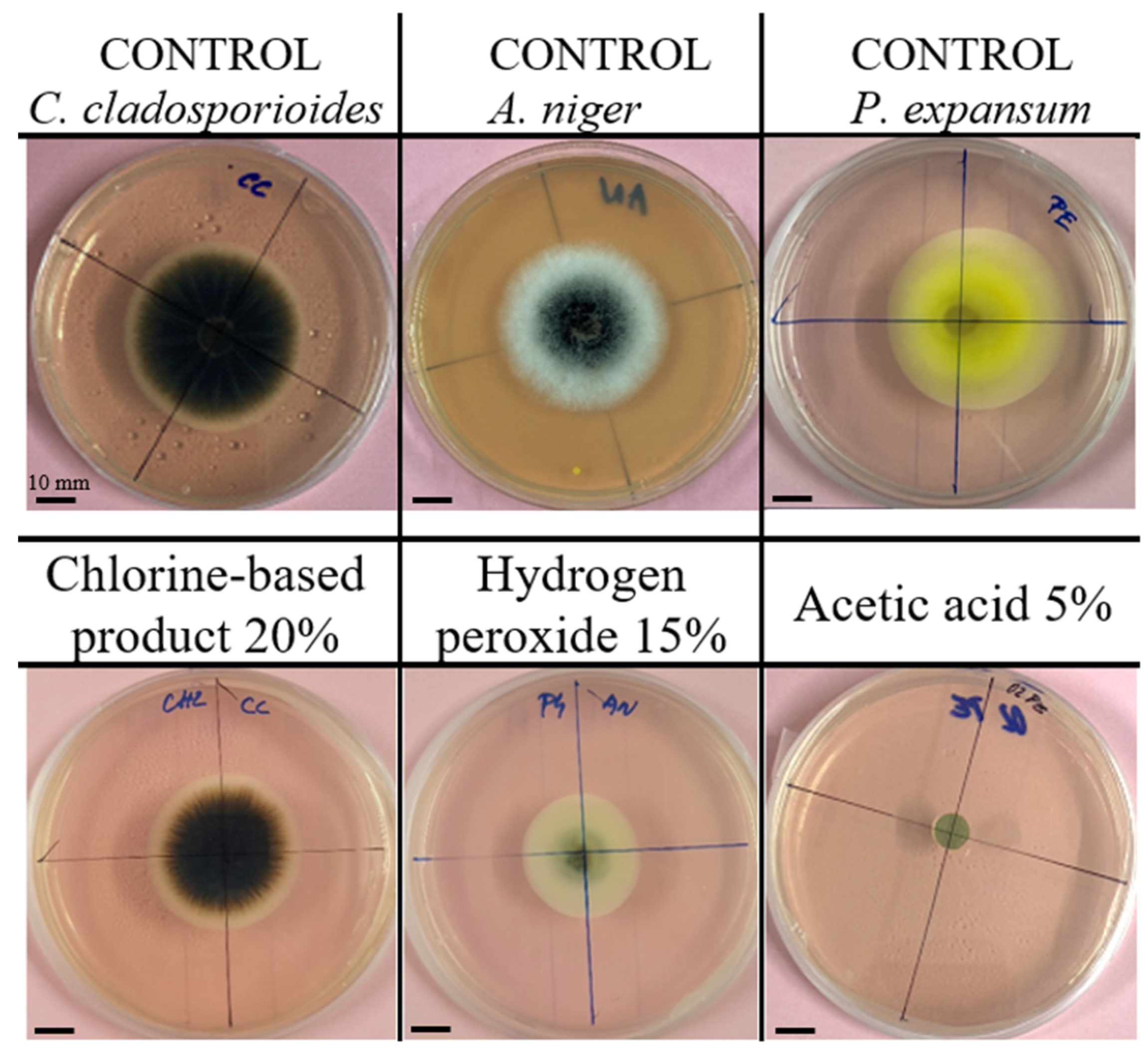
3.1. Representative Images of Fungal Growth

To support the interpretation of the inhibition tests, representative photographs of fungal growth after 8 days of incubation are presented in Figure 1. The incubation was carried out on malt agar at 21 °C in 90 mm diameter Petri dishes (the fungal disk has a diameter of 8 mm). The images include untreated controls of A. niger, P. expansum, and C. cladosporioides and examples of growth inhibition by selected chemical agents. These photographs illustrate the typical morphology of the fungal colonies and the inhibitory effects observed under the test conditions.

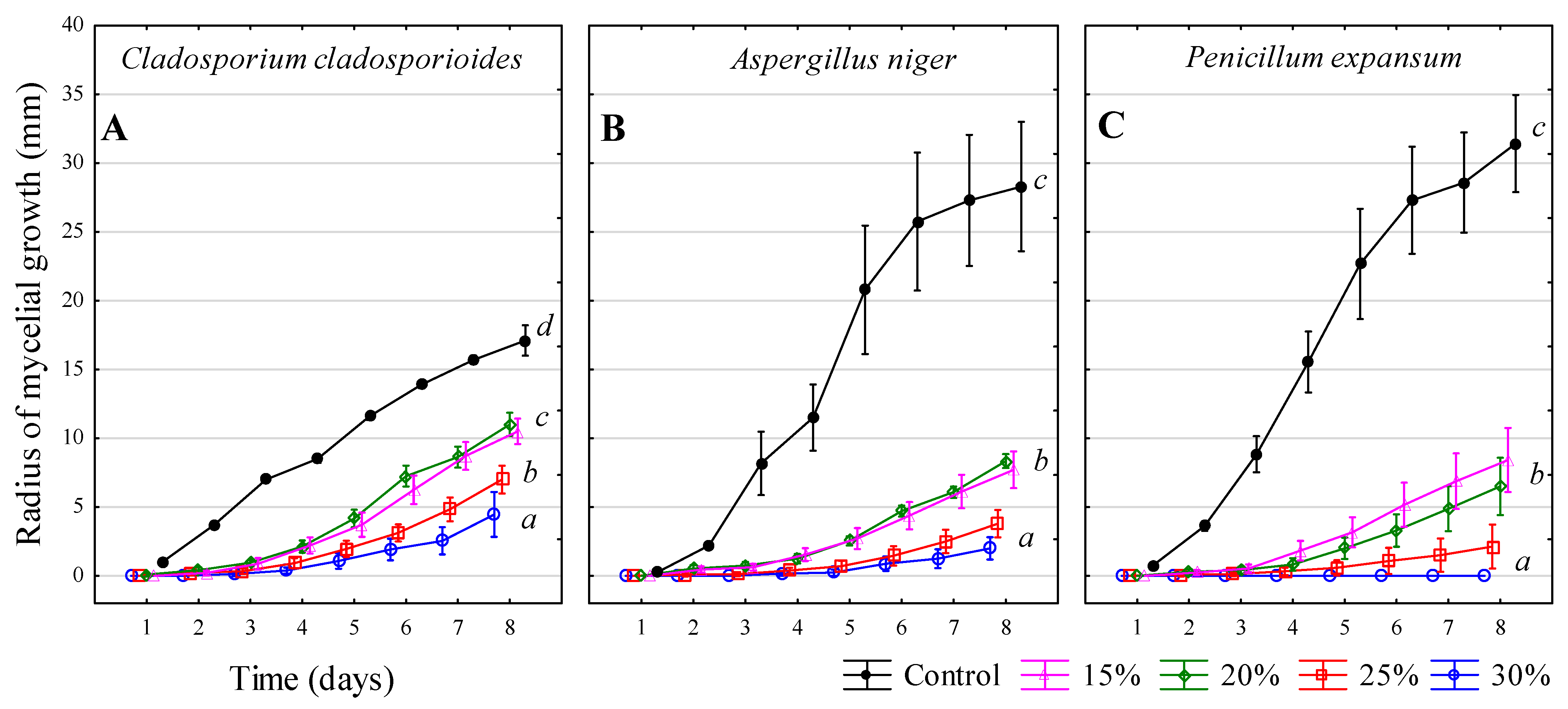
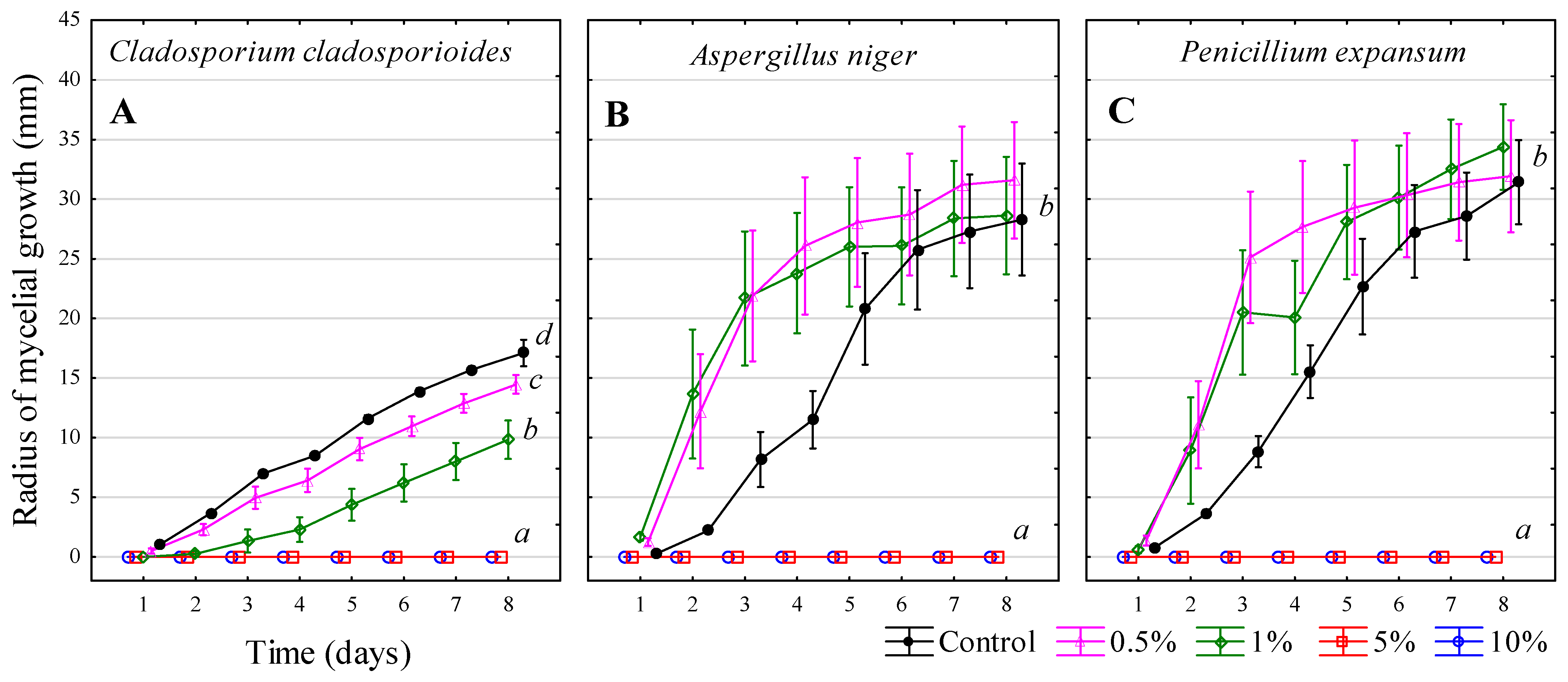
3.2. Sulphur Dioxide SO2–Solution NH4HSO3

For C. cladosporioides (Figure 2A), the control sample exhibited significantly higher growth on day 8. The 0.5% solution led to a marked reduction in growth (58% effectiveness), while the 1%, 2%, and 4% solutions displayed significantly lower values, with no statistical differences among them. The 4% solution achieved complete (100%) effectiveness. In the case of A. niger (Figure 2B), the 0.5% solution unexpectedly showed significantly higher growth values than the control. The 1% solution demonstrated noticeably less growth compared to the control on day 8, with an effectiveness of 82%. The 2% and 4% solutions exhibited the most substantial reduction in growth, achieving 94% and 100% effectiveness, respectively. For P. expansum (Figure 2C), on day 8, the 0.5% solution displayed values statistically similar to the control. A considerable reduction was observed with the 1% solution (36% effectiveness), while the 2% solution exhibited significantly lower values (90% effectiveness). The 4% solution demonstrated complete (100%) effectiveness.
Sulphur dioxide is commonly used in the wine industry not only for disinfecting barrels but also as a preservative in wine. However, its use may be associated with certain risks, as it can irritate the respiratory tract and eyes [27] or cause corrosion of metal parts [28]; its use should follow strict safety protocols in enclosed environments. SO2 exerts its antifungal activity primarily through reactions with cellular thiol groups, disrupting redox homeostasis and inducing oxidative stress. It interferes with the structural integrity of the fungal cell wall by targeting components such as chitosan, which also plays a role in pigment retention and membrane stability. SO2 further impairs cellular metabolism and enzymatic function, contributing to growth inhibition. The antifungal effect is influenced by the fungus’s ability to activate antioxidative defense mechanisms, including glutathione-mediated responses [29,30]. While sulphur dioxide exhibited a strong fungicidal effect, its effectiveness depends on the concentration. Overall, the 4% solution proved to be the most effective, completely stopping fungal growth and spread. In contrast, the 0.5% solution was almost ineffective, showing results comparable to the control samples.
The C. cladosporioides was the most sensitive to the preparation, with the 0.5% solution demonstrating 58% effectiveness, while the results for P. expansum and A. niger were inconclusive. To achieve optimal inhibitory effectiveness, it is recommended that the highest concentration of the tested preparation be used.
Jiang et al. [31] mentioned that low levels of SO2 may actually stimulate growth, as sulphur is an essential element for the growth of microorganisms. This could explain why the 0.5% solution appeared to stimulate the growth of certain filamentous fungi. Davidson et al. [32] stated that a 0.1% concentration of sulphur dioxide has minimal effect and only moderates fungal growth.
Kazemi et al. [33] studied the effect of sulphur on the toxic Aspergillus flavus in vitro, observing complete inhibition at a concentration of 15 g∙L−1. In the case of A. niger, higher concentrations were found to completely prevent growth.
Everitt and Sullivan [34] conducted tests showing that each sulphur compound has varying levels of effectiveness against filamentous fungi. According to Davidson, Sofos, and Branen [32], temperature also influence effectiveness—sulphur dioxide is less effective at higher temperatures.

3.3. Hydrogen Peroxide—H2O2

For C. cladosporioides (Figure 3A), the control sample exhibited the highest growth on day 8. The 15% and 20% hydrogen peroxide solutions resulted in a noticeable reduction in growth (approximately 37% effectiveness). Lower fungal growth was observed with the 25% solution, while the 30% hydrogen peroxide solution showed the lowest growth, achieving 74% effectiveness. In the case of A. niger (Figure 3B), the 15% and 20% solutions demonstrated significantly lower growth in millimetres compared to the control (72% effectiveness). The 25% hydrogen peroxide solution further reduced growth, while the statistically lowest values were recorded with the 30% solution, showing 93% effectiveness. For P. expansum (Figure 3C), the 15% and 20% hydrogen peroxide solutions significantly reduced growth compared to the control (75% effectiveness), with no statistical difference between them. The 25% solution demonstrated a stronger inhibitory effect (93%), while the 30% solution achieved complete inhibition (100% effectiveness), completely stopping fungal growth. Statistically, these concentrations were not significantly different from each other.
Hydrogen peroxide is a weak acid and a strong oxidizing agent that exerts antifungal activity by inducing oxidative stress, damaging fungal cell membranes, DNA, essential enzymes, and other biomolecules of microorganisms. It can also affect ligninolytic enzymes such as laccase and manganese peroxidase, with low concentrations stimulating and high concentrations inhibiting their activity, ultimately limiting fungal growth [35,36]. Although it is unstable and harder to store, hydrogen peroxide decomposes into water and oxygen, making it environmentally friendly and non-polluting to wastewater [37]. However, it may also have corrosive effects and, in high concentrations, it poses risks of skin, eye, and respiratory irritation [38].
Sharaf-Eldin and Geösel [39] confirmed that even a 1% hydrogen peroxide solution can slow fungal growth. Cerioni et al. [40] found that 400 mmol∙L−1 H2O2 completely inhibited the growth of P. expansum conidia, which corresponds to a significantly lower dose compared to the concentrations used in this study. Muhialdin, Hassan, and Sadon [35] reported that a 5% hydrogen peroxide solution completely inhibited the growth of P. expansum. They also noted that hydrogen peroxide degrades in MRS agar, indicating that the type of growth medium used may affect test results.
Martin and Maris [41] observed that various combinations of hydrogen peroxide with acids could inhibit microorganisms. However, it is worth considering whether hydrogen peroxide may have decomposed before making contact with the filamentous fungus, potentially reducing its effectiveness. This assumption is supported by the observation that only the 30% hydrogen peroxide solution was able to nearly halt fungal growth.

3.4. Copper(II) Sulphate Pentahydrate—CuSO4·5H2O

For C. cladosporioides (Figure 4A), the control sample exhibited the highest growth on day 8. The 1% solution led to a significant reduction in growth, demonstrating 85% effectiveness, while the 2.5%solution showed a markedly stronger inhibition, achieving 95% effectiveness. The 5% and 10% solutions achieved complete inhibition (100% effectiveness), with zero growth recorded. In the case of A. niger (Figure 4B), the 1% solution was statistically indistinguishable from the control on day 8. The 2.5% solution demonstrated clear inhibition, with 92% effectiveness. The highest concentrations, 5% and 10% solutions, both achieved complete inhibition (100% effectiveness). For P. expansum (Figure 4C), the control sample exhibited the highest growth. The 1% solution demonstrated noticeably lower values, with an inhibitory effectiveness of 90%. Higher concentrations of solutions, 2.5%, 5%, and 10%, clearly inhibited growth, each achieving 100% effectiveness.
Blue vitriol (copper(II) sulphate) exerts antifungal effects by releasing Cu2+ ions, which disrupt fungal cell membranes, denature proteins, and interfere with essential enzymatic processes. Copper ions also promote the formation of reactive oxygen species (ROS), leading to oxidative damage of cellular components. The overall mechanism results in impaired metabolism, membrane disintegration, and growth inhibition [42]. Copper(II) sulphate exhibits a strong inhibitory effect, with the 1% solution (10 g∙L−1) significantly reducing fungal growth in C. cladosporioides and P. expansum. The concentration level required to completely prevent growth ranged from 2.5–5% solution of blue vitriol (25 g∙L−1 to 50 g∙L−1).
Araujo et al. [43] confirmed that filamentous fungi of the Aspergillus genus are sensitive to copper sulphate. Cerioni et al. [44] studied the inhibitorty effects of oxidative compounds, including copper sulphate. They found that a concentration of 300 mmol∙L−1 copper sulphate had no impact on the viability of conidia. However, in this experiment, even 40 mmol∙L−1 (equivalent to 10 g∙L−1–1% solution) significantly affected filamentous fungus viability.
Henry et al. [45] used copper oxide nanoparticles, which proved to be highly effective against A. niger and Penicillium chrysogenum. Similarly, Pařil et al. [46] investigated copper nanoparticles against wood-decaying fungi, finding that a concentration of 3 g∙L−1 was effective against the tested fungi. These findings suggest that copper-based treatments could be beneficial for maintaining the condition of barrels. Although blue vitriol can have corrosive effects, it is compatible with stainless steel and plastic [47].

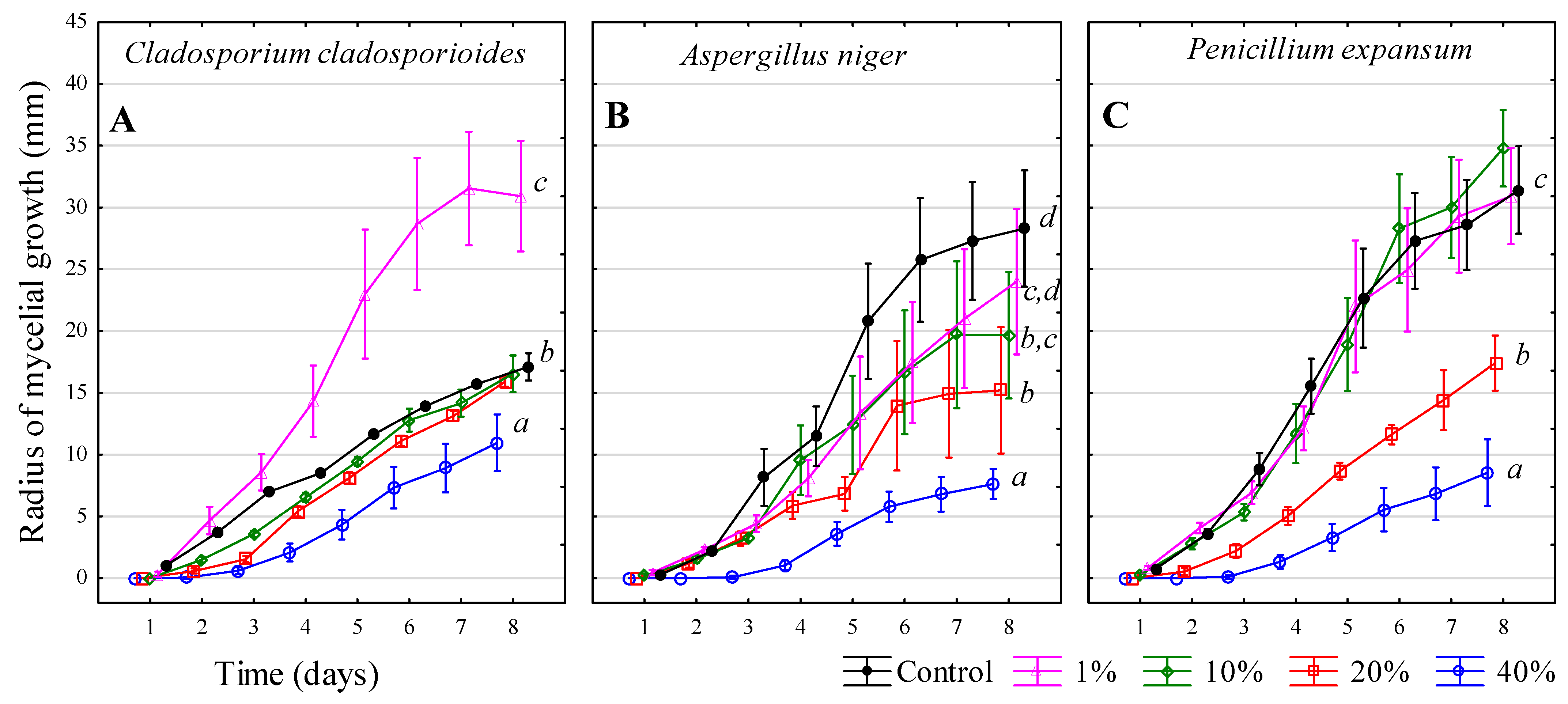
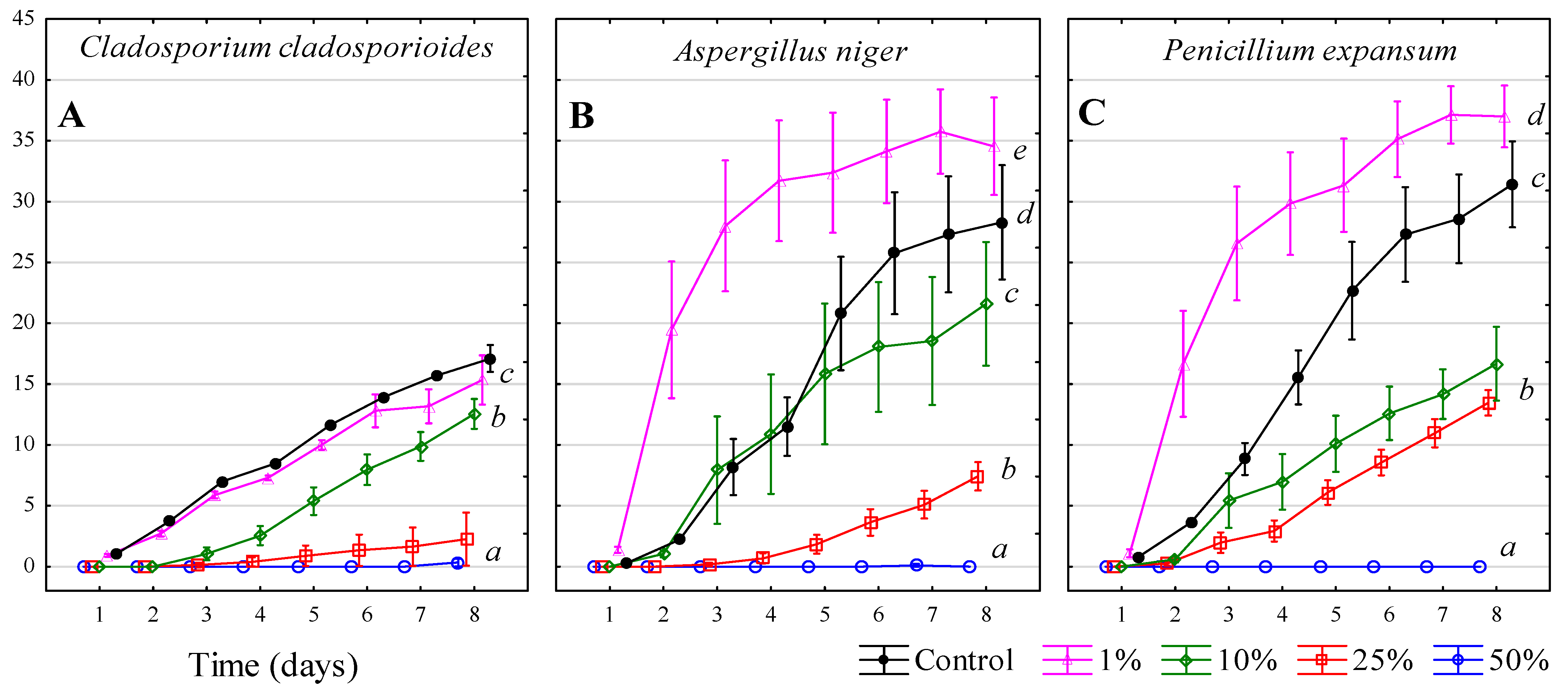
3.5. Chlorine-Based Products

For C. cladosporioides (Figure 5A), the 1% solution exhibited the highest values, with the filamentous fungus spreading faster than in the control. The 10% and 20% solutions showed statistically similar values to the control on day 8. The most significant inhibition was observed at 40% solution, with an effectiveness of 36%. For A. niger (Figure 5B), the control and the 1% solution showed statistically similar values on day 8. This concentration did not differ significantly from the 10% solution. The 20% solution demonstrated significantly higher effectiveness (46%), while the most statistically effective was the 40% solution, with 73% effectiveness on day 8. For P. expansum (Figure 5C), the control did not differ significantly from the 1% and 10% solutions. A noticeably lower growth rate was recorded at 20% solution (45% effectiveness), and the lowest growth was clearly observed at 40%, showing an inhibitory effect of 73%.
Floor and wall cleaner + Cl
Figure 5. The effect of chlorine-based products (floor and wall cleaner + Cl) on the growth of selected filamentous fungi. (A) Cladosporium cladosporioides, (B) Aspergillus niger, (C) Penicillium expansum. Legend: Mean values (n = 6) on the last day of measurement were divided into homogeneous groups (marked by letters a, b, c, d) based on Fisher’s LSD test (α = 0.05).
Microbiolres 16 00182 g005
Chlorine-based disinfectants act through various mechanisms to inactivate fungal spores. Chlorine and chlorine dioxide (ClO2) disrupt spores by damaging the cell wall and membrane, leading to leakage of intracellular contents and subsequent cell death [48]. Chlorine inhibits fungal growth by reacting with cell wall components, particularly chitosan, which reduces the amount of active chlorine available and supports cell wall integrity. Chitosan also contributes to melanin production and pigment retention, both important for fungal survival. Chlorine further induces oxidative stress by generating ROS inside the cell. In response, fungi activate antioxidant enzymes such as superoxide dismutase and catalase to counteract the damage [49]. While this preparation exhibits antifungal effects, these effects only occur at sufficiently high concentrations. The strongest effect was observed at the tested 40% solution (concentration of 400 g∙L−1–48 g∙L−1 of active chlorine), which slowed fungal growth but did not completely halt it. Achieving full inhibition would require a higher concentration.
The manufacturer’s recommended dose of 10% solution (100 g∙L−1–12 g∙L−1 of active chlorine) did not significantly slow growth, as the values remained close to the control. Lower recommended doses had minimal to no effect and, in some cases, even a negative impact. The observed stimulation of fungal growth at low concentrations of chlorine-based agents may be attributed to the phenomenon of hormesis, a biphasic dose–response in which low levels of a stressor activate protective or metabolic pathways, while higher concentrations exert an inhibitory effect [50,51]. ClO2 appears to be a more effective disinfectant compared to active chlorine. It is a strong oxidizing agent with broad-spectrum bactericidal activity, demonstrating high efficacy against most bacteria and pathogenic microorganisms. Its low risk of resistance development and minimal formation of halogenated by-products make it suitable for various applications, including water treatment, food safety, healthcare, public health, and environmental protection. In contrast, active chlorine is cheaper and more readily available but may lead to the formation of harmful chlorinated compounds [52].
For C. cladosporioides (Figure 6A), the 1% solution showed values statistically similar to the control on day 8, indicating no significant difference. The 10% solution demonstrated a significantly lower value (27% effectiveness). The most clearly effective results were observed with the 25% and 50% solutions, which did not differ statistically from each other. The 25% solution achieved 87% effectiveness, while the 50% solution reached 98% effectiveness, completely preventing fungal growth. For A. niger (Figure 6B), the 1% solution significantly promoted growth, showing an increase greater than that of the control. The 10% solution demonstrated significantly lower growth compared to the control on day 8. A substantial reduction in growth was observed with the 25% solution (74% effectiveness), while the 50% solution exhibited the highest inhibitory effect (100% effectiveness). For P. expansum (Figure 6C), the 1% solution resulted in the highest growth, significantly greater than the control. A significant reduction compared to the control was observed with the 10% and 25% solutions (47% and 57% effectiveness, respectively), with no statistical difference between them. The 50% solution completely inhibited fungal growth (100% effectiveness).
NaClO-based product (Savo)
Figure 6. The effect of NaClO-based product on the growth of selected filamentous fungi. (A) Cladosporium cladosporioides, (B) Aspergillus niger, (C) Penicillium expansum. Legend: Mean values (n = 6) on the last day of measurement were divided into homogeneous groups (marked by letters a, b, c, d, e) based on Fisher’s LSD test (α = 0.05).
Microbiolres 16 00182 g006
Savo is a disinfectant based on NaClO, a chlorine-containing compound known for its strong antimicrobial activity and extensive use in household, industrial, public, and medical settings. Its effectiveness varies with concentration [53]. In all three cases, the 50% solution (corresponding to approximately 23.5 g∙L−1 NaClO) completely stopped fungal growth and spread. The 25% solution was most effective against C. cladosporioides but showed a weaker effect on P. expansum. Filamentous fungi differ in their resistance to this preparation, with the greatest inhibition observed in C. cladosporioides, while A. niger and P. expansum exhibited higher resistance. Notably, the 1% solution lacked inhibitory properties and even had a stimulatory effect, with values exceeding those of the control, suggesting a possible growth-promoting effect. Low concentrations of NaClO may act as mild oxidative stressors, temporarily activating cellular defence and metabolic pathways, which can paradoxically promote fungal growth. This biphasic dose–response phenomenon, known as hormesis, has been reported in fungal pathogens exposed to fungicides and other chemical agents [50,51].
Araujo, Gonçalves Rodrigues, and Pina-Vaz [43] confirmed that filamentous fungi of the Aspergillus genus are sensitive to NaClO. Reynolds et al. [54] evaluated the effectiveness of a 2.4% NaClO solution, which is approximately equivalent to a 50% dilution of the tested product, and found it effectively reduced filamentous fungi of the Penicillium, Cladosporium, and Aspergillus genera. Cerioni, de los Ángeles Lazarte, Villegas, Rodríguez-Montelongo, and Volentini [44] identified the minimum inhibitory concentration for P. expansum to be 50 mg∙L−1, which is significantly lower than the concentrations that completely inhibited P. expansum in this study.
Visconti et al. [55] recommended using 0.1–1% NaClO or 1–3 mg∙L−1 of free chlorine for C. cladosporioides, 0.1–1.6% NaClO or 100–1300 mg∙L−1 of free chlorine for A. niger, and 50 ppm (0.005%) free chlorine for P. expansum, or 500 ppm (0.05%) when applied to surfaces. However, these values proved insufficient in this test, requiring higher concentrations for complete inhibition. Visconti et al. [56] reported that using 0.2% active chlorine for 2.5 min resulted in the complete inactivation of C. cladosporioides. Nevertheless, the relevance of free chlorine values is limited in this context, as chlorine use in the wine industry should generally be avoided due to the risk of cork taint in wine.
Chlorine remains one of the most effective and widely used disinfectants in the food industry. One of its key advantages is that its use is well-established and well-understood. When comparing chlorine-based products, Savo appears to be more effective than floor and wall cleaner + Cl. After recalculating, a 50% solution of Savo contains approximately 45 g∙L−1 of active chlorine, which completely prevented fungal spread. In contrast, 400 g∙L−1 of the cleaner product (containing 48 g∙L−1 of active chlorine) only limited and slowed down fungal growth.
The higher effectiveness of Savo may be attributed to its additional components, such as trace amounts of sodium hydroxide, which help stabilize pH and prevent the breakdown of hypochlorite. However, these products also contain volatile halogen compounds, making them unsuitable for use in the wine industry due to the risk of cork taint. Furthermore, they are characterized by a strong “pool-like” odor, which is undesirable in winemaking environments.

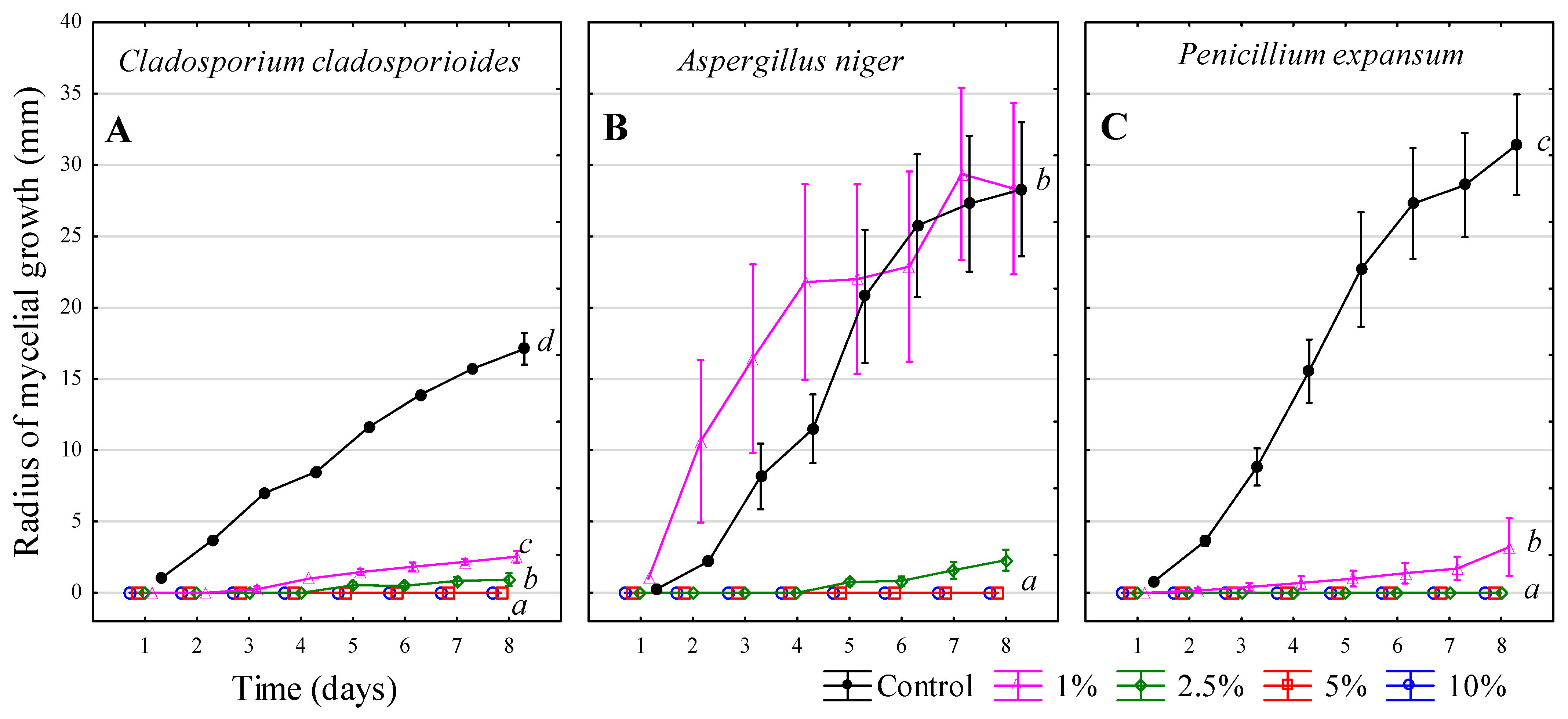
3.6. Chlorine-Free Product (FUNGISAN)
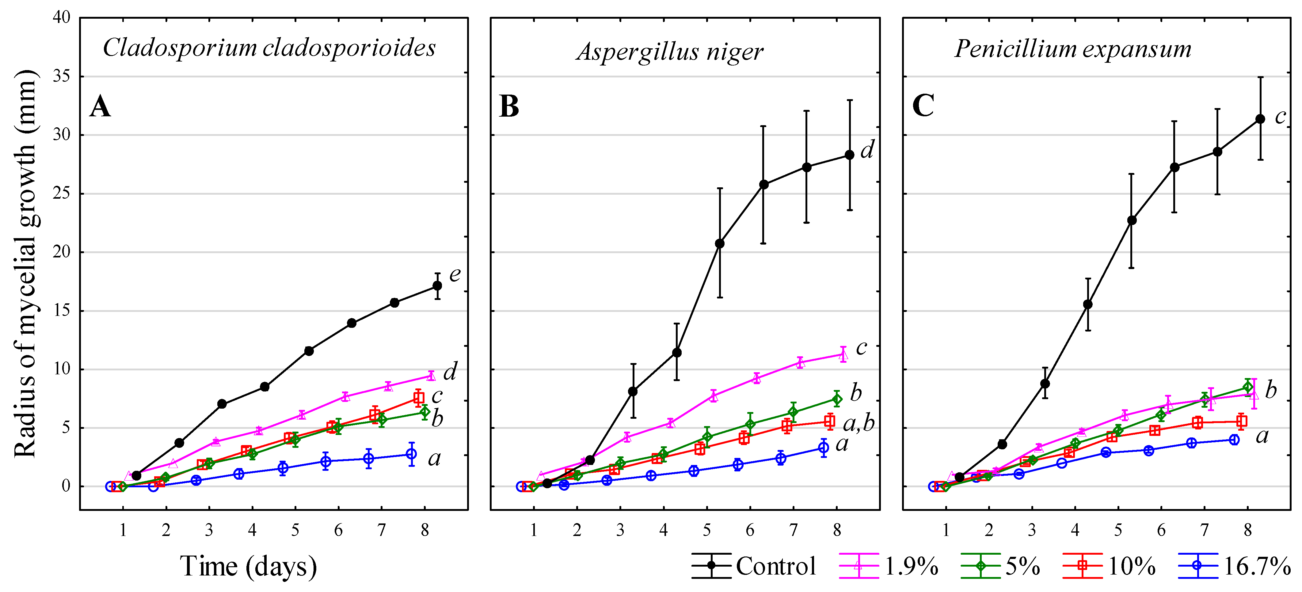
For C. cladosporioides (Figure 7A), the control showed the highest growth on day 8. At a 1.9% solution, growth was significantly reduced (45% effectiveness). On day 8, the 5% solution (63% effectiveness) and the 10% solution (56% effectiveness) differed statistically, with the 5% solution showing lower growth. The highest inhibitory effectiveness (84%) was clearly achieved with the 16.7% solution. For A. niger (Figure 7B), the control values were visibly the highest. On day 8, the 1.9% solution demonstrated clearly lower growth with 60% effectiveness. Statistically lower values were observed at 5% and 10% solutions (73% and 80% effectiveness, respectively). The highest inhibition was recorded at the 16.7% solution, with 88% effectiveness. For P. expansum (Figure 7C), the control exhibited the highest growth. The 1.9% and 5% solutions did not differ statistically from each other and showed significantly reduced fungal growth, both with 74% effectiveness. Similar growth was observed at the 10% and 16.7% solutions, both achieving 87% effectiveness.
FungiSAN is a chlorine-free preparation that has proven effective against filamentous fungi. The manufacturer recommends a dilution ratio of 1:19 (5% w/v); however, this dose appears to be insufficient. While this concentration effectively slowed fungal growth, it did not completely prevent its spread. Better results were achieved with higher concentrations, with the 16.7% solution being the most effective-though not entirely inhibitory.
FungiSAN contains 4.5 g∙kg−1 of zinc pyrithione, 4.5 g∙kg−1 of 2-octyl-2H-isothiazol-3-one (OIT), and 8 g∙kg−1 of terbutryn. Zinc pyrithione, functioning as a zinc ionophore, increases intracellular Zn2+ levels, leading to mismetalation and inactivation of iron–sulfur proteins essential for energy metabolism, while also impairing nutrient transport via membrane depolarization [57]. OIT, an isothiazolinone biocide, reacts with cellular nucleophiles via an activated N–S bond, disrupting enzymatic and metabolic functions and compromising cell integrity [58]. Terbutryn disrupts respiratory enzymatic systems by inhibiting electron transport processes, thereby reducing fungal metabolic capacity and growth [59]. Reeder, Xu, Youngquist, Schwartz, Rust, and Saunders [57] demonstrated that zinc pyrithione inhibits growth by increasing cellular copper levels, leading to damage to iron–sulphur protein clusters essential for fungal metabolism. Li et al. [60] also found that combining zinc pyrithione with silver nanoparticles had inhibitory effects on fungal growth. Although promoted as a chlorine-free alternative, FungiSAN contains isothiazolinones and terbutryn, which are toxic to aquatic life and require careful handling and disposal.

3.7. Ethanol—C2H5OH

For C. cladosporioides (Figure 8A), fungal growth on day 8 showed no significant difference between 96%, 86.4%, and 76.8% ethanol compared to the control, indicating no inhibitory effect. The highest growth on day 8 was recorded with 67.2% ethanol, significantly exceeding the control. For A. niger (Figure 8B), the control showed the highest growth on day 8. Statistically similar values and significantly lower growth were observed with 67.2% ethanol (24% effectiveness) and 96% ethanol (40% effectiveness). Clearly reduced growth was recorded with 86.4% ethanol, while the most statistically effective inhibition (67%) was observed with 76.8% ethanol. For P. expansum (Figure 8C), the control exhibited the highest growth on day 8. The 67.2% ethanol concentration resulted in significantly reduced growth (26% effectiveness), with similar values observed for 86.4% ethanol (37% effectiveness). Significantly lower growth was recorded with 96% and 86.4% ethanol, with the best inhibitory effect (51%) shown by 76.8% ethanol.
Ethanol is biodegradable and generally considered safe. However, it is highly volatile and flammable, requiring careful storage and handling. Its antifungal mechanism is primarily based on disrupting the fungal cell membrane, leading to loss of permeability and cell disintegration. Ethanol denatures proteins in the cell wall, cytoplasmic membrane, and cytoplasm, inhibits nutrient uptake (e.g., glucose and ammonium ions), and targets mitochondrial membranes by altering phospholipid phase transition, increasing spore mortality. Its effectiveness depends on concentration, temperature, moisture, and fungal development stage, with ~70% ethanol showing optimal activity due to protein denaturation and membrane damage [61]. Ethanol demonstrated an overall lower inhibitory effect compared to other tested substances. For some filamentous fungi, medium concentrations (76.8% and 86.4%) were more effective than 96% ethanol. The lowest tested concentration (67.2%) had a weak effect or did not slow fungal growth at all. The filamentous fungi varied in their resistance, with C. cladosporioides being the most resistant, showing only minimal inhibition.
According to these studies [62,63], alcohol appears to be inactive against spores. Sequeira et al. [64] demonstrated antifungal activity at 30%, 70%, and 100% ethanol concentrations, with 70% ethanol showing fungicidal properties against A. niger and C. cladosporioides. Dantigny et al. [65] noted that ethanol appears effective in inhibiting fungal growth. Dao and Dantigny [61] reported qualitative growth inhibition at ethanol concentrations ranging from 0–12%, depending on the filamentous fungus species. Between 0–4%, growth was observed in all studied filamentous fungi (P. expansum), but at 8%, fungal growth was halted.

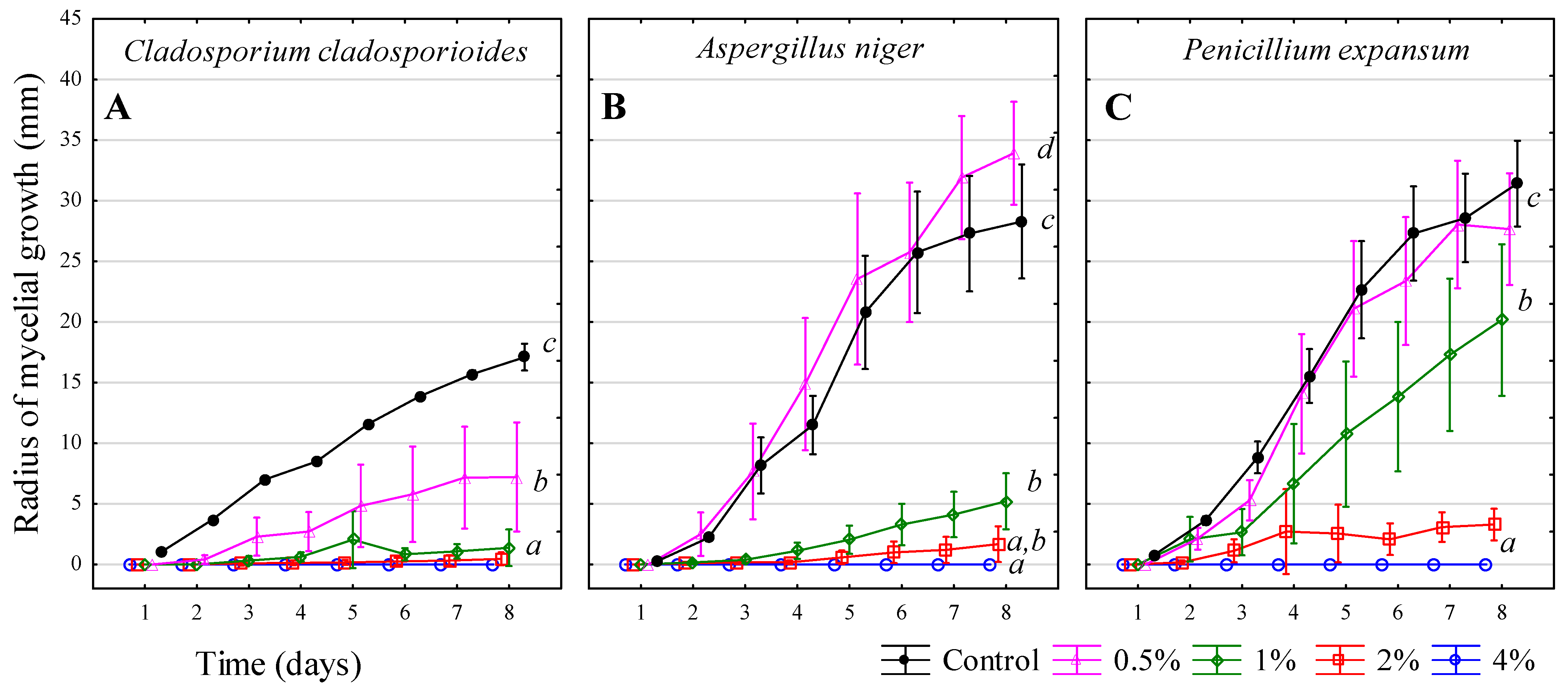
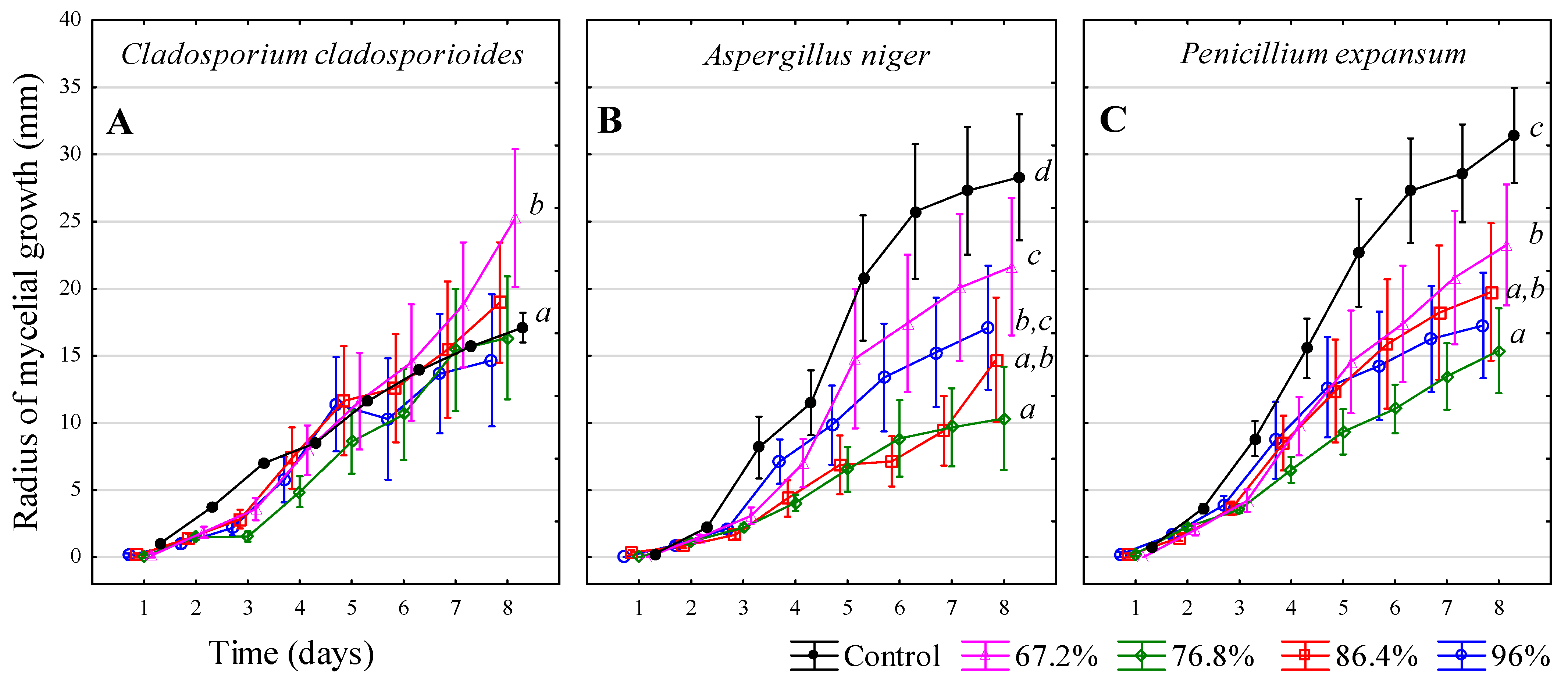
3.8. Acetic Acid—CH3COOH

For C. cladosporioides (Figure 9A), the control sample showed the highest growth on day 8. Significantly lower growth was observed, with the 0.5% solution achieving 15% effectiveness. Clearly different values were recorded for the 1% solution, which demonstrated 43% effectiveness. The 5% and 10% acetic acid solutions exhibited a proven 100% inhibitory effect. For A. niger (Figure 9B), similar values were measured for the control, 0.5%, and 1% solutions, which showed no inhibitory effect and even promoted growth. Complete suppression of growth occurred with the 5% and 10% solutions, both demonstrating 100% effectiveness. For P. expansum (Figure 9C), the control and 0.5% and 1% acetic acid solutions showed no significant differences on day 8. However, the 5% and 10% solutions clearly inhibited growth, both achieving 100% effectiveness.
Acetic acid is a weak organic acid and the main component of vinegar [66,67]. It exhibits antimicrobial activity primarily through the acidification of the extracellular environment, which impairs microbial metabolism and inhibits cellular proliferation. Furthermore, the undissociated form of acetic acid is able to penetrate fungal cell membranes and dissociates within the cytoplasm, leading to intracellular acidification, disruption of enzymatic function, oxidative stress, and loss of membrane integrity. Although acetic acid is biodegradable and generally regarded as safe at low concentrations, its concentrated form is corrosive and may cause dermal irritation [68,69]. The acid is stable and can be stored for long periods. Its effectiveness ranges from low to moderate and is mostly effective against bacteria and filamentous fungi [69].
C. cladosporioides was the most susceptible to acetic acid, with all tested concentrations showing some inhibitory effect. Surprisingly, A. niger and P. expansum exhibited enhanced growth at low acetic acid concentrations (0.5% and 1%) compared to the control. This may be explained by the hormetic effect, a biphasic dose–response where low doses of stress-inducing agents stimulate microbial growth or metabolism, while higher doses are inhibitory. Similar stimulatory effects of weak organic acids at low concentrations have been observed in fungal systems [70]. In contrast, A. niger and P. expansum were more resistant, with the 1% solution showing no inhibitory effect. The 5% solution, however, was able to completely halt the growth and spread of all tested filamentous fungi.
This result was also confirmed by Al-Sawah et al. [71], who used a different inhibition method (well diffusion test) and observed pronounced inhibition zones with a 5% white vinegar solution. Niamat et al. [72] also confirmed the inhibitory effects of white vinegar against A. niger. Alcano et al. [73] found inhibitory effects at concentrations of 36–44 mM, which are significantly lower than the concentrations identified in this study. Furthermore, acetic acid at concentrations of 1%, 3%, and 5% has been shown to completely stop the growth of A. flavus [74].
Not only acetic acid but also other organic acids, such as lactic, formic, oxalic, and propionic acids, have fungicidal properties [75]. The study by Niamat, Imtiaz, and Khan [72] also showed that different types of vinegar (white distilled, grape, apple) can serve as alternatives to synthetic antifungal agents. Thanks to their content of active compounds like phenols and flavonoids, these vinegars exhibit antifungal and antioxidant properties, helping to mitigate the drawbacks of synthetic products. Among the tested types, apple vinegar showed the most effective results against A. niger.
Acetic acid can also cause corrosion of metal components [76]. This raises the question of whether using acetic acid in winemaking is safe. Common vinegar may contain acetic acid bacteria and is, therefore, not suitable for disinfecting winemaking tools or barrels, as it could lead to undesirable effects under unfavourable conditions [67]. For safe disinfection, pure acetic acid (of at least 80–100% purity, whether technical or food-grade quality) must be used as it does not contain microorganisms. The acid should then be diluted to the desired concentration.
PAA also appears to be a promising alternative, as it is a strong oxidizing agent formed by mixing hydrogen peroxide with acetic acid. PAA exhibits stronger antimicrobial effects than hydrogen peroxide due to its combined oxidative action and ability to destroy cellular structures. However, PAA is unstable and requires careful storage, decomposing completely into acetic acid, hydrogen peroxide, and water [77]. Abdelrhim et al. [78] confirmed the inhibitory effects of PAA, while Hosotani et al. [79] mentioned that 80 mg∙L−1 of PAA is highly effective against filamentous fungi.

3.9. Summary

A 40% v/v solution of ammonium hydrogen sulphite was used for sulphur dioxide inhibition, and a 4% v/v solution completely inhibited filamentous fungal growth. C. cladosporioides proved to be the most sensitive to sulphur dioxide. For hydrogen peroxide, the only concentration that inhibited the growth of P. expansum was 30% w/v. The lowest effectiveness of hydrogen peroxide was observed against C. cladosporioides, indicating that higher concentrations would be necessary for complete inhibition.
CuSO4·5H2O (blue vitriol) was able to completely inhibit fungal growth at a concentration of 50 g∙L−1 (5% w/v solution), while 25 g∙L−1 (5% w/v solution) also proved very effective, particularly against P. expansum, which was the most sensitive to copper sulphate. Overall, copper sulphate demonstrated the best cost-effectiveness ratio, achieving excellent inhibition results while remaining economically accessible.
The chlorine-based product floor and wall cleaner + Cl, tested at its strongest concentration of 40% w/v solution (400 g∙L−1–48 g∙L−1 active chlorine), was able to slow fungal growth but not stop it entirely. This concentration was four times higher than the manufacturer’s recommended dose and still proved insufficient. The lowest effectiveness was observed against C. cladosporioides, while the product demonstrated twice the effectiveness against A. niger and P. expansum. Savo, a NaClO-based product, completely halted the growth of filamentous fungi at a concentration containing 23.5 g∙L−1 of NaClO. The highest effectiveness was recorded against C. cladosporioides. The chlorine-free product FungiSAN exhibited good fungicidal properties, but the manufacturer’s recommended dose was insufficient to completely inhibit fungal growth. A higher dosage would be necessary to achieve full inhibition. Better results were observed against A. niger and P. expansum.
Ethanol demonstrated the weakest inhibitory effects, as no minimum effective concentration capable of completely halting filamentous fungal growth was identified. No inhibitory effect was observed at any tested ethanol concentration against C. cladosporioides. For acetic acid, the growth inhibition threshold was identified at a 5% solution, using a commercial white vinegar product. C. cladosporioides displayed higher sensitivity to the acetic acid solution compared to the other tested filamentous fungi.
Although the tested compounds demonstrated effective antifungal activity against filamentous fungi, it is important to consider their environmental impact. Some substances, such as copper(II) sulphate and sodium hypochlorite, are known to be toxic to aquatic organisms and pose a potential risk if released into wastewater. In contrast, ethanol and acetic acid are biodegradable, but their use at higher concentrations requires caution due to volatility and possible corrosive effects. Future applications should aim to balance antimicrobial efficacy with environmental safety, for example, by using lower doses or selecting eco-friendly alternatives.

4. Conclusions

Based on the analysis conducted, it can be concluded that the effectiveness of individual preparations varies depending on their concentration and specific properties. It is also evident that the filamentous fungi C. cladosporioides, A. niger, and P. expansum exhibit different sensitivities to various inhibitory agents.
When selecting a disinfectant, it is essential to consider its effectiveness, safety, and suitability for the specific environment. An inappropriate choice may not only be ineffective but could even promote fungal growth. Key factors to consider include pH stability, volatility, toxicity, activity spectrum, temperature resistance, corrosiveness, surface activity, and interactions with organic matter. Additionally, the efficiency and economic cost of the product must be carefully evaluated.
These findings have broad applications not only in the wine industry but also in sectors such as food production, brewing, pharmaceuticals, healthcare, and raw material storage. They are particularly relevant in situations where microbial contamination poses a risk to quality and safety.
The study provides a foundation for selecting effective sanitation methods and optimizing disinfection processes, ultimately contributing to improved product quality, safer working environments, and enhanced public health protection.

Author Contributions

Writing—original draft preparation, writing—review and editing, conceptualization, K.K.; methodology and investigation, R.H. and A.K.; formal analysis, F.M.; project administration, funding acquisition, J.S.; supervision, M.B. All authors have read and agreed to the published version of the manuscript.

Funding

This research was funded by the project “Inhibition of mould in cellar management” IGA-ZF/2025-SI1-007, co-financed by Mendel University in Brno.

Institutional Review Board Statement

Not applicable.

Informed Consent Statement

Not applicable.

Data Availability Statement

The raw data supporting the conclusions of this article will be made available by the authors on request.

Conflicts of Interest

The authors declare no conflicts of interest.

Abbreviations

The following abbreviations are used in this manuscript:
CCMCzech Collection of Microorganisms
CH3COOHacetic acid
C2H5OHethanol
ClO2chlorine dioxide
CuSO4·5H2Ocopper(II) sulphate pentahydrate
e.g.,exempli gratia
NaClOsodium hypochlorite
NH4HSO3ammonium bisulphate
OIT2-octyl-2H-isothiazol-3-one
LSDleast significant difference
PAAperacetic acid
ROSreactive oxygen species

References

  1. Le Montagner, P.; Etourneau, L.; Ballestra, P.; Dols-Lafargue, M.; Albertin, W.; Maupeu, J.; Moine, V.; Renouf, V.; Masneuf-Pomarède, I. Critical areas for Brettanomyces bruxellensis contamination and biofilm formation in the cellar: On the origin of wine spoilage. OENO One 2024, 58. [Google Scholar] [CrossRef]
  2. Liu, D.; Zhang, P.; Chen, D.; Howell, K. From the vineyard to the winery: How microbial ecology drives regional distinctiveness of wine. Front. Microbiol. 2019, 10, 2679. [Google Scholar] [CrossRef]
  3. Aires, C.; Maioto, R.; Inês, A.; Dias, A.A.; Rodrigues, P.; Egas, C.; Sampaio, A. Microbiome and Microbiota Within Wineries: A Review. Microorganisms 2025, 13, 538. [Google Scholar] [CrossRef]
  4. Abdo, H.; Catacchio, C.R.; Ventura, M.; D’Addabbo, P.; Alexandre, H.; Guilloux-Bénatier, M.; Rousseaux, S. The establishment of a fungal consortium in a new winery. Sci. Rep. 2020, 10, 7962. [Google Scholar] [CrossRef]
  5. Ortiz-Villeda, B.; Lobos, O.; Aguilar-Zuniga, K.; Carrasco-Sánchez, V. Ochratoxins in wines: A review of their occurrence in the last decade, toxicity, and exposure risk in humans. Toxins 2021, 13, 478. [Google Scholar] [CrossRef]
  6. Wu, H.; Wong, J.W.C. Mechanisms of indoor mold survival under moisture dynamics, a special water treatment approach within the indoor context. Chemosphere 2022, 302, 134748. [Google Scholar] [CrossRef]
  7. Stadler, E.; Fischer, U. Sanitization of oak barrels for wine—A review. J. Agric. Food Chem. 2020, 68, 5283–5295. [Google Scholar] [CrossRef] [PubMed]
  8. Magyar, D.; Kállai, Z.; Sipiczki, M.; Dobolyi, C.; Sebők, F.; Beregszászi, T.; Bihari, Z.; Kredics, L.; Oros, G. Survey of viable airborne fungi in wine cellars of Tokaj, Hungary. Aerobiologia 2018, 34, 171–185. [Google Scholar] [CrossRef]
  9. Caillaud, D.; Keirsbulck, M.; Leger, C.; Leynaert, B.; of the Outdoor Mould ANSES Working Group. Outdoor mold and respiratory health: State of science of epidemiological studies. J. Allergy Clin. Immunol. Pract. 2022, 10, 768–784.e763. [Google Scholar] [CrossRef] [PubMed]
  10. Hurraß, J.; Heinzow, B.; Aurbach, U.; Bergmann, K.-C.; Bufe, A.; Buzina, W.; Cornely, O.A.; Engelhart, S.; Fischer, G.; Gabrio, T. Medical diagnostics for indoor mold exposure. Int. J. Hyg. Environ. Health 2017, 220, 305–328. [Google Scholar] [CrossRef]
  11. La Placa, L.; Tsitsigiannis, D.; Camardo Leggieri, M.; Battilani, P. From grapes to wine: Impact of the vinification process on ochratoxin A contamination. Foods 2023, 12, 260. [Google Scholar] [CrossRef]
  12. Zjalic, S.; Markov, K.; Loncar, J.; Jakopovic, Z.; Beccaccioli, M.; Reverberi, M. Biocontrol of occurrence Ochratoxin A in wine: A review. Toxins 2024, 16, 277. [Google Scholar] [CrossRef]
  13. Cosme, F.; Filipe-Ribeiro, L.; Nunes, F.M. Wine stabilisation: An overview of defects and treatments. Chem. Biochem. Winemak. Wine Stab. Aging 2021, 21, 175–204. [Google Scholar]
  14. Considine, J.A.; Frankish, E.J. Chapter 6—Microbiology and methods. In A Complete Guide to Quality in Small-Scale Wine Making, 2nd ed.; Considine, J.A., Frankish, E.J., Eds.; Academic Press: Cambridge, MA, USA, 2023; pp. 93–109. [Google Scholar]
  15. Marx, C.; Oberholster, A. Optimizing concentrations and contact times of cleaning and sanitizing agents for inactivating winery spoilage microorganisms. In Proceedings of the BIO Web of Conferences; EDP Science: Les Ulis, France, 2019; p. 02009. [Google Scholar]
  16. Marx, C.J. Winery Cleaning and Sanitization: Optimized Chemistries for Managing Fermentation Soils and the Sulfur Dioxide Fumigation of Winery Cooperage; University of California, Davis: Davis, CA, USA, 2021. [Google Scholar]
  17. Freitag, A.; Cluff, M.; Pretorius, W.; Bothma, C.; Hugo, A.; Hugo, C. Chemical, Microbial, and Sensory Effects of Natural Preservatives as Sulfur Dioxide Replacers in Boerewors. J. Food Process. Preserv. 2024, 2024, 4336909. [Google Scholar] [CrossRef]
  18. Damiano, C.; Intrieri, D.; Sonzini, P.; Rizzato, S.; Di Natale, C.; Paolesse, R.; Gallo, E. Nickel (0) Complexes as Promising Chemosensors for Detecting the “Cork Taint” in Wine. Eur. J. Inorg. Chem. 2022, 2022, e202101013. [Google Scholar] [CrossRef]
  19. Baker, J.; Rana, Y.S.; Chen, L.; Beary, M.A.; Balasubramaniam, V.; Snyder, A.B. Superheated steam can rapidly inactivate bacteria, but manual operation of commercial units resulted in limited efficacy during dry surface sanitization. J. Food Prot. 2025, 88, 100461. [Google Scholar] [CrossRef]
  20. Oztoprak, N.; Kizilates, F.; Percin, D. Comparison of steam technology and a two-step cleaning (water/detergent) and disinfecting (1,000 resp. 5,000 ppm hypochlorite) method using microfiber cloth for environmental control of multidrug-resistant organisms in an intensive care unit. GMS Hyg. Infect. Control 2019, 14, Doc15. [Google Scholar]
  21. Breniaux, M.; Renault, P.; Meunier, F.; Ghidossi, R. Study of high power ultrasound for oak wood barrel regeneration: Impact on wood properties and sanitation effect. Beverages 2019, 5, 10. [Google Scholar] [CrossRef]
  22. Guzzon, R.; Bernard, M.; Barnaba, C.; Bertoldi, D.; Pixner, K.; Larcher, R. The impact of different barrel sanitation approaches on the spoilage microflora and phenols composition of wine. J. Food Sci. Technol. 2017, 54, 810–821. [Google Scholar] [CrossRef] [PubMed]
  23. Epelle, E.I.; Macfarlane, A.; Cusack, M.; Burns, A.; Okolie, J.A.; Mackay, W.; Rateb, M.; Yaseen, M. Ozone application in different industries: A review of recent developments. Chem. Eng. J. 2023, 454, 140188. [Google Scholar] [CrossRef]
  24. Mousavi, B.; Hedayati, M.T.; Hedayati, N.; Ilkit, M.; Syedmousavi, S. Aspergillus species in indoor environments and their possible occupational and public health hazards. Curr. Med. Mycol. 2016, 2, 36. [Google Scholar] [CrossRef]
  25. Chen, L.; Ingham, B.; Ingham, S. Survival of Penicillium expansum and patulin production on stored apples after wash treatments. J. Food Sci. 2004, 69, C669–C675. [Google Scholar] [CrossRef]
  26. Segers, F.J.; Meijer, M.; Houbraken, J.; Samson, R.A.; Wösten, H.A.; Dijksterhuis, J. Xerotolerant Cladosporium sphaerospermum are predominant on indoor surfaces compared to other Cladosporium species. PLoS ONE 2015, 10, e0145415. [Google Scholar] [CrossRef]
  27. Khalaf, E.; Mohammadi, M.; Sulistiyani, S.; Ramirez, A.; Al-Attabi, A.; Almulla, A.; Asban, P.; Farhadi, M.; Derikondi, M. Effects of sulfur dioxide inhalation on human health: A review. Rev. Environ. Health 2022, 39, 331–337. [Google Scholar] [CrossRef]
  28. Cai, Y.; Zhao, Y.; Ma, X.; Zhou, K.; Chen, Y. Influence of environmental factors on atmospheric corrosion in dynamic environment. Corros. Sci. 2018, 137, 163–175. [Google Scholar] [CrossRef]
  29. Zhao, X.; Zhou, J.; Tian, R.; Liu, Y. Microbial volatile organic compounds: Antifungal mechanisms, applications, and challenges. Front. Microbiol. 2022, 13, 922450. [Google Scholar] [CrossRef]
  30. Wang, T.; Yang, Y.; Liu, M.; Liu, H.; Liu, H.; Xia, Y.; Xun, L. Elemental sulfur inhibits yeast growth via producing toxic sulfide and causing disulfide stress. Antioxidants 2022, 11, 576. [Google Scholar] [CrossRef]
  31. Jiang, C.; Shi, J.; Chen, X.; Liu, Y. Effect of sulfur dioxide and ethanol concentration on fungal profile and ochratoxin a production by Aspergillus carbonarius during wine making. Food Control 2015, 47, 656–663. [Google Scholar] [CrossRef]
  32. Davidson, P.M.; Sofos, J.N.; Branen, A.L. Antimicrobials in Food; CRC press: Boca Raton, FL, USA, 2005. [Google Scholar]
  33. Kazemi, A.; Sedaghat, E.; Fani, S.R.; Moradi, M.; Nadi, M. Effect of sulfur on toxigenic Aspergillus flavus in vitro. J. Nuts 2023, 14, 273–282. [Google Scholar] [CrossRef]
  34. Everitt, E.L.; Sullivan, M. The fungistatic and fungicidal action of certain organic sulphur compounds. J. Wash. Acad. Sci. 1940, 30, 125–131. [Google Scholar]
  35. Muhialdin, B.J.; Hassan, Z.; Sadon, S.K. Biopreservation of food by lactic acid bacteria against spoilage fungi. Ann. Food Sci. Technol. 2011, 12, 45–57. [Google Scholar]
  36. Wiberth, C.-C.; Casandra, A.-Z.C.; Zhiliang, F.; Gabriela, H. Oxidative enzymes activity and hydrogen peroxide production in white-rot fungi and soil-borne micromycetes co-cultures. Ann. Microbiol. 2019, 69, 171–181. [Google Scholar] [CrossRef]
  37. Petigara, B.R.; Blough, N.V.; Mignerey, A.C. Mechanisms of hydrogen peroxide decomposition in soils. Environ. Sci. Technol. 2002, 36, 639–645. [Google Scholar] [CrossRef]
  38. Mosquera-Romero, S.; Prévoteau, A.; Vanwonterghem, I.; Arends, J.B.; Dominguez, L.; Rousseau, D.P.; Rabaey, K. Hydrogen peroxide in bioelectrochemical systems negatively affects microbial current generation. J. Appl. Electrochem. 2021, 51, 1463–1478. [Google Scholar] [CrossRef]
  39. Sharaf-Eldin, M.A.; Geösel, A. Efficacy of hydrogen peroxide on postharvest quality of white button mushroom. Egypt. J. Hort 2016, 43, 1–17. [Google Scholar]
  40. Cerioni, L.; Rapisarda, V.A.; Hilal, M.; Prado, F.E.; Rodriguez-Montelongo, L. Synergistic antifungal activity of sodium hypochlorite, hydrogen peroxide, and cupric sulfate against Penicillium digitatum. J. Food Prot. 2009, 72, 1660–1665. [Google Scholar] [CrossRef]
  41. Martin, H.; Maris, P. Synergism between hydrogen peroxide and seventeen acids against five agri-food-borne fungi and one yeast strain. J. Appl. Microbiol. 2012, 113, 1451–1460. [Google Scholar] [CrossRef]
  42. Robinson, J.R.; Isikhuemhen, O.S.; Anike, F.N. Fungal–metal interactions: A review of toxicity and homeostasis. J. Fungi 2021, 7, 225. [Google Scholar] [CrossRef]
  43. Araujo, R.; Gonçalves Rodrigues, A.; Pina-Vaz, C. Susceptibility pattern among pathogenic species of Aspergillus to physical and chemical treatments. Med. Mycol. 2006, 44, 439–443. [Google Scholar] [CrossRef]
  44. Cerioni, L.; de los Ángeles Lazarte, M.; Villegas, J.M.; Rodríguez-Montelongo, L.; Volentini, S.I. Inhibition of Penicillium expansum by an oxidative treatment. Food Microbiol. 2013, 33, 298–301. [Google Scholar] [CrossRef]
  45. Henry, P.; Halbus, A.F.; Athab, Z.H.; Paunov, V.N. Enhanced antimould action of surface modified copper oxide nanoparticles with phenylboronic acid surface functionality. Biomimetics 2021, 6, 19. [Google Scholar] [CrossRef]
  46. Pařil, P.; Baar, J.; Čermák, P.; Rademacher, P.; Prucek, R.; Sivera, M.; Panáček, A. Antifungal effects of copper and silver nanoparticles against white and brown-rot fungi. J. Mater. Sci. 2017, 52, 2720–2729. [Google Scholar] [CrossRef]
  47. Zatkalíková, V.; Markovičová, L. The effect of long-term exposure in mixed sulfuric acid and copper sulfate solution on corrosion behavior of austenitic stainless steels. Proc. J. Phys. Conf. Ser. 2024, 2712, 012003. [Google Scholar] [CrossRef]
  48. Xu, X.; Cao, R.; Li, K.; Wan, Q.; Wu, G.; Lin, Y.; Huang, T.; Wen, G. The protective role and mechanism of melanin for Aspergillus niger and Aspergillus flavus against chlorine-based disinfectants. Water Res. 2022, 223, 119039. [Google Scholar] [CrossRef]
  49. Wang, Y.; Wu, G.; Wan, Q.; Wang, J.; Wen, G. Comparisons on the evaluation methods of chlorine resistance fungi in drinking water. Environ. Res. 2025, 278, 121650. [Google Scholar] [CrossRef]
  50. Kasatkina, E.A.; Shumilov, O.I.; Kirtsideli, I.Y.; Makarov, D.V. Hormesis and Low Toxic Effects of Three Lanthanides in Microfungi Isolated from Rare Earth Mining Waste in Northwestern Russia. Toxics 2023, 11, 1010. [Google Scholar] [CrossRef]
  51. Garzon, C.D.; Flores, F.J. Hormesis: Biphasic dose-responses to fungicides in plant pathogens and their potential threat to agriculture. In Fungicides–Showcases of Integrated Plant Disease Management from Around the World; InTech: London, UK, 2013; pp. 311–328. [Google Scholar]
  52. Jiang, Y.; Qiao, Y.; Jin, R.; Jia, M.; Liu, J.; He, Z.; Liu, Z. Application of chlorine dioxide and its disinfection mechanism. Arch. Microbiol. 2024, 206, 400. [Google Scholar] [CrossRef]
  53. Đurđević-Milošević, D.; Petrović, A.; Elez, J.; Kalaba, V.; Gagula, G. Environmental protection through the rational use of sodium hypochlorite as a fungicide. In Proceedings of the XV International Mineral Processing and Recycling Conference, IMPRC, Belgrade, Serbia, 17–19 May 2023; pp. 542–547. [Google Scholar]
  54. Reynolds, K.; Boone, S.; Bright, K.; Gerba, C. Efficacy of sodium hypochlorite disinfectant on the viability and allergenic properties of household mold. J. Allergy Clin. Immunol. 2004, 113, S180. [Google Scholar] [CrossRef]
  55. Visconti, V.; Coton, E.; Rigalma, K.; Dantigny, P. Effects of disinfectants on inactivation of mold spores relevant to the food industry: A review. Fungal Biol. Rev. 2021, 38, 44–66. [Google Scholar] [CrossRef]
  56. Visconti, V.; Rigalma, K.; Coton, E.; Dantigny, P. Impact of the physiological state of fungal spores on their inactivation by active chlorine and hydrogen peroxide. Food Microbiol. 2021, 100, 103850. [Google Scholar] [CrossRef]
  57. Reeder, N.L.; Xu, J.; Youngquist, R.; Schwartz, J.; Rust, R.; Saunders, C. The antifungal mechanism of action of zinc pyrithione. Br. J. Dermatol. 2011, 165, 9–12. [Google Scholar] [CrossRef]
  58. Silva, V.; Silva, C.; Soares, P.; Garrido, E.M.; Borges, F.; Garrido, J. Isothiazolinone biocides: Chemistry, biological, and toxicity profiles. Molecules 2020, 25, 991. [Google Scholar] [CrossRef]
  59. Heri, W.; Pfister, F.; Carroll, B.; Parshley, T.; Nabors, J.B. Production, development, and registration of triazine herbicides. Triazine Herbic. 2008, 50, 584. [Google Scholar]
  60. Li, W.; Chen, M.; Li, Y.; Sun, J.; Liu, Y.; Guo, H. Improving mildew resistance of soy meal by nano-Ag/TiO2, zinc pyrithione and 4-cumylphenol. Polymers 2020, 12, 169. [Google Scholar] [CrossRef]
  61. Dao, T.; Dantigny, P. Control of food spoilage fungi by ethanol. Food Control 2011, 22, 360–368. [Google Scholar] [CrossRef]
  62. Palombo, E.A. Ethanol treatment does not inactivate spore-forming bacteria–A cautionary note about the safe transport of bacteria prior to identification by MALDI-TOF MS. J. Microbiol. Methods 2020, 172, 105893. [Google Scholar] [CrossRef]
  63. Oh, E.; Shin, H.; Han, S.; Do, S.J.; Shin, Y.; Pi, J.H.; Kim, Y.; Ko, D.-H.; Lee, K.H.; Choi, H.-J. Enhanced biocidal efficacy of alcohol based disinfectants with salt additives. Sci. Rep. 2025, 15, 3950. [Google Scholar] [CrossRef]
  64. Sequeira, S.O.; Phillips, A.J.; Cabrita, E.J.; Macedo, M.F. Ethanol as an antifungal treatment for paper: Short-term and long-term effects. Stud. Conserv. 2017, 62, 33–42. [Google Scholar] [CrossRef]
  65. Dantigny, P.; Guilmart, A.; Radoi, F.; Bensoussan, M.; Zwietering, M. Modelling the effect of ethanol on growth rate of food spoilage moulds. Int. J. Food Microbiol. 2005, 98, 261–269. [Google Scholar] [CrossRef]
  66. Khalifa, S.A.; El-Shabasy, R.M.; Tahir, H.E.; Abo-Atya, D.M.; Saeed, A.; Abolibda, T.; Guo, Z.; Zou, X.; Zhang, D.; Du, M. Vinegar, the beneficial food additive: Production, safety, possibilities, and applications from ancient to modern times. Food Funct. 2024, 15, 10262–10282. [Google Scholar] [CrossRef]
  67. Román-Camacho, J.J.; García-García, I.; Santos-Dueñas, I.M.; García-Martínez, T.; Mauricio, J.C. Latest trends in industrial vinegar production and the role of acetic acid bacteria: Classification, metabolism, and applications—A comprehensive review. Foods 2023, 12, 3705. [Google Scholar] [CrossRef]
  68. Lourenço, A.; Pedro, N.A.; Salazar, S.B.; Mira, N.P. Effect of acetic acid and lactic acid at low pH in growth and azole resistance of Candida albicans and Candida glabrata. Front. Microbiol. 2019, 9, 3265. [Google Scholar] [CrossRef]
  69. Zinn, M.-K.; Bockmühl, D. Did granny know best? Evaluating the antibacterial, antifungal and antiviral efficacy of acetic acid for home care procedures. BMC Microbiol. 2020, 20, 1–9. [Google Scholar] [CrossRef]
  70. Kuroki, M.; Shiga, Y.; Narukawa-Nara, M.; Arazoe, T.; Kamakura, T. Extremely low concentrations of acetic acid stimulate cell differentiation in rice blast fungus. Iscience 2020, 23, 100786. [Google Scholar] [CrossRef]
  71. Al-Sawah, I.; Walid, E.; Hazem, J.; Essama, R.; Ashraf, R.; Azab, S.; Fakhry, S.; Mubarak, M.; Ahmed, N.; Nassar, H. Effect of some essential oils, some plant extracts and white vinegar against some plant pathogenic fungi. Sci. J. Fac. Sci. Menoufia Univ. 2024, 28, 71–80. [Google Scholar] [CrossRef]
  72. Niamat, H.; Imtiaz, A.; Khan, M.A. Efficacy of Different Vinegars as Antifungal Agents Against Aspergillusniger. Lahore Garrison Univ. J. Life Sci. 2019, 3, 140–146. [Google Scholar] [CrossRef]
  73. Alcano, M.d.J.; Jahn, R.C.; Scherer, C.D.; Wigmann, É.F.; Moraes, V.M.; Garcia, M.V.; Mallmann, C.A.; Copetti, M.V. Susceptibility of Aspergillus spp. to acetic and sorbic acids based on pH and effect of sub-inhibitory doses of sorbic acid on ochratoxin A production. Food Res. Int. 2016, 81, 25–30. [Google Scholar] [CrossRef]
  74. Qassim, R.A.; Ahmad, S.O. Inhibitory Effect of Acetic Acid on Aspergillus flavus Growth and Kojic Acid and Aflatoxin B1 Production in Stored Corn. Tikrit J. Agric. Sci. 2018, 18, 60–69. [Google Scholar]
  75. Morgunov, I.G.; Kamzolova, S.V.; Dedyukhina, E.G.; Chistyakova, T.I.; Lunina, J.N.; Mironov, A.A.; Stepanova, N.N.; Shemshura, O.N.; Vainshtein, M.B. Application of organic acids for plant protection against phytopathogens. Appl. Microbiol. Biotechnol. 2017, 101, 921–932. [Google Scholar] [CrossRef]
  76. Talukdar, A.; Rajaraman, P.V. Investigation of Acetic Acid Effect on Carbon Steel Corrosion in CO2-H2S Medium: Mechanistic Reaction Pathway and Kinetics. ACS Omega 2020, 5, 11378–11388. [Google Scholar] [CrossRef]
  77. Liu, B.; Huang, B.; Ma, X.; Huang, H.; Zou, C.; Liu, J.; Luo, Q.; Wang, C.; Liang, J. Recent advances in peracetic acid-based advanced oxidation processes for emerging pollutants elimination: A review. J. Environ. Chem. Eng. 2024, 12, 112927. [Google Scholar] [CrossRef]
  78. Abdelrhim, A.S.; Dawood, M.F.; Galal, A.A. Hydrogen peroxide-mixed compounds and/or microwave radiation as alternative control means against onion seed associated pathogens, Aspergillus niger and Fusarium oxysporum. J. Plant Pathol. 2022, 104, 49–63. [Google Scholar] [CrossRef]
  79. Hosotani, Y.; Nakamura, N.; Kito, H.; Inatsu, Y. Effectiveness of sodium hypochlorite and peracetic acid for spoilage-causing molds on sweet potato slices. Food Sci. Technol. Res. 2023, 29, 257–267. [Google Scholar] [CrossRef]
Figure 1. Representative photographs of fungal growth after 8 days.
Microbiolres 16 00182 g001
Figure 2. The effect of sulphur-based products on the growth of selected filamentous fungi. (A) Cladosporium cladosporioides, (B) Aspergillus niger, (C) Penicillium expansum. Legend: Mean values (n = 6) on the last day of measurement were divided into homogeneous groups (marked by letters a, b, c, d) based on Fisher’s LSD test (α = 0.05).
Microbiolres 16 00182 g002
Figure 3. The effect of hydrogen peroxide on the growth of selected filamentous fungi. (A) Cladosporium cladosporioides, (B) Aspergillus niger, (C) Penicillium expansum. Legend: Mean values (n = 6) on the last day of measurement were divided into homogeneous groups (marked by letters a, b, c, d) based on Fisher’s LSD test (α = 0.05).
Microbiolres 16 00182 g003
Figure 4. The effect of copper(II) sulphate pentahydrate on the growth of selected filamentous fungi. (A) Cladosporium cladosporioides, (B) Aspergillus niger, (C) Penicillium expansum. Legend: Mean values (n = 6) on the last day of measurement were divided into homogeneous groups (marked by letters a, b, c, d) based on Fisher’s LSD test (α = 0.05).
Microbiolres 16 00182 g004
Figure 7. The effect of chlorine-free product on the growth of selected filamentous fungi. (A) Cladosporium cladosporioides, (B) Aspergillus niger, (C) Penicillium expansum. Legend: Mean values (n = 6) on the last day of measurement were divided into homogeneous groups (marked by letters a, b, c, d, e) based on Fisher’s LSD test (α = 0.05).
Microbiolres 16 00182 g007
Figure 8. The effect of ethanol on the growth of selected filamentous fungi. (A) Cladosporium cladosporioides, (B) Aspergillus niger, (C) Penicillium expansum. Legend: Mean values (n = 6) on the last day of measurement were divided into homogeneous groups (marked by letters a, b, c, d) based on Fisher’s LSD test (α = 0.05).
Microbiolres 16 00182 g008
Figure 9. The effect of acetic acid on the growth of selected filamentous fungi. (A) Cladosporium cladosporioides, (B) Aspergillus niger, (C) Penicillium expansum. Legend: Mean values (n = 6) on the last day of measurement were divided into homogeneous groups (marked by letters a, b, c, d) based on Fisher’s LSD test (α = 0.05).
Microbiolres 16 00182 g009
Table 1. Concentrations used for the different inhibitors included in the study.
Inhibitors1234
Sulphur dioxide–solution NH4HSO34% v/v2% v/v1% v/v0.5% v/v
Hydrogen peroxide—H2O230% w/v25% w/v20% w/v15% w/v
Copper(II) sulphate pentahydrate—
CuSO4·5H2O
10% w/v
(100 g∙L−1)
5% w/v
(50 g∙L−1)
2.5% w/v
(25 g∙L−1)
1% w/v
(10 g∙L−1)
Chlorine-based product
Floor and wall cleaner + Cl
40% w/v
(400 g∙L−1)
20% w/v
(200 g∙L−1)
10% w/v
(100 g∙L−1 *)
1% w/v
(10 g∙L−1 *)
NaClO-based product—Savo 50% w/v25% w/v10% w/v1% w/v
Chlorine-free product—FungiSAN16.7% w/v
(1:5)
10% w/v
(1:9 *)
5% w/v
(1:19 *)
1.9% w/v
(1:50)
Ethanol—C2H5OH96% v/v84.4% v/v76.8% v/v67.2% v/v
Acetic acid—CH3COOH10% v/v5% v/v1% v/v0.5% v/v
* Recommended concentrations/dosages.
Disclaimer/Publisher’s Note: The statements, opinions and data contained in all publications are solely those of the individual author(s) and contributor(s) and not of MDPI and/or the editor(s). MDPI and/or the editor(s) disclaim responsibility for any injury to people or property resulting from any ideas, methods, instructions or products referred to in the content.

