Communication

Identification, Antimicrobial Susceptibility and Clinical Significance of Klebsiella variicola Strains

by
Alicja Sękowska
1,*,
Yulian Konechnyi
2 and
Andrés Carrazco-Montalvo
3
1
Department of Microbiology, Ludwik Rydygier Collegium Medicum in Bydgoszcz, Nicolaus Copernicus University in Toruń, 85-094 Bydgoszcz, Poland
2
Department of Microbiology, Danylo Halytsky Lviv National Medical University, 79010 Lviv, Ukraine
3
Centro de Referencia Nacional de Genómica, Secuenciación y Bioinformática, Instituto Nacional de Investigación en Salud Pública “Leopoldo Izquieta Pérez”, Quito 170403, Ecuador
*
Author to whom correspondence should be addressed.
Microbiol. Res. 2025, 16(6), 123; https://doi.org/10.3390/microbiolres16060123
Submission received: 7 April 2025 / Revised: 19 May 2025 / Accepted: 5 June 2025 / Published: 10 June 2025

Abstract

:
Klebsiella variicola strains are Gram-negative rod-shaped bacteria that usually cause bloodstream, urinary and respiratory tract infections. The aim of the study was to identify K. variicola strains, evaluate the susceptibility of strains to selected antimicrobials, and detect their resistance mechanisms to β-lactams. Strain identification was performed using the mass spectrometry method. DNA sequencing was performed for selected strains. Susceptibility to selected antimicrobials was assessed using an automated method. The presence of an antimicrobial resistance mechanism and genes encoding ESβL was determined using the double-disc synergy test and genotypic methods. Most of the 108 analyzed strains were susceptible to imipenem (99.1%), meropenem (96.3%) and amikacin (96.3%). Over 12% of strains produced ESβL and were multidrug-resistant. Although K. variicola strains remain susceptible to antibiotics, there is a constant need to monitor their susceptibility to selected antimicrobials. The isolation of multidrug-resistant K. variicola strains underscores the critical importance of accurate species identification. This species may be clinically significant, as certain strains can also produce enzymes that pose significant threats today.

1. Introduction

Klebsiella variicola (KVA) is a part of the Klebsiella pneumoniae complex (KPNc) and belongs to one of the seven phylogroups within this complex. The KPNc is currently classified into the following members: K. pneumoniae, K. quasipneumoniae subsp. quasipneumoniae, K. variicola subsp. variicola, K. quasipneumoniae subsp. similipneumoniae, K. variicola subsp. tropica, K. quasivariicola and K. africana [1]. Due to the presence of a capsule, these bacteria typically form mucoid colonies. KVA isolates exhibit positive reactions for lactose and L-sorbose fermentation, as well as citrate utilization [2]. However, biochemical or automated identification methods often misidentify KVA as other species within the complex. KVA is commonly found in natural environment. While traditionally considered less pathogenic, numerous studies have reported its capability to cause serious infections, including bloodstream [3,4,5,6,7,8,9,10,11], urinary tract [3,7,9] and respiratory tract infections [4,5,11]. Although these bacteria is rarely isolated in cases of meningitis, intra-abdominal, or endodontic infections [6,11,12], it is worth noting that in recent years, KVA multidrug-resistant (MDR) strains have been found more and more often.
The aim of this study was to establish a reliable identification method for KVA strains and evaluate their susceptibility to antimicrobials along with the mechanisms of resistance to β-lactams. Selected strains were subjected to Principal Component Analysis (PCA) followed by Random Amplified Polymorphic DNA (RAPD) analysis for comparison. Whole genome sequencing (WGS) was performed on the selected KVA strains.

2. Materials and Methods

The study analyzed 108 K. variicola (KVA) strains isolated from various clinical specimens collected from hospitalized patients. The strains were obtained from infection cases including urine (38), blood (15), wound swabs (14), bronchoalveolar lavage (11), pus (9), vascular prostheses (9), bile (4), peritoneal fluid (4), and tissue (1). Additionally, three strains were isolated from cases of gastrointestinal tract colonization. All specimens were collected during routine diagnostic procedures in the microbiology laboratory.
The strains were identified using MALDI-TOF MS with the MALDI Biotyper Microflex LT/SH system (Bruker, Bremen, Germany), with each strain analyzed in triplicate. For identification, Bruker Biotyper software version 7.0.0.1 was used. The susceptibility of the strains to selected antimicrobials was determined using the automated Phoenix M50 system, with NMIC-408 Panels (Becton-Dickinson, Franklin Lakes, NJ, USA), and interpreted according to EUCAST Recommendations [13].
Extended-spectrum β-lactamase (ESβL) enzymes activities were detected by double disc synergy test. For strains resistant to carbapenems, the ability to produce carbapenemases was determined using Carbapenem Inactivation Method [14], and double disc tests with EDTA and boronic acid.
The presence of ESβL genes was evaluated using standard PCR. Bacterial DNA was isolated using the GeneMATRIX kit (EURx, Gdańsk, Poland). blaCTX-M and blaSHV genes were detected according to Jemima and Verghese [15] and blaTEM gene according to Bali et al. [16]. PCR products were detected by electrophoresis in 1.5% agarose (Sigma, Aizu, Japan) gel containing MidoriGreen (Nippon Genetics GmbH, Dueren, Germany) and 1× TBE buffer (Bio-Rad, Irvine, CA, USA). Electrophoresis conditions were as follows: 80 V and 85 min.
Simultaneously, the genes encoding ESβL and carbapenemases were investigated using the eazyplex® SuperBug CRE test (AmplexDiagnostics GmbH, Gars am Inn, Germany).
For ESβL-positive KVA strains, PCA was performed based on the peaks acquired from the MALDI-TOF instrument. Additionally, dendrogram clustering was carried out using MALDI Biotyper Compass Explorer software to visualize the hierarchical relationship between the analyzed KVA isolates, adopting default settings according to the manufacturer’s instructions. The closely related KVA strains identified in the PCA were compared using the RAPD method with the primer AP5 (5′CGGGTC ATTTATTGTACCCCTAGTCACGGC3′) (Genomed, Warszawa, Poland). PCR products were detected by electrophoresis in a 1.5% agarose gel (Sigma, Japan) containing MidoriGreen (Nippon Genetics GmbH, Germany) and 1× TBE buffer (Bio-Rad, USA). Electrophoresis conditions were as follows: 60 V and 150 min.
For the selected MDR KVA strains (one isolated from a wound swab and ESβL-positive; one isolated from blood and ESβL-positive; and one isolated from urine and ESβL-negative in the eazyplex® SuperBug CRE test but ESβL-positive in standard PCR; strains number 8, 9 and 10, respectively), WGS was performed. Moreover, all ESβL-positive KVA strains exhibited similar MALDI TOF spectra and drug susceptibility profiles. For library preparation, 300 ng of genomic DNA was used following the DNA PCR-Free Prep protocol (Illumina, San Diego, CA, USA), in accordance with the manufacturer’s guidelines. Paired-end sequencing (2 × 150 bp) was performed on an Illumina NovaSeq platform (Illumina, CA, USA). The FASTQ files underwent quality analysis using the FastQC tool. Adapters, duplicates, and low-quality sequences were removed using Trimmomatic. A de novo assembly was performed using Spades v3.11.1 https://github.com/ablab/spades (accessed on 30 January 2024). For species identification and confirmation, we utilized the tools: Ribosome MLST from PubMLST and Pathogen Watch. The sequences were deposited in GenBank under BioProject Number PRJNA1071560.

3. Results

A total of 40 (37.0%) KVA strains were isolated as a monoculture: 17 from urine, 14 from blood sample, 4 from pus, 2 from peritoneal fluid, 2 from prostheses and 1 from bile. In the mass spectrometry, the obtained identification index for 79 (73.1%) KVA strains was over 2.300 and for 29 (26.9%) strains, the score ranged between 2.000 and 2.299.
Most of the analyzed KVA strains were susceptible to antimicrobials. Specifically, the majority showed susceptibility to imipenem (99.1%), meropenem and amikacin (96.3%). However, the lowest susceptibility rates were observed with cefuroxime (23.6%). Antimicrobial susceptibility results of the analyzed strains are presented in Table 1.
With the phenotypic method for ESβL detection, 12 (11.1%) ESβL-positive strains were detected. In strain No. 13, no inhibition zones were observed, making interpretation impossible. Six (5.6%) strains were resistant to at least one carbapenem, but none produced carbapenemases. All three ESβL genes were confirmed in four strains. The detailed data on β-lactamases genes detected, among the examined strains, are presented in Table 2.
In the PCA, only five strains were closely related. Two strains isolated from urine samples of two patients (No. 6 and 7) were closely related (level 0.3). They were also similar to the strain isolated from blood (No. 9) at a similarity level of 0.4 (purple colour). Additionally, two strains cultured from a wound swab and BAL (No. 5 and 11) were closely related (level 0.4) (blue colour) (Figure 1).
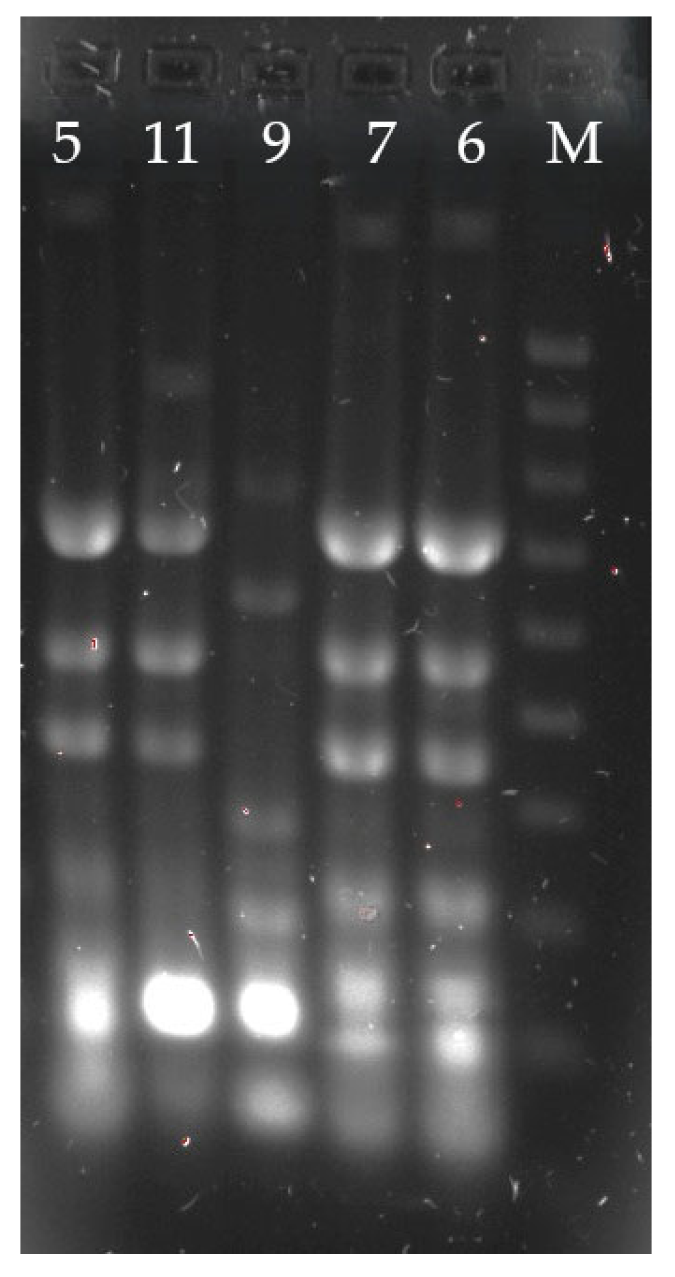
As for the RAPD technique, an identical electrophoretic profile of the strains was obtained only for strains isolated from urine (strains No. 6 and 7) (Figure 2). These strains were cultured from patients hospitalized at the same clinic at the same time. Both strains were isolated in monoculture. The remaining strains were cultured from patients hospitalized at different times and in different clinics.
The ribosomal MLST analysis showed that all the samples matched 100% with Klebsiella variicola subsp. variicola, specifically with ribosomal sequence type (rST) 44139. These findings were confirmed independently using the PathogenWatch platform.
The genome assemblies were as follows:
Sample MEDLv-3793_AS4 was assembled into 5,870,980 base pairs, spread across 19 contigs, with a GC content of 57.1%.
Sample MEDLv-3934_AS4 had 5,701,052 base pairs, distributed in 28 contigs, and a GC content of 57.3%.
Sample MEDLv-3958_AS4 was assembled into 5,711,018 base pairs across 28 contigs, with a GC content also 57.3%.

4. Discussion

KVA was initially discovered in 2004 by Rosenblueth et al. [17] as a novel species of Klebsiella typically isolated from plants. Over the years, an increasing number of reports have identified KVA as a causative agent of human infections. Therefore, the reliable and rapid identification of these strains is crucial and holds significant clinical relevance. The reported frequency of KVA isolation varies, ranging from 2.1% to 11% [18,19]. KVA is associated with various infections, with the most common being bacteraemia, including catheter-related infections [10,20,21,22]. In the study by Legese et al. [10], KVA was the second (18.1%) most frequently isolated pathogen from blood, following K. pneumoniae (KPN). Imai et al. [8] observed that the most common source of bloodstream infection was the abdominal cavity (47.4%), followed by the urinary tract. Long et al. [21] analyzed 13 KVA strains, finding that over 30% were cultured from urine. Conversely, Harada et al. [23] noted that while bloodstream infections were predominant (62.5%), the primary sources were bile ducts and urinary tract. In this study, the majority of strains were isolated from urine, blood, and wound swabs, accounting for nearly 64% of cases. KVA primarily affects immunocompromised patients, including those with oncological conditions, diabetic and those hospitalized in Intensive Care Units (ICU) [5,8,22,24,25]. Several authors have reported outbreaks of KVA in neonatal and children settings [24,25]. In this study, only two strains were isolated from neonates. The majority of analyzed strains were cultured from surgical (over 30%) and ICU patients (almost 18%), with approximately 9% originating from oncological patients.
KVA may also colonize in patients, particularly the gastrointestinal or upper respiratory tract [26,27]. Due to their low nutritional requirements, these bacteria can also inhabit hospital environments [28]. Tsukada et al. isolated KVA strains producing IMP-1 carbapenemases from waste room sinks and hospital room sinks. In a university hospital, KVA strains were isolated from sink syphons and the pre-surgery room working table (unpublished data). The increasing frequency of KVA strain isolation from infections underscores the importance of reliable identification and differentiation within the KPNc. Mass spectrometry is a highly effective method for identifying these bacteria [10,26,29]. Ohama et al. [26] highlighted the importance of the library version in their study. The authors compared two versions of the library, version 4.0.0.0 and 9.0.0.0, for Matrix Assisted Laser Desorption/Ionization (MALDI, Bruker Daltonik) analysis and obtained differing results. In the first version, only 12 strains were identified as KVA, whereas in the second version, 26 strains were identified. Similarly, Imai et al. [8] employed the same method using version 4.0.0.1 of the library, which initially identified the strains as KPN. However, a PCR analysis later revealed that these strains were misidentified. Conversely, Long et al. [30] initially identified strains using version 6.0.0.0. of the MALDI TOF Biotyper library as KPN, but the WGS confirmed them as KVA. In this work, among the 1777 (0.73%) strains analyzed, 13 were identified as KVA. Rodríguez-Medina et al. [4] pointed out that library version 4.0.0.0. did not include spectra for KVA, whereas version 6.0.0.0 contained spectra for both species. Voellmy et al. [29] emphasized the importance of updating the library for accurate identification of KVA strains using mass spectrometry. Challenges arise in distinguishing other species from the KPNc due to their close relationship. Some authors suggest that whole genome sequencing is the best identification for KVA, but this method is often unavailable for routine diagnostics. In this work, only three isolates underwent WGS, and the identification was consistent with mass spectrometry.
The analyzed strains were mostly multidrug-susceptible (MDS) to antimicrobials. More than 95% of KVA strains were susceptible to imipenem, meropenem, and amikacin. Similar results were obtained by other authors [9,26]. Ohama et al. [26] analyzed 26 MDS strains of KVA. In this study, all of the analyzed strains were susceptible to ampicillin-sulbactam, piperacillin-tazobactam, cefmetazole, ceftriaxone, ceftazidime, cefepime, meropenem, levofloxacin, gentamicin, and amikacin. None of the analyzed KVA strains produced ESβL. In contrast, Legese et al. [10] reported cephalosporin resistance rates ranging from 62.3% to 68.8% among 228 strains. Isolates were cultured from blood and the study included 77 strains. Conversely, Huang et al. and Zurfluh et al. reported MDR KVA strains producing carbapenemases [5,31]. In our study, although we did not identify strains producing carbapenemases, 12% of the analyzed strains were found to be MDR. Previous studies have described various mechanisms of antimicrobial resistance in KVA; however, the number of MDR strains reported has been limited. An exception is the study by Legese et al. [10], which identified 71.4% of MDR KVA among the 77 analyzed strains. In this study, more than 90% of the strains were susceptible to amikacin, gentamicin and levofloxacin, and almost 90% were susceptible to ciprofloxacin. In the available literature, articles typically present clinical cases or susceptibility to antimicrobials in various strains. An exception is the study by Voellmy et al. [29]. The authors analyzed 9899 KVA strains, of which 97.1% were susceptible to 3rd and 4th generation cephalosporins, 97.8% to ciprofloxacin, 98.3% to gentamicin, and 99.9% to carbapenems. On the other hand, Legese et al. [10] reported 81.8% susceptibility to amikacin, 31.2% to gentamicin, and 62.3% to ciprofloxacin. In contrast, Garza-Ramos et al. [18] noted the highest number of KVA strains susceptible to piperacillin-tazobactam, fluoroquinolones and tetracycline. All 24 strains were susceptible to the aforementioned antimicrobials. Differences in the susceptibility of KVA strains to antimicrobials may be associated with the frequency of use of specific drugs and the frequency of isolation of MDR strains in the hospital.
In the literature, there are articles describing the isolation of both KVA and KPN strains from the same sample simultaneously. For instance, Morales-León et al. [22] isolated strains of both species from catheter, while Huang et al. [5] isolated them from sputum. All of these strains were MDR. In our hospital, these two species were isolated from three cases. In the first case, strains were isolated from pus: a KPN ESβL-negative and a KVA ESβL-positive strain. In the second patient, isolates were cultured from urine and were MDS. In the third case, strains were isolated from a prosthesis: a KPN ESβL-positive MDR strain and a KVA ESβL-negative strain.
The results of the study indicate that more than 12% of KVA strains produced ESβLs. The results obtained in the CRE test and PCR were largely consistent for most strains. However, we observed three discrepancies. For two strains (No. 8 and 13), the blaSHV and blaTEM genes were detected in PCR, while blaCTX-M1 was detected in the eazyplex® SuperBug CRE test. For one strain (No. 10), the blaSHV gene was detected in PCR, but the eazyplex® SuperBug CRE result was negative. In all three strains, these differences may be attributed to the limitations of the eazyplex® SuperBug CRE assay, which only detects ESβLs from the CTX-M 1 and -9 groups. For strains numbered 8 and 13, the amplification cycle is particularly crucial, as it is strictly defined for each gene; any deviation may result in a false negative outcome. Such a scenario cannot be ruled out for these strains. PCR enables the detection of various genes encoding ESβLs, but it requires time and the use of multiple primers. Therefore, the eazyplex® assay can serve as an alternative to PCR, particularly for MDR strains. In the study by Watanabe et al. [9], all 31 KVA strains isolated were ESβL-negative. Garza-Ramos et al. [18] observed a high percentage of ESβL-positive KVA strains, reaching 56.5%. In our study, the majority of ESβL-positive strains produced CTX-M (over 76% or 84% depending on the method) enzymes. This finding is not unexpected, given that CTX-M enzymes have been predominant in Poland for several years. However, in the study by Garza-Ramos et al. [18], the most commonly identified ESβL was SHV (91.6%), with only one strain producing the CTX-M enzyme. Variations in the prevalence of ESβL-producing KVA strains may be attributed to their epidemic potential and their ability to acquire resistance genes from other hospital strains.

5. Conclusions

While KVA strains continue to exhibit susceptibility to antimicrobials, ongoing monitoring of their susceptibility to antibacterial drugs is essential. The isolation of MDR KVA strains underscores the importance of precisely identifying even less frequently isolated species of Enterobacterales, as they may hold clinical importance and have the potential to produce enzymes that pose significant threats. Further research is warranted to comprehensively understand the virulence and role of these bacteria in human infections.

Author Contributions

Conceptualization, A.S.; methodology, A.S. and Y.K.; software, A.S.; validation, A.S.; formal analysis, A.S.; investigation, A.S. and Y.K.; resources, A.S. and A.C.-M.; data curation, A.S.; writing—original draft preparation, A.S.; writing—review and editing, A.S. and A.C.-M.; visualization, A.S. and A.C.-M.; supervision, A.S.; project administration, A.S.; funding acquisition, A.S., Y.K. and A.C.-M. All authors have read and agreed to the published version of the manuscript.

Funding

Sequencing of K. variicola strains was funded by FEMS, grant number 2668.

Institutional Review Board Statement

The study was conducted in accordance with the Declaration of Helsinki and approved by the Ludwik Rydygier Collegium Medicum in Bydgoszcz and Nicolaus Copernicus University in Toruń (No: 270/2023), approval date 11 July 2023.

Informed Consent Statement

Not applicable.

Data Availability Statement

The data presented in this study are available on a reasonable request from the corresponding author.

Conflicts of Interest

The authors declare no conflicts of interest.

References

  1. Dong, N.; Yang, X.; Chan, E.W.-C.; Zhang, R.; Chen, S. Klebsiella Species: Taxonomy, Hypervirulence and Multidrug Resistance. EBioMedicine 2022, 79, 103998. [Google Scholar] [CrossRef]
  2. Alves, M.S.; da Silva Dias, R.C.; de Castro, A.C.D.; Riley, L.W.; Moreira, B.M. Identification of Clinical Isolates of Indole-Positive and Indole-Negative Klebsiella spp. J. Clin. Microbiol. 2006, 44, 3640–3646. [Google Scholar] [CrossRef] [PubMed]
  3. Potter, R.F.; Lainhart, W.; Twentyman, J.; Wallace, M.A.; Wang, B.; Burnham, C.A.; Rosen, D.A.; Dantas, G. Population Structure, Antibiotic Resistance, and Uropathogenicity of Klebsiella variicola. mBio 2018, 9, e02481-18. [Google Scholar] [CrossRef] [PubMed]
  4. Rodríguez-Medina, N.; Barrios-Camacho, H.; Duran-Bedolla, J.; Garza-Ramos, U. Klebsiella variicola: An Emerging Pathogen in Humans. Emerg. Microbes Infect. 2019, 8, 973–988. [Google Scholar] [CrossRef] [PubMed]
  5. Huang, L.; Fu, L.; Hu, X.; Liang, X.; Gong, G.; Xie, C.; Zhang, F.; Wang, Y.; Zhou, Y. Co-Occurrence of Klebsiella variicola and Klebsiella pneumoniae Both Carrying Bla KPC from a Respiratory Intensive Care Unit Patient. Infect. Drug Resist. 2021, 14, 4503–4510. [Google Scholar] [CrossRef]
  6. Kiley, J.L.; Mende, K.; Beckius, M.L.; Kaiser, S.J.; Carson, M.L.; Lu, D.; Whitman, T.J.; Petfield, J.L.; Tribble, D.R.; Blyth, D.M. Resistance Patterns and Clinical Outcomes of Klebsiella pneumoniae and Invasive Klebsiella variicola in Trauma Patients. PLoS ONE 2021, 16, e0255636. [Google Scholar] [CrossRef]
  7. Lo, C.; Masud, S.; Deans, G.D. Pyelonephritis and Bacteremia Caused by Klebsiella variicola Following Renal Transplantation. Case Rep. Infect. Dis. 2021, 2021, 9988396. [Google Scholar] [CrossRef]
  8. Imai, K.; Ishibashi, N.; Kodana, M.; Tarumoto, N.; Sakai, J.; Kawamura, T.; Takeuchi, S.; Taji, Y.; Ebihara, Y.; Ikebuchi, K.; et al. Correction to: Clinical Characteristics in Blood Stream Infections Caused by Klebsiella pneumoniae, Klebsiella variicola, and Klebsiella quasipneumoniae: A Comparative Study, Japan, 2014–2017. BMC Infect. Dis. 2022, 22, 425. [Google Scholar] [CrossRef]
  9. Watanabe, N.; Watari, T.; Otsuka, Y.; Yamagata, K.; Fujioka, M. Clinical Characteristics and Antimicrobial Susceptibility of Klebsiella pneumoniae, Klebsiella variicola and Klebsiella quasipneumoniae Isolated from Human Urine in Japan. J. Med. Microbiol. 2022, 71, 001546. [Google Scholar] [CrossRef]
  10. Legese, M.H.; Asrat, D.; Swedberg, G.; Hasan, B.; Mekasha, A.; Getahun, T.; Worku, M.; Shimber, E.T.; Getahun, S.; Ayalew, T.; et al. Sepsis: Emerging Pathogens and Antimicrobial Resistance in Ethiopian Referral Hospitals. Antimicrob. Resist. Infect. Control. 2022, 11, 83. [Google Scholar] [CrossRef]
  11. Guo, L.-Y.; Li, Y.; Zhu, L.; Dong, F.; Ji, Y.-Q.; Yao, K.-H.; Gang, L. The First Community-Acquired Klebsiella variicola Bacterial Meningitis Identified by Metagenomic Next Generation Sequencing. Pediatr. Infect. Dis. J. 2023, 42, e186–e188. [Google Scholar] [CrossRef] [PubMed]
  12. Nakamura-Silva, R.; Macedo, L.M.D.; Cerdeira, L.; Oliveira-Silva, M.; Silva-Sousa, Y.T.C.; Pitondo-Silva, A. First Report of Hypermucoviscous Klebsiella variicola Subsp. Variicola Causing Primary Endodontic Infection. Clin. Microbiol. Infect. 2021, 27, 303–304. [Google Scholar] [CrossRef]
  13. Eucast: EUCAST. Available online: https://www.eucast.org/ (accessed on 3 October 2023).
  14. van der Zwaluw, K.; de Haan, A.; Pluister, G.N.; Bootsma, H.J.; de Neeling, A.J.; Schouls, L.M. The Carbapenem Inactivation Method (CIM), a Simple and Low-Cost Alternative for the Carba NP Test to Assess Phenotypic Carbapenemase Activity in Gram-Negative Rods. PLoS ONE 2015, 10, e0123690. [Google Scholar] [CrossRef]
  15. Jemima, S.A.; Verghese, S. Multiplex PCR for Bla(CTX-M) & Bla(SHV) in the Extended Spectrum Beta Lactamase (ESBL) Producing Gram-Negative Isolates. Indian J. Med. Res. 2008, 128, 313–317. [Google Scholar] [PubMed]
  16. Bali, E.B.; Açık, L.; Sultan, N. Phenotypic and Molecular Characterization of SHV, TEM, CTX-M and Extended-Spectrum-Lactamase Produced by Escherichia coli, Acinobacter baumannii and Klebsiella Isolates in a Turkish Hospital. Afr. J. Microbiol. Res. 2010, 4, 650–654. [Google Scholar]
  17. Rosenblueth, M.; Martínez, L.; Silva, J.; Martínez-Romero, E. Klebsiella variicola, a Novel Species with Clinical and Plant-Associated Isolates. Syst. Appl. Microbiol. 2004, 27, 27–35. [Google Scholar] [CrossRef]
  18. Garza-Ramos, U.; Silva-Sánchez, J.; Martínez-Romero, E.; Tinoco, P.; Pina-Gonzales, M.; Barrios, H.; Martínez-Barnetche, J.; Gómez-Barreto, R.E.; Tellez-Sosa, J. Development of a Multiplex-PCR Probe System for the Proper Identification of Klebsiella variicola. BMC Microbiol. 2015, 15, 64. [Google Scholar] [CrossRef]
  19. Brisse, S.; van Himbergen, T.; Kusters, K.; Verhoef, J. Development of a Rapid Identification Method for Klebsiella pneumoniae Phylogenetic Groups and Analysis of 420 Clinical Isolates. Clin. Microbiol. Infect. 2004, 10, 942–945. [Google Scholar] [CrossRef]
  20. Dahl, S.S.; Thorsteinsson, M.; Lambine, T.-L.; Penninga, L. Severe Sepsis Caused by a Gas-Forming Clostridium perfringens and Klebsiella variicola Liver Abscess Following Total Pancreatectomy. BMJ Case Rep. 2020, 13, e238896. [Google Scholar] [CrossRef]
  21. Long, D.-L.; Wang, Y.-H.; Wang, J.-L.; Mu, S.-J.; Chen, L.; Shi, X.-Q.; Li, J.-Q. Fatal Community-Acquired Bloodstream Infection Caused by Klebsiella variicola: A Case Report. World J. Clin. Cases 2022, 10, 2474–2483. [Google Scholar] [CrossRef]
  22. Morales-León, F.; Opazo-Capurro, A.; Caro, C.; Lincopan, N.; Cardenas-Arias, A.; Esposito, F.; Illesca, V.; Rioseco, M.L.; Domínguez-Yévenes, M.; Lima, C.A.; et al. Hypervirulent and Hypermucoviscous Extended-Spectrum β-Lactamase-Producing Klebsiella pneumoniae and Klebsiella variicola in Chile. Virulence 2021, 12, 35–44. [Google Scholar] [CrossRef] [PubMed]
  23. Harada, S.; Aoki, K.; Yamamoto, S.; Ishii, Y.; Sekiya, N.; Kurai, H.; Furukawa, K.; Doi, A.; Tochitani, K.; Kubo, K.; et al. Clinical and Molecular Characteristics of Klebsiella pneumoniae Isolates Causing Bloodstream Infections in Japan: Occurrence of Hypervirulent Infections in Health Care. J. Clin. Microbiol. 2019, 57, e01206-19. [Google Scholar] [CrossRef]
  24. Farzana, R.; Jones, L.S.; Rahman, M.A.; Andrey, D.O.; Sands, K.; Portal, E.; Watkins, W.J.; Pervin, M.; Banerjee, M.; Walsh, T.R. Outbreak of Hypervirulent Multidrug-Resistant Klebsiella variicola Causing High Mortality in Neonates in Bangladesh. Clin. Infect. Dis. 2019, 68, 1225–1227. [Google Scholar] [CrossRef] [PubMed]
  25. Piepenbrock, E.; Higgins, P.G.; Wille, J.; Xanthopoulou, K.; Zweigner, J.; Jahn, P.; Reuter, S.; Skov, R.; Eichhorn, J.; Seifert, H. Klebsiella variicola Causing Nosocomial Transmission among Neonates—An Emerging Pathogen? J. Med. Microbiol. 2020, 69, 396–401. [Google Scholar] [CrossRef]
  26. Ohama, Y.; Nomura, Y.; Mizoguchi, M.; Higurashi, Y.; Okamoto, K.; Harada, S. Accurate Identification of Klebsiella variicola by MALDI-TOF Mass Spectrometry in Clinical Microbiology Laboratories. Microbiol. Spectr. 2022, 10, e0284422. [Google Scholar] [CrossRef]
  27. Kim, S.J.; Jo, J.; Ko, K.S. Lipid A Modification-Induced Colistin-Resistant Klebsiella variicola from Healthy Adults. J. Med. Microbiol. 2023, 72, 001680. [Google Scholar] [CrossRef] [PubMed]
  28. Tsukada, M.; Miyazaki, T.; Aoki, K.; Yoshizawa, S.; Kondo, Y.; Sawa, T.; Murakami, H.; Sato, E.; Tomida, M.; Otani, M.; et al. The Outbreak of Multispecies Carbapenemase-Producing Enterobacterales Associated with Pediatric Ward Sinks: IncM1 Plasmids Act as Vehicles for Cross-Species Transmission. Am. J. Infect. Control. 2024, 52, 801–806. [Google Scholar] [CrossRef]
  29. Voellmy, I.K.; Lang, C.; Gasser, M.; Kronenberg, A.; Swiss Centre for Antibiotic Resistance (ANRESIS). Antibiotic Resistance Surveillance of Klebsiella pneumoniae Complex Is Affected by Refined MALDI-TOF Identification, Swiss Data, 2017 to 2022. Eurosurveillance 2022, 27, 2200104. [Google Scholar] [CrossRef]
  30. Long, S.W.; Linson, S.E.; Ojeda Saavedra, M.; Cantu, C.; Davis, J.J.; Brettin, T.; Olsen, R.J. Whole-Genome Sequencing of Human Clinical Klebsiella pneumoniae Isolates Reveals Misidentification and Misunderstandings of Klebsiella pneumoniae, Klebsiella variicola, and Klebsiella quasipneumoniae. mSphere 2017, 2, e00290-17. [Google Scholar] [CrossRef]
  31. Zurfluh, K.; Poirel, L.; Nordmann, P.; Klumpp, J.; Stephan, R. First Detection of Klebsiella variicola Producing OXA-181 Carbapenemase in Fresh Vegetable Imported from Asia to Switzerland. Antimicrob. Resist. Infect. Control. 2015, 4, 38. [Google Scholar] [CrossRef]
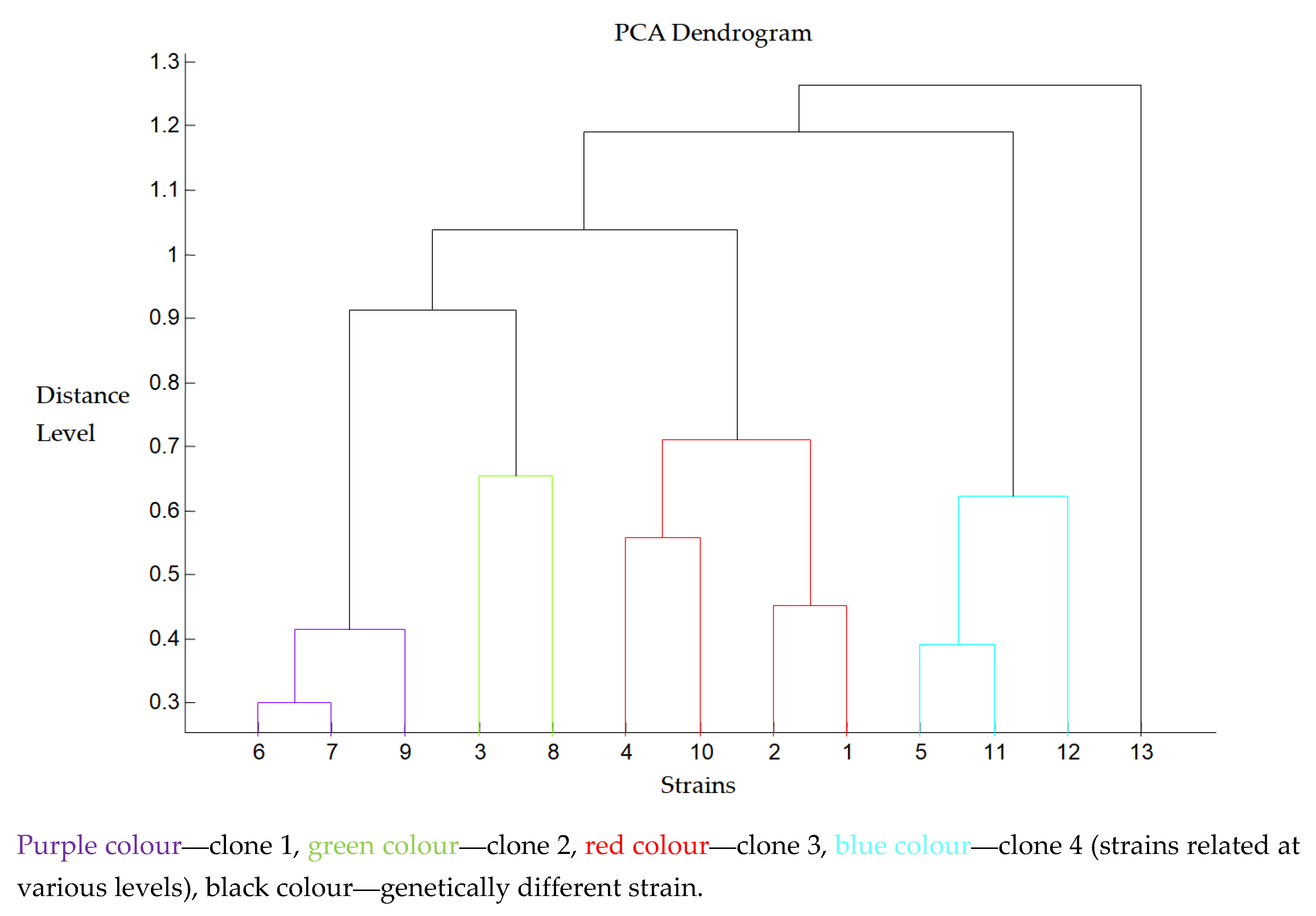
Figure 1. PCA dendrogram of ESβL-positive KVA strains (n = 13).
Microbiolres 16 00123 g001
Figure 2. RAPD pattern for selected KVA strains.
Microbiolres 16 00123 g002
Table 1. Antimicrobial susceptibility of the KVA strains (n = 108).
AntimicrobialPercentage of Strains (%)
SusceptibleSusceptible
(High Exposure)
Resistant
Amoxicillin-clavulanate57.116.226.6
Piperacillin-tazobactam82.40.916.7
Cefuroxime23.650.026.6
Cefotaxime87.9-12.1
Ceftazidime85.51.813.0
Cefepime85.20.913.9
Imipenem99.1-0.9
Meropenem96.30.92.8
Ertapenem94.5-5.5
Gentamicin92.6-7.4
Amikacin96.3-3.7
Tobramycin88.6-10.4
Ciprofloxacin89.82.77.7
Levofloxacin90.51.87.7
Trimethoprim-sulfamethoxazole85.0-15.0
Table 2. The β-lactamases genes detected amongst the analyzed KVA strains (n = 13).
StrainStandard PCRCRE Test
1SHVCTX-M 1
2SHV-
3/5/11CTX-M, SHVCTX-M 1
4CTX-M, TEMCTX-M 1
6/7/9/13CTX-M, SHV, TEMCTX-M 1
8CTX-MCTX-M 1
10CTX-M, TEM-
12TEMCTX-M 1
Disclaimer/Publisher’s Note: The statements, opinions and data contained in all publications are solely those of the individual author(s) and contributor(s) and not of MDPI and/or the editor(s). MDPI and/or the editor(s) disclaim responsibility for any injury to people or property resulting from any ideas, methods, instructions or products referred to in the content.