Brief Report

Disaggregated Helicobacter pylori Biofilm Impairs Bactericidal Activity and Bacterial Phagocytosis by Human Neutrophils

by
Clara Lourdes Tovar-Robles
1,
Yolanda Romo-Lozano
1,*,
Daniel Cervantes-García
1,2 and
Rodolfo González-Segovia
1,*
1
Centro de Ciencias Básicas, Universidad Autónoma de Aguascalientes, Av. Universidad No. 940, Aguascalientes 20131, Mexico
2
Secretaría de Ciencia, Humanidades, Tecnología e Innovación, Ciudad de México 03940, Mexico
*
Authors to whom correspondence should be addressed.
Microbiol. Res. 2025, 16(6), 121; https://doi.org/10.3390/microbiolres16060121
Submission received: 4 April 2025 / Revised: 4 June 2025 / Accepted: 6 June 2025 / Published: 8 June 2025

Abstract

:
Helicobacter pylori (H. pylori), a prevalent human pathogen affecting nearly half the global population, is a major contributor to chronic gastritis, peptic ulcer, and gastric cancer. H. pylori develops biofilms (BFs) allowing bacteria to evade the immune response. Differences in composition between planktonic and biofilm cells influence the host’s immune response, yet the specific biofilm components modulating this response remain uncharacterized. Considering the above, this study evaluated the effect of in vitro-generated H. pylori BF on the antibacterial activity of neutrophils. This work utilized sonication to obtain disaggregated H. pylori BF (d-BF-Hp) to challenge human neutrophils, assessing their bactericidal and phagocytic activity against Staphylococcus aureus. S. aureus survival in the presence of neutrophils was enhanced by 10 μg/mL of d-BF-Hp’s protein. Conversely, S. aureus survival was significantly lower at 30 µg/mL compared to 10 µg/mL d-BF-Hp. Furthermore, 10 and 30 µg/mL of d-BF-Hp significantly reduced the neutrophil phagocytosis rate. Our findings suggest that d-BF-Hp components diminish neutrophil bactericidal activity, although this effect was not observed at higher d-BF-Hp concentrations. Increased d-BF-Hp concentrations proportionally reduced neutrophil phagocytic capacity. Future work should explore the mechanisms underlying the alteration of neutrophil microbicidal properties.

1. Introduction

Helicobacter pylori is a microaerophilic, Gram-negative, slow-growing, helical-shaped bacterium with abundant flagella [1]. H. pylori infection is a risk factor for chronic gastritis and peptic ulcer that affects 50% of the world’s population [2]. The most important virulence factors identified in this bacterium are cytotoxin-associated gene A (CagA) and vacuolating cytotoxin A (VacA). CagA is considered a marker of the most virulent strains of H. pylori and is associated with cell proliferation and inflammation in the gastric mucosa. Meanwhile, VacA causes the formation of cytoplasmic vacuoles and apoptosis in gastric cells [3]. H. pylori has the ability to develop biofilms (BFs), which are complex matrices formed by deposits of exopolysaccharides, amino acids, lipids, and extracellular DNA [4]. Through nuclear magnetic resonance analysis, it has been determined that the BF of H. pylori is formed predominantly by polysaccharides with 1,4-mannosyl linkages [5]. Immersed in the matrix are also heterogeneous bacterial cells, as well as pores and interstitial spaces that function as water channels for irrigation [6]. Moreover, cellular structures can be found, such as outer membrane vesicles, flagella, and pili [6]. BF protects microorganisms against extreme environments and external agents [7]. Additionally, it has been shown that the BF of some bacterial pathogens can promote immune evasion processes, such as the production of the leukotoxin leucocidin AB present in the BF of S. aureus, which interferes with the phagocytic activity of macrophages and neutrophils [8,9]. BF formation is one of the challenges facing standard treatments for H. pylori, as it affects their eradication efficacy through its role in antibiotic resistance and immune tolerance. Despite recent interest in H. pylori infection and management, BFs are still understudied [10].
H. pylori colonization generates a significant infiltration of neutrophils in the gastric mucosa [11]. The pathophysiological relevance of the levels of this infiltration has traditionally been used to classify the severity of gastric tissue damage [12]. Neutrophils are immune cells that importantly control infectious processes using microbicidal processes such as phagocytosis, degranulation, and extracellular traps (NETs) to combat bacterial pathogens [13]. It has been demonstrated that neutrophils are attracted to BF through chemotactically active molecules such as lipopolysaccharides, peptidoglycans, microbial DNA, and other pathogen-associated molecular patterns (PAMPs) [14]. The interaction of the BF of H. pylori and neutrophils has been poorly studied, although is known that neutrophil-activating protein (NAP) is upregulated in BF cells compared to planktonic cells [5]. The NAD molecule is released around the gastric epithelial monolayer, where it contributes to the inflammatory process by increasing the adhesion and activation of neutrophils [15]. To enhance the understanding of the immune evasion mechanisms of the bacteria, it is important to study the possible existence of H. pylori BF components that could interfere with the antibacterial activity of neutrophils. In this work, the effect of H. pylori BF generated in vitro and disaggregated by sonication on the antibacterial activity of neutrophils was studied using S. aureus as a prokaryotic model to evaluate this activity.

2. Materials and Methods

2.1. Reagents and Culture Media

Reagents: Cyclodextrin (CCD) (Sigma-Aldrich, Inc., St. Louis, MO, USA), glucose (Chemical Technique, Mexico City, México), crystal violet (Karal S.A. de C.V., León, Gto., Mexico), Coomassie Blue (Sigma-Aldrich, Inc., St. Louis, MO, USA), Lymphoprep (Fresenius Kabi Norge AS, Halden, Norway), Histopaque-1119 (Sigma-Aldrich, Inc., St. Louis, MO, USA), Bovine Serum Albumin (BSA) (Sigma-Aldrich, Inc., St. Louis, MO, USA), Trypan blue (Sigma, Steinheim, Germany), and Wright-Giemsa (Golden Bell, Mexico City, Mexico).
Culture media: Brucella broth (BB) (Becton, Dickinson, Sparks, MD, USA), H. pylori selection antibiotics (Selective supplement DENT, Oxoid Ltd., Basingstoke, Hampshire, UK), Brain–Heart Infusion (BHI) broth and BHI agar (BHIA) (BD Bioxon, Becton Dickinson, México), Mannitol Salt Agar (MSA) (BD Bioxon, Becton Dickinson, Mexico), Blood Agar Base (BD Bioxon, Becton Dickinson, Cuautitlan Izcalli, Mexico, Mexico), and Roswell Park Memorial Institute (RPMI)-1640 medium with phenol red (Sigma-Aldrich, Inc., St. Louis, MO, USA).

2.2. Characterization of H. pylori BF Formation In Vitro

A clinical isolate of H. pylori, A97, which was previously obtained from a gastric biopsy, was used. Molecular analysis confirmed that this strain has two key virulence factors: the cytotoxin-associated gene (cagA+) and the vacuolating toxin (vacA) genotype s2/m2. Bacteria were cultured on Blood Agar Base supplemented with 5% defibrinated sheep blood, and incubated at 37 °C under microaerophilic conditions. For BF formation, BB supplemented with 0.1% CCD, 0.3% glucose, and 4 µL/mL of H. pylori selective antibiotics was dispensed into 24-well culture plates (Celltreat®, Ayer, MA, USA). Subsequently, 20 µL of bacterial suspensions, incubated for 72 h in BB and adjusted to 1.5 × 10⁶ bacteria/mL (McFarland turbidity standard), were added to each well to a final volume of 1 mL. The culture plates were then incubated at 37 °C in a microaerophilic environment for five days. To confirm that the culture conditions used allowed the formation of BF, this structure was identified from 3 wells of the culture plate, using the method described by O’Toole based on the staining of the BF matrix with crystal violet [16].

2.3. Preparation of Disaggregated H. pylori BF

The BF of H. pylori cultured in a 24-well plate was recovered by carefully removing the supernatant. The wells were washed with 200 µL of phosphate-buffered saline (PBS) [1.75 mM NaH2PO4, 8 mM Na2HPO4, and 75 mM NaCl, pH 6.5]. After the removal of the initial PBS, 200 µL of fresh PBS was added to the wells to facilitate BF removal by gentle scraping. The resuspended BF was collected in a sterile container and kept in an ice bath for subsequent disaggregation through sonication. Sonication was carried out using a Sonicor ultrasonic processor (UP 400A 50/60Hz cycles, Sonicor Instrument Co., West Babylon, NY, USA) with 5 pulses of 10 s at level 4. The disaggregated H. pylori BF (d-BF-Hp) obtained corresponds to a mixture of cellular debris together with components of the ultrasonic disintegration of the BF matrix and was used in the tests to assess the microbicidal activity of neutrophils.
The protein concentration of the d-BF-Hp was quantified using the Bradford method, with BSA as the standard [17]. To ensure that the obtained d-BF-Hp was free from viable H. pylori cells or contamination with other bacteria, samples were cultured on blood agar (5%) and incubated at 37 °C under microaerophilic conditions for 24 h. Additionally, samples were cultured on BHIA and incubated at 37 °C for 24 h. The presence and integrity of the proteins in the d-BF-Hp were further assessed by sodium dodecyl sulfate–polyacrylamide gel electrophoresis (SDS-PAGE). The gel was stained with Coomassie Blue and the d-BF-Hp was stored at −20 °C until further use.

2.4. Staphylococcus aureus

A S. aureus isolate, previously obtained in the laboratory, was used, with its identification as catalase+, coagulase+, and thermonuclease+ confirmed in accordance with the Official Mexican Standard (NOM-210-SSA1-2014) [18]. For bacterial suspensions, the isolate was incubated for 24 h at 37 °C in 5 mL of BHI. After incubation, bacterial cells were harvested by centrifugation at 1409× g for 5 min, and the pellet was resuspended in 5 mL of PBS. The optical density (OD) at 600 nm was measured in triplicate from a 1:3 dilution of this suspension using a spectrophotometer (Jenway 7305, Bibby Scientific, Stone, Staffs, UK). Bacterial concentration (bacteria/mL) was determined by interpolating the OD values against an OD curve generated from McFarland standards (1 to 10). Bacterial suspensions were then adjusted to 1 × 10⁶ bacteria/mL in PBS. This cell concentration was verified by quantifying colony-forming units (CFU) after seeding 100 µL of serial dilutions of S. aureus on MSA plates and incubating at 37 °C for 24 h.

2.5. Interaction of Human Neutrophils with Disaggregated H. pylori BF

The protocol for obtaining blood samples was approved by the Institutional Bioethics Committee of the Universidad Autónoma de Aguascalientes (office SG-1783-2023, project identification PIBB24-1) on 30 October 2023. All adult volunteers signed an informed consent prior to blood donation for research purposes. Neutrophils were purified using a discontinuous density gradient prepared with Lymphoprep and Histopaque-1119, as previously described [19]. Contaminating erythrocytes were lysed by osmotic shock in a 0.15 M NH4Cl solution. Cells were washed twice with PBS and resuspended in 1 mL of supplemented RPMI-1640 medium containing phenol red. Cell quantification was performed using a Neubauer chamber, and neutrophil viability was determined by the Trypan blue exclusion test. Only preparations with neutrophil purity and viability greater than 95% were used.
To test the effect of the d-BF-Hp, 1 × 10⁶ neutrophils were used. These cells were stabilized in RPMI for 30 min at 37 °C. Subsequently, they were incubated with d-BF-Hp (10 and 30 µg of total protein/mL) for 30 min at 37 °C. Finally, S. aureus (1 × 10⁶) was added to achieve a 1:1 multiplicity of infection (MOI) with neutrophils, and the mixture was incubated at 37 °C for 1 h. The cellular pellets were obtained by centrifugation at 300× g for 10 min at 4 °C to determine phagocytic cells, or at 8050× g for 7 min at 4 °C to determine CFU. All assays were performed in duplicate in independent trials.

2.6. Determination of Phagocytic Cells by Optical Microscopy

To evaluate the effect of d-BF-Hp on phagocytosis, the number of S. aureus-phagocytosing cells was quantified by optical microscopy. The cellular pellets obtained after interaction were gently resuspended in 30 μL of RPMI. Smears prepared on sterile, degreased slides were fixed with absolute methanol, stained with Wright-Giemsa, and examined under an optical microscope. Images were captured by a digital camera (Motic BA310, Hong Kong, China) adapted to the microscope and analyzed using the Motic Images Plus Program, version 2.0. For each experimental condition, 200 cells were counted on each slide to determine the percentage of neutrophils that bound to or engulfed bacteria. Control groups included neutrophils with or without d-BF-Hp or S. aureus bacteria.

2.7. Viability of S. aureus

The cellular pellets obtained by centrifugation at 8050× g at the end of the interaction were resuspended in 1 mL of sterile distilled water and mixed vigorously for 3 min to lyse the neutrophils and release intracellular bacteria. Dilutions (1 × 10⁻³) of the samples were plated by streaking on MSA and incubated for 24 h at 37 °C to obtain CFU. The results were expressed as a percentage of the CFU obtained in each treatment. In addition to CFU quantification, a spot dilution test (in duplicate) was performed to evaluate bacterial growth in each dilution and to demonstrate bacterial viability. For this purpose, three 5 µL aliquots of each cellular suspension were spotted onto a Petri dish with MSA. The drops were allowed to dry to prevent displacement. Plates were incubated at 37 °C for 24 h [20].

2.8. Statistical Analysis

Data were analyzed with GraphPad Prism 8.0 software (GraphPad Software Inc., La Jolla, CA, USA) and presented as the mean ± standard error of the mean (SEM) from triplicate experiments. One-way analysis of variance (ANOVA), followed by a Bonferroni test, was used for group comparisons [21]. Statistical significance was considered when p < 0.05.

3. Results

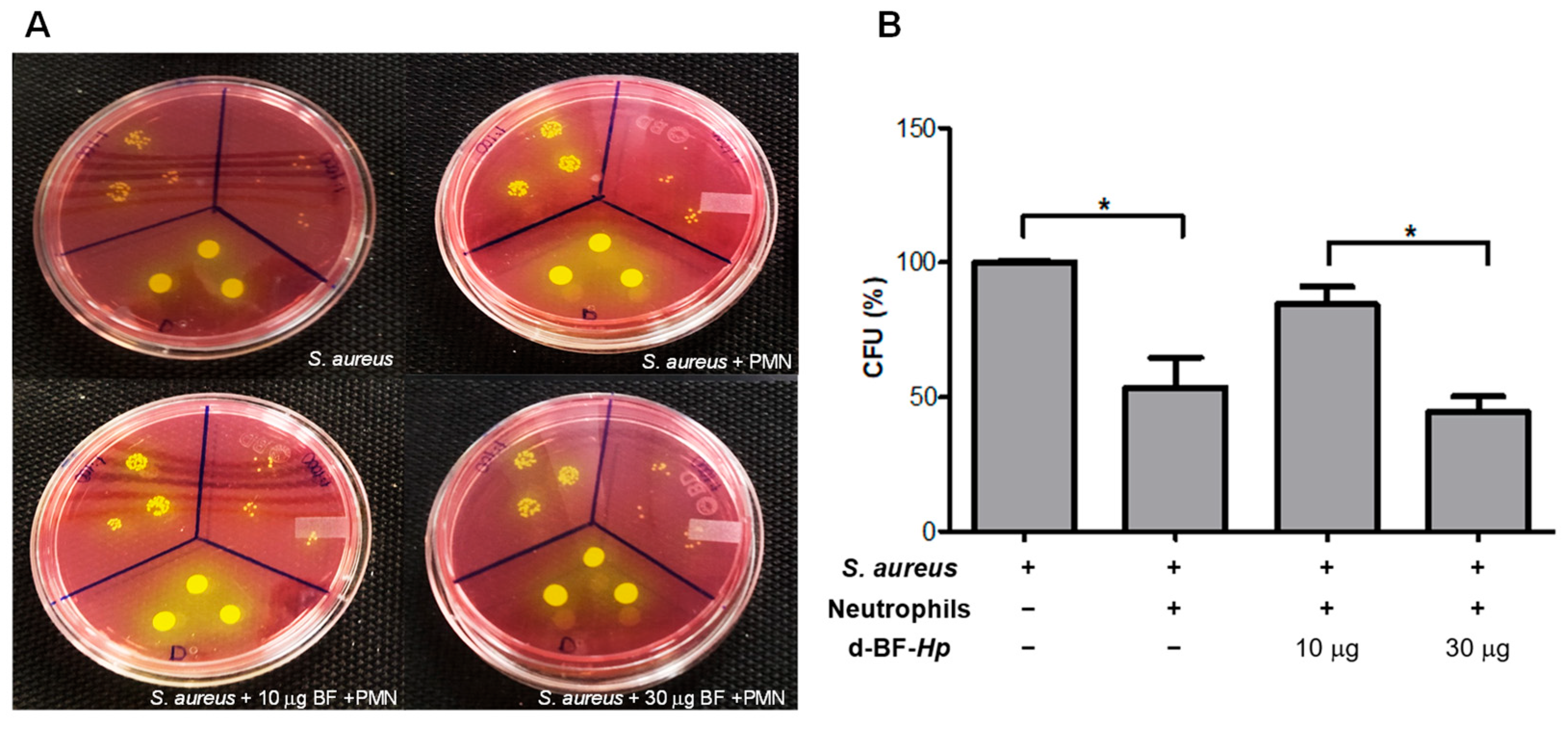
3.1. Disaggregated BF H. pylori Increase S. aureus Survival in Human Neutrophils

To evaluate the effect of d-BF-Hp on bacterial survival after phagocytosis in neutrophils, we assessed the CFU percentage of S. aureus within neutrophils following d-BF-Hp stimulation (Figure 1). As expected, the viability of S. aureus was significantly reduced by 53.5% ± 27.75 when compared to the CFU of S. aureus in the absence of neutrophils. Notably, 10 µg/mL of d-BF-Hp enhanced the survival of S. aureus in the presence of neutrophils, as its viability showed a significant increase compared to S. aureus when exposed to neutrophils. However, in the presence of 30 µg/mL of d-BF-Hp, S. aureus survival was significantly lower than that observed with 10 µg/mL of d-BF-Hp. But, it is similar to the survival obtained when the bacteria are challenged only with neutrophils (p > 0.05).

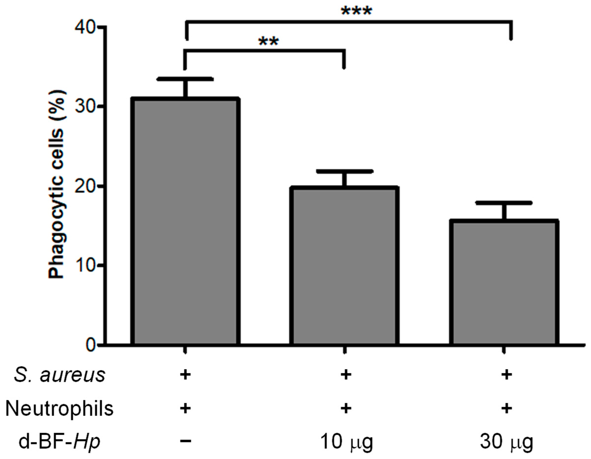
3.2. Disaggregated H. pylori BF Reduces the Phagocytosis Rate of S. aureus by Neutrophils

To analyze the effect of d-BF-Hp on neutrophil phagocytosis, neutrophils were incubated with 10 and 30 µg/mL of d-BF-Hp and then co-cultured with S. aureus at a MOI of 1. Phagocytosis of S. aureus by neutrophils was observed in 31 ± 6.9% of the cells. However, in the presence of 10 and 30 µg/mL of d-BF-Hp, the percentage of phagocytic cells significantly reduced to 19.75 ± 6.1% and 15.63 ± 6.43%, respectively, compared to the treatment without d-BF-Hp (Figure 2).

4. Discussion

Helicobacter pylori infection elicits an immune response with immunomodulatory actions promoting the persistence of gastric colonization. This modulatory response involves the evasion of pattern recognition receptors, manipulation of immune cell activity, and downregulation of adaptive responses [22].
The ability of H. pylori to develop BF may favor the persistence of the infection, as BF impedes the effect of antibiotics and the function of the immune response [23]. Strains capable of forming BF exhibit a higher level of resistance as compared to those of strains incapable of developing these structures [4]. In this study, we sought to analyze in vitro the effect that H. pylori BF components may have on the microbicidal action of neutrophils, considering the use of a S. aureus strain as an indicator of antibacterial activity.
Quantification of S. aureus CFU obtained from neutrophil cultures stimulated with d-BP-Hp allowed the evaluation of the effect of BF components on bacterial survival. The increased viability of S. aureus observed in neutrophils stimulated with the lowest concentration of BF components used in the study (10 µg/mL) indicates that elements of this bacterial structure may interfere with the bactericidal action of neutrophils. There are no previous reports like this study of the activity on neutrophils with H. pylori BF. In other bacteria, such as Pseudomonas aeruginosa, it has been shown that some quorum-sensing molecules associated with BF may function as inhibitors of reactive oxygen species production in neutrophils [24]. However, it is striking that in our study, the increase in H. pylori BF components (30 µg/mL) has almost no effect on the bactericidal activity of neutrophils. This phenomenon could be related to an overstimulation of neutrophils caused by the enrichment of PAMPs due to the increased concentration that could increase bactericidal activity, through mechanisms such as the formation of NETs [25].
The finding that components of H. pylori BP reduce the bactericidal activity of neutrophils may be a mechanism contributing to the chronic persistence of the bacteria. Since in vivo, it has been shown that H. pylori develops associated with a dense BF that adheres to the surface of the gastric mucosa [26]. BF components could diffuse from the luminal space to internal sites of the gastric mucosa, crossing the epithelial barrier, and then control the bactericidal activity of infiltrated neutrophils. It has been shown that molecules produced in bacterial BFs can generate alterations in the permeability of epithelial cells, as is the case with quorum-sensing molecules of the N-acyl homoserine lactone family that can modify the organization of intercellular junction proteins such as occludin, E-cadherin, and zonula occludens-1 [27].
In the phagocytosis analysis, a decrease in the phagocytic activity of neutrophils was identified depending on the increase in d-BF-Hp. This decrease could be related to the masking of PAMPs by BF polysaccharides, generating the inhibition of opsonization processes, thus altering the phagocytic capacity of neutrophils [28]. However, it is feasible to consider the possibility that within the components of the BF of H. pylori, factors are present that could directly alter the neutrophilic phagocytic capacity. These types of alterations have been documented in the BF of bacteria such as S. aureus, in which the presence of protein factors such as alpha-toxin (Hla) and leukotoxin leukocidin AB, has been identified as factors that cause dysfunction in the phagocytic response of macrophages and neutrophils [8,9].
Future studies could focus on characterizing the H. pylori BF components related to the alteration of the microbicidal activity of neutrophils, including the mechanism associated with this process. However, it is crucial to consider the compositional variations that bacterial BF presents, since it is known that the same strain presents different components depending on the environmental conditions in which it develops [29]. Furthermore, the characterization of such components will facilitate the design of rapid methods for the detection of infections of H. pylori in advanced stages, not only in the initial stages [30].

5. Conclusions

In this in vitro study, it was observed that the components of the d-BF-Hp, including structural elements, cellular derivatives, and signaling molecules, among others, altered the bactericidal and phagocytic capacity of neutrophils against S. aureus. This suggests that some d-BF-Hp components can directly or indirectly modify the microbicidal actions of immune cells. Such a mechanism could favor immune evasion by contributing to H. pylori’s persistence during gastric colonization. Further research is needed to identify the precise d-BF-Hp components that are responsible for altering neutrophil microbicidal activity.

Author Contributions

Conceptualization, R.G.-S., Y.R.-L., and D.C.-G.; methodology, R.G.-S. and Y.R.-L.; formal analysis, C.L.T.-R., R.G.-S., Y.R.-L., and D.C.-G.; investigation, C.L.T.-R., Y.R.-L., and R.G.-S.; resources, R.G.-S. and Y.R.-L.; data curation, C.L.T.-R., D.C.-G., and Y.R.-L.; writing—original draft preparation, C.L.T.-R., R.G.-S., Y.R.-L., and D.C.-G.; writing—review and editing, R.G.-S., Y.R.-L., and D.C.-G.; supervision of project, Y.R.-L. and R.G.-S.; project administration, R.G.-S. and Y.R.-L.; funding acquisition, R.G.-S. and Y.R.-L. All authors have read and agreed to the published version of the manuscript.

Funding

This work was funded by the Universidad Autónoma de Aguascalientes [grant number PIBB 22-4 González- Segovia R. and PIBB24-1 to Romo-Lozano Y.]. Meanwhile, Clara Lourdes Tovar-Robles was supported by Consejo Nacional de Humanidades, Ciencias y Tecnologías (CONAHCYT) of México via grant 166856.

Institutional Review Board Statement

The protocol was approved by the Institutional Bioethics Committee of the Universidad Autónoma de Aguascalientes approved the protocol for obtaining blood samples (office SG-1783-2023, project identification PIBB24-1) on [30 October 2023].

Informed Consent Statement

Informed consent was obtained from all subjects involved in the study.

Data Availability Statement

Data may be shared and should be requested from the corresponding author.

Acknowledgments

The authors are also thankful to Aylin Mayela Avila Colis and Angelina Lizbeth Montañez Flores for technical assistance and support.

Conflicts of Interest

The authors declare no conflicts of interest.

Abbreviations

The following abbreviations are used in this manuscript:
BBBrucella broth
BFBiofilm
BHIBrain–heart infusion broth
BHIABrain–heart infusion agar
CFUColony-forming Units
d-BF-HpDisaggregated H. pylori biofilm
MSAMannitol Salt Agar
NETsNeutrophil Extracellular Traps
PAMPsPathogen-associated Molecular Patterns
RPMIRoswell Park Memorial Institute 1640

References

  1. Li, N.; Xie, C.; Lu, N.H. Transforming growth factor-β: An important mediator in Helicobacter pylori-associated pathogenesis. Front. Cell. Infect. Microbiol. 2015, 5, 77. [Google Scholar] [CrossRef] [PubMed]
  2. Zeng, J.; Xie, C.; Zhang, L.; Liu, X.; Chan, M.T.V.; Wu, W.K.K.; Chen, H. Host Cell Antimicrobial Responses against Helicobacter pylori Infection: From Biological Aspects to Therapeutic Strategies. Int. J. Mol. Sci. 2022, 23, 10941. [Google Scholar] [CrossRef] [PubMed]
  3. Muzaheed. Helicobacter pylori Oncogenicity: Mechanism, Prevention, and Risk Factors. Sci. World J. 2020, 2020, 3018326. [CrossRef]
  4. Fauzia, K.A.; Effendi, W.I.; Alfaray, R.I.; Malaty, H.M.; Yamaoka, Y.; Mifthussurur, M. Molecular Mechanisms of Biofilm Formation in Helicobacter pylori. Antibiotics 2024, 13, 976. [Google Scholar] [CrossRef]
  5. Yang, F.L.; Hassanbhai, A.M.; Chen, H.Y.; Huang, Z.Y.; Lin, T.L.; Wu, S.H.; Ho, B. Proteomannans in biofilm of Helicobacter pylori ATCC 43504. Helicobacter 2011, 16, 89–98. [Google Scholar] [CrossRef]
  6. Krzyżek, P.; Grande, R.; Migdał, P.; Paluch, E.; Gościniak, G. Biofilm Formation as a Complex Result of Virulence and Adaptive Responses of Helicobacter pylori. Pathogens 2020, 9, 1062. [Google Scholar] [CrossRef] [PubMed]
  7. Liu, X.; Yao, H.; Zhao, X.; Ge, C. Biofilm Formation and Control of Foodborne Pathogenic Bacteria. Molecules 2023, 28, 2432. [Google Scholar] [CrossRef]
  8. Scherr, T.D.; Hanke, M.L.; Huang, O.; James, D.B.; Horswill, A.R.; Bayles, K.W.; Fey, P.D.; Torres, V.J.; Kielian, T. Staphylococcus aureus Biofilms Induce Macrophage Dysfunction Through Leukocidin AB and Alpha-Toxin. mBio 2015, 6, e01021-15. [Google Scholar] [CrossRef]
  9. Bhattacharya, M.; Berends, E.T.M.; Zheng, X.; Hill, P.J.; Chan, R.; Torres, V.J.; Wozniak, D.J. Leukocidins and the Nuclease Nuc Prevent Neutrophil-Mediated Killing of Staphylococcus aureus Biofilms. Infect. Immun. 2020, 88, e00372-20. [Google Scholar] [CrossRef]
  10. Al-Fakhrany, O.M.; Elekhnawy, E. Helicobacter pylori in the post-antibiotics era: From virulence factors to new drug targets and therapeutic agents. Arch. Microbiol. 2023, 205, 301. [Google Scholar] [CrossRef]
  11. Ieni, A.; Barresi, V.; Rigoli, L.; Fedele, F.; Tuccari, G.; Caruso, R.A. Morphological and Cellular Features of Innate Immune Reaction in Helicobacter pylori Gastritis: A Brief Review. Int. J. Mol. Sci. 2016, 17, 109. [Google Scholar] [CrossRef] [PubMed]
  12. Bacha, D.; Walha, M.; Ben Slama, S.; Ben Romdhane, H.; Bouraoui, S.; Bellil, K.; Lahmar, A. Chronic gastritis classifications. Tunis Med. 2018, 96, 405–410. [Google Scholar] [PubMed]
  13. Aristotelous, A.C. Biofilm neutrophils interactions under hypoxia: A mathematical modeling study. Math. Biosci. 2022, 352, 108893. [Google Scholar] [CrossRef] [PubMed]
  14. Hirschfeld, J. Dynamic interactions of neutrophils and biofilms. J. Oral. Microbiol. 2014, 6, 26102. [Google Scholar] [CrossRef]
  15. Codolo, G.; Coletta, S.; D’Elios, M.M.; de Bernard, M. HP-NAP of Helicobacter pylori: The Power of the Immunomodulation. Front. Immunol 2022, 13, 944139. [Google Scholar] [CrossRef]
  16. O’Toole, G.A. Microtiter dish biofilm formation assay. J. Vis. Exp. 2011, 47, 2437. [Google Scholar] [CrossRef]
  17. Bradford, M.M. A rapid and sensitive method for the quantitation of microgram quantities of protein utilizing the principle of protein-dye binding. Anal. Biochem. 1976, 7, 248–254. [Google Scholar] [CrossRef]
  18. Secretaría de Salud. Norma Oficial Mexicana NOM-210-SSA1-2014, Productos y Servicios. Métodos de Prueba Microbiológicos. Determinación de Microorganismos Indicadores. Determinación de Microorganismos Patógenos. Available online: https://dof.gob.mx/nota_detalle_popup.php?codigo=5398468 (accessed on 6 November 2023).
  19. Curtiellas-Piñol, V.; Ventura-Juárez, J.; Ruiz-Baca, E.; Romo-Lozano, Y. Morphological changes and phagocytic activity during the interaction of human neutrophils with Sporothrix schenckii: An in vitro model. Microb. Pathog. 2019, 129, 56–63. [Google Scholar] [CrossRef]
  20. Martínez-Montelongo, J.H.; Medina-Ramírez, I.E.; Romo-Lozano, Y.; González-Gutiérrez, A.; Macías-Díaz, J.E. Development of nano-antifungal therapy for systemic and endemic mycoses. J. Fungi 2021, 7, 158. [Google Scholar] [CrossRef]
  21. Prichard, A.; Khuu, L.; Whitmore, L.C.; Irimia, D.; Allen, L.H. Helicobacter pylori-infected human neutrophils exhibit impaired chemotaxis and a uropod retraction defect. Front. Immunol 2022, 13, 1038349. [Google Scholar] [CrossRef]
  22. Sirit, I.S.; Peek, R.M., Jr. Decoding the ability of Helicobacter pylori to evade immune recognition and cause disease. Cell Mol. Gastroenterol. Hepatol. 2025, 19, 101470. [Google Scholar] [CrossRef] [PubMed]
  23. Wu, X.; Wu, D.; Cui, G.; Lee, K.H.; Yang, T.; Zhang, Z.; Liu, Q.; Zhang, J.; Chua, E.G.; Chen, Z. Association between biofilm formation and structure and antibiotic resistance in H. pylori. Infect. Drug Resist. 2024, 17, 2501–2512. [Google Scholar] [CrossRef]
  24. Bjarnsholt, T.; Jensen, P.Ø.; Burmølle, M.; Hentzer, M.; Haagensen, J.A.J.; Hougen, H.P.; Calum, H.; Madsen, K.G.; Moser, C.; Molin, S.; et al. Pseudomonas aeruginosa tolerance to tobramycin, hydrogen peroxide and polymorphonuclear leukocytes is quorum-sensing dependent. Microbiology 2005, 151, 373–383. [Google Scholar] [CrossRef] [PubMed]
  25. Thiam, H.R.; Wong, S.L.; Wagner, D.D.; Waterman, C.M. Cellular mechanisms of NETosis. Annu. Rev. Cell Dev. Biol. 2020, 36, 191–218. [Google Scholar] [CrossRef]
  26. Rizzato, C.; Torres, J.; Kasamatsu, E.; Camorlinga-Ponce, M.; Bravo, M.M.; Canzian, F.; Kato, I. Potential Role of Biofilm Formation in the Development of Digestive Tract Cancer With Special Reference to Helicobacter pylori Infection. Front. Microbiol. 2019, 10, 846. [Google Scholar] [CrossRef]
  27. Coquant, G.; Grill, J.P.; Seksik, P. Impact of N-Acyl-Homoserine Lactones, Quorum Sensing Molecules, on Gut Immunity. Front. Immunol. 2020, 11, 1827. [Google Scholar] [CrossRef] [PubMed]
  28. Langereis, J.D.; Weiser, J.N. Shielding of a lipooligosaccharide IgM epitope allows evasion of neutrophil-mediated killing of an invasive strain of nontypeable Haemophilus influenzae. MBio 2014, 5, e01478-14. [Google Scholar] [CrossRef]
  29. Campoccia, D.; Ravaioli, S.; Mirzaei, R.; Bua, G.; Daglia, M.; Arciola, C.R. Interactions of neutrophils with the polymeric molecular components of the biofilm matrix in the context of implant-associated bone and joint infections. Int. J. Mol. Sci. 2023, 24, 17042. [Google Scholar] [CrossRef]
  30. Aziz, F.; Kazmi, S.U. Thin layer immunoassay: An economical approach to diagnose Helicobacter pylori infection in gastroduodenal ulcer disease patients of Pakistan, a comparative analysis. Diagnostics 2023, 13, 517. [Google Scholar] [CrossRef]
Figure 1. Staphylococus aureus CFU percentage with human neutrophils. (A) Spot dilution test for viability control of S. aureus treated with S. aureus alone, S. aureus with neutrophils, S. aureus with neutrophils and 10 μg of d-BF-Hp, S. aureus with neutrophils and 30 μg of d-BF-Hp. (B) Effect of d-BF-Hp on S. aureus survival post-neutrophils phagocytosis. Mean comparisons are indicated (* p < 0.05).
Microbiolres 16 00121 g001
Figure 2. Effect of d-BF-Hp on neutrophil phagocytosis of S. aureus. Mean comparisons are indicated (** p < 0.01, *** p < 0.001).
Microbiolres 16 00121 g002
Disclaimer/Publisher’s Note: The statements, opinions and data contained in all publications are solely those of the individual author(s) and contributor(s) and not of MDPI and/or the editor(s). MDPI and/or the editor(s) disclaim responsibility for any injury to people or property resulting from any ideas, methods, instructions or products referred to in the content.

