Article

Distribution of Treponema Species in Active Digital Dermatitis Lesions and Non-Lesional Skin of Dairy Cattle

1
Clinic of Ruminants, University Veterinary Hospital, University of Veterinary Medicine and Pharmacy in Košice, Komenského 73, 041 81 Košice, Slovakia
2
Department of Microbiology and Immunology, University of Veterinary Medicine and Pharmacy in Košice, Komenského 73, 041 81 Košice, Slovakia
3
Department of Morphological Disciplines, University of Veterinary Medicine and Pharmacy in Košice, Komenského 73, 041 81 Košice, Slovakia
4
Clinic of Swine, University Veterinary Hospital, University of Veterinary Medicine and Pharmacy in Košice, Komenského 73, 041 81 Košice, Slovakia
*
Author to whom correspondence should be addressed.
Microbiol. Res. 2025, 16(6), 119; https://doi.org/10.3390/microbiolres16060119
Submission received: 6 April 2025 / Revised: 2 June 2025 / Accepted: 3 June 2025 / Published: 5 June 2025

Abstract

:
This study examined the prevalence, distribution, and detection methods linked to Treponema species associated with active bovine digital dermatitis (BDD) in dairy cattle. Tissue, surface swabs, interdigital space swabs, and faecal samples were collected from 20 Holstein-Friesian cows from a farm in Eastern Slovakia. Molecular analysis revealed that all cows tested positive for at least one Treponema species. The most prevalent species were Treponema medium (100%), Treponema pedis (95%), and Treponema brennaborense (75%). Distribution analysis demonstrated significant differences in the occurrence of these species across sampling methods, with T. pedis being more prevalent in tissue biopsies and surface swabs (p < 0.001), and T. brennaborense in surface swabs (p < 0.001). A comparison of qualitative real-time PCR and standard PCR revealed that real-time PCR detected T. pedis and T. brennaborense in 70% and 75% of tissue samples, respectively, while standard PCR failed to detect T. brennaborense. Furthermore, real-time PCR showed a significantly higher prevalence of T. brennaborense (p < 0.001). These findings underscore the enhanced sensitivity of real-time PCR in detecting T. brennaborense and highlight the complex distribution of Treponema species in BDD lesions, which may inform the development of more effective control strategies for BDD.

1. Introduction

Bovine digital dermatitis (BDD) is an infectious skin disease in cattle, widely regarded as the leading cause of infectious lameness in dairy cows. Initially described by Cheli and Mortellaro in the 1970s [1], the prevalence of BDD at the herd level has been reported to reach up to 93%, though this may vary due to factors such as breed, housing type, parity, and hoof trimming frequency [2,3].
BDD is characterized by ulcerative lesions located above the heel bulbs [4]. These lesions are often extremely painful, and can lead to reduced milk yield, impaired reproductive performance [5], or premature culling of affected animals [6,7]. BDD monitoring is typically carried out during milking and involves visual inspection of the feet. Diagnosis and classification are subsequently based on clinical observations [8]. Accurate identification of the disease stage is therefore essential for determining appropriate treatment strategies and implementing effective herd-level control measures [9]. Moreover, it enables a more precise estimation of the economic impact of BDD [10], which includes treatment costs and reduced production performance [11], and decreases within-herd prevalence [12].
Originally characterized as a multifactorial polymicrobial disease in dairy cattle, BDD has also been documented in beef cattle, sheep, and, more recently, in goats and elk [13]. Spirochaetal bacteria, particularly Treponema species, are considered the primary pathogens in clinical BDD. Forty-five species of Treponema have been identified in BDD lesions [14], with the most prevalent and abundant species belonging to three phylogroups: Treponema medium, Treponema phagedenis, and Treponema pedis [15]. The evidence suggests geographical variations in the distribution of Treponema phylotypes, particularly Treponema brennaborense and Treponema socranski, which are either found in relatively small quantities or absent altogether [16].
Many BDD-associated pathogens are believed to reside in the foot skin, gastrointestinal tract, and ruminant faeces, and may be present in the surrounding environment. The polymicrobial nature of BDD suggests that the composition of the foot skin microbiota and the interactions among its bacterial members may play a role in the occurrence and progression of lesions [17].
Due to the challenges associated with cultivating Treponema species, PCR-based molecular methods have become the preferred approach for detecting these bacteria, offering a more efficient and rapid alternative [18]. In particular, the more specific and sensitive real-time PCR is commonly employed for both qualitative and quantitative analyses of larger sample sets [19]. Another technique used to explore the diversity and dynamics of bacterial populations associated with various stages of BDD is amplicon sequencing. However, this method has limitations, particularly in terms of potential taxonomic imprecision at the species level [20].
The objective of this study was to examine the prevalence of Treponema species such as T. medium, T. pedis, and T. brennaborense in active (M2) stage BDD lesions in dairy cattle. In addition, the study aimed to investigate their distribution on the lesion surface, in the interdigital space, and in faeces, given that these locations can contain potential sources of infection, using both standard methods and real-time PCR. It also aimed to compare diagnostic techniques and sampling methods.

2. Materials and Methods

2.1. Assessment of Bovine Digital Dermatitis Lesions and Sample Collection

Tissue samples from the M2 BDD lesions, surface swabs from the lesions, swabs from the interdigital space, and faecal samples were collected from 20 Holstein-Friesian dairy cattle (mean age 3 ± 1 years); additionally, 20 swabs of the interdigital space and faecal samples were collected from BDD-negative dairy cattle; all samples were obtained from a commercial farm in Eastern Slovakia in November 2022. The lying boxes with concrete surfaces were lined with straw. Dairy cows on the farm regularly underwent claw trimming twice a year by professional trimmers. Foot baths were not used on this farm. The prevalence of BDD in the herd (comprising 335 dairy cattle) was 7.8% during the initial sampling. BDD diagnosis was made through a standard visual examination of the plantar and palmar aspects of the feet. Several lesions extended into the interdigital cleft, occasionally affecting the surface of the interdigital skin, or extended dorsally onto the accessory digits. All BDD lesions were classified as being at the classical ulcerative stage, referred to as the ‘M2’ stage, as defined by Döpfer et al. [21].
The sampling procedure was as follows: each cow was, first, restrained in the trimming chute, and prior to sampling, the BDD lesions were gently washed with water to remove any debris adhering to the skin surface. To prevent potential cross-contamination between animals, nitrile gloves were changed between each animal. Sample collection was performed by a single veterinary doctor, following a standardized methodology. From each selected cow, a total of four samples were collected at the same time point, as previously described. Local anaesthesia was administered before sampling using 4 mL of procaine subcutaneously (Procamidor 20 mg/mL, VetViva Richter GmbH, Wels, Austria). Tissue biopsy samples were obtained using a sterile biopsy punch needle (ø 6 mm, with a maximum coring depth of 6 mm; Jørgen Kruuse A/S, Denmark). For comparison of real-time PCR and standard PCR, a portion of each tissue biopsy sample (0.3 × 0.3 cm) was placed in RNA Later (Qiagen, Manchester, UK) and stored at −80 °C, while the remaining portion was stored at −20 °C until processing. Surface samples from the lesion were collected using a sterile nylon flocked swab (COPAN Diagnostics, Murrieta, CA, USA), which was gently passed over the active lesion for approximately 10 s. The interdigital space was similarly sampled, applying sufficient pressure to ensure adequate collection. Faecal samples were obtained directly from the rectum using clean, individual rectal sleeves, and then transferred to sterile 13 mL tubes (Sarstedt AG & Co. KG, Nümbrecht, Germany). Swab and faecal samples were transported on ice and stored at −20°C, without the use of media during the retention period, for subsequent PCR analysis.

2.2. DNA Extraction and Standard PCR Assay

DNA isolation from tissue biopsy samples was performed using DNAzol® Direct (Molecular Research Center Inc., Cincinnati, SA, USA) according to the manufacturer’s protocol. In brief, the tissue biopsy was placed in a 0.5 mL microtube containing 100 µL of DNAzol® Direct, and the samples were incubated for 15 min at 95 °C with agitation at 400 rpm. DNA from lesion surface swabs, interdigital space swabs, and faecal samples was extracted using the Quick-DNA Faecal/Soil Microbe Miniprep Kit (Zymo Research, Irvine, CA, USA), following the manufacturer’s instructions. The concentration and purity of the extracted DNA were assessed using a Nanodrop Eight spectrophotometer (Thermo Fisher Scientific, Waltham, MA, USA) by measuring absorbance ratios at 260/280 nm and 260/230 nm. The purity values ranged between 1.8 and 2.2.
To amplify the DNA samples, standard PCR was carried out with a total reaction volume of 30.6 μL, which included 1 μL of sample (50 ng), OneTaq 2X Master Mix with standard buffer (New England Biolabs, Foster City, CA, USA), 0.6 μL of primers (with an input concentration of 33 μM), and molecular water. Negative control (molecular water) and the positive controls, namely, T. pedis DSM 18691, T. brennaborense DSM 12168, and Treponema denticola DSM 14222 (DSMZ, Braunschweig, Germany), were included in each corresponding PCR reaction. DNA amplification was performed using a TProfessional Basic thermal cycler (Biometra GmbH, Göttingen, Germany) with primers as described by Brandt et al. [22]. PCR reaction for the detection of T. denticola strains, Treponema vincentii strains, T. medium ssp. bovis, and T. phagedenis ssp. vaccae (flaB2 gene) was performed with an initial denaturation step at 94 °C for 5 min, followed by 45 cycles consisting of denaturation (94 °C for 30 s), annealing (63 °C for 30 s), and extension (72 °C for 40 s) followed by a final extension step at 72 °C for 5 min. The amplification program for the detection of T. pedis (flaB gene) and T. brennaboresne (16S r RNA gene) was the same and included an initial denaturation at 94 °C for 5 min, followed by 35 cycles consisting of denaturation (94 °C for 30 s), annealing (61 °C for 30 s), and extension (72 °C for 40 s), followed by a final extension step at 72 °C for 5 min.
Separation of the amplification products was performed using 2% agarose gel electrophoresis, with visualization achieved through the use of the intercalating dye GelRed (Biotium, Inc., Hayward, CA, USA) under UV light with a wavelength of 254 nm (UV-transilluminator UVT-20 SE, Herolab GmbH, Wiesloch, Germany) (Supplementary Figures S1–S3). Positive samples were subsequently sent for Sanger sequencing (Microsynth, Wien, Austria). The resulting sequences were processed and compared using Geneious 8.0.5 software (Biomatters, Auckland, New Zealand). Sequence homology was assessed using the Basic Local Alignment Search Tool (BLASTn) available at https://blast.ncbi.nlm.nih.gov/Blast.cgi (accessed on 19 February 2024).

2.3. Qualitative Real-Time PCR

Homogenization of tissue biopsy samples and the isolation and purification of total RNA were performed as described by Karaffová et al. [23]. RNA purity and concentration were assessed with a NanoPhotometer P-Class P 300 spectrophotometer (Implen, Munich, Germany) by measuring absorbance ratios at 260/280 nm and 260/230 nm. The purity values ranged between 1.8 and 2.2. Gene expression of the selected treponemal species T. pedis (gene coding protein flagellin B2) and T. brennaborense (gene coding specific fragment of 16S rRNA) was measured. The primer sequences used for real-time PCR shown in Table 1 were designed using the web-based program Primer3 available at https://primer3.ut.ee/ (accessed on 24 August 2022). All primer sets allowed DNA amplification efficiencies between 94% and 100%. Amplification and detection of specific products was performed using the Power SYBR™ Green PCR Master Mix (Thermo Scientific, Waltham, MA, USA) on a LightCycler 480 II (Roche, Basel, Switzerland). Real-time PCR to detect mRNA expression for both treponemal genes was carried out over 40 cycles under the following conditions: initial denaturation at 94 °C for 3 min, subsequent denaturation at 93 °C for 45 s, annealing at 60 °C for 30 s, and a final extension step at 72 °C for 2 min. A melting curve from 50 °C to 95 °C with readings at every 0.5 °C was recorded for each individual real-time PCR plate. All reactions for real-time PCR were performed in duplicate, and mean values of duplicates were used for further analysis. It was also confirmed that the efficiency of amplification for each selected gene was essentially 100% in the exponential phase of the reaction, for which the quantification cycle (Cq) was calculated.

Sequence Data Collection

Both genes were obtained, aiming to identify the sequences of each Treponema genus, from 3 public nucleic acid databases, including GenBank available at http://www.ncbi.nlm.nih.gov/ (accessed on 22 August 2022), the Silva comprehensive ribosomal RNA database Silva available at http://www.arb-silva.de/ (accessed on 10 December 2022), and the Ribosomal Database Project (RDP) available at http://rdp.cme.msu.edu/ (accessed on 10 December 2022), using the following search terms: “Treponema spp.”, “T. pedis”, “T. brennaborense”, “flagellin and 16s rRNA”. Possible chimeric sequences were found using Chimera Slayer and UCHIME in the Mothur package [24,25], and were removed. The database record information associated with each of the sequences was evaluated, and the sequences not of Treponema spp. origin were removed manually.

2.4. Statistical Analysis

The Chi-square test (with Yates’ correction) was employed to assess the presence of specific Treponema species (T. pedis, T. brennaborense, and T. medium) across different collection methods (tissue biopsy, surface swab, interdigital space swab, and faecal sample). The test was also used to compare the detection of T. pedis and T. brennaborense between standard PCR and qualitative real-time PCR methods. Specifically, the presence of each bacterium was compared across different collection methods and diagnostic techniques. The number of positive cases for a given bacterium detected by one collection method was compared with the number detected by the others, and the number of positive cases identified by one diagnostic method was compared with the determination of the others. All statistical analyses were conducted using R software version 4.4.1 (R Core Team, Vienna, Austria, 2016). A p-value of < 0.05 was considered statistically significant in all analyses. The Yates correction was applied in the Chi-square test to account for the expected small frequencies in the contingency tables, reducing the likelihood of Type I errors in the statistical analyses.

3. Results

3.1. Prevalence of Treponemes

In the molecular analysis, the overall prevalence of Treponema species in the 20 dairy cattle with active stage (M2) BDD, regardless of the sampling method or PCR variant used, was determined. Each cow was found to harbour at least one of the Treponema species investigated. T. medium, the dominant species associated with BDD, was detected in 100% of samples, while T. pedis was identified in 95% (19/20) of samples. T. brennaborense, which is more commonly linked to contamination from the gastrointestinal tract, was present in 75% (15/20) of samples.

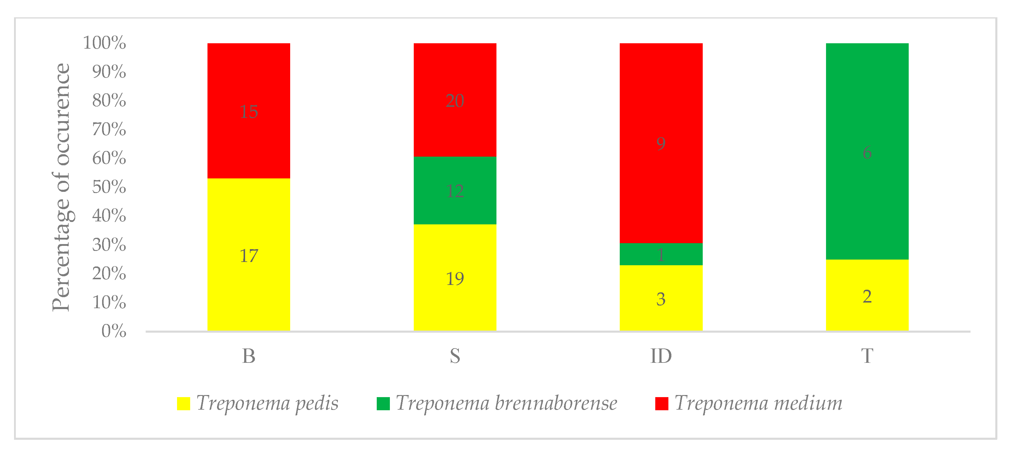
3.2. Evaluation of Treponemal Distribution

The analysis of the distribution of selected Treponema species across different sampling methods yielded significant results regarding their presence (Figure 1). The occurrence of T. pedis in tissue biopsy and surface swab samples was significantly higher (p < 0.001), compared to its presence in interdigital space swabs or faecal samples. Statistical comparison of sampling types for T. brennaborense revealed a significantly higher prevalence of this bacterium in surface swab samples (p < 0.001). For T. medium, there was a significantly higher occurrence in tissue biopsy samples, surface swabs, and interdigital space swabs compared to faecal samples (p < 0.001, p < 0.001, and p < 0.005, respectively). Notably, the presence of T. brennaborense was not detected in tissue biopsy samples, while T. medium was not found in faecal samples.
Regarding the sequencing of the amplified products using selected primer pairs, BLAST analysis revealed an average homology of 98.8% for T. medium, 99.7% for T. pedis, and 99.1% for T. brennaborense.
As a negative control group was utilized in this study, we analysed conventional PCR results from swabs of the interdigital space collected from BDD-negative cows, sampled prior to the detection of any BDD-positive cases during herd-level screening. In this control group, T. medium was detected in 20% of the samples, and T. pedis in 5%. None of the faecal samples tested positive. These findings are consistent with the overall prevalence observed in healthy cows within the herd, based on interdigital space samples.

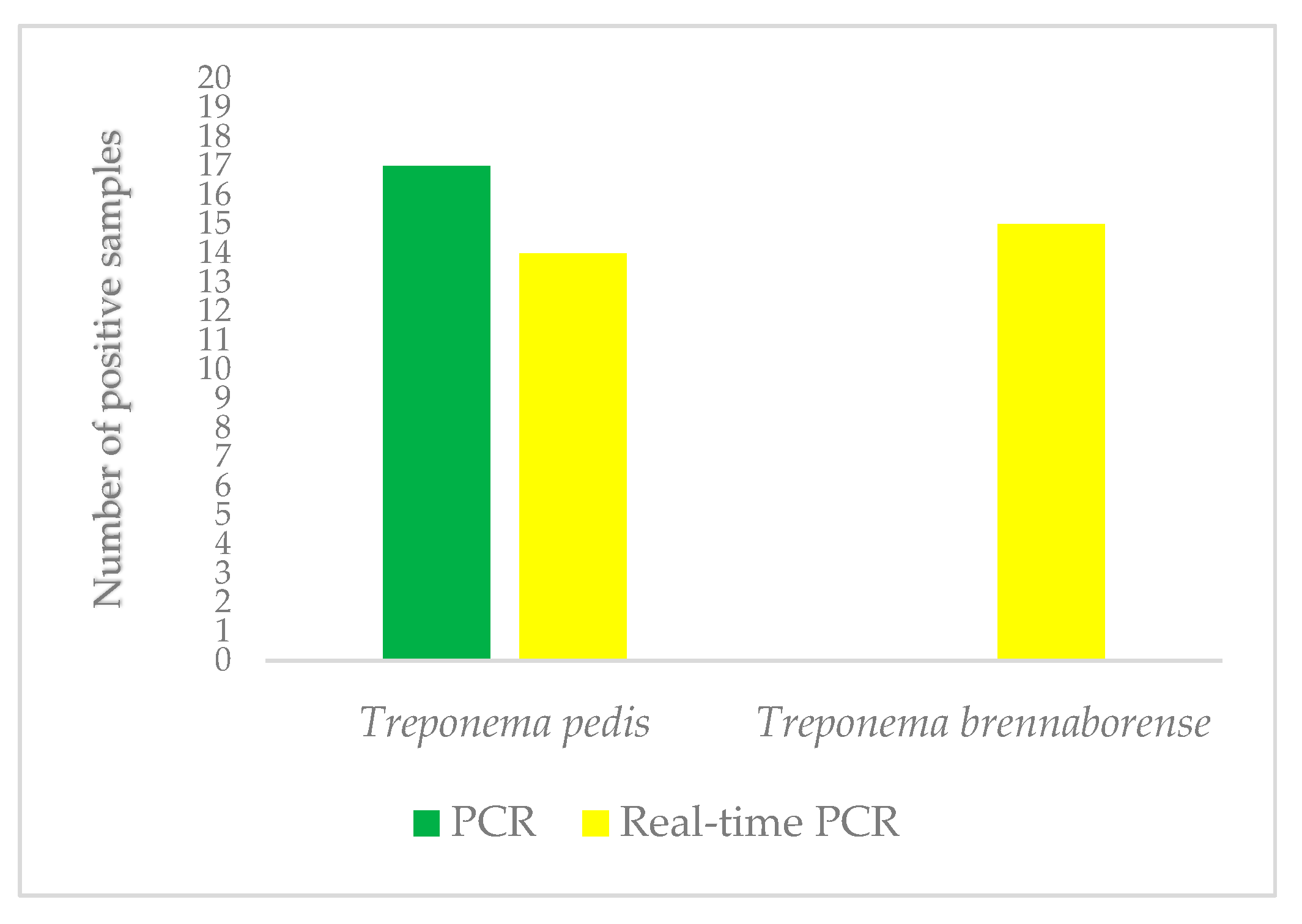
3.3. Comparison of Standard and Real-Time PCR in the Detection of Treponema Pedis and Treponema Brennaborense

The presence of T. pedis and T. brennaborense in tissue biopsy samples was successfully detected using qualitative real-time PCR. Of the 20 tissue biopsy samples from BDD lesions, 70% (14/20) tested positive for T. pedis, while T. brennaborense was detected in 75% (15/20) of the samples (Table 2). In contrast, none of the samples were positive for T. brennaborense when analysed using standard PCR (Figure 2). Additionally, the results of qualitative real-time PCR showed a lower detection rate for T. pedis compared to standard PCR. Statistical analysis between PCR methods for tissue samples revealed no significant differences for T. pedis, but a significantly higher prevalence of T. brennaborense was observed with the qualitative real-time PCR method (p < 0.001).

4. Discussion

Cattle lameness is commonly linked to bovine digital dermatitis (BDD), which, in addition to reducing production performance, significantly increases the economic costs associated with the treatment of this disease [11]. Whether the cattle are dairy or feedlot, the clinical manifestations and microbial composition remain similar, with the lesions typically located at a characteristic plantar site between the heel bulbs [20,26]. Culture-independent molecular methods have been employed to identify bacteria associated with BDD, and those which are crucial components of the BDD microbiome, due to their nature and relatively low abundance in healthy skin. Treponema species are generally predicted to dominate the bacterial population and play a pivotal role in the pathogenesis of this disease, with T. phagedenis, T. medium, and T. pedis being the most frequently detected species [16]. Given that several factors can influence not only the prevalence of BDD but also the distribution of different Treponema phylotypes [3,27], this study analysed the Treponema phylotypes associated with BDD in Slovak farm, along with various sampling and PCR methods.
The presence and abundance of spirochetes in BDD lesions are not consistent across studies [28]. T. pedis has been identified as one of the dominant species in the studies of Mamuad et al. [29] and Moreira et al. [30]. However, Canales et al. [31] reported that it was detected in only 22% of biopsy samples from active lesions in Chilean dairy cows. In contrast, the prevalence in this study was 95% (surface swabs), which supports the hypothesis of geographic variation in the prevalence of Treponema phylotypes. Mamuad et al. [29] found that T. phagedenis and T. medium were the most abundant species in dairy cows with BDD, and a similar pattern was observed in the study by Canales et al. [31], in which the results for T. medium were very similar to our findings (95%). However, T. phagedenis was not detected in any dairy cows in that study. In contrast, the studies of Dias and De Buck [32], Canales et al. [31], and Mamuad et al. [29] found the prevalence of T. phagedenis to be 100%, 63%, and 68.96%, respectively, which does not align with our results. In a study by Marčekova et al. [33], which investigated the presence of Treponema spp. in skin samples from four farms in Slovakia as a pilot project in the territory, the authors confirmed the presence of T. phagedenis in one sample (1/36). This finding may indicate geographic and regional differences compared to the results of other studies, ones in which T. phagedenis is most frequently found in M2 lesions; this would be inconsistent with the findings of our own study. The failure to detect T. phagedenis could explain the claim by Krull et al. [34] that T. phagedenis is significantly abundant in the early stages of lesions. T. pedis and T. medium are frequently co-isolated with T. phagedenis [35]. A larger sample size could help clarify the reasons for the absence of T. phagedenis association in certain samples, as the present study was constrained by the limited prevalence of bovine digital dermatitis (BDD) during the research period. According to Frosth et al. [36], one potential explanation for the low detection rate may lie in the atypical nature of some lesions, or in technical factors such as tissue manipulation and DNA extraction processes.
T. brennaborense has been detected by in situ hybridization in several cases of digital dermatitis lesions [37], but in other instances, it was often undetected, leading to the erroneous assumption that this species of Treponema does not significantly contribute to the pathogenesis of BDD [38]. However, the role of T. brennaborense in BDD remains unclear, as it has been found in several cases [14]. In this study, a 75% incidence of T. brennaborense was observed in biopsies, which contradicts the findings of Wilson-Welder et al. [39], who reported it as being less common. However, this finding was confirmed in positive skin samples (1/36) in the study by Marčeková et al. [33], which is the only study conducted in Slovakia to date; this is comparable to our results detected by standard PCR, as it is the same method used in this study. Tissue invasion by multiple Treponema species, due to their virulence factors, contributes to the formation of BDD lesions, often in conjunction with other bacteria as secondary infections [40]. However, according to Dias et al. [41], P. levii, F. necrophorum, and B. pyogenes did not act as secondary invaders, but appeared to precede colonization by Treponema spp. Thus, there is a need to reassess the role and interplay of T. brennaborense in the pathogenesis of BDD. Additionally, this study found differences between standard PCR and real-time PCR methods, with qualitative real-time PCR showing a significantly higher prevalence of T. brennaborense, likely due to its higher sensitivity. In contrast, T. pedis was detected more frequently using standard PCR.
Non-invasive swab sampling appears to be an effective method used for assessing the bacterial composition associated with digital dermatitis in herds [32,42]. In contrast, more invasive sampling procedures, such as tissue biopsies, are less practical for commercial feedlots, as they require local anaesthesia and increase the risk of complications such as bleeding, tissue scarring, or secondary infections [43]. Interestingly, Moreira et al. [30] suggest that the Treponema species most relevant to the pathogenesis of digital dermatitis tend to penetrate deeper into the affected tissue.
In our study, both invasive and non-invasive methods for sampling (M2) BDD lesions were employed. The results of the standard PCR analyses of presence for all three Treponema species clearly indicated a higher prevalence of positive samples obtained via the non-invasive surface swab method, compared to tissue biopsies. These findings highlight the high prevalence of Treponema species in BDD lesions. The abundance of T. phagedenis, T. medium, and T. pedis was found to be higher in lesion swabs than in biopsy samples, consistent with the results of Dias and De Buck [32]. Our results similarly showed that T. pedis was detected in 95% of surface swabs and in 85% and 70% of positive biopsies samples using standard PCR and qualitative real-time PCR, respectively. This indicates that, although these species are considered invasive, their DNA can still be detected in surface swabs, as reported by Dias and De Buck [32]. Therefore, surface swabs not only serve as an effective diagnostic tool for detecting BDD but also provide valuable insights into the bacterial communities present on the surface, potentially guiding topical treatments targeting specific active stages of the disease [42].
Molecular methods have been instrumental in detecting Treponema species associated with bovine digital dermatitis (BDD). However, the absence of culture-based techniques raises concerns about the viability of these treponemes in bovine faeces [44]. Faecal matter has been considered a potential reservoir for BDD infection, given the presence of Treponema species in the gastrointestinal tract and the frequent exposure of cattle foot skin to faecal matter [45,46]. Bell et al. [44] observed that Treponema species can survive in faeces for up to one day, which, while relatively brief, provides enough time for cattle to come into contact with fresh faeces containing BDD-associated treponemes. Furthermore, cattle often walk directly behind each other and may stand in faeces shortly after defecation, especially in walkways and chutes. These factors create multiple opportunities for cattle feet to interact with fresh faeces.
In this study, 30% of faecal samples were positive for T. brennaborense, and 10% for T. pedis. These findings contrast with the results of Dias et al. [47] and Evans et al. [48], who reported no positive samples from faecal material, challenging the hypothesis that faecal matter serves as a significant reservoir for BDD treponemes. While T. medium was not detected in faecal samples in this study, it is important to note that the percentage of positive faecal samples is still low compared to the findings of Zinicola et al. [46], who reported a relative abundance of less than 0.005% of Treponema species in the faecal microbiome of cattle, based on deep sequencing of the 16S rRNA gene. These findings highlight the uncertainty surrounding the potential for faecal transmission of Treponema species.
The presence of BDD-associated treponemes in faeces could stem from the gastrointestinal tract, but they may also originate from other biological secretions, or from contact between faeces and BDD lesions, or from environmental sources [45]. An interesting observation made by Potterton et al. [49] suggested that cattle with dirtier hocks tend to have fewer hock lesions, potentially indicating a protective effect of dirt. This hypothesis is partly supported by Dias et al. [47], who found that unhygienic conditions might be more closely linked to skin damage, which increases the risk of BDD, but did not suggest that such conditions directly serve as transmission routes or reservoirs for BDD pathogens.
Treponema species have been detected in most healthy skin samples [2], supporting the findings of Dias et al. [47], whose comparison of bacterial counts on healthy skin versus lesions suggests that colonization of healthy skin may be a transient event, likely resulting from environmental contamination during the resting period. This finding opens the door for longitudinal studies to better understand the dynamics of Treponema spp. colonization, which could help determine whether skin colonization is transient or persistent over time. Such research would be invaluable for understanding how Treponema species, particularly those associated with BDD, behave and establish in different stages of infection.
As Beninger et al. [16] noted, the number of unique Treponema species and their absolute abundance are typically higher in active stages and ulcerative lesions compared to chronic stages, healing stages, or healthy skin in dairy cattle. This is evident in the results from interdigital space samples in the current study. Despite the source for these samples being areas without clinical signs of BDD, 15% were positive for T. pedis, 45% for T. medium, and 5% for T. brennaborense. The fact that none of the positive samples from the interdigital space overlapped with faecal samples from the same cows suggests that while the interdigital space may indeed be contaminated by faeces, it is not simply a direct reflection of faecal contamination.
There is substantial variation in Treponema phylotypes and species across individual cattle and across studies [34,50]. These studies have consistently shown much higher Treponema abundance in BDD lesions compared to healthy skin. Interestingly, Beninger et al. [16] also pointed out that it is not uncommon to detect a high proportion of at least one Treponema species even in healthy skin samples, suggesting that certain species might transiently colonize healthy skin, without this necessarily indicating active disease. Additionally, lesions such as “non-healing” hoof lesions, including toe necrosis, white line disease, and sole ulcers, have also tested positive for Treponema species, further complicating the picture. In the work by Evans et al. [51], bacteria isolated from these lesions were thought to have an environmental causality. This highlights the complexity of Treponema colonization and the challenge in distinguishing between pathogenic and non-pathogenic bacteria.
One area that would greatly benefit from further exploration is the development of more refined methods to distinguish between viable and non-viable Treponema bacteria. A suitable approach capable of accurately identifying viable bacteria would allow researchers to gain a better understanding of the dynamics of Treponema colonization and its role in the transmission and maintenance of BDD within the cattle herd, as suggested by Dias et al. [37]. This could be crucial for determining how best to control and manage BDD, particularly in the context of environmental contamination and transmission between animals. Roelofs et al. [52] emphasize that monitoring prevalence at the national level is essential for controlling this disease and mitigating its impact on dairy cow productivity, as systemic exposure adversely affects both reproduction and milk production [53]. According to Robcis et al. [54], a key consideration is the consensus on integrating both individual and collective approaches to therapy and management, addressing infectious as well as non-infectious causes of lameness.
Of the 20 interdigital space samples from BDD-positive cows, 15% tested positive for T. pedis and 45% for T. medium, and T. brennaborense was detected in 5% of the samples. In samples from BDD-negative cows, PCR showed a lower detection rate: 4% for T. pedis and 20% for T. medium. In contrast, none of the faecal samples from BDD-negative cows tested positive. Among BDD-positive cows, T. pedis was identified in 10% of faecal samples, while T. brennaborense was present in 30%. In the case of biopsies, negative controls from BDD-negative cows were not included, as a non-invasive approach was preferred over an invasive one. Invasive sampling is limited by both the number of samples that can be collected from a single animal and the feasibility of repeated sampling. Additionally, the creation of wounds that may serve as entry points for secondary infections, the risk of complications such as bleeding, and the increased duration of restraint in a fixation device represent significant limitations.
Our study had several limitations. It was conducted in a single commercial dairy herd where BDD was endemic, and cattle were exposed to standard farm management practices, including a lack of foot-bathing. Another limitation was the prevalence of BDD at the time of sampling, which influenced the number of available samples. Future studies involving multiple herds across Slovakia are recommended to provide a deeper understanding of the pathogenesis and aetiology of BDD.

5. Conclusions

In this study, the prevalence of only three Treponema species, namely, T. medium, T. pedis, and T. brennaborense, was detected in BDD lesions in the active stage (M2). Their distribution on the surface of the lesion, in the interstitial spaces, and in faeces, which may serve as a potential source of infection, was also confirmed. In addition, the absence of T. phagedenis, which is associated with active stages of BDD, was an interesting finding of this study, suggesting possible geographic or regional variation. Standard and real-time PCR methods were used to detect T. pedis and T. brennaborensne, and the real-time PCR method proved to be more reliable for the detection of T. pedis, whereas surface swabs tested with standard PCR yielded more compelling results, despite the cost disparity. For T. brennaborense, real-time PCR demonstrated higher sensitivity, although the precise role of the species in the emergence of BDD and its interactions with other Treponema species necessitate further investigation. Therefore, it may be concluded that it is prudent to carefully consider the selection of sampling methods, taking into account the conditions of dairy farms, the prevalence of treponemes, the importance of animal welfare, and the effectiveness of the results. This approach should align with the broader goal of establishing a gold standard method for diagnosing and understanding BDD.

Supplementary Materials

The following supporting information can be downloaded at: https://www.mdpi.com/article/10.3390/microbiolres16060119/s1, Figure S1: Gel electrophoresis pattern for Treponema medium isolates products after amplification; Figure S2: Gel electrophoresis pattern for Treponema pedis isolates products after amplification; Figure S3: Gel electrophoresis pattern for Treponema brennaborense isolates products after amplification.

Author Contributions

Conceptualization, S.M. and M.S.; methodology, M.S., N.Š.H., V.K. and M.M.; validation, M.S.; formal analysis, S.M., M.S. and P.G.; investigation, S.M, M.S. and N.Š.H.; resources, M.M. and P.M.; data curation, S.M., M.S. and N.Š.H.; writing—original draft preparation, S.M., M.S. and V.K.; writing—review and editing, P.M.; visualization, S.M. and M.S.; supervision, P.M. and M.M.; project administration, P.M.; funding acquisition, P.M. All authors have read and agreed to the published version of the manuscript.

Funding

This research was funded by the Slovak Research and Development Agency, which provided financial support for this research under the number APVV-19-0462 and by the Scientific Grant Agency of the Ministry of Education of the Slovak Republic under grant number VEGA 1/0650/24.

Institutional Review Board Statement

The animal study protocol was approved by the Ethics Committee of the University of Veterinary Medicine and Pharmacy in Kosice under protocol code EKVP/2023-03 for studies involving animals.

Informed Consent Statement

Not applicable.

Data Availability Statement

The data presented in this study are available on request from the corresponding author.

Conflicts of Interest

The authors declare no conflicts of interest.

References

  1. Cheli, R.; Mortellaro, C. La dermatite digitale del bovino. In Proceedings of the 8th International Conference on Diseases of Cattle, Piacenza, Milan, Italy, 9–13 September 1974; pp. 208–213. [Google Scholar] [CrossRef]
  2. Caddey, B.; De Buck, J. Meta-Analysis of Bovine Digital Dermatitis Microbiota Reveals Distinct Microbial Community Structures Associated With Lesions. Front. Cell. Infect. Microbiol. 2021, 11, 685861. [Google Scholar] [CrossRef] [PubMed]
  3. Salem, S.E.; Monir, A.; Abdallah, H.M.; Mahmoud, A.F.; Elsheikh, H.A.; Refaai, W.; Ahmed, A.B.; Elsheikh, H.E.M.; El-Sheikh, M.; Mesalam, A. Digital dermatitis in dairy cattle in Egypt: Herd-level risk factors and Treponema spp. prevalence across lesion M-scores. Vet. J. 2025, 309, 1062289. [Google Scholar] [CrossRef] [PubMed]
  4. Read, D.H.; Walker, R.L. Papillomatous digital dermatitis (Footwarts) in California dairy cattle: Clinical and gross pathologic findings. J. Vet. Diagn. Investig. 1998, 10, 67–76. [Google Scholar] [CrossRef] [PubMed]
  5. Evans, N.J.; Murray, R.D.; Carter, S.D. Bovine digital dermatitis: Current concepts from laboratory to farm. Vet. J. 2016, 211, 3–13. [Google Scholar] [CrossRef]
  6. Döpfer, D.; Holzhauer, M.; Boven, M. The dynamics of digital dermatitis in populations of dairy cattle: Model-based estimates of transition rates and implications for control. Vet. J. 2012, 193, 648–653. [Google Scholar] [CrossRef]
  7. Biemans, F.; Bijma, P.; Boots, N.M.; de Jong, M.C.M. Digital dermatitis in dairy cattle: The contribution of different disease classes to transmission. Epidemics 2018, 23, 76–84. [Google Scholar] [CrossRef]
  8. Stokes, J.E.; Leach, K.A.; Main, D.C.; Whay, H.R. The reliability of detecting digital dermatitis in the milking parlour. Vet. J. 2012, 193, 679–684. [Google Scholar] [CrossRef]
  9. Gomez, A.; Cook, N.B.; Socha, M.T.; Döpfer, D. First-lactation performance in cows affected by digital dermatitis during the rearing period. J. Dairy Sci. 2015, 98, 4487–4498. [Google Scholar] [CrossRef]
  10. Afonso, J.S.; Oikonomou, G.; Carter, S.; Clough, H.E.; Griffiths, B.E.; Rushton, J. Diagnosis of bovine digital dermatitis: Exploring the usefulness of indirect ELISA. Front. Vet. Sci. 2021, 8, 728691. [Google Scholar] [CrossRef]
  11. Cortes, J.A.; Hendrick, S.; Janzen, E.; Pajor, E.A.; Orsel, K. Economic impact of digital dermatitis, foot rot, and bovine respiratory disease in feedlot cattle. Transl. Anim. Sci. 2021, 5, txab076. [Google Scholar] [CrossRef]
  12. Schulz, T.; Gundelach, Y.; Feldmann, M.; Hoedemaker, M. Early detection and treatment of lame cows. Tierärztliche Prax. Ausg. G Großtiere/Nutztiere 2016, 44, 5–11. [Google Scholar] [CrossRef]
  13. Wilson-Welder, J.H.; Alt, D.P.; Nally, J.E. The etiology of digital dermatitis in ruminants: Recent perspectives. Vet. Med. 2015, 6, 155–164. [Google Scholar] [CrossRef]
  14. Nally, J.E.; Hornsby, R.L.; Alt, D.P.; Whitelegge, J.P. Phenotypic and proteomic characterization of treponemes associated with bovine digital dermatitis. Vet. Microbiol. 2019, 235, 35–42. [Google Scholar] [CrossRef] [PubMed]
  15. Espiritu, H.M.; Mamuad, L.L.; Jin, S.J.; Kim, S.H.; Kwon, S.W.; Lee, S.S.; Lee, S.M.; Cho, Y.I. Genotypic and Phenotypic Characterization of Treponema phagedenis from Bovine Digital Dermatitis. Microorganisms 2020, 8, 1520. [Google Scholar] [CrossRef] [PubMed]
  16. Beninger, C.; Naqvi, S.A.; Naushad, S.; Orsel, K.; Luby, C.; Derakhshani, H.; Khafipour, E.; De Buck, J. Associations between digital dermatitis lesion grades in dairy cattle and the quantities of four Treponema species. Vet. Res. 2018, 49, 111. [Google Scholar] [CrossRef]
  17. Bay, V.; Gillespie, A.; Ganda, E.; Evans, N.J.; Carter, S.D.; Lenzi, L.; Lucaci, A.; Haldenby, S.; Barden, M.; Griffiths, B.E.; et al. The bovine foot skin microbiota is associated with host genotype and the development of infectious digital dermatitis lesions. Microbiome 2023, 11, 4. [Google Scholar] [CrossRef]
  18. Zojaji, V.; Mokhtari, A.; Mohamadnia, A. Molecular Isolation of Treponema spp. from Ovine Footrot Lesions, Finding Evidence for Contagious Ovine Digital Dermatitis. Iran. J. Vet. Surg. 2024, 19, 118–124. [Google Scholar] [CrossRef]
  19. Anklam, K.; Kulow, M.; Yamazaki, W.; Döpfer, D. Development of real-time PCR and loop-mediated isothermal amplification (LAMP) assays for the differential detection of digital dermatitis associated treponemes. PLoS ONE 2017, 12, e0178349. [Google Scholar] [CrossRef]
  20. Caddey, B.; Orsel, K.; Naushad, S.; Derakhshani, H.; De Buck, J. Identification and Quantification of Bovine Digital Dermatitis-Associated Microbiota across Lesion Stages in Feedlot Beef Cattle. mSystems 2021, 6, e0070821. [Google Scholar] [CrossRef]
  21. Döpfer, D.; Koopmans, A.; Meijer, F.A.; Szakáll, I.; Schukken, Y.H.; Klee, W.; Bosma, R.B.; Cornelisse, J.L.; van Asten, A.J.; ter Huurne, A.A. Histological and bacteriological evaluation of digital dermatitis in cattle, with special reference to spirochaetes and Campylobacter faecalis. Vet. Rec. 1997, 140, 620–623. [Google Scholar] [CrossRef]
  22. Brandt, S.; Apprich, V.; Hackl, V.; Tober, R.; Danzer, M.; Kainzbauer, C.; Gabriel, C.; Stanek, C.; Kofler, J. Prevalence of bovine papillomavirus and Treponema DNA in bovine digital dermatitis lesions. Vet. Microbiol. 2011, 148, 161–167. [Google Scholar] [CrossRef] [PubMed]
  23. Karaffová, V.; Marcinková, E.; Bobíková, K.; Herich, R.; Revajová, V.; Stašová, D.; Kavuľová, A.; Levkutová, M.; Levkut, M.; Lauková, A.; et al. TLR4 and TLR21 expression, MIF, IFN-β, MD-2, CD14 activation, and sIgA production in chickens administered with EFAL41 strain challenged with Campylobacter jejuni. Folia Microbiol. 2017, 62, 89–97. [Google Scholar] [CrossRef]
  24. Schloss, P.D.; Westcott, S.L.; Ryabin, T.; Hall, J.R.; Hartmann, M.; Hollister, E.B.; Lesniewski, R.A.; Oakley, B.B.; Parks, D.H.; Robinson, C.J.; et al. Introducing mothur: Open-source, platform-independent, community-supported software for describing and comparing microbial communities. Appl. Environ. Microbiol. 2009, 75, 7537–7541. [Google Scholar] [CrossRef] [PubMed]
  25. Edgar, R.C.; Haas, B.J.; Clemente, J.C.; Quince, C.; Knight, R. UCHIME improves sensitivity and speed of chimera detection. Bioinformatics 2011, 27, 2194–2200. [Google Scholar] [CrossRef]
  26. Sullivan, L.E.; Evans, N.J.; Blowey, R.W.; Grove-White, D.H.; Clegg, S.R.; Duncan, J.S.; Carter, S.D. A molecular epidemiology of treponemes in beef cattle digital dermatitis lesions and comparative analyses with sheep contagious ovine digital dermatitis and dairy cattle digital dermatitis lesions. Vet. Microbiol. 2015, 178, 77–87. [Google Scholar] [CrossRef]
  27. Cook, N. LifeStep—A Lesion Oriented, Life Cycle Approach to Preventing Lameness in Dairy Herds. In Proceedings of the KvaegKongress, Herning, Denmark, 1 February 2016; pp. 1–11. Available online: https://www.landbrugsinfo.dk/kvaeg/dansk-kvaeg-kongres/sider/bilag-kk-nigel-b-cook-02.pdf (accessed on 20 January 2022).
  28. Hesseling, J.; Legione, A.R.; Stevenson, M.A.; McCowan, C.I.; Pyman, M.F.; Finochio, C.; Nguyen, D.; Roic, C.L.; Thiris, O.L.; Zhang, A.J.; et al. Bovine digital dermatitis in Victoria, Australia. Aust. Vet. J. 2019, 97, 404–413. [Google Scholar] [CrossRef]
  29. Mamuad, L.L.; Seo, B.J.; Faruk, M.S.A.; Espiritu, H.M.; Jin, S.J.; Kim, W.I.; Lee, S.S.; Cho, Y.I. Treponema spp., the dominant pathogen in the lesion of bovine digital dermatitis and its characterization in dairy cattle. Vet. Microbiol. 2020, 245, 108696. [Google Scholar] [CrossRef]
  30. Moreira, T.F.; Facury Filho, E.J.; Carvalho, A.U.; Strube, M.L.; Nielsen, M.W.; Klitgaard, K.; Jensen, T.K. Pathology and bacteria related to digital dermatitis in dairy cattle in all year round grazing system in Brazil. PLoS ONE 2018, 13, e0193870. [Google Scholar] [CrossRef]
  31. Canales, N.; Bustamante, H.; Wilson-Welder, J.; Thomas, C.; Ramirez, E.; Salgado, M. First Molecular Confirmation of Treponema spp. in Lesions Consistent with Digital Dermatitis in Chilean Dairy Cattle. Pathogens 2022, 11, 510. [Google Scholar] [CrossRef]
  32. Dias, A.P.; De Buck, J. Detection and quantification of bacterial species DNA in bovine digital dermatitis lesions in swabs and fine-needle aspiration versus biopsies. Front. Vet. Sci. 2022, 9, 1040988. [Google Scholar] [CrossRef]
  33. Marčeková, P.; Mad’ar, M.; Styková, E.; Kačírová, J.; Sondorová, M.; Mudroň, P.; Žert, Z. The presence of Treponema spp. in equine hoof canker biopsies and skin samples from bovine digital dermatitis lesions. Microorganisms 2021, 9, 2190. [Google Scholar] [CrossRef] [PubMed]
  34. Krull, A.C.; Shearer, J.K.; Gorden, P.J.; Cooper, V.L.; Phillips, G.J.; Plummer, P.J. Deep sequencing analysis reveals temporal microbiota changes associated with development of bovine digital dermatitis. Infect. Immun. 2014, 82, 3359–3373. [Google Scholar] [CrossRef] [PubMed]
  35. Brodard, I.; Alsaaod, M.; Gurtner, C.; Jores, J.; Steiner, A.; Kuhnert, P. A filter-assisted culture method for isolation of Treponema spp. from bovine digital dermatitis and their identification by MALDI-TOF MS. J. Vet. Diagn. Investig. 2021, 33, 801–805. [Google Scholar] [CrossRef] [PubMed]
  36. Frosth, S.; Eriksson, H.K.; Rosander, A. Development of a multiplex quantitative PCR assay for simultaneous detection of Treponema phagedenis, Treponema pedis, Treponema medium, and ‘Treponema vincentii’ and evaluation on bovine digital dermatitis biopsies. Vet. Res. Commun. 2023, 47, 1937–1947. [Google Scholar] [CrossRef]
  37. Klitgaard, K.; Boye, M.; Capion, N.; Jensen, T.K. Evidence of multiple Treponema phylotypes involved in bovine digital dermatitis as shown by 16S rRNA gene analysis and fluorescence in situ hybridization. J. Clin. Microbiol. 2008, 46, 3012–3020. [Google Scholar] [CrossRef]
  38. Nordhoff, M.; Moter, A.; Schrank, K.; Wieler, L.H. High prevalence of treponemes in bovine digital dermatitis-a molecular epidemiology. Vet. Microbiol. 2008, 131, 293–300. [Google Scholar] [CrossRef]
  39. Wilson-Welder, J.H.; Alt, D.P.; Nally, J.E. Digital Dermatitis in Cattle: Current Bacterial and Immunological Findings. Animals 2015, 5, 1114–1135. [Google Scholar] [CrossRef]
  40. Corlevic, A.T.; Beggs, D.S. Host Factors Impacting the Development and Transmission of Bovine Digital Dermatitis. Ruminants 2022, 2, 90–100. [Google Scholar] [CrossRef]
  41. Dias, A.P.; Orsel, K.; Gammariello, C.S.; De Buck, J. Sequential emergence and quantitative dynamics of key bacterial species preceding digital dermatitis lesion onset in dairy cattle. Vet. Microbiol. 2025, 302, 110378. [Google Scholar] [CrossRef]
  42. Wong, N.S.T.; Malmuthge, N.; Gellatly, D.; Nordi, W.M.; Alexander, T.W.; Ortega Polo, R.; Janzen, E.; Schwartzkopf-Genswein, K.; Jelinski, M. Characterization of the hoof bacterial communities in feedlot cattle affected with digital dermatitis, foot rot or both using a surface swab technique. Anim. Microbiome 2024, 6, 2. [Google Scholar] [CrossRef]
  43. Nischal, U.; Nischal, K.C.; Khopkar, U. Techniques of skin biopsy and practical considerations. J. Cutan. Aesthetic Surg. 2008, 1, 107–111. [Google Scholar] [CrossRef]
  44. Bell, J.; Crosby-Durrani, H.E.; Blowey, R.W.; Carter, S.D.; Evans, N.J. Survival of bovine digital dermatitis treponemes in conditions relevant to the host and farm environment. Anaerobe 2023, 82, 102766. [Google Scholar] [CrossRef] [PubMed]
  45. Klitgaard, K.; Strube, M.L.; Isbrand, A.; Jensen, T.K.; Nielsen, M.W. Microbiota Analysis of an Environmental Slurry and Its Potential Role as a Reservoir of Bovine Digital Dermatitis Pathogens. Appl. Environ. Microbiol. 2017, 83, e00244-17. [Google Scholar] [CrossRef] [PubMed]
  46. Zinicola, M.; Lima, F.; Lima, S.; Machado, V.; Gomez, M.; Döpfer, D.; Guard, C.; Bicalho, R. Altered microbiomes in bovine digital dermatitis lesions, and the gut as a pathogen reservoir. PLoS ONE 2015, 10, e0120504. [Google Scholar] [CrossRef] [PubMed]
  47. Dias, A.P.; Orsel, K.; De Buck, J. Quantifying and mapping digital dermatitis-associated bacteria in lesion and nonlesion body sites and dairy farm environment. J. Dairy Sci. 2024, 107, 3252–3268. [Google Scholar] [CrossRef]
  48. Evans, N.J.; Timofte, D.; Isherwood, D.R.; Brown, J.M.; Williams, J.M.; Sherlock, K.; Carter, S.D. Host and environmental reservoirs of infection for bovine digital dermatitis treponemes. Vet. Microbiol. 2012, 156, 102–109. [Google Scholar] [CrossRef]
  49. Potterton, S.L.; Green, M.J.; Millar, K.M.; Brignell, C.J.; Harris, J.; Whay, H.R.; Huxley, J.N. Prevalence and characterisation of, and producers’ attitudes towards, hock lesions in UK dairy cattle. Vet. Rec. 2011, 169, 634. [Google Scholar] [CrossRef]
  50. Klitgaard, K.; Foix Bretó, A.; Boye, M.; Jensen, T.K. Targeting the treponemal microbiome of digital dermatitis infections by high-resolution phylogenetic analyses and comparison with fluorescent in situ hybridization. J. Clin. Microbiol. 2013, 51, 2212–2219. [Google Scholar] [CrossRef]
  51. Evans, N.J.; Blowey, R.W.; Timofte, D.; Isherwood, D.R.; Brown, J.M.; Murray, R.; Paton, R.J.; Carter, S.D. Association between bovine digital dermatitis treponemes and a range of ’non-healing’ bovine hoof disorders. Vet. Rec. 2011, 168, 214. [Google Scholar] [CrossRef]
  52. Roelofs, L.; Frössling, J.; Rosander, A.; Bjerketorp, J.; Belaghi, R.A.; Hansson, I.; Frosth, S. Digital dermatitis in Swedish dairy herds assessed by ELISA targeting Treponema phagedenis in bulk tank milk. BMC Vet. Res. 2024, 20, 168. [Google Scholar] [CrossRef]
  53. Mellado, M.; Saavedra, E.; Gaytán, L.; Veliz, F.G.; Macías-Cruz, U.; Avendano-Reyes, L.; García, E. The effect of lameness-causing lesions on milk yield and fertility of primiparous Holstein cows in a hot environment. Livest. Sci. 2018, 217, 8–14. [Google Scholar] [CrossRef]
  54. Robcis, R.; Ferchiou, A.; Berrada, M.; Raboisson, D. Management of digital dermatitis in dairy herds: Optimization and time allocation. Animals 2023, 13, 1988. [Google Scholar] [CrossRef] [PubMed]
Figure 1. Distributions of the three treponemes in the analyses of samples from different sampling types, using standard PCR: percentage (%) of occurrence of each Treponema spp. and presence in biopsies. T—tissue sample, S—surface swab, ID—interdigital space swab, F—faecal sample.
Microbiolres 16 00119 g001
Figure 2. Results of the comparison between standard and real-time PCR for Treponema detection, including the number of positive samples.
Microbiolres 16 00119 g002
Table 1. List of primers used in real-time PCR for the detection of selected species of Treponema.
PrimerSequence 5′–3′Self-Complementarity IndexMelting
Temperature
Product SizeReferences
T. pedis flaB FwGCAAGTTCCGCACAATTTAA358 °C121 bpIn this study
T. pedis flaB RevTTCTTTGGTCCATGTTTGCA258 °C
T. bren. 16S rRNAFwCAAAGCAAACGTGATAAGTGT360 °C117 bp
T.bren. 16S rRNA RevTCGCGTACCATCGAATTAAA258 °C
Table 2. Treponema spp. detection results, using qualitative real-time PCR and standard PCR, for biopsy samples.
Treponema pedisTreponema brennaborense
No.Real-Time PCRPCRReal-Time PCRPCR
1NegativePositivePositiveNegative
2PositivePositivePositiveNegative
3PositivePositiveNegativeNegative
4PositivePositivePositiveNegative
5PositivePositivePositiveNegative
6NegativePositivePositiveNegative
7PositivePositivePositiveNegative
8PositiveNegativePositiveNegative
9NegativePositivePositiveNegative
10PositivePositivePositiveNegative
11PositiveNegativeNegativeNegative
12PositivePositiveNegativeNegative
13PositivePositivePositiveNegative
14PositivePositivePositiveNegative
15NegativePositivePositiveNegative
16NegativeNegativeNegativeNegative
17PositivePositivePositiveNegative
18PositivePositivePositiveNegative
19PositivePositivePositiveNegative
20NegativePositiveNegativeNegative
Disclaimer/Publisher’s Note: The statements, opinions and data contained in all publications are solely those of the individual author(s) and contributor(s) and not of MDPI and/or the editor(s). MDPI and/or the editor(s) disclaim responsibility for any injury to people or property resulting from any ideas, methods, instructions or products referred to in the content.

