Article

HIV-1 and Antiretroviral Therapy Modulate HERV Pol and Syncytin Gene Expression in Mothers and Newborns

1
Department of Public Health and Pediatric Sciences, University of Turin, 10126 Turin, Italy
2
Pediatric Laboratory, Department of Pediatrics, Regina Margherita Children’s Hospital, 10126 Turin, Italy
*
Authors to whom correspondence should be addressed.
These authors contributed equally to this work.
Microbiol. Res. 2025, 16(6), 116; https://doi.org/10.3390/microbiolres16060116
Submission received: 30 April 2025 / Revised: 29 May 2025 / Accepted: 1 June 2025 / Published: 3 June 2025

Abstract

:
Background: Human endogenous retroviruses (HERVs) are remnants of ancestral retroviral infections integrated into the human genome, some of which maintain a residual active expression and retain physiological relevance. HIV-1 infection and antiretroviral therapy (ART) are known to modulate HERV expression, yet their specific effects during pregnancy remain poorly understood. This study aimed to investigate the peripartum transcriptional activity of selected HERV sequences in HIV-1-positive women receiving ART and their newborns exposed to the therapy and HIV-1-negative healthy controls. Methods: We quantified the expression of pol regions of HERV-H, -K, and -W and of Syncytin 1 and Syncytin 2 in peripheral blood samples collected at delivery using real-time PCR. Results: In HIV-1-positive mothers on ART therapy, we observed a significant downregulation in the pol gene expression of HERV-H, HERV-K, and HERV-W, as well as of Syncytin 1 and Syncytin 2, compared to healthy mothers. In contrast, no differences in the expression of the different targets were found in the two groups of newborns. All the HERV genes analyzed were also found to be expressed at significantly higher levels in the newborns compared to their mothers. Discussion: The results obtained suggest that antiretroviral therapy may influence and modulate HERV expression during pregnancy in both the mother and the fetus.

1. Introduction

The human genome comprises numerous repetitive elements, with human endogenous retroviruses (HERVs) accounting for approximately 8% of the total sequences [1]. Over the course of the last 100 million years, repeated infections by now-extinct exogenous retroviruses have led to the integration of these retroviral sequences into the human germline [2,3]. Unlike modern retroviruses, which primarily infect somatic cells, these ancient retroviruses occasionally infected primate germ cells, a rare event that allowed the stable integration of the viral genetic material into the host genome [4]. Once integrated, the viral sequence became a permanent part of the host genome and have been inherited vertically through generations, according to Mendelian inheritance principles [5].
Structurally, HERVs retain the classical features of exogenous retroviruses, including a proviral organization with two long terminal repeats (LTRs) flanking a central region containing the gag, pro, pol, and env genes [6,7]. Although most transposable elements are largely silenced by transcriptional repression mechanisms, such as histone hypermethylation, many have co-evolved with their hosts and are subject to selection pressure [8]. In fact, the epigenetic silencing of these elements may reflect an ancient adaptive mechanism: organisms first developed defense systems to suppress their expression, which later evolved into more refined systems to regulate gene activity. As a result, despite the presence of control mechanisms, many human endogenous retroviruses still retain a residual expression capacity, producing both coding and non-coding RNAs that influence various aspects of host biology [9]. Some maintain the ability to be transcribed and a few encode proteins that are co-opted for pivotal physiological functions. One of the most striking examples of HERV contribution to vertebrate physiology is the syncytins familyEnv proteins encoded by different HERVs across all mammals. These genes have undergone convergent evolution and have been co-opted for critical roles in placental development. In humans, two key Env loci encode the proteins Syncytin 1 and Syncytin 2: Syncytin 1 plays a central role in the formation and maintenance of the syncytiotrophoblast layer of the placenta [10,11], while Syncytin 2 is believed to be involved in promoting maternal immune tolerance toward the fetus [12].
Beyond their protein-coding functions, the thousands of HERV sequences scattered throughout the genome have significantly shaped primate genome evolution by serving as sources of regulatory elements. HERVs participate in gene regulatory networks, acting both as cis and trans elements, influencing the expression of host genes at multiple levels [8,13]. When unable to produce functional proteins, HERVs often continue to generate non-coding RNAs (ncRNAs), such as microRNAs and long non-coding RNAs. These transcripts can act as cis-regulatory elements by modulating gene expression either independently, by offering recognition sites for RNA-binding proteins, or in cooperation with cellular transcription factors [2]. HERVs can act as promoters of neighboring host genes and influence innate and adaptive immune responses [8].
From an immunological perspective, HERV-derived products exhibit hybrid characteristics resembling both self-antigens and microbial molecules. Their capacity to either evade immune detection or provoke immune responses appears context-dependent, influenced by specific expression conditions. This dual potential has spurred growing interest in investigating HERV sequences as potential contributors to autoimmune pathogenesis [14].
Moreover, other immune stimuli have been shown to influence HERV expression, suggesting a highly complex and not yet fully understood interaction between these elements and the immune system [2]. Theoretically, the innate immune pathways activated by HERV-derived products overlap with those involved in the first-line defense against exogenous viral infections, including both cytoplasmic and membrane-bound pattern recognition receptors (PRRs) [14].
The typical genomic structure shared by retroviruses suggests that proteins encoded by one retrovirus might act in trans on others, potentially compensating for defects in another virus [15]. Following the introduction of protease inhibitors targeting HIV-1 into clinical practice, some researchers speculated that proteases encoded by human endogenous retroviruses (HERVs), which might be resistant to these drugs, could compensate for the inhibited HIV-1 protease. The HERV-K10 protease has been shown to be drug-resistant and capable of processing the HIV-1 Gag protein at the correct cleavage site [16]. However, when HERV-K protease was incorporated into HIV-1 virions lacking their native protease, no functional complementation was observed, and Gag-Pol precursors were cleaved at incorrect sites [17]. In contrast, HERV-K10 integrase can partially complement integrase-deficient HIV-1 virions, although infectivity remains significantly lower than that of wild-type HIV-1 [18]. The Env protein of HERV-W, Syncytin 1, plays a critical role in placental syncytiotrophoblast formation [15] by acting as a fusogenic protein that promotes cytotrophoblast cell fusion. This process is vital for maintaining the placenta’s structural integrity and functional capacity, directly influencing embryo implantation and pregnancy maintenance [19]
Syncityn 1 retains characteristics of a functional retroviral envelope protein. It is notable that the production of infectious particles by env-defective HIV-1 strains has been reported, whereby HERV-W Env mediates the infection of CD4-negative T lymphocytes [20], suggesting that HERV-derived env expression may contribute to expanded HIV-1 tropism during infection [21].
Antiretroviral therapy (ART) has been demonstrated to reduce the plasma HIV-1 viral load to below 50 copies/mL. The quantification of HERV-K RNA in the plasma of HIV-1-infected patients undergoing antiretroviral therapy has demonstrated a positive correlation between the expression of this class of endogenous retroviruses and the HIV-1 viral load [15]. It remains unclear whether ART directly inhibits HERV-K expression or whether this effect is secondary to reduced HIV-1 activity. Protease inhibitors targeting HIV-1 do not appear to affect HERV-K gene products; however, nucleoside reverse transcriptase inhibitors may exert a modulatory effect [15].
The present study aims to further explore the interaction between HIV-1 infection, antiretroviral therapy, and HERV expression by analyzing the transcriptional activity of selected HERV sequences in peripheral blood collected at delivery from two groups of mother–newborn pairs. The first group consists of HIV-1-positive mother–infant pairs receiving antiretroviral therapy (ART), while the second group consists of HIV-1-negative, ART-naïve control pairs.
The analyzed genes include the pol regions of HERV-W, HERV-K, and HERV-H, as well as Syncytin 1 and Syncytin 2. This preliminary analysis aims to evaluate the potential influence of both HIV-1 infection and antiretroviral therapy on the peripartum expression of these endogenous retroviral elements.

2. Materials and Methods

2.1. Population and Samples Collection

This study involved 37 women (mean age 32.3 years) and their newborns. Of the women, 17 had been diagnosed with HIV-1, either prior to or during pregnancy, and were undergoing antiretroviral therapy (ART) at the Sant’Anna Obstetric and Gynecological Hospital in Turin, Italy. The remaining 20 women were not HIV-1-positive and, therefore, had not received ART; consequently, their infants were not exposed to antiretroviral drugs during intrauterine life. The two groups were comparable in terms of the gestational age at delivery and the maternal age at birth.
All the HIV-1-positive women were treated with a combination antiretroviral therapy, comprising two or more active substances belonging to different classes. The antiretroviral drugs were as follows: integrase inhibitors (INs) in 58.8% of the women; nucleoside reverse transcriptase inhibitors (NRTIs) in 100% of the women; non-nucleoside reverse transcriptase inhibitors (NNRTIs) in 23.5% of the women; and protease inhibitors (PIs) in 17.6% of the women. For 13 out of the 17 women, it was possible to evaluate the viral load of HIV-1.
Samples were collected at the time of delivery, including a peripheral blood sample from each mother and a cord blood sample from the placenta of each newborn.

2.2. RNA Extraction

For the analysis, total RNA was extracted from 200 μL of blood, to which 0.8 mL of RNApro solution (Biomole, Turin, Italy) was added. Nucleic acid extraction was performed using the Maxwell instrument with the Maxwell® 16 LEV simplyRNA Kit, according to the manufacturer’s instructions (Promega Corporation, Madison, WI, USA). The RNA obtained, eluted in 50 μL of nuclease free water, was frozen at −80 °C until use. The concentration of RNA in the extracted eluates was then measured using the NanoDrop 2000™ instrument (Thermo Fisher Scientific, Waltham, MA, USA). This instrument was also used to assess the quality of the RNA.

2.3. Reverse Transcription and Real Time PCR

To amplify and quantify the expression of specific mRNAs, the total RNA was first reverse transcribed into cDNA using a GeneAmp PCR System 9700 thermal cycler (Applied Biosystems, Foster City, CA, USA). The reverse transcription was performed according to the following temperature profile: 25 °C for 5 min, 42 °C for 60 min, and 70 °C for 15 min. The resulting cDNA samples were stored at −20 °C until further use.
The quantification of the selected genes was carried out by real-time PCR. For each target, 4 μL of cDNA (equivalent to 50 ng) was amplified in a total reaction volume of 20 μL, including 16 μL of PCR mix. Gene-specific primer and probe kits were used for GAPDH, HERV-K, HERV-W, HERV-H, Syncytin 1, and Syncytin 2, using optimized primers and probes by Biomole (Turin, Italy): HERV-K PP-054, HERV-W PP-055, HERV-H PP-056, SINC1 PP-112, and SINC2 PP-113.
Amplification reactions were performed on the ABI PRISM 7500 Real-Time PCR System (Life Technologies, Austin, TX, USA) using a 96-well plate format. The thermal cycling conditions were as follows: 95 °C for 2 min and then 45 cycles of 95 °C for 15 s and 60 °C for 1 min.

2.4. Relative Quantification with ΔΔCT Method

The ΔΔCT method was used for the relative quantification (RQ). The analysis proceeds by normalizing the cycle threshold (CT) value of the target gene to that of the reference gene for both the treated (test) and control (calibrator) samples. This yields the following: Δ(test) = CT(target, test) − CT(ref, test) and ΔCT(cal) = CT(target, cal) − CT(ref, cal). Next, the ΔCT value of the test sample is normalized against the ΔCT of the calibrator to obtain the following: ΔΔCT = ΔCT(test) − ΔCT(cal). The relative expression ratio is then calculated using the following formula: ratio = 2−ΔΔCT.
This ratio quantifies the fold change in the expression of the target gene in the test sample relative to the calibrator, normalized to the reference gene. A ΔΔCT value of greater than 0 (i.e., ΔCT(test) > ΔCT(cal)) results in a ratio of less than 1, indicating the downregulation of the target gene in the treated sample. Conversely, a ΔΔCT value of less than 0 (i.e., ΔCT(test) < ΔCT(cal)) yields a ratio of greater than 1, reflecting the upregulation of the target gene in the treated sample. This method allows for the quantification of gene expression differences across samples [22].

2.5. Statistical Analysis

GraphPad Software, Version 7, was used for the data analysis. The Mann–Whitney test was used to compare the transcriptional levels of HERV-H-pol, HERV-K-pol, HERV-W-pol, Syncytin 1, and Syncytin 2 between the HIV-1-positive mothers and the healthy controls and between the corresponding newborns. A p-value of <0.05 was considered statistically significant.

3. Results

In a cohort of 17 HIV-1-positive mothers undergoing antiretroviral therapy (ART) and their newborns, we quantified the transcriptional activity of the pol genes of HERV-K, HERV-W, and HERV-H, as well as the Syncytin 1 and Syncytin 2 genes, in maternal and neonatal whole blood collected at delivery. These data were compared to those from a control group of 20 HIV-1-negative women not receiving ART and their newborns, matched for the maternal age and the gestational age at delivery.

3.1. Expression of Pol Genes of HERV-K, HERV-W, and HERV-H and of Syncytin 1 and Syncytin 2 Genes in HIV-1-Negative Mothers and Their Newborns

The expression levels of pol genes of HERV-K, HERV-W, and HERV-H in HIV-1-negative mothers (healthy mothers) were significantly reduced compared to the expression levels in their newborns, while no significant differences were observed in the expression of Syncytin 1 and Syncytin 2 in the two groups (Figure 1).

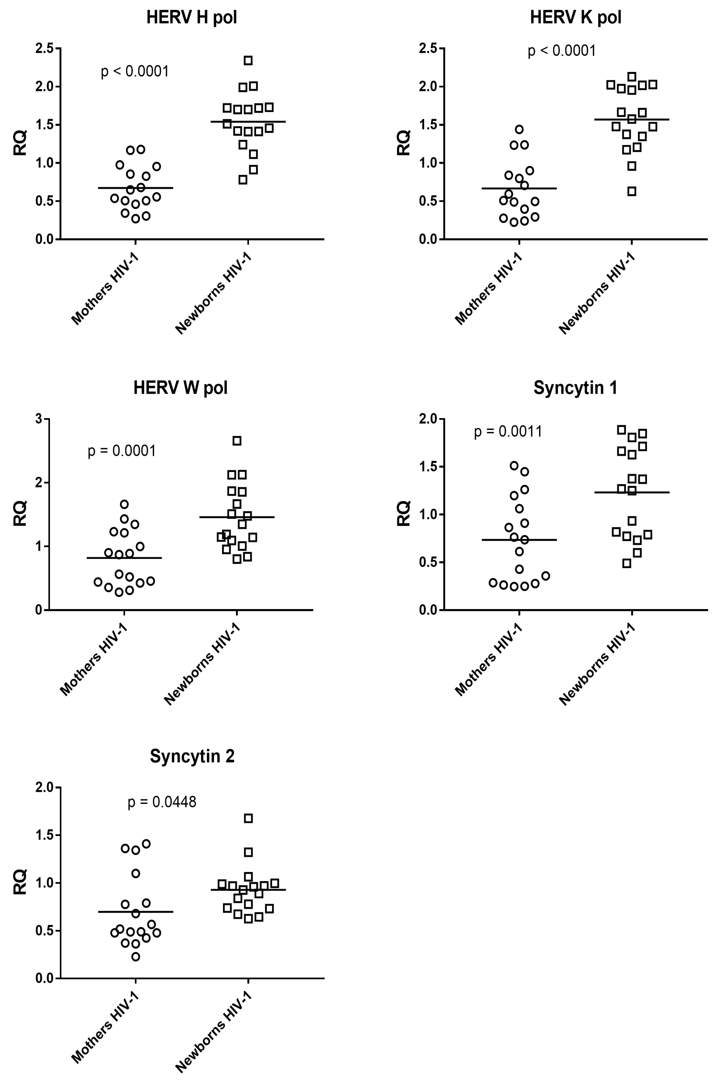
3.2. Expression of Pol Genes of HERV-K, HERV-W, and HERV-H and of Syncytin 1 and Syncytin 2 Genes in HIV-1-Positive Mothers with Antiretroviral Therapy and Their Newborns

The expression levels of pol genes of HERV-K, HERV-W, and HERV-H, as well as of Syncytin 1 and Syncytin 2 in HIV-1-positive mothers undergoing antiretroviral therapy was significantly reduced compared to the expression levels in their newborns (Figure 2).

3.3. Expression of Pol Genes of HERV-K, HERV-W, and HERV-H and of Syncytin 1 and Syncytin 2 Genes in HIV-1-Infected Mothers on Antiretroviral Therapy and in Healthy Mothers

In the whole blood of HIV-1-infected mothers on antiretroviral therapy, all the analyzed targets showed a statistically significant reduced expression compared to healthy mothers (Figure 3).

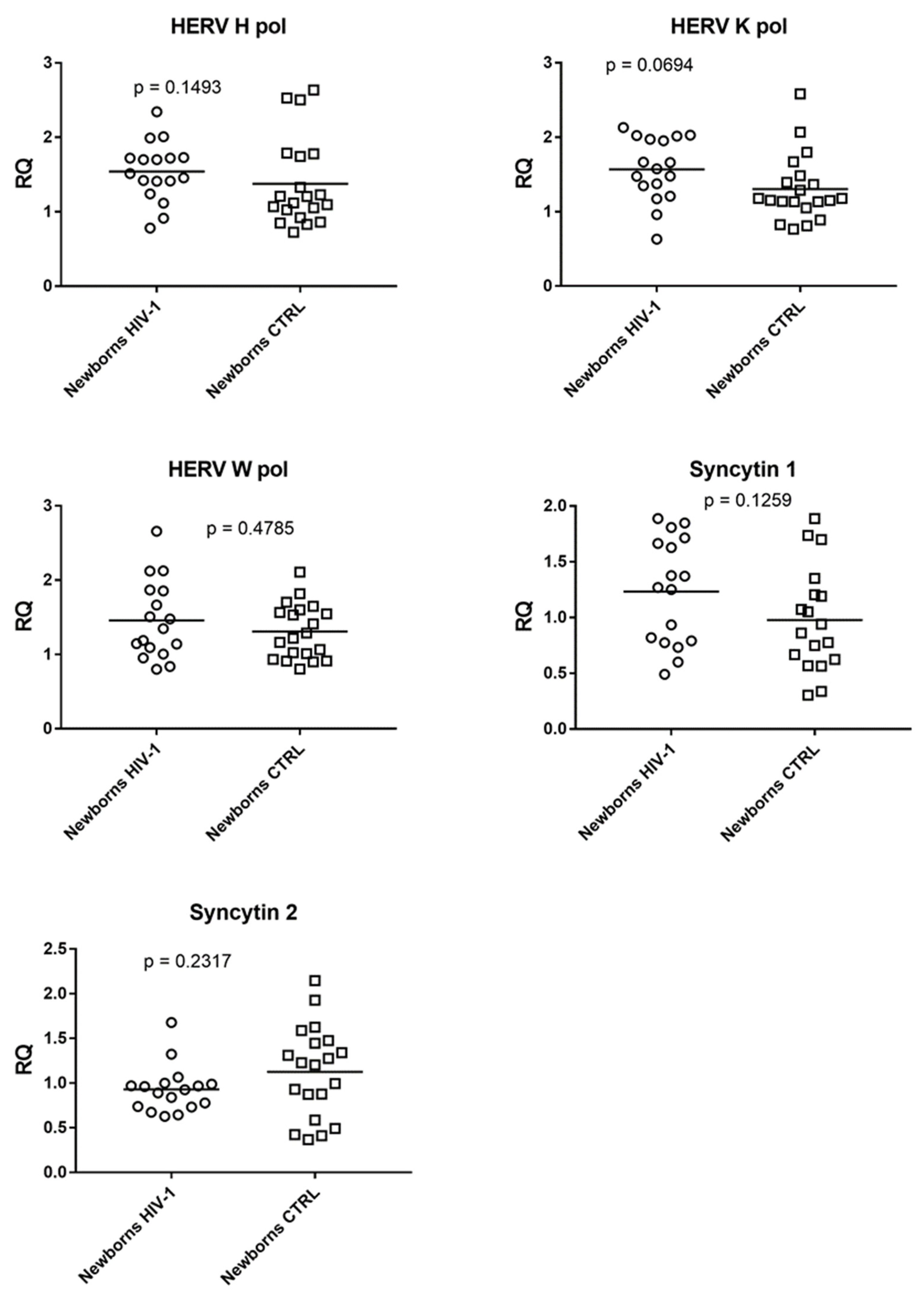
3.4. Expression of Pol Genes of HERV-K, HERV-W, and HERV-H and of Syncytin 1 and Syncytin 2 Genes in Infants Exposed to Antiretroviral Therapy and in Not-Exposed Infants

In the cord blood samples, no significant differences were observed in the expression of the pol genes of HERV-K, HERV-W, and HERV-H, as well as of the Syncytin 1 and Syncytin 2 genes, in infants exposed to ART in utero compared to those who were not exposed (Figure 4).

4. Discussion

Human endogenous retroviruses (HERVs) are retrovirus-derived sequences that have been integrated into the human genome over millions of years [2,3]. While the majority are inactive, some HERVs retain the capacity to express retroviral proteins, such as the syncytins, which are essential for the development of the placental syncytiotrophoblast [10]. The presence of homologous retroviral sequences with similar functions across all eutherian mammal genomes underscores their evolutionary conservation and important role in placental physiology.
Among the known modulators of HERV expression, both HIV-1 infection and antiretroviral therapy (ART) have been shown to exert significant effects. HIV-1, as an exogenous retrovirus, can activate the transcription of specific HERV families, whereas ART appears to suppress their expression [21]. These dynamic regulatory mechanisms raise important questions regarding the potential impact of ART not only on viral suppression but also on endogenous retroviral activity.
Our study aims to further investigate the interaction between HIV-1, antiretroviral therapy, and HERV activity by analyzing the transcriptional levels of selected HERV sequences in peripheral blood collected at delivery from two groups of mother–newborn pairs. The first group includes HIV-1-positive women and their infants, all receiving ART, while the second group comprises HIV-1-negative, ART-naïve controls.
Given that HIV-1-positive women represent the only population currently receiving ART during pregnancy, this group offers a unique opportunity to explore the relationship between ART exposure and HERV expression. The analysis focuses on the pol regions of HERV-W, HERV-K, and HERV-H, along with the genes encoding Syncytin 1 and Syncytin 2. This preliminary investigation aims to assess the potential impact of HIV-1 infection and ART on the peripartum expression of these endogenous retroviral elements.
In our study, we observed a significantly increased expression of the pol genes of HERV-H, -K, and -W in umbilical cord blood compared to maternal peripheral blood inboth healthy subjects and in HIV-1-infected mother–newborn pairs. Moreover, both Syncytin 1 and Syncytin 2 were also found to be significantly increased in cord blood compared to maternal blood in HIV-1-infected mother–newborn pairs.
These results support the hypothesis that, during gestation, the transcription of endogenous retroviruses is typically enhanced in fetal-origin tissues and cells and only to a lesser extent in maternal-origin tissues. This hypothesis is further corroborated by other studies [23]. This difference in HERV expression can be at least partially attributed to the high transcriptional activity of endogenous retroviruses in stem cells; notably, cord blood is particularly rich in stem cell progenitors [23].
The comparison between the two groups of mothers in our study revealed that HERV expression is consistently lower in HIV-1-exposed mothers undergoing antiretroviral therapy than in healthy, unexposed mothers. These findings support the hypothesis that antiretroviral therapy may exert an inhibitory effect not only on the expression and activity of the exogenous HIV-1 retrovirus but also on various endogenous retroviral sequences. Several studies in the literature have already supported this hypothesis [24,25,26,27,28], although they have been conducted primarily on HERV-K and in population groups different from the one analyzed in our study.
The quantification of HERV-K RNA in the plasma of HIV-1-infected patients undergoing antiretroviral therapy has demonstrated a positive correlation between the expression of this class of endogenous retroviruses and the HIV-1 viral load [15]. Patients who have achieved optimal HIV-1 viral load suppression have been observed to have up to twofold lower levels of HERV-K RNA in their plasma compared to those with poorly controlled HIV-1 infection [29]. Some studies have demonstrated the long-term suppression of HERV-K in patients receiving effective antiretroviral therapy, with the HIV-1 viral load remaining undetectable (i.e., <50 copies/mL). In contrast, patients receiving ineffective therapy have not shown such suppression. Indeed, in these cases, plasma HERV-K levels have been observed to rise rapidly, often preceding an increase in the HIV-1 viral load [21].
In our study, 13 out of the 17 women involved (76.5%) showed good infection control, with an undetectable viral load (<20 RNA copies/mL), while in the remaining 4 women, the viral load at the end of pregnancy was unknown. This finding suggests, on the one hand, that there was a low probability that the HERV expression levels in the HIV-1-positive women receiving ART were altered by the action of HIV-1 proteins and virions, and on the other hand, that the effectiveness of the antiretroviral therapy may have also contributed to the inhibition of HERV expression.
A significant, dose-dependent inhibition of reverse transcriptase (RT) and, thus, of the pol gene of endogenous HERV-K sequences also emerges from the study of Tyagi et al. [30]. This study specifically analyzed the inhibition of HERV-K by different classes of antiretroviral drugs. Comparing the results of this study with the therapy received by the women in our research, it appears that all of them were treated with nucleoside reverse transcriptase inhibitors (NRTIs) as a part of their combination antiretroviral therapy, which, in Tyagi et al.’s study, showed the effective inhibition of HERV-K reverse transcriptase. Similarly, effective inhibition was also observed for non-nucleoside reverse transcriptase inhibitors (NNRTIs), used by 23.5% of the women in our study, and for integrase inhibitors (INIs), used by 58.8% of the participants. As for protease inhibitors (PIs), which were used by only three women in this study (17.6%), Tyagi et al.’s research highlighted a greater specificity of action toward HIV-1 compared to the other drug classes.
Several studies have investigated the transcriptional activity of HERV-W env in HIV-1–infected individuals undergoing antiretroviral therapy. These investigations revealed no statistically significant difference in the expression of HERV-W env between HIV-1-infected subjects and healthy controls [28,31]. Similarly, no difference was observed in the expression of HERV-W env between subjects who were treated and those who were not treated for infection. The authors hypothesize that the observed discrepancy from the original hypothesis may be due to a potential mismatch between the cell types responsible for HERV-W production (predominantly monocytes and B cells) and those responsible for HIV-1 production (predominantly T cells). Indeed, since RNA was extracted from whole peripheral blood of HIV-1–infected patients, the resulting data may not accurately reflect the activity of specific cell types. During the course of HIV-1 infection, an alteration of immune cell populations is observed, characterized by the hyperactivation and increased apoptosis of B cells [32]. Consequently, it is possible that this alteration is exclusive to HIV-1-infected cells and may not be discernible through the analysis of the patients’ whole blood [28]. In a related study, no reduction in HERV-W expression was observed in infected individuals undergoing antiretroviral therapy in comparison to those not receiving therapy [28]. However, the authors acknowledge that the small number of subjects in the untreated population considered in the study may be a significant confounding factor in their analysis. They do not exclude the possibility that HERV-W expression in vivo may be suppressed by antiretroviral therapy [28].
In contrast, the comparison between the two groups of newborns revealed that the expression levels of the different targets did not show statistically significant differences.
These findings suggest the absence of an inhibitory effect on HERV sequences by antiretroviral therapy at the fetal and neonatal levels, in contrast to what was observed at the maternal level. Furthermore, despite the demonstrated effectiveness of antiretroviral therapy in the majority of the women in the study, it is not possible to exclude an indirect modulatory action by the HIV-1 virus present at the maternal level. The interpretation of these results is, therefore, complex, as it is not possible to fully separate the effects, potentially due to exposure to therapy from those related to maternal infection.
The study by L. H. S. Nali et al. compared the expression of HERV-K and HERV-W in HIV-1-infected women and healthy women, as well as in their respective children under 1 year of age. All the women recruited were undergoing antiretroviral therapy during pregnancy and had undetectable viral loads, and all the children were found to be negative for the infection. This study revealed a significantly lower level of HERV-W expression in the children exposed to therapy compared to those not exposed. In contrast, the significantly higher expression of HERV-W was observed in HIV-1-infected mothers exposed to therapy compared to healthy mothers [24]. Though the results of this study appear to be in contrast with those of our study, the two studies are not perfectly comparable, especially due to differences in the type of sample collected and the timing of the collection. In our study, the samples were collected at the time of delivery from maternal peripheral blood and from cord blood, whereas in the study by L. H. S. Nali et al., the samples were taken from maternal and infant peripheral blood at a later period, within the first year of life, with a mean age of 5.5 months [22]. The expression of pol genes from HERV-H, HERV-K, and HERV-W, although active throughout the first decade of life, is actually higher in newborns, while it is lower and stable in infants and children [23,33].

5. Conclusions

In the population analyzed in this study, which included 17 mother–newborn pairs exposed to maternal HIV-1 infection and antiretroviral therapy during pregnancy, the levels of endogenous retrovirus (HERV) expression showed some significant differences compared to those found in healthy mother–newborn pairs, who were not exposed to antiretroviral therapy.
The HIV-1-infected mothers undergoing ART showed significantly lower expression levels of the pol genes of HERV-H and HERV-K, as well as the env genes of Syncytin 1 and Syncytin 2, compared to the healthy mothers not receiving ART. However, no differences in the expression of the different targets were found in the two groups of newborns.
All the HERV genes analyzed were also found to be expressed at significantly higher levels in the newborns compared to their mothers.
Despite the small size of the study population and the inability to completely eliminate the confounding effect of simultaneous exposure to antiretroviral therapy and maternal retroviral infection, the results obtained suggest that antiretroviral therapy may influence and modulate HERV expression during pregnancy in both the mother and the fetus.

Author Contributions

Conceptualization, P.-A.T. and M.B.; writing—original draft, review, and editing, A.P., I.G. and M.B.; formal analysis, C.C., A.C. and P.M.; data curation, A.P., I.G. and S.G.; supervision, M.B. All authors have read and agreed to the published version of the manuscript.

Funding

This research did not receive any specific grant from funding agencies in the public, commercial, or not-for-profit sectors.

Institutional Review Board Statement

The study was carried out in accordance with the principles of the Helsinki Declaration (World Medical Association General Assembly, Seoul, Korea, October 2008). The study protocol was approved by the ethics committee of the Azienda Ospedaliera-Universitaria Città della Salute e della Scienza, Turin (code n°0067257–2018).

Informed Consent Statement

Informed consent was obtained from all the subjects involved in the study.

Data Availability Statement

Data will be made available on request.

Acknowledgments

The authors would like to thank Alessio Massaro and Elena Trevisan for samples recruitment.

Conflicts of Interest

The authors declare no conflicts of interest.

References

  1. Johnson, W.E. Origins and evolutionary consequences of ancient endogenous retroviruses. Nat. Rev. Microbiol. 2019, 17, 355–370. [Google Scholar] [CrossRef] [PubMed]
  2. Grandi, N.; Tramontano, E. Human endogenous retroviruses are ancient acquired elements still shaping innate immune responses. Front. Immunol. 2018, 9, 2039. [Google Scholar] [CrossRef] [PubMed]
  3. Chabukswar, S.; Grandi, S.; Tramontano, E. Prolonged activity of HERV-K(HML2) in Old World Monkeys accounts for recent integrations and novel recombinant variants. Front Microbiol. 2022, 13, 1040792. [Google Scholar] [CrossRef]
  4. Xue, B.; Sechi, L.A.; Kelvin, D.J. Human Endogenous Retrovirus K (HML-2) in Health and Disease. Front Microbiol. 2020, 11, 1690. [Google Scholar] [CrossRef] [PubMed]
  5. Bolze, P.A.; Mommert, M.; Mallet, F. Contribution of Syncytins and Other Endogenous Retroviral Envelopes to Human Placenta Pathologies. Prog. Mol. Biol. Transl. Sci. 2017, 145, 111–162. [Google Scholar]
  6. Dopkins, N.; Singh, B.; Michael, S.; Zhang, P.; Marston, J.L.; Fei, T.; Singh, M.; Feschotte, C.; Collins, N.; Bendall, M.L.; et al. Ribosomal profiling of human endogenous retroviruses in healthy tissues. BMC Genom. 2024, 25, 5. [Google Scholar] [CrossRef]
  7. Fan, J.; Qin, Z. Roles of Human Endogenous Retrovirus-K-Encoded Np9 in Human Diseases: A Small Protein with Big Functions. Viruses 2024, 16, 581. [Google Scholar] [CrossRef]
  8. Nishihara, H. Transposable elements as genetic accelerators of evolution: Contribution to genome size, gene regulatory network rewiring and morphological innovation. Genes Genet Syst. 2020, 94, 269–281. [Google Scholar] [CrossRef]
  9. Lavialle, C.; Cornelis, G.; Dupressoir, A.; Esnault, C.; Heidmann, O.; Vernochet, C.; Heidmann, T. Paleovirology of ‘syncytins’, retroviral env genes exapted for a role in placentation. Philos. Trans. R Soc. Lond B Biol. Sci. 2013, 368, 1626. [Google Scholar] [CrossRef]
  10. Gholami Barzoki, M.; Shatizadeh Malekshahi, S.; Heydarifard, Z.; Mahmodi, M.J.; Soltanghoraee, H. The important biological roles of Syncytin-1 of human endogenous retrovirus W (HERV-W) and Syncytin-2 of HERV-FRD in the human placenta development. Mol. Biol. Rep. 2023, 50, 7901–7907. [Google Scholar] [CrossRef]
  11. Holder, B.S.; Tower, C.L.; Abrahams, V.M.; Aplin, J.D. Syncytin 1 in the human placenta. Placenta 2012, 33, 460–466. [Google Scholar] [CrossRef]
  12. Zhou, H.; Zhao, C.; Wang, P.; Yang, W.; Zhu, H.; Zhang, S. Regulators involved in trophoblast syncytialization in the placenta of intrauterine growth restriction. Front. Endocrinol. 2023, 14, 1107182. [Google Scholar] [CrossRef] [PubMed]
  13. Göke, J.; Ng, H.H. Retrotransposons and their contribution to regulation and innovation of the transcriptome. EMBO Rep. 2016, 17, 1131–1144. [Google Scholar] [CrossRef]
  14. Chisca, M.; Larouche, J.D.; Xing, Q.; Kassiotis, G. Antibodies against endogenous retroviruses. Immunol. Rev. 2024, 328, 300–313. [Google Scholar] [CrossRef]
  15. Van der Kuyl, A.C. HIV infection and HERV expression: A review. Retrovirology 2012, 9, 6. [Google Scholar] [CrossRef]
  16. Towler, E.M.; Gulnik, S.V.; Bhat, T.N.; Xie, D.; Gustschina, E.; Sumpter, T.R.; Robertson, N.; Jones, C.; Sauter, M.; Mueller-Lantzsch, N.; et al. Functional characterization of the protease of human endogenous retrovirus, K10: Can it complement HIV-1 protease? Biochemistry 1998, 37, 17137–17144. [Google Scholar] [CrossRef]
  17. Padow, M.; Lai, L.; Fisher, R.J.; Zhou, J.C.; Wu, X.; Kappes, J.C.; Towler, E.M. Analysis of human immunodeficiency virus type 1 containing HERV-K protease. AIDS Res. Hum. Retroviruses 2000, 16, 1973–1980. [Google Scholar] [CrossRef]
  18. Ogata, T.; Okui, N.; Sakuma, R.; Kobayashi, N.; Kitamura, Y. Integrase of human endogenous retrovirus K-10 supports the replication of replication-incompetent Int- human immunodeficiency virus type 1 mutant. Jpn. J. Infect. Dis. 1999, 52, 251–252. [Google Scholar] [CrossRef]
  19. Chen, Y.; Xu, Y.; Zhang, Y.; Yang, D.; Sun, Y. Functions of the fusogenic and non-fusogenic activities of Syncytin-1 in human physiological and pathological processes. Biochem. Biophys. Res. Commun. 2025, 761, 151746. [Google Scholar] [CrossRef] [PubMed]
  20. An, D.S.; Xie, Y.; Chen, I.S.Y. Envelope Gene of the Human Endogenous Retrovirus HERV-W Encodes a Functional Retrovirus Envelope. J. Virol. 2001, 75, 3488–3489. [Google Scholar] [CrossRef]
  21. Contreras-Galindo, R.; Almodóvar-Camacho, S.; González-Ramírez, S.; Lorenzo, E.; Yamamura, Y. Short communication: Comparative longitudinal studies of HERV-K and HIV-1 RNA titers in HIV-1-infected patients receiving successful versus unsuccessful highly active antiretroviral therapy. AIDS Res. Hum. Retroviruses 2007, 23, 1083–1086. [Google Scholar] [CrossRef]
  22. Livak, K.J.; Schmittgen, T.D. Analysis of relative gene expression data using real-time quantitative PCR and the 2−ΔΔCT Method. Methods 2001, 25, 402–408. [Google Scholar] [CrossRef] [PubMed]
  23. Bergallo, M.; Marozio, L.; Botta, G.; Tancredi, A.; Daprà, V.; Galliano, I.; Montanari, P.; Coscia, A.; Benedetto, C.; Tovo, P.A. Human Endogenous Retroviruses Are Preferentially Expressed in Mononuclear Cells from Cord Blood Than from Maternal Blood and in the Fetal Part of Placenta. Front Pediatr. 2020, 14, 244. [Google Scholar] [CrossRef]
  24. Nali, L.H.S.; Oliveira, A.C.S.; Alves, D.O.; Caleiro, G.S.; Nunes, C.F.; Gerhardt, D.; Succi, R.C.M.; Romano, C.M.; Machado, D.M. Expression of human endogenous retrovirus K and W in babies. Arch. Virol. 2017, 162, 857–861. [Google Scholar] [CrossRef]
  25. Garcia-Montojo, M.; Fathi, S.; Norato, G.; Smith, B.R.; Rowe, D.B.; Kiernan, M.C.; Vucic, S.; Mathers, S.; van Eijk, R.P.A.; Santamaria, U.; et al. Inhibition of HERV-K (HML-2) in amyotrophic lateral sclerosis patients on antiretroviral therapy. J. Neurol. Sci. 2021, 15, 117358. [Google Scholar] [CrossRef]
  26. Li, M.; Yu, F.; Zhu, B.; Xiao, J.; Yan, C.; Yang, X.; Liang, X.; Wang, F.; Zhang, H.; Zhang, F. Interactions between human immunodeficiency virus and human endogenous retroviruses. J. Virol. 2025, 99, e02319-24. [Google Scholar] [CrossRef]
  27. Serrao, E.; Wang, C.-H.; Frederick, T.; Lee, C.-L.; Anthony, P.; Arribas-Layton, D.; Baker, K.; Millstein, J.; Kovacs, A.; Neamati, N. Alteration of select gene expression patterns in individuals infected with HIV-1. J. Med. Virol. 2014, 86, 678–686. [Google Scholar] [CrossRef]
  28. Morandi, E.; Tanasescu, R.; Tarlinton, R.E.; Constantin-Teodosiu, D.; Gran, B. Do antiretroviral drugs protect from multiple sclerosis by inhibiting expression of MS-associated retrovirus? Front. Immunol. 2019, 10, 1–11. [Google Scholar] [CrossRef]
  29. Contreras-Galindo, R.; González, M.; Almodovar-Camacho, S.; González-Ramírez, S.; Lorenzo, E.; Yamamura, Y. A new Real-Time-RT-PCR for quantitation of human endogenous retroviruses type K (HERV-K) RNA load in plasma samples: Increased HERV-K RNA titers in HIV patients with HAART non-suppressive regimens. J. Virol. Methods 2006, 136, 51–57. [Google Scholar] [CrossRef]
  30. Tyagi, R.; Li, W.; Parades, D.; Bianchet, M.A.; Nath, A. Inhibition of human endogenous retrovirus-K by antiretroviral drugs. Retrovirology 2017, 14, 21. [Google Scholar] [CrossRef]
  31. Lennox, J.L.; Landovitz, R.J.; Ribaudo, H.J.; Ofotokun, I.; Na, L.H.; Godfrey, C.; Kuritzkes, D.R.; Sagar, M.; Brown, T.T.; Cohn, S.E.; et al. Efficacy and tolerability of 3 nonnucleoside reverse transcriptase inhibitor-sparing antiretroviral regimens for treatment-naïve volunteers infected with HIV-1: A Randomized, controlled equivalence trial. Ann. Intern. Med. 2014, 161, 461–471. [Google Scholar] [CrossRef] [PubMed]
  32. Moir, S.; Fauci, A.S. B cells in HIV infection and disease. Nat. Rev. Immunol. 2009, 9, 235–245. [Google Scholar] [CrossRef] [PubMed]
  33. Bergallo, M.; Galliano, I.; Pirra, A.; Daprà, V.; Licciardi, F.; Montanari, P.; Coscia, A.; Bertino, E.; Tovo, P.-A. Transcriptional activity of human endogenous retroviruses is higher at birth in inversed correlation with gestational age. Infect. Genet. Evol. 2019, 68, 273–279. [Google Scholar] [CrossRef] [PubMed]
Figure 1. Transcriptional activity of HERV-H, HERV-K, and HERV-W pol genes and Syncytin 1 and Syncytin 2 genes in the maternal whole blood of HIV-1-negative mothers and in their newborns. RQ: relative quantification; Mothers CTRL: healthy mothers; Newborn CTRL: newborns not exposed to antiretroviral therapy; line: mean value.
Microbiolres 16 00116 g001
Figure 2. Transcriptional activity of HERV-H, HERV-K, and HERV-W pol genes and Syncytin 1 and Syncytin 2 genes in the maternal whole blood of HIV-1-infected mothers on antiretroviral therapy and in their newborns. RQ: relative quantification; Mothers HIV-1: HIV-1-infected mothers on antiretroviral therapy; Newborn HIV-1: newborns exposed to antiretroviral therapy; line: mean value.
Microbiolres 16 00116 g002
Figure 3. Transcriptional activity of HERV-H, HERV-K, and HERV-W pol genes and Syncytin 1 and Syncytin 2 genes in the maternal whole blood of HIV-1-infected mothers on antiretroviral therapy and in healthy mothers. RQ: relative quantification; Mothers HIV-1: HIV-1-infected mothers on antiretroviral therapy; Mothers CTRL: healthy mothers; line: mean value.
Microbiolres 16 00116 g003
Figure 4. Transcriptional activity of HERV-K, HERV-H, and HERV-W pol genes and of Syncytin 1 and Syncytin 2 genes in cord blood from infants exposed to antiretroviral therapy and those not exposed to antiretroviral therapy. RQ: relative quantification; Newborn HIV-1: newborns exposed to antiretroviral therapy; Newborns CTRL: newborns not exposed to antiretroviral therapy; line: mean value.
Microbiolres 16 00116 g004
Disclaimer/Publisher’s Note: The statements, opinions and data contained in all publications are solely those of the individual author(s) and contributor(s) and not of MDPI and/or the editor(s). MDPI and/or the editor(s) disclaim responsibility for any injury to people or property resulting from any ideas, methods, instructions or products referred to in the content.

