Review

Ellagitannins and Their Derivatives: A Review on the Metabolization, Absorption, and Some Benefits Related to Intestinal Health

by
Erick M. Raya-Morquecho
1,
Pedro Aguilar-Zarate
2,*,
Leonardo Sepúlveda
1,
Mariela R. Michel
2,
Anna Iliná
3,
Cristóbal N. Aguilar
1 and
Juan A. Ascacio-Valdés
1,*
1
Bioprocesses & Bioproducts Research Group, Food Research Department, School of Chemistry, Autonomous University of Coahuila, Saltillo 25280, Mexico
2
Laboratorio Nacional CONAHCYT Para la Evaluación de Productos Bióticos-LaNAEPBi, Unidad de Servicio Tecnológico Nacional de México/I. T. de Ciudad Valles, San Luis Potosí 79010, Mexico
3
Nanobioscience Group, Chemistry School, Autonomous University of Coahuila, Saltillo 25280, Mexico
*
Authors to whom correspondence should be addressed.
Microbiol. Res. 2025, 16(6), 113; https://doi.org/10.3390/microbiolres16060113
Submission received: 10 April 2025 / Revised: 22 May 2025 / Accepted: 30 May 2025 / Published: 2 June 2025

Abstract

:
Ellagitannins are bioactive phenolic acids found in various fruits, plants, and beverages such as wine and spirits. This review aims to discuss the metabolism, absorption, and some health benefits related to the intestinal activity of these molecules, as well as some supplements developed from them. Ellagitannins are first biodegraded to ellagic acid and then to urolithins, which are more easily absorbed. This process is mediated by specific enzymes and intestinal microbiota. Not all individuals can metabolize ellagitannins into urolithins due to differences in the composition of the intestinal microbiota, resulting in three phenotypes: metabotypes A, B, and 0. In recent decades, ellagitannins and their derivatives (ellagic acid and urolithins) have gained significant attention for their potential benefits against various digestive diseases, including irritable bowel syndrome, peptic ulcers, gastritis, colon cancer, esophageal cancer, and pancreatic cancer. As a result, nutraceutical supplements have been developed to treat these conditions, representing significant and promising applications of these compounds in digestive health.

1. Introduction

The digestive system plays a fundamental role in nutrition and the overall well-being of the human body. However, the global burden of mortality from digestive diseases has increased over the past three decades, making them one of the most common reasons for medical consultations [1]. These diseases encompass a wide range of conditions, are very common, and have a high incidence in the population [2]. They can be caused by various factors, ranging from genetics to lifestyle and diet [3]. Therefore, it is essential to diagnose and treat them accurately to improve patients’ quality of life, both in terms of health and economic impact [4,5]. This growing impact has sparked greater interest in their study, particularly in the search for bioactive compounds such as ellagitannins, which represent a promising alternative to conventional treatments due to their low toxicity and minimal side effects [6].
Ellagitannins are bioactive phenolic compounds classified as hydrolyzable tannins [7]. They are currently considered phenolic acids belonging to the hydroxybenzoic acid derivatives [8]. They are characterized by their low molecular weight, approximately between 300 and 3000 g/mol, and the various structural couplings they can present [9,10,11]. Ellagitannins are found in a wide variety of plants, fruits, and some everyday consumer products. These dietary compounds are not essential for metabolism and therefore do not provide nutritional value; however, their intake is of great importance, as those who consume them show a lower incidence of chronic diseases such as cardiovascular conditions and certain types of cancer [12]. Various studies have described their antioxidant, antifungal, antibacterial, antitumor, and anti-inflammatory properties [13,14].
The chemical structure and degradation mechanisms of these compounds have been extensively studied, as their molecular bonds responsible for their stability can be hydrolyzed into subunits when exposed to acids, bases, and certain enzymes [15]. Ellagitannins are ingested through foods that go through the six stages of the digestive system: ingestion, propulsion, mechanical breakdown, chemical digestion, absorption, and excretion [16]. These bioactives undergo initial structural damage upon contact with digestive enzymes that break some of their bonds; nevertheless, research has shown that ellagitannins reach the colon almost intact due to their structural complexity. There, they are metabolized by the gut microbiota to form urolithins, which are better absorbed and can exert greater beneficial effects on health [17].
Therefore, this review aims to contextualize current knowledge about ellagitannins, focusing on their biotransformation into urolithins, the enzymes and microorganisms involved, and their mechanisms of action in the gastrointestinal tract. Special attention will be given to the evidence supporting their beneficial effects on intestinal disorders, highlighting their potential as natural therapeutic agents for gut health and the development of nutraceuticals.

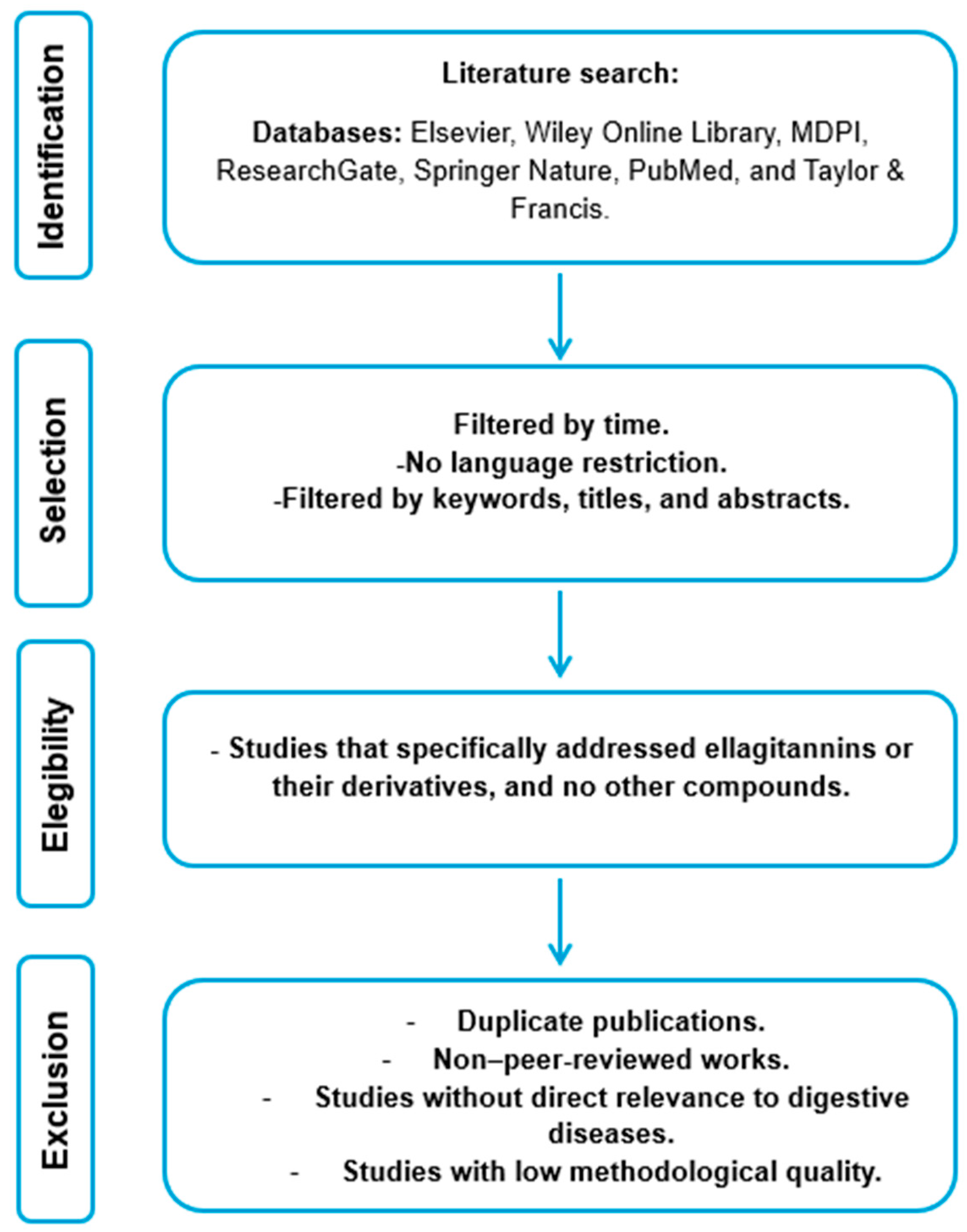
2. Research Methodology

This review article is based on an exhaustive search of a total of 143 articles published between 2005 and 2025, covering topics from the basic and structural aspects of ellagitannins to specific and recent data on their effects on diseases. The literature search was conducted across several reputable scientific databases, including Elsevier, Wiley Online Library, MDPI, ResearchGate, Springer Nature, PubMed, and Taylor & Francis. The following keywords were used: “ellagitannins”, “ellagic acid”, “urolithins”, and “digestive diseases”.
Inclusion criteria encompassed peer-reviewed publications, original research articles, review articles, book chapters, and books published in English or Spanish that explored the relationship between ellagitannins and disorders of the digestive system. Only studies specifically addressing ellagitannins or their derivatives, and no other compounds, were considered.
Exclusion criteria included duplicate publications, non-peer-reviewed works, studies lacking direct relevance to digestive diseases, and studies of low methodological quality. Additionally, retracted or withdrawn studies were excluded (Figure 1).

3. Chemical Structure and Classification of Ellagitannins

There is a great structural diversity of ellagitannins (Table 1). To date, over 1000 different coupling configurations have been identified, contingent on the variation in the position, stereochemistry, and frequency of the hexahydroxydiphenic acid units, the degree of galloylation, and/or the anomeric stereochemistry of the sugar residues [18].
Ellagitannins can be classified as monomeric, dimeric, oligomeric, polymeric, or C-glycosidic, depending on the number of glucose molecules in the structure [19]. Ellagitannin monomers are composed of a glucose molecule linked to the residues of hexahydroxydiphenic acid, with the potential for gallic acid to be present. In contrast, dimers, oligomers, and polymers are formed through the polymerization of monomeric ellagitannins [20].
Table 1. Classification of ellagitannins.
Type of EllagitanninNameChemical StructureMolecular FormulaMolecular Weight (g/mol)Reference
MonomericPunicalaginMicrobiolres 16 00113 i001C48H28O301084.71 [21]
Corilagin Microbiolres 16 00113 i002C27H22018634.5 [22]
Geraniin Microbiolres 16 00113 i003C41H28O27952.6 [23]
Casuarictin Microbiolres 16 00113 i004C41H28O26936.6 [24]
DimericSanguin H-6 Microbiolres 16 00113 i005C82H54O521871.27[25,26]
Coriariin A Microbiolres 16 00113 i006C82H58O521875.3 [27]
Cornusiin E Microbiolres 16 00113 i007C82H58O521875.3 [28]
Oenothein B Microbiolres 16 00113 i008C68H48O441569.1[29]
OligomericNupharin C Microbiolres 16 00113 i009C82H58OH521875.307[30]
Lambertianin C Microbiolres 16 00113 i010C123H80O782805.81[31]
Agrimoniin Microbiolres 16 00113 i011C82H54O521871.3[32]
Hirtellin A Microbiolres 16 00113 i012C82H58O521875.3[33]
C-glycosidicCastalagin Microbiolres 16 00113 i013C41H26O26934.63[34,35]
Vescalagin Microbiolres 16 00113 i014C41H26O26934.63[36]
Casuarinin Microbiolres 16 00113 i015C41H28O26936.6[37]
Grandinin Microbiolres 16 00113 i016C46H34O301066.7[38]

4. Sources of Ellagitannins

Ellagitannins and their derivatives, including glucosides, arabinosides, rhamnoses, acetylated esters, and free ellagic acid, are widely distributed in nature, as is the case in some fruits of the Rosaceae family, including raspberries, blackberries, strawberries, currants, and grapes [39].
In addition to the previously mentioned fruits, pomegranates, rambutans, and other fruits, as well as certain seeds such as pistachios, walnuts, cashews, pecans, and acorns, are also significant sources of ellagitannins [40]. Due to their chemical composition, ellagitannins are found in various plants, many of which are used for medicinal purposes in Asia. Agrimonia pilosa contains the ellagitannin agrimoniin. Similarly, the camellia (Camellia japonica) contains camelliatannin A, the dogwood (Cornus officinalis) contains cornusin A, the geranium (Geranium thunbergii) contains geraniin, the avens (Geum japonicum) contains gemin A, the sweetgum tree (Liquidambar formosana) contains casuarictin, and the mallotus (Mallotus japonicus) contains mallotusinic acid [41].
Other sources of ellagitannins are products, including tea and honey. Alcoholic beverages like red wine, cider, whiskey, and brandy also contain ellagitannins. The presence of ellagitannins is attributed to the processing of these products in wooden barrels, such as oak, which allows for the release of ellagitannins, particularly C-glucosides, including acutissimin A and B, epiacutissimin A and B, as well as mongolicain A. During the aging process, these compounds are released from the wood, enhancing certain organoleptic characteristics of these beverages [42,43].

5. Biosynthesis of Ellagitannins

The biosynthesis of these secondary metabolites in plants occurs in cytoplasmic vacuoles or the cell wall. Their production is closely related to biological functions such as defense against microorganisms (fungi, bacteria, and viruses) and herbivorous animals, as well as protection against ultraviolet rays from the sun, explaining why these molecules have high antioxidant capacity [44]. It has been reported that genetics, species, plant maturity, humidity, luminosity, pruning, foliation by herbivores, and seasonal changes have a profound impact on the qualitative and quantitative content of ellagitannins [45].
The proposed biosynthesis pathway of punicalagin is as follows (Figure 2): phosphoenolpyruvic acid (PEP) and D-erythrose-4-phosphate (E4P) produce 3-deoxy-D-arabinoheptulosonate-7-phosphate (DAHP) through the action of 3-deoxy-D-arabinoheptulosonate-7-phosphate synthase (DAHPS). The conversion of 3-deoxy-D-arabinoheptulosonate-7-phosphate (DAHP) to 3-dehydroquinate is facilitated by the action of 3-dehydroquinate synthase (DHQS). The bifunctional enzyme 3-dehydroquinate dehydratase/shikimate dehydrogenase (DHD/SDH) catalyzes the conversion of 3-dehydroquinate to 3-dehydroshikimic acid, which in turn produces shikimic acid and gallic acid. Under the action of UDP-glucose: gallate glucosyltransferase (UGT), the latter produces β-glucogallin, better known as 1-O-galloyl-β-D-glucopyranose. Β-glucogallin O-galloyltransferase then converts this into digalloylglucose. A series of reactions catalyzed by galloyltransferase (GT) produce pentagalloylglucose, wherein the galloyl groups are oxidatively coupled to form the hexahydroxydiphenoyl (HHDP) group. It is subsequently metabolized into punicalagin (monomeric ellagitannin) by the action of pentagalloylglucose oxygen oxidoreductase (POR) [46,47,48].

6. Degradation of Ellagitannins

Ellagitannins are molecules that, due to their structural composition, can be hydrolyzed into progressively simpler compounds. Until recently, the degradation mechanism of these molecules was not well understood. However, recent studies have identified several pathways that elucidate how these molecules degrade [50,51].
Ellagitannins can be hydrolyzed in vitro by promoting the hydrolysis of the ester bonds between the sugar core and the HHDP units, releasing hexahydroxydiphenic acid and glucose units. Because of this separation, spontaneous lactonization occurs, forming ellagic acid molecules (Figure 3) [52].
Figure 3. Diagram of punicalagin degradation by ellagitannase (EAH) action [53].
Microbiolres 16 00113 g003

6.1. Acid Hydrolysis

The recovery and purification of bioactive phytochemicals from plant materials is typically carried out through various extraction methods. The isolation of ellagic acid is achieved by extracting a plant fraction rich in ellagitannins using different solvent mixtures. In addition to the previous step, a hydrolysis process of the ellagitannins is performed using concentrated acids, commonly HCl and H2SO4. The composition and purity of ellagic acid are monitored through high-performance liquid chromatography (HPLC) [54].
The optimization of ellagitannin acid hydrolysis was evaluated by subjecting ellagitannin samples to three HCl concentrations (1.2, 2, and 4 M) for varying durations (1, 2, 4, 6, and 20 h) at a temperature of 85 °C. To prepare the samples, 5, 8.3, or 16.6 mL of 37% HCl was added and diluted with 50 mL of methanol. The degradation of ellagitannins was assessed by detecting the increase in ellagic acid using HPLC [55].

6.2. Microbial Biodegradation

The hydrolysis of ellagitannins through microbial enzymes has been a topic of debate in the scientific community. It has been observed that microbial tannase is active on galloyl residues, as well as on hexahydroxydiphenoyl and other ellagitannin residues [56]. Other enzymes, such as phenol oxidase and decarboxylase, have also been associated with the degradation of gallotannins and ellagitannins. In other reports, crude enzymatic extracts obtained from the fermentation of ellagitannins have been observed to exhibit activities distinct from those of tannase and ellagitannase. These include polyphenol oxidase, glycosyl hydrolases, cellulase, xylanase, and β-glucosidase [57].
Recent studies have demonstrated a new activity, ellagitannin acyl hydrolase (EAH), where an enzyme can biodegrade ellagitannins by promoting the hydrolysis of ester bonds between the sugar core and HHDP units. This enzyme has been tested in solid-state fermentation against other enzymes, including cellulase, xylanase, polyphenol oxidase, tannase, β-glucosidase, and α-L-arabinofuranosidase. The enzyme with the highest activity value (U/L) at the moment of maximum ellagic acid accumulation was found to be ellagitannase, which confirms that this enzyme is directly associated with ellagitannin degradation [58].

6.3. Products of Degradation

6.3.1. Gallic Acid

Gallic acid is the main unit of polyphenols, also known as 3,4,5-trihydroxybenzoic acid. It is a phenolic acid found in various plant sources such as plants, fruits, and beverages like coffee, red wine, and green tea [59]. This compound is attributed to various pharmacological effects such as antioxidant, anti-inflammatory, anticancer, cardiovascular, neurodegenerative, and antidiabetic properties [60,61].

6.3.2. Ellagic Acid

Ellagic acid is a heterotetracyclic organic compound resulting from the dimerization of gallic acid through oxidative aromatic coupling with intramolecular lactonization between both carboxylic acid groups of the resulting barrel, with an approximate molecular weight of 338.2 g/mol [62]. Ellagic acid is a phenolic nutraceutical molecule that can be present in free form in some plant species, including plants, fruits, and vegetables. It can be found in these plant materials as a secondary metabolite or derived from the degradation of its precursors, the ellagitannins [63]. It is considered a thermally stable molecule (melting point of 350 °C) due to the four rings of the molecule representing lipophilic dominance and the four phenolic groups representing the hydrophilic area, which causes high insolubility in water. However, it is soluble in organic acids. Due to its phenolic nature, it tends to react by forming complexes with other molecules, such as proteins, alkaloids, and polysaccharides [64].
Among its biological functions, its antioxidant capacity is particularly noteworthy due to its ability to eliminate free radicals and promote the synthesis of antioxidant enzymes, including superoxide dismutase (SOD), glutathione peroxidase (GPx), glutathione reductase (GR), and catalase (CAT). Additionally, it exhibits antibacterial, antifungal, antiviral, hepatoprotective, and cardioprotective activities [65]. Furthermore, it displays anti-inflammatory properties by regulating the formation of proinflammatory cytokines (IL-6, IL-8, IL-12, TNF-α, etc.). Additionally, it exhibits chemopreventive, neuroprotective, antiviral, antidiabetic, gastroprotective, anti-hyperlipidemic, and antidepressant activities [66].

6.3.3. Urolithins

Urolithins (Uros) are a class of metabolites that originate from ellagitannins and ellagic acid. These metabolites possess a common benzopyran-6-one nucleus, and their number of hydroxyl groups is reduced as they are metabolized in decreasing order [67].
Only 13 urolithins and their conjugated metabolites (glucuronides, sulfates, among others) have been reported (Figure 4). Their presence has been identified in different human fluids and tissues (blood, feces, urine, breast, and colon) [68].
Due to the low bioavailability of these compounds, a significant portion of them reaches the colon, where they are metabolized by the intestinal microbiota [69].

7. Action of the Gut Microbiota in the Degradation of Ellagitannins

The gut microbiota comprises a consortium of microorganisms that live in symbiosis with humans and play a central role in food digestion and host metabolic regulation [70,71]. A significant proportion of ingested food remains undigested by host digestive enzymes, and the unabsorbed residues reach the colon, where many microorganisms with advanced metabolic machinery can facilitate the absorption process [72].
This is the case with ellagic acid, which exhibits extremely low bioavailability and is converted into urolithins by the action of the gut microbiota. Urolithins exert significant bioactivity through cleavage, decarboxylation, and dehydroxylation reactions of the lactone ring, starting with pentahydroxy-Uro (Uro-M5), followed by tetrahydroxy-Uros (Uro-D, Uro-E, and Uro-M6) and trihydroxy-Uros (Uro-C and Uro-M7), culminating in dihydroxy-Uros (Uro-A and isoUro-A) and monohydroxy-Uro (Uro-B), which is regularly detected when isoUro-A is also produced [73].

7.1. Bacteria Capable of Degrading Ellagitannins into Ellagic Acid and Urolithins

Recent studies have focused on identifying specific bacterial strains capable of degrading ellagitannins, such as punicalagin, to enhance the bioavailability of these beneficial compounds (Table 2).
In one study, thirty lactic acid bacteria were isolated from two food products: aged cheese and dry-cured fermented sausage. The bacteria’s ability to biotransform a pomegranate peel extract rich in punicalagin and a food supplement with a high punicalagin content was evaluated. Among the isolated strains, 27 were discarded, and three strains with the highest activity in the degradation of ellagitannins were selected for further analysis. Lactiplantibacillus plantarum 89, Lacticaseibacillus paracasei 116, and Enterococcus faecium 126 were identified. The ellagic acid content in the experimental units was significantly higher, with concentrations up to 25 and 18 times higher than those found in the pomegranate extract. This evidence demonstrates a correlation between lactic acid bacteria and the degradation of ellagitannins [74].
Several bacterial strains of the Eggerthellaceae family have been identified as capable of converting ellagitannins and ellagic acid into urolithins. Among these strains are Gordonibacter urolithinfaciens DSM27213 and Gordonibacter pamelaeae DSM19378, isolated from the feces of healthy individuals. These strains can convert ellagitannins and ellagic acid into urolithin M5, urolithin M6, urolithin C, and isourolithin A under stationary culture and anaerobic conditions (Figure 5) [75,76].
Table 2. Some urolithin-producing bacterial strains.
FamilyBacterial StrainUrolithin ProducedSource of InsulationReference
Streptococcaceae Streptococcus thermophilus FUA329Urolithin AHuman breast milk[77]
BifidobacteriaceaeBifidobacterium pseudocatenulatum INIA P815Urolithin A and BHuman milk and feces and feces of infants[78]
StreptococcaceaeLactococcus garvieae FUA009Urolithin AHuman feces[79]
EggerthellaceaeGordonibacter KGMB12511 TUrolithin CHuman feces[80]
EnterococcusEnterococcus faecium FUA027Urolithin AHuman feces[81]
EggerthellaceaeEllagibacter isourolithinifaciens DSM 104140 TUrolithins M5, M6, C and isourolithin AN/E[82]
EggerthellaceaeGordinobzacter urolithinfaciens DSM 27213 TUrolithin M5, M6 and CN/E[82]
EggerthellaceaeCEBAS 4A1Urolithins M6, C and isourolithin AHuman feces[83]
Source of insulation: (N/E) not specified.
Another study reported the isolation of four intestinal strains (CEBAS 4A1, 4A2, 4A3, 4A4) of the same species, which were found to be capable of transforming ellagic acid into urolithins M6, C, and isourolithin A [84]. Furthermore, Beltrán et al. (2018) identified a bacterium proposed to belong to a new genus named Ellagibacter, within the species Ellagibacter isourolithinifaciens, with the capacity to degrade ellagic acid into urolithins [85].
It is crucial to emphasize that the number of bacteria comprising the gut microbiota is 10 times greater than that of human cells [86]. It indicates that additional microorganisms or enzymes are likely involved in this biotransformation process, which has not yet been identified [87].

7.2. Urolithin Metabotypes

It has been demonstrated that the bacterial strains responsible for converting ellagitannins/ellagic acid into urolithins are not present in the gut microbiota of all individuals. This results in significant discrepancies among individuals in the positive effects that these compounds can have on health. Consequently, a classification of individuals capable of producing specific types of urolithins has been established [88].
Individuals capable of metabolizing ellagitannins and ellagic acid into urolithins are classified into three phenotypes, designated metabotypes (Figure 6). Individuals who produce urolithin A are classified as metabotype A, while those who produce urolithin B and/or isourolithin A and urolithin A are classified as metabotype B. Metabotype B in adult populations ranges from 20% to 30%. Individuals unable to produce detectable levels of urolithins are grouped into metabotype 0 [89]. The distribution of metabotypes is influenced by several factors, including age, diet, physical activity, health status, sex, and body mass index [90].
The digestive system is responsible for the physical and chemical breakdown of food through a complex series of processes to release its nutrients in a manner that allows for optimal metabolism, transport to related organs, and the expulsion of remaining waste [91]. It comprises the digestive tract, which can be described as a tube with a total length of 8 to 9 m, starting at the mouth and ending at the anus. This tract includes the pharynx, esophagus, stomach, and small and large intestines [92]. Additionally, it consists of accessory organs, namely the tongue, teeth, salivary glands, liver, gallbladder, and pancreas [93].
In terms of human anatomy, the digestive system operates in a sequential manner, with strict control over the transition from one process to the next. These processes correspond to the mouth (oral processing), the stomach (gastric processing), the small intestine (intestinal processing), and the large intestine or colon (fermentation) [94]. Following the ingestion of food, oral processing is the first step, considered a mechanical process as it breaks down food into small particles, which are hydrated by saliva to form the food bolus. This process includes mastication and salivation [95]. After oral processing, ingested food mixes with gastric juices containing acid, pepsin, lipase, and salts, with a pH value of approximately 2. Through this process, food is degraded both physically and chemically due to the peristalsis of the stomach walls and acid/enzymatic reactions [96].
The small intestine is where starch digestion and absorption occur. It is divided into the duodenum, jejunum, and ileum. A large portion of starch is digested in the duodenum, where it receives digestive secretions from the pancreas and liver, and is subsequently absorbed in the jejunum and ileum [97]. The large intestine is characterized by slow flow and a neutral or slightly acidic pH. It has three main functions: the absorption of water and electrolytes, the absorption of vitamins, and the solidification and storage of feces prior to expulsion [98].

8. Digestive System Diseases

Digestive diseases encompass a broad range of chronic disorders that primarily cause issues in the stomach and intestines (Figure 7), significantly and negatively impacting the quality of life of affected individuals [99]. These types of diseases pose a substantial health threat, and their detection and treatment result in a considerable economic burden on the healthcare sector, with little to no reduction in prevalence and incidence [100]. In 2019, digestive diseases accounted for more than one-third of prevalent diseases and approximately one-fifth of incident disease cases [101].
In the United States alone in 2015, the annual healthcare expenditure on gastrointestinal diseases reached $135.9 billion. There were 54.4 million outpatient visits with a primary diagnosis of gastrointestinal disease, 266,600 new cases of gastrointestinal cancer were diagnosed, and there were 144,300 cancer-related deaths [102].
The following section describes some of the most common digestive diseases and some alternative treatments with ellagitannins.

9. Impact of Ellagitannins, Ellagic Acid, and Urolithins on Intestinal Health

Several studies have shown that ellagitannins and their derivatives, such as ellagic acid and urolithins, have beneficial effects on digestive system health. (Table 3) presents the reported effects of these compounds on various intestinal diseases.

9.1. Effect of Ellagitannins on Irritable Bowel Syndrome

Irritable bowel syndrome (IBS) is one of the most common intestinal conditions. It significantly impacts the daily lives of affected individuals and places a substantial economic burden on society [109]. The main symptoms of IBS include abdominal pain and alterations in bowel movements, primarily diarrhea or constipation, which cannot be explained by any structural or biochemical anomaly [110].
A randomized, double-blind, placebo-controlled trial involving 44 individuals with irritable bowel syndrome evaluated the effect of ellagic acid on gastrointestinal symptoms. The participants were randomly assigned to two groups: one group received 180 mg of ellagic acid per day (n = 22), and the other group received a placebo (n = 22) for two months. After the experiment, it was observed that ellagic acid consumption had a beneficial effect, as it reduced the symptoms of irritable bowel syndrome. It was attributed to the anti-inflammatory and antioxidant properties of ellagic acid [111].
Ellagitannins and their derivatives are emerging as promising preventive alternatives or adjuvant therapies for various health conditions. However, in the case of irritable bowel syndrome, more scientific evidence is still needed to support their beneficial effects. Additional clinical studies are required, as well as the establishment of effective and safe dosages.

9.2. Ellagitannins and Their Activity Against Gastric Ulcers

Gastric ulcers are defects in the gastric wall that occur when there is a disruption in the mucosal barrier lining the stomach [112]. Gastric ulcers occur when mucosal protection is compromised by harmful factors such as hypersecretion of gastric acid, nonsteroidal anti-inflammatory drugs (13% of cases), and H. pylori infection, which is associated with damage such as hemorrhagic erosion, neutrophil infiltration, lymphoid follicles, and epithelial damage [113].
The gastroprotective activity of casuarinin, an ellagitannin extracted from Melaleuca leucadendra, was evaluated in a previous study. They conducted a study with mice in which ethanol-induced ulceration was induced, and casuarinin was administered in three concentrations (25, 50, and 100 mg/kg), resulting in a reduction in ulcer area by 45%, 78%, and 99%, respectively, compared to the group not receiving the ellagitannin. Casuarinin (100 mg/kg) increased mucin content by up to 1.8 times and decreased acidity by 48%. Histological data demonstrated that ellagitannin exhibited a protective effect against tissue modifications caused by the induced ulcer [114].
In the case of the positive effects of ellagitannins on gastric ulcers, this is one of the conditions discussed in this review for which a greater number of studies have been conducted, mainly in vitro in cells and in vivo in rodents. However, evidence from human studies is still limited.

9.3. Ellagitannins Against Gastritis

Gastritis is defined as inflammation of the mucosa and submucosa of the stomach and is one of the most prevalent diseases globally, with an estimated 1.8 to 2.1 million cases annually [115]. It can be classified into two types: non-atrophic, which causes mucosal damage that may be extensive and/or severe and can be restored with some sequelae or progress to atrophic phenotypes; and atrophic, characterized by long-lasting periods, non-self-limiting, and resulting from harmful agents that drastically modify the resident population of gastric glands [116].
Several studies have reported that ellagitannins possess antibacterial and anti-inflammatory activities [117]. The anti-inflammatory effect of ellagitannins from strawberries was demonstrated, although not exclusively related to their ability to inhibit the NF-κB pathway, a key factor in inflammatory diseases such as gastritis. They used the two pure ellagitannins most prevalent in strawberries (agrimoniin and casuarictin), showing that agrimoniin moderates IL-8 secretion by inhibiting the NF-kB pathway. Additionally, they demonstrated for the first time that casuarictin, in its pure state, possesses anti-inflammatory properties, specifically targeting the NF-kB pathway in human gastric epithelial cells [118].
In a study by Piazza et al. (2023), ellagitannins extracted from leaves of Castanea sativa L., commonly known as a chestnut tree (castalagin and vescalagin), were used against gastritis [119]. Approximately 1% w/w of the dry extract was employed. In this assay, GES-1 cells infected with pathogenic strains of H. pylori, highly associated with this condition [120], were used since they activate NF-kB and IL-8 expression at the epithelial level. Ellagitannins inhibited IL-8 release (IC50 ≈ 28 µg/mL and 11 µM, respectively), and their anti-inflammatory activity was partly due to decreased NF-kB signaling. Moreover, ellagitannins reduced bacterial growth and cell adhesion.
Although ellagitannins have shown positive effects in the prevention and treatment of gastritis in preclinical studies, due to their antioxidant, anti-inflammatory, and cytoprotective properties, most of the evidence comes from in vitro or animal models. The lack of clinical trials in humans represents a significant limitation for translating these findings into clinical practice.

9.4. Ellagitannins Against Colon Cancer

Ellagitannins exhibit a wide range of biologically and clinically significant activities, with the potential to promote health and applications in medicine, including cancer prevention and treatment [121]. Colon cancer is the third most prevalent disease worldwide, with the highest mortality rate. Its prognosis is dependent on the clinical status of the patient. According to the World Health Organization, 1.93 million cases were reported in 2020 [122].
Ellagitannins extracted from raspberry seeds were evaluated against HT-29 colon cancer cells in a previous study. Ellagic acid was observed to induce a slight cell cycle arrest in the G1 phase, while urolithins were found to arrest the cell cycle in the G2/M phase and increase p21 expression. The apoptotic cells were identified using the Annexin V-FITC/PI assay. The observed variations in mitochondrial membrane potential and the activation of caspases 8 and 9 indicated that both extrinsic and intrinsic apoptotic pathways might be involved. The anticancer activity of ellagitannins, ellagic acid, and urolithins A and B was demonstrated by their ability to halt the cell cycle and induce apoptosis in human colon cancer cells (HT-29) [123].
The impact of urolithin A on SW620 colorectal cancer cells was investigated in a separate study. They demonstrated urolithin A-induced autophagy in damaged cells at submicromolar concentrations. Moreover, the dose-dependent reduction in cell proliferation and matrix metalloproteinase-9 activity observed in this study suggests that urolithin A may have a role in treating colorectal cancer. The inhibition of autophagy by Atg5-siRNA and caspases by Z-VAD-FMK suppressed cell death induced by urolithin A [124].

9.5. Ellagitannins Against Esophageal Cancer

Esophageal cancer is one of the most frequently occurring neoplasms, ranking eighth worldwide and third among gastrointestinal cancers [125]. It is regarded as one of the most aggressive digestive tract diseases, with tumors developing in the surrounding tissues of the esophagus, the muscular tube through which food is transported from the throat to the stomach. This results in a high morbidity and mortality rate of [126]. Esophageal cancer is classified into two main types: adenocarcinoma and squamous cell carcinoma. Squamous cell carcinoma is the most prevalent type, accounting for approximately 90% of cases. The two types of cancer do not share epidemiological, histopathological, or etiological characteristics [127].
High antitumor activity of ellagic acid against esophageal squamous cell carcinoma has been demonstrated. The inhibitory effect of ellagic acid on carcinoma cell survival was concentration- and time-dependent. The compound significantly reduced luciferase activity (reporter gene) induced by STAT3, a transcription factor involved in the tumorigenesis of various tumors. Ellagic acid inhibited both endogenous and cytokine-induced activation of STAT3 in esophageal squamous carcinoma cells. Furthermore, it was indicated that ellagic acid could suppress the expression of RNF6, an E3 ligase of SHP-1. It was also observed that the overexpression of RNF6 could significantly diminish the inhibitory effects of ellagic acid on cancer cell survival. It suggests that STAT3 signaling can be inhibited by modulating RNF6/SHP-1. The findings of this study indicate that ellagic acid may be a promising therapeutic option for the treatment of esophageal squamous cell carcinoma [128].

9.6. Ellagitannins Against Pancreatic Cancer

Pancreatic cancer is one of the most aggressive diseases and is considered nearly incurable since symptoms occur in very advanced stages, making diagnosis and treatment very challenging [129]. It is estimated that 50% of patients die within the first six months. The American Cancer Society reports a five-year relative survival rate of 9% for all combined stages. For patients diagnosed at an advanced stage (approximately 53%), the five-year survival rate is estimated at 3% [130].
The efficacy of pomegranate extract (Punica granatum) was evaluated on pancreatic cancer cells at concentrations of 5, 10, and 20 mg/mL. The extract demonstrated the ability to attenuate angiogenesis through tumor morphology, weight, and hemoglobin concentration changes [131].
The impact of urolithin A was investigated in mice xenografted with subcutaneous injections of pancreatic ductal adenocarcinoma cell lines. The mice were orally administered a dose of 20 mg/kg/day of urolithin A orally via a probe once the subcutaneous tumors had already formed. The treatment resulted in a reduction and cessation of tumor growth. Additionally, they collaborated with genetically modified mice (Ptf1acre/+; LSL KrasG12D/+; Tgfbr2flox/flox—PKT mice), which spontaneously develop pancreatic cancer at an early age (4–5 weeks). The mice were orally administered 20 mg/kg/day of urolithin A through a probe, increasing overall survival from approximately 59 to 71 days [132].

9.7. Ellagitannins Against Liver Cancer

Hepatocellular carcinoma (HCC) is the most prevalent form of liver cancer, accounting for 90% of all cases. It is a highly malignant neoplasm that typically arises in individuals with a history of chronic liver disease and/or cirrhosis. It is the fifth most common cancer in men and the seventh most common cancer in women [133]. Globally, HCC represents the most common malignant neoplasm. Liver cancer is frequently diagnosed in advanced stages, which often results in a poor prognosis. For the treatment of liver cancer, chemotherapy and immunotherapy are the most common options. However, natural compounds may help patients achieve better outcomes with lower systemic toxicity and fewer side effects [134].
The chemopreventive efficacy of ellagic acid against N-nitroso diethylamine-induced hepatocarcinogenesis a potent hepatocarcinogenic dialkyl N-nitrosamine found in tobacco smoke, cured meats, and some beverages was evaluated in rats. Rats were classified into four groups: a normal control group, a group injected with a single dose (200 mg/kg body weight) of N-nitrosodiethylamine, a group administered ellagic acid orally daily at a dose of 50 mg/kg body weight for 7 days before and 14 days after N-nitrosodiethylamine administration, and a fourth group that received the same dose of ellagic acid for 21 days after N-nitrosodiethylamine administration. It was observed that in hepatocellular carcinoma rats, ellagic acid significantly decreased the activities of liver antioxidant enzymes. Additionally, oral administration of ellagic acid as a protective agent promoted a significant increase in tested antioxidant enzyme activities and total serum protein levels, concomitant with a reduction in tumor markers such as arginase and α-fucosidase, as well as liver enzymes and total and direct bilirubin levels. Similarly, oral application of ellagic acid as a therapeutic agent inhibited partial DNA fragmentation produced by hepatocellular carcinoma in the rat model [135].
Although various preclinical studies have demonstrated promising antitumor effects of ellagitannins and their derivatives in different types of cancer affecting the digestive system, such as colon, esophageal, pancreatic, and liver cancer, most of this evidence comes from in vitro or animal models. There is a clear need for clinical studies in humans to confirm these findings and to assess their safety, efficacy, and bioavailability. Additionally, it is important to elucidate the underlying mechanisms of action and explore potential synergistic effects with conventional cancer therapies.

10. Food Products with Ellagitannins and Their Derivatives as Bioactive Ingredients

A patent was developed for an antioxidant product based on camu-camu fruits, showing high potential for use in the food or pharmaceutical industry. The patent delineates the optimal conditions for the extraction of bioactive compounds, commencing with the selection of fruit (soluble solids/acidity, seed removal, and fruit dehydration), grinding, sieving (0.2 to 0.8 mm), and the mixing of the obtained flour with an acidic solution, followed by agitation and subsequent decantation to separate the liquid phase from the solid phase, which underwent a freeze-drying process. The product described is notable for its high concentration of vitamins and polyphenols, including ellagic acid (100 to 200 mg/100 g) and ellagitannins such as castalagin, vescalagin, ellagitannin, pedunculagin, HHDP-galloyl-glucose, Di-HHDP-galloyl-glucose, and tri-galloyl-HHDP-glucose (350 to 450 mg/100 g), among other compounds. The total polyphenol concentration of the product is estimated to be between 3500 and 5000 milligrams per 100 g [136].
A patent outlines the formulation of nutritional and medical products involving a medium-chain triglyceride, lecithin, and a compound from the urolithin family (urolithin A). This combination gives urolithin A a superior bioavailability and pharmacokinetic profile compared to urolithin A alone in a simple saline solution suspension. Specifically, this composition is applicable in treating and managing healthy individuals who would benefit from improved muscle function, including those with a deteriorated physique, low endurance capacity, and muscle atrophy. Furthermore, this composition offers an alternative treatment for conditions such as diabetes, obesity, reduced metabolic rate, metabolic syndrome, cardiovascular diseases, stress, anxiety disorders, and others [137].
A patent presents a method for obtaining ellagic acid from a polyphenolic extract of pomegranate, particularly from its seeds. This method has high potential as a nutritional or food supplement due to the multiple beneficial effects of ellagic acid. Initially, pomegranate seeds are immersed in an ethanol solution on multiple occasions, after which the resulting fractions are stored and combined. The solid fraction is discarded, and the liquid fraction is concentrated at a 20:1 ratio. Subsequently, further extractions with acetic ether are combined and concentrated to produce a solid material, which is then reduced to a dry powder. The final product for oral use can be presented in the form of hard gelatin capsules, where the active ingredient is integrated with an inert solid diluent (calcium carbonate, calcium phosphate, etc.), or as soft gelatin capsules, where the active ingredient is integrated with water or an oily medium. Each capsule contains approximately 200 mg of ellagic acid in a bioavailable form [138].
A patent describes the development of functional nutritional products containing pomegranate extract for the prevention and/or treatment of cardiovascular diseases, arterial plaque buildup, hypertension, and metabolic syndrome. The mentioned products include orange juice supplemented with pomegranate extract, functional dairy products with pomegranate extract, functional cooked ham with pomegranate extract, and functional canned tuna with pomegranate extract. In all products, the pomegranate extract content ranged from approximately 400 to 4000 mg/kg, with punicalagin content at 50% w/w. These products were evaluated using the ABTS technique to determine their antioxidant activity [139].
It is important to highlight that the use of this type of molecule may cause mild side effects, although there is very little literature addressing this issue [140]. However, most available studies in the scientific literature mainly emphasize their positive effects, which outweigh the reported adverse events in both frequency and clinical relevance [141,142]. This trend suggests a favorable safety profile when these compounds are used in food matrices or nutraceutical products under controlled conditions and at appropriate doses [143].

11. Future Perspectives and Conclusions

Ellagitannins and their derivatives, such as ellagic acid and, particularly, urolithins, have emerged as promising alternatives for the prevention and adjunctive treatment of various digestive system disorders. This is attributable to their potent antioxidant, anti-inflammatory, and anticancer properties, which have been extensively documented in numerous preclinical and clinical studies. Moreover, these compounds can be sourced from agro-industrial residues, promoting sustainable resource utilization and offering the possibility of developing economically accessible products for the general population, thereby providing relief from the high costs associated with conventional pharmacological treatments.
However, significant challenges limit their immediate clinical application. Among these, the low oral bioavailability of ellagitannins and the marked interindividual variability in the gut microbiome’s capacity to convert these compounds into urolithins—metabolites with enhanced biological activity and more potent therapeutic effects—stand out. This variability complicates dose standardization and the prediction of therapeutic responses. Therefore, it is imperative to deepen the molecular understanding of this microbial biotransformation and explore strategies to modulate it, such as supplementation with specific probiotics, to maximize the therapeutic potential of these compounds. Additionally, more controlled, long-term clinical trials are needed to validate their efficacy and safety, establishing clear protocols for their use in various digestive pathologies.
Finally, although the demand for and commercialization of nutraceuticals and dietary supplements containing ellagitannins have increased significantly, a lack of specific regulation remains that ensures the quality, efficacy, safety, and traceability of these products. Consequently, the development and implementation of robust regulatory standards and guidelines are a priority to protect consumers and foster confidence in these products. This should be accompanied by educational campaigns targeted at healthcare professionals and the public, promoting responsible use grounded in scientific evidence. In this way, ellagitannins and their derivatives could become valuable and accessible tools within the therapeutic arsenal against digestive diseases, contributing to sustainable improvements in public health.

Author Contributions

E.M.R.-M.: Conceptualization, Investigation, Writing—Original Draft, Visualization. P.A.-Z.: Conceptualization, Investigation, Writing—Review and Editing, Visualization, Resources. L.S.: Writing—Review and Editing. M.R.M.: Writing—Review and Editing, A.I.: Writing—Review and Editing, C.N.A.: Writing—Review and Editing, Supervision and J.A.A.-V.: Conceptualization, Investigation, Writing—Review and Editing, Visualization. All authors have read and agreed to the published version of the manuscript.

Funding

This work was financially supported by Tecnológico Nacional de México through the following projects: 20024.24-P Estudio de la degradación de elagitaninos por enzimas glicolíticas and 22037.25-P Estudio de la degradación de elagitaninos con enzimas digestivas asociadas a su degradación.

Data Availability Statement

Not applicable.

Acknowledgments

The authors thank Secretaria de Ciencia, Humanidades, Tecnología e Innovación (SECIHTI) of Mexico for the scholarship granted as financial support with scholarship number 1319543 and the Department of Food Research of the Autonomous University of Coahuila.

Conflicts of Interest

The authors declare that they have no known competing financial interests or personal relationships that could have appeared to influence the work reported in this paper.

References

  1. Aldhaleei, W.A.; Wallace, M.B.; Bi, Y.; Rusk, A.M.; Bhagavathula, A.S. Racial, Ethnic, and Geographic Disparities in Digestive Diseases Mortality in the United States, 2000–2019. Clin. Gastroenterol. Hepatol. 2025, 23, 59–68. [Google Scholar] [CrossRef] [PubMed]
  2. Zhao, H.; Zheng, X.; Lin, G.; Wang, X.; Lu, H.; Xie, P.; Jia, S.; Shang, Y.; Wang, Y.; Bai, P.; et al. Effects of air pollution on the development and progression of digestive diseases: An umbrella review of systematic reviews and meta-analyses. BMC Public Health 2025, 25, 183. [Google Scholar] [CrossRef]
  3. Wang, Y.; Huang, Y.; Chase, R.C.; Li, T.; Ramai, D.; Li, S.; Huang, X.; Antwi, S.O.; Keaveny, A.P.; Pang, M. Global Burden of Digestive Diseases: A Systematic Analysis of the Global Burden of Diseases Study, 1990 to 2019. Gastroenterology 2023, 165, 773–783. [Google Scholar] [CrossRef] [PubMed]
  4. Bajaj, J.S.; Barbara, G.; DuPont, H.L.; Mearin, F.; Gasbarrini, A.; Tack, J. New concepts on intestinal microbiota and the role of the non-absorbable antibiotics with special reference to rifaximin in digestive diseases. Dig. Liver Dis. 2018, 50, 741–749. [Google Scholar] [CrossRef]
  5. Su, Q.; Wang, F.; Chen, D.; Chen, G.; Li, C.; Wei, L. Deep convolutional neural networks with ensemble learning and transfer learning for automated detection of gastrointestinal diseases. Comput. Biol. Med. 2022, 150, 106054. [Google Scholar] [CrossRef]
  6. Anwanwan, D.; Singh, S.K.; Singh, S.; Saikam, V.; Singh, R. Challenges in liver cancer and possible treatment approaches. Biochim. Et Biophys. Acta Rev. Cancer 2020, 1873, 188314. [Google Scholar] [CrossRef]
  7. Márquez-López, A.; del Carmen Chavéz-Parga, M.; González Hernández, J.C. Aspectos generales sobre los elagitaninos y su conversión a ácido elágico. Cienc. Nicolaita 2019, 77, 36–58. [Google Scholar]
  8. Ebrahimi, P.; Lante, A. Polyphenols: A Comprehensive Review of their Nutritional Properties. Open Biotechnol. J. 2021, 15, 164–172. [Google Scholar] [CrossRef]
  9. Aguilar-Zárate, P.; Wong-Paz, J.E.; Michel, M.; Buenrostro-Figueroa, J.; Díaz, H.R.; Ascacio, J.A.; Contreras-Esquivel, J.C.; Gutiérrez-Sánchez, G.; Aguilar, C.N. Characterisation of Pomegranate-Husk Polyphenols and Semi-Preparative Fractionation of Punicalagin. Phytochem. Anal. 2017, 28, 433–438. [Google Scholar] [CrossRef]
  10. Izábal-Carvajal, A.L.; Sepúlveda, L.; Chávez-González, M.L.; Torres-León, C.; Aguilar, C.N.; Ascacio-Valdés, J.A. Extraction of Bioactive Compounds via Solid-State Fermentation Using Aspergillus niger GH1 and Saccharomyces cerevisiae from Pomegranate Peel. Waste 2023, 1, 806–814. [Google Scholar] [CrossRef]
  11. Márquez, D.; Suárez, Á. El uso de taninos condensados como alternativa nutricional y sanitaria en rumiantes. Rev. De Med. Vet. 2008, 16, 87–110. [Google Scholar]
  12. Vega García, C.C.; García Niño, W.R. Efectos benéficos de los compuestos antioxidantes de la granada (Punica granatum L) en patologías asociadas con el estrés oxidante. Tequio 2022, 5, 67–94. [Google Scholar] [CrossRef]
  13. Bustamante, A.; García-Díaz, D.; Jiménez, P.; Valenzuela, R.; Pando, M.E.; Echeverría, F. Potential therapeutic effect for liver steatosis of polyphenols obtained from pomegranate peel. Rev. Chil. De Nutr. 2022, 49, 89–99. [Google Scholar] [CrossRef]
  14. Wang, D.; Özen, C.; Abu-Reidah, I.M.; Chigurupati, S.; Patra, J.K.; Horbanczuk, J.O.; Józwik, A.; Tzvetkov, N.T.; Uhrin, P.; Atanasov, A.G. Vasculoprotective effects of pomegranate (Punica granatum L.). Front. Pharmacol. 2018, 9, 1663–9812. [Google Scholar] [CrossRef]
  15. Aguilar-Zárate, P.; Gutiérrez-Sánchez, G.; Michel, M.R.; Bergmann, C.W.; Buenrostro-Figueroa, J.J.; Ascacio-Valdés, J.A.; Contreras-Esquivel, J.C.; Aguilar, C.N. Production of a Fungal Punicalagin-Degrading Enzyme by Solid-State Fermentation: Studies of Purification and Characterization. Foods 2023, 12, 903. [Google Scholar] [CrossRef]
  16. Mackie, A. The digestive tract: A complex system. Interdiscip. Approaches Food Dig. 2019, 1, 11–27. [Google Scholar] [CrossRef]
  17. Yang, Y.; Lee, P.K.; Wong, H.C.; Zhao, D. Oral supplementation of Gordonibacter urolithinfaciens promotes ellagic acid metabolism and urolithin bioavailability in mice. Food Chem. 2024, 437, 137953. [Google Scholar] [CrossRef]
  18. Banc, R.; Rusu, M.E.; Filip, L.; Popa, D.S. The Impact of Ellagitannins and Their Metabolites through Gut Microbiome on the Gut Health and Brain Wellness within the Gut–Brain Axis. Foods 2023, 12, 270. [Google Scholar] [CrossRef]
  19. Niemetz, R.; Gross, G.G. Enzymology of gallotannin and ellagitannin biosynthesis. Phytochemistry 2005, 66, 2001–2011. [Google Scholar] [CrossRef]
  20. Klewicka, E.; Sójka, M.; Ścieszka, S.; Klewicki, R.; Milczarek, A.; Lipińska, L.; Kołodziejczyk, K. The antimycotic effect of ellagitannins from raspberry (Rubus idaeus L.) on Alternaria alternata. Eur. Food Res. Technol. 2020, 246, 1341–1349. [Google Scholar] [CrossRef]
  21. Xu, J.; Cao, K.; Liu, X.; Zhao, L.; Feng, Z.; Liu, J. Punicalagin regulates signaling pathways in inflammation-associated chronic diseases. Antioxidants 2022, 11, 29. [Google Scholar] [CrossRef] [PubMed]
  22. Gupta, A.; Singh, A.K.; Kumar, R.; Ganguly, R.; Rana, H.K.; Pandey, P.K.; Sethi, G.; Bishayee, A.; Pandey, A.K. Corilagin in cancer: A critical evaluation of anticancer activities and molecular mechanisms. Molecules 2019, 24, 3399. [Google Scholar] [CrossRef] [PubMed]
  23. Ito, H. Metabolites of the ellagitannin geraniin and their antioxidant activities. Planta Medica 2011, 77, 1110–1115. [Google Scholar] [CrossRef] [PubMed]
  24. Goenka, S.; Ceccoli, J.; Simon, S.R. Anti-melanogenic activity of ellagitannin casuarictin in B16F10 mouse melanoma cells. Nat. Prod. Res. 2021, 35, 1830–1835. [Google Scholar] [CrossRef]
  25. Davidson, M.; Louvet, F.; Meudec, E.; Landolt, C.; Grenier, K.; Périno, S.; Ouk, T.S.; Saad, N. Optimized Single-Step Recovery of Lipophilic and Hydrophilic Compounds from Raspberry, Strawberry, and Blackberry Pomaces Using a Simultaneous Ultrasound-Enzyme-Assisted Extraction (UEAE). Antioxidants 2023, 12, 1793. [Google Scholar] [CrossRef]
  26. Puljula, E.; Walton, G.; Woodward, M.J.; Karonen, M. Antimicrobial Activities of Ellagitannins against Clostridium perfringens, Escherichia coli, Lactobacillus plantarum and Staphylococcus aureus. Molecules 2020, 25, 3714. [Google Scholar] [CrossRef]
  27. Feldman, K.S.; Lawlor, M.D. Ellagitannin chemistry. The first total synthesis of a dimeric ellagitannin, coriariin A. J. Am. Chem. Soc. 2020, 122, 7396–7397. [Google Scholar] [CrossRef]
  28. Niemetz, R.; Schilling, G.; Gross, G.G. Biosynthesis of the dimeric ellagitannin, cornusiin E, in Tellima grandiflora. Phytochemistry 2023, 64, 109–114. [Google Scholar] [CrossRef]
  29. Schepetkin, I.A.; Ramstead, A.G.; Kirpotina, L.N.; Voyich, J.M.; Jutila, M.A.; Quinn, M.T. Therapeutic Potential of Polyphenols from Epilobium Angustifolium (Fireweed). Phytother. Res. 2016, 30, 1287–1297. [Google Scholar] [CrossRef]
  30. Mora, J.; Pott, D.M.; Osorio, S.; Vallarino, J.G. Regulation of Plant Tannin Synthesis in Crop Species. Front. Genet. 2022, 13, 1664–8021. [Google Scholar] [CrossRef]
  31. Kähkönen, M.; Kylli, P.; Ollilainen, V.; Salminen, J.P.; Heinonen, M. Antioxidant activity of isolated ellagitannins from red raspberries and cloudberries. J. Agric. Food Chem. 2012, 60, 1167–1174. [Google Scholar] [CrossRef] [PubMed]
  32. Grochowski, D.M.; Skalicka-Wózniak, K.; Orhan, I.E.; Xiao, J.; Locatelli, M.; Jakub, P.P.; Granica, S.; Tomczyk, M. A comprehensive review of agrimoniin. Phytochem. Med. Food 2017, 1401, 166–180. [Google Scholar] [CrossRef] [PubMed]
  33. Orabi, M.A.A.; Taniguchi, S.; Terabayashi, S.; Hatano, T. Hydrolyzable tannins of tamaricaceous plants. IV: Micropropagation and ellagitannin production in shoot cultures of Tamarix tetrandra. Phytochemistry 2011, 72, 1978–1989. [Google Scholar] [CrossRef] [PubMed]
  34. 34. Da Silva, R.D.C.V.; Bolda Mariano, L.N.; Bidinha, E.R.; Bueno De Almeida, C.L.; Cechinel-Filho, V.; Santos Zanuncio, V.S.; Silva, D.B.; Gasparotto Junior, A.; De Souza, P. Ethyl Acetate Fraction from Leandra dasytricha (A. Gray) Cong. Leaves Promotes Vasodilatation and Reduces Blood Pressure in Normotensive and Hypertensive Rats. Evid. Based Complement. Altern. Med. 2021, 2021, 7203934. [Google Scholar] [CrossRef]
  35. Karonen, M.; Ahern, J.R.; Legroux, L.; Suvanto, J.; Engström, M.T.; Sinkkonen, J.; Salminen, J.P.; Hoste, H. Ellagitannins Inhibit the Exsheathment of Haemonchus contortus and Trichostrongylus colubriformis Larvae: The Efficiency Increases Together with the Molecular Size. J. Agric. Food Chem. 2020, 68, 4176–4186. [Google Scholar] [CrossRef]
  36. Araújo, A.R.; Camero, S.; Taboada, P.; Reis, R.L.; Pires, R.A. Vescalagin and castalagin reduce the toxicity of amyloid-beta42 oligomers through the remodelling of its secondary structure. Chem. Commun. 2020, 56, 3187–3190. [Google Scholar] [CrossRef]
  37. Wakamori, S.; Matsumoto, S.; Kusuki, R.; Ikeuchi, K.; Yamada, H. Total Synthesis of Casuarinin. Org. Lett. 2020, 22, 3392–3396. [Google Scholar] [CrossRef]
  38. Kaneshima, T.; Myoda, T.; Nakata, M.; Fujimori, T.; Toeda, K.; Nishizawa, M. Antioxidant activity of C-Glycosidic ellagitannins from the seeds and peel of camu-camu (Myrciaria dubia). LWT Food Sci. Technol. 2016, 69, 76–81. [Google Scholar] [CrossRef]
  39. Landete, J.M. Ellagitannins, ellagic acid and their derived metabolites: A review about source, metabolism, functions, and health. Food Res. Int. 2011, 44, 1150–1160. [Google Scholar] [CrossRef]
  40. Mateș, L.; Banc, R.; Zaharie, F.A.; Rusu MEy Popa, D.-S. Mechanistic Insights into the Biological Effects and Antioxidant Activity of Walnut (Juglans regia L.) Ellagitannins: A Systematic. Antioxidants 2024, 13, 974. [Google Scholar] [CrossRef]
  41. Lipińska, L.; Klewicka, E.; Sójka, M. Structure, occurrence and biological activity of ellagitannins: A general review. Acta Sci. Pol. Technol. Aliment. 2014, 13, 289–299. [Google Scholar] [CrossRef] [PubMed]
  42. Gadrat, M.; Lavergne, J.; Emo, C.; Teissedre, P.L.; Chira, K. Validation of a mass spectrometry method to identify and quantify ellagitannins in oak wood and cognac during aging in oak barrels. Food Chem. 2021, 342, 128223. [Google Scholar] [CrossRef] [PubMed]
  43. Rasines-Perea, Z.; Jacquet, R.; Jourdes, M.; Quideau, S.; Teissedre, P.L. Ellagitannins and flavano-ellagitannins: Red wines tendency in different areas, barrel origin and ageing time in barrel and bottle. Biomolecules 2019, 9, 316. [Google Scholar] [CrossRef] [PubMed]
  44. Sánchez-Loredo, E.; Sepúlveda, L.; Wong-Paz, J.E.; Palomo-Ligas, L.; Rodríguez-Herrera, R.; Aguilar, C.N.; Ascacio-Valdés, J.A. Elagitaninos de Eucalyptus camaldulensis y su potencial uso en la industria alimentaria. Explor. Foods Foodomics 2024, 2, 83–100. [Google Scholar] [CrossRef]
  45. Karlińska, E.; Masny, A.; Cieślak, M.; Macierzyński, J.; Pecio, Ł.; Stochmal, A.; Kosmala, M. Ellagitannins in roots, leaves, and fruits of strawberry (Fragaria × ananassa Duch.) vary with developmental stage and cultivar. Sci. Hortic. 2021, 275, 109665. [Google Scholar] [CrossRef]
  46. Olivas-Aguirre, F.J.; Wall-Medrano, A.; González-Aguilar, G.A.; López-Díaz, J.A.; Álvarez-Parrilla, E.; De La Rosa, L.A.; Ramos-Jiménez, A. Taninos hidrolizables; bioquímica, aspectos nutricionales y analíticos y efectos en la salud. Nutr. Hosp. 2015, 31, 55–66. [Google Scholar] [CrossRef]
  47. Phane Quideau, S. Chemistry and Biology of Ellagitannins: An Underestimated Class of Bioactive Plant Polyphenols; World Scientific: Singapore, 2009. [Google Scholar] [CrossRef]
  48. Yamada, H.; Wakamori, S.; Hirokane, T.; Ikeuchi, K.; Matsumoto, S. Structural revisions in natural ellagitannins. Molecules 2018, 23, 1901. [Google Scholar] [CrossRef]
  49. Feng, L.; Yin, Y.; Yang, X.; Tang, H.; Jiao, Q. Dynamic variations in punicalagin and related metabolic substances in pomegranate fruit and leaves during development periods. Hortic. J. 2019, 88, 444–454. [Google Scholar] [CrossRef]
  50. Aguilar-Zarate, P.; Wong-Paz, J.E.; Buenrostro-Figueroa, J.J.; Ascacio, J.A.; Contreras-Esquivel, J.C.; Aguilar, C.N. Ellagitannins: Bioavailability, Purification and Biotechnological Degradation. Mini Rev. Med. Chem. 2018, 18, 1244–1252. [Google Scholar] [CrossRef]
  51. Ascacio-Valdés, J.A.; Aguilera-Carbó, A.F.; Buenrostro, J.J.; Prado-Barragán, A.; Rodríguez-Herrera, R.; Aguilar, C.N. The complete biodegradation pathway of ellagitannins by Aspergillus niger in solid-state fermentation. J. Basic Microbiol. 2016, 56, 329–336. [Google Scholar] [CrossRef]
  52. Buenrostro-Figueroa, J.; Gutiérrez-Sánchez, G.; Prado-Barragán, L.A.; Rodríguez-Herrera, R.; Aguilar-Zárate, P.; Sepúlveda, L.; Ascacio-Valdés, J.A.; Tafolla-Arellano, J.C.; Aguilar, C.N. Influence of culture conditions on ellagitannase expression and fungal ellagitannin degradation. Bioresour. Technol. 2021, 337, 125462. [Google Scholar] [CrossRef] [PubMed]
  53. Aguilar-Zárate, P.; Wong-Paz, J.E.; Rodríguez-Duran, L.V.; Buenrostro-Figueroa, J.; Michel, M.; Saucedo-Castañeda, G.; Favela-Torres, E.; Ascacio-Valdés, J.A.; Contreras-Esquivel, J.C.; Aguilar, C.N. On-line monitoring of Aspergillus niger GH1 growth in a bioprocess for the production of ellagic acid and ellagitannase by solid-state fermentation. Bioresour. Technol. 2017, 247, 412–418. [Google Scholar] [CrossRef] [PubMed]
  54. Evtyugin, D.D.; Magina, S.; Evtuguin, D.V. Recent Advances in the Production and Applications of Ellagic Acid and Its Derivatives A Review. Molecules 2020, 25, 2745. [Google Scholar] [CrossRef] [PubMed]
  55. Vrhovsek, U.; Palchetti, A.; Reniero, F.; Guillou, C.; Masuero, D.; Mattivi, F. Concentration and mean degree of polymerization of Rubus ellagitannins evaluated by optimized acid methanolysis. J. Agric. Food Chem. 2006, 54, 4469–4475. [Google Scholar] [CrossRef]
  56. De León-Medina, J.C.; Sepúlveda, L.; Buenrostro-Figueroa, J.J.; Mata-Gómez, M.A.; Flores-Gallegos, A.C.; Rodríguez-Herrera, R.; Aguilar, C.N.; Ascacio-Valdés, J.A. Production and evaluation of ellagitannase activity using a pure geraniin substrate. Food Bioprod. Process. 2025, 149, 112–117. [Google Scholar] [CrossRef]
  57. Sójka, M.; Nowakowska, A.; Hejduk, A. Influencia de la clarificación enzimática, la filtración y la pasteurización en el contenido de elagitaninos y antocianinas en los jugos de frambuesa. Eur. Food Res. Technol. 2024, 250, 351–359. [Google Scholar] [CrossRef]
  58. De León-Medina, J.C.; Buenrostro-Figueroa, J.J.; Sepúlveda, L.; Aguilar, C.N.; Ascacio-Valdés, J.A. Fungal biodegradation of ellagitannins extracted from rambutan peel. Food Bioprod. Process. 2023, 141, 81–90. [Google Scholar] [CrossRef]
  59. Bai, J.; Zhang, Y.; Tang, C.; Hou, Y.; Ai, X.; Chen, X.; Zhang, Y.; Wang, X.; Meng, X. Gallic acid: Pharmacological activities and molecular mechanisms involved in inflammation-related diseases. Biomed. Pharmacother. 2021, 133, 110985. [Google Scholar] [CrossRef]
  60. Shabani, S.; Rabiei, Z.; Amini-Khoei, H. Exploring the multifaceted neuroprotective actions of gallic acid: A review. Int. J. Food Prop. 2020, 23, 736–752. [Google Scholar] [CrossRef]
  61. Xu, Y.; Tang, G.; Zhang, C.; Wang, N.; Feng, Y. Gallic acid and diabetes mellitus: Its association with oxidative stress. Molecules 2021, 26, 7115. [Google Scholar] [CrossRef]
  62. Guzmán Martínez, O.; Isasmendi Cortés, M.; Biviano Pérez, C.; Dorantes Bautista, G.; Aguirre García, M.; González Pérez, M. Estudio In Silico de las Interacciones del Ácido Elágico y los Aminoácidos de los Tejidos Humanos usando Química Cuántica. Cienc. Lat. Rev. Científica Multidiscip. 2023, 7, 1354–1365. [Google Scholar] [CrossRef]
  63. Najmi, A.; Nikrad, N.; Ghaffari Sarghein, M.; Hosseinpour dogolsar, M.; Alizadeh, M. Ellagic acid supplementation on oxidative stress, antioxidative capacity and inflammation biomarkers: A systematic review and dose–response meta-analysis of randomized controlled trials. J. Funct. Foods 2024, 122, 106492. [Google Scholar] [CrossRef]
  64. McClements, D.J.; Öztürk, B. Utilization of Nanotechnology to Improve the Application and Bioavailability of Phytochemicals Derived from Waste Streams. J. Agric. Food Chem. 2022, 70, 6884–6900. [Google Scholar] [CrossRef] [PubMed]
  65. Tang, Y.; Jiang, X. Effect of solubilization with surfactant on the antioxidant activity of ellagic acid. Tenside Surfactants Deterg. 2024, 61, 250–258. [Google Scholar] [CrossRef]
  66. Abd-Elghany, A.A.; Mohamad, E.A. Chitosan-coated niosomes loaded with ellagic acid present antiaging activity in a skin cell line. ACS Omega 2023, 8, 16620–16629. [Google Scholar] [CrossRef]
  67. Romo-Vaquero, M.; Cortés-Martín, A.; Loria-Kohen, V.; Ramírez-de-Molina, A.; García-Mantrana, I.; Collado, M.C.; Espín, J.C.; Selma, M.V. Deciphering the Human Gut Microbiome of Urolithin Metabotypes: Association with Enterotypes and Potential Cardiometabolic Health Implications. Mol. Nutr. Food Res. 2019, 63, 1800958. [Google Scholar] [CrossRef]
  68. González-Sarrías, A.; García-Villalba, R.; Romo-Vaquero, M.; Alasalvar, C.; Örem, A.; Zafrilla, P.; Tomás-Barberán, F.A.; Selma, M.V.; Espín, J.C. Clustering according to urolithin metabotype explains the interindividual variability in the improvement of cardiovascular risk biomarkers in overweight-obese individuals consuming pomegranate: A randomized clinical trial. Mol. Nutr. Food Res. 2017, 61, 1600830. [Google Scholar] [CrossRef]
  69. Isla, K.K.Y.; Tanae, M.M.; de Lima-Landman, M.T.R.; de Magalhães, P.M.; Lapa, A.J.; Souccar, C. Vasorelaxant effects of ellagitannins isolated from Cuphea carthagenensis. Planta Medica 2024, 90, 276–285. [Google Scholar] [CrossRef]
  70. Álvarez, J.; Fernández Real, J.M.; Guarner, F.; Gueimonde, M.; Rodríguez, J.M.; Saenz de Pipaon, M.; Sanz, Y. Gut microbes and health. Gastroenterol. Y Hepatol. 2021, 44, 519–535. [Google Scholar] [CrossRef]
  71. Fehily, S.R.; Basnayake, C.; Wright, E.K.; Kamm, M.A. The gut microbiota and gut disease. Intern. Med. J. 2021, 51, 1594–1604. [Google Scholar] [CrossRef]
  72. Espín, J.C.; Jarrín-Orozco, M.P.; Osuna-Galisteo, L.; Ávila-Gálvez, M.Á.; Romo-Vaquero, M.; Selma, M.V. Perspective on the Coevolutionary Role of Host and Gut Microbiota in Polyphenol Health Effects: Metabotypes and Precision Health. Mol. Nutr. Food Res. 2024, 68, 2400526. [Google Scholar] [CrossRef] [PubMed]
  73. García-Villalba, R.; Giménez-Bastida, J.A.; Cortés-Martín, A.; Ávila-Gálvez, M.Á.; Tomás-Barberán, F.A.; Selma, M.V.; Espín, J.C.; González-Sarrías, A. Urolithins: A Comprehensive Update on their Metabolism, Bioactivity, and Associated Gut Microbiota. Mol. Nutr. Food Res. 2022, 66, 2101019. [Google Scholar] [CrossRef] [PubMed]
  74. Caballero, V.; Estévez, M.; Tomás-Barberán, F.A.; Morcuende, D.; Martín, I.; Delgado, J. Biodegradation of Punicalagin into Ellagic Acid by Selected Probiotic Bacteria: A Study of the Underlying Mechanisms by MS-Based Proteomics. J. Agric. Food Chem. 2022, 70, 16273–16285. [Google Scholar] [CrossRef] [PubMed]
  75. Watanabe, H.; Kishino, S.; Kudoh, M.; Yamamoto, H.; Ogawa, J. Evaluation of electron-transferring cofactor mediating enzyme systems involved in urolithin dehydroxylation in Gordonibacter urolithinfaciens DSM 27213. J. Biosci. Bioeng. 2020, 129, 552–557. [Google Scholar] [CrossRef]
  76. Selma, M.V.; Beltrán, D.; García-Villalba, R.; Espín, J.C.; Tomás-Barberán, F.A. Description of urolithin production capacity from ellagic acid of two humans intestinal Gordonibacter species. Food Funct. 2014, 5, 1779–1784. [Google Scholar] [CrossRef]
  77. Liu, Q.; Liu, S.; Ye, Q.; Hou, X.; Yang, G.; Lu, J.; Hai, Y.; Shen, J.; Fang, Y. Un nuevo Streptococcus thermophilus FUA329 aislado de leche materna humana capaz de producir urolitina A a partir de ácido elágico. Foods 2022, 11, 3280. [Google Scholar] [CrossRef]
  78. Gaya, P.; Peirotén, Á.; Medina, M.; Álvarez, I.; Landete, J.M. Bifidobacterium pseudocatenulatum INIA P815: The first bacterium able to produce urolithins A and B from ellagic acid. J. Funct. Foods 2018, 45, 95–99. [Google Scholar] [CrossRef]
  79. Mi, H.; Liu, S.; Hai, Y.; Yang, G.; Lu, J.; He, F.; Zhao, Y.; Xia, M.; Hou, X.; Fang, Y. Lactococcus garvieae FUA009, una nueva bacteria intestinal capaz de producir el metabolito bioactivo urolitina A a partir del ácido elágico. Foods 2022, 11, 2621. [Google Scholar] [CrossRef]
  80. Kim, H.S.; Kim, J.S.; Suh, M.K.; Eom, M.K.; Do, H.E.; Lee, J.H.; Park, S.H.; Kang, S.W.; Lee, D.H.; Yoon, H.; et al. Gordonibacter faecis sp. nov., que produce urolitina C a partir de ácido elágico, aislado de heces de sujetos coreanos sanos. Arch. Microbiol. 2024, 206, 108. [Google Scholar] [CrossRef]
  81. Zhang, X.; Fang, Y.; Yang, G.; Hou, X.; Hai, Y.; Xia, M.; He, F.; Zhao, Y.; Liu, S. Isolation and characterization of a novel human intestinal Enterococcus faecium FUA027 capable of producing urolithin A from ellagic acid. Front. Nutr. 2022, 9, 1039697. [Google Scholar] [CrossRef]
  82. García-Villalba, R.; Beltrán, D.; Frutos, M.D.; Selma, M.V.; JCEspín, J.C.; Tomás-Barberán, F.A. Metabolism of different dietary phenolic compounds by the urolithin-producing human-gut bacteria Gordonibacter urolithinfaciens and Ellagibacter isourolithinifaciens. Food Funct. 2020, 11, 7012–7022. [Google Scholar] [CrossRef] [PubMed]
  83. Selma, M.V.; Beltrán, D.; Luna, M.C.; Romo-Vaquero, M.; García-Villalba, R.; Mira, A.; Espín, J.C.; Tomás-Barberán, F.A. Isolation of Human Intestinal Bacteria Capable of Producing the Bioactive Metabolite Isourolithin A from Ellagic Acid. Front. Microbiol. 2017, 8, 1521. [Google Scholar] [CrossRef] [PubMed]
  84. Selma, M.V.; González-Sarrías, A.; Salas-Salvadó, J.; Andrés-Lacueva, C.; Alasalvar, C.; Örem, A.; Tomás-Barberán, F.A.; Espín, J.C. The gut microbiota metabolism of pomegranate or walnut ellagitannins yields two urolithin-metabotypes that correlate with cardiometabolic risk biomarkers: Comparison between normoweight, overweight-obesity and metabolic syndrome. Clin. Nutr. 2017, 37, 897–905. [Google Scholar] [CrossRef] [PubMed]
  85. Beltrán, D.; Romo-Vaquero, M.; Espín, J.C.; Tomás-Barberán, F.A.; Selma, M.V. Ellagibacter isourolithinifaciens gen. Nov., sp. Nov., a new member of the family Eggerthellaceae, isolated from human gut. Int. J. Syst. Evol. Microbiol. 2018, 68, 1707–1712. [Google Scholar] [CrossRef]
  86. Álvarez-Calatayud, G.; Guarner, F.; Requena, T.; Marcos, A. Diet and microbiota. Impact Health. Nutr. Hosp. 2018, 35, 11–15. [Google Scholar] [CrossRef]
  87. Lu, C.; Li, X.; Gao, Z.; Song, Y.; Shen, Y. Urolithins and intestinal health. Drug Discov. Ther. 2022, 16, 105–111. [Google Scholar] [CrossRef]
  88. Xia, M.; Hua, Z.; Zhao, Y.; Zhang, G.; Hou, X.; Yang, G.; Liu, S.; Fang, Y. Improvement of Urolithin A Yield by In Vitro Cofermentation of Streptococcus thermophilus FUA329 with Human Gut Microbiota from Different Urolithin Metabotypes. J. Agric. Food Chem. 2024, 72, 3008–3016. [Google Scholar] [CrossRef]
  89. Tomás-Barberán, F.A.; García-Villalba, R.; González-Sarrías, A.; Selma, M.V.; Espín, J.C. Ellagic acid metabolism by human gut microbiota: Consistent observation of three urolithin phenotypes in intervention trials, independent of food source, age, and health status. J. Agric. Food Chem. 2014, 62, 6535–6538. [Google Scholar] [CrossRef]
  90. Al-Harbi, S.A.; Abdulrahman, A.O.; Zamzami, M.A.; Khan, M.I. Urolithins: The Gut Based Polyphenol Metabolites of Ellagitannins in Cancer Prevention, a Review. Front. Nutr. 2021, 8, 647582. [Google Scholar] [CrossRef]
  91. Roa, I.; Meruane, M. Desarrollo del Aparato Digestivo. Int. J. Morphol. 2012, 30, 1285–1294. [Google Scholar] [CrossRef]
  92. Sensoy, I. A review on the food digestion in the digestive tract and the used in vitro models. Curr. Res. Food Sci. 2021, 4, 308–319. [Google Scholar] [CrossRef] [PubMed]
  93. Srivastava, A.; Kumari, M.; Gond, D.P. Chapter 19—Basic overview of human physiology. In Smart Healthcare for Disease Diagnosis and Prevention; Academic Press: Cambridge, MA, USA, 2020; pp. 193–212. [Google Scholar] [CrossRef]
  94. Boland, M. Human digestion—A processing perspective. J. Sci. Food Agric. 2016, 96, 2275–2283. [Google Scholar] [CrossRef] [PubMed]
  95. Li, C.; Hu, Y.; Li, S.; Yi, X.; Shao, S.; Yu, W.; Li, E. Biological factors controlling starch digestibility in human digestive system. Food Sci. Hum. Wellness 2023, 12, 351–358. [Google Scholar] [CrossRef]
  96. Guo, Q.; Ye, A.; Singh, H.; Rousseau, D. Destructuring and restructuring of foods during gastric digestion. Compr. Rev. Food Sci. Food Saf. 2020, 19, 1658–1679. [Google Scholar] [CrossRef]
  97. Montoro-Huguet, M.A.; Belloc, B.; Domínguez-Cajal, M. Small and large intestine (I): Malabsorption of nutrients. Nutrients 2021, 13, 1254. [Google Scholar] [CrossRef]
  98. Greenwood-Van Meerveld, B.; Johnson, A.C.; Grundy, D. Gastrointestinal Physiology and Function. In Gastrointestinal Pharmacology; Handbook of Experimental Pharmacology; Springer: Cham, Switzerland, 2017; Volume 239, pp. 1–16. [Google Scholar] [CrossRef]
  99. Mathews, S.C.; Izmailyan, S.; Brito, F.A.; Yamal, J.M.; Mijail, O.; Revere, F.L. Prevalence and Financial Burden of Digestive Diseases in a Commercially Insured Population. Clin. Gastroenterol. Hepatol. 2022, 20, 1480–1487. [Google Scholar] [CrossRef]
  100. Rose, T.C.; Pennington, A.; Kypridemos, C.; Chen, T.; Subhani, M.; Hanefeld, J.; Ricciardiello, L.; Barr, B. Analysis of the burden and economic impact of digestive diseases and investigation of research gaps and priorities in the field of digestive health in the European Region-White Book 2: Executive summary. United Eur. Gastroenterol. J. 2022, 10, 657–662. [Google Scholar] [CrossRef]
  101. Wang, R.; Li, Z.; Liu, S.; Zhang, D. Global, regional, and national burden of 10 digestive diseases in 204 countries and territories from 1990 to 2019. Front. Public Health 2023, 11, 2296–2565. [Google Scholar] [CrossRef]
  102. Peery, A.F.; Crockett, S.D.; Murphy, C.C.; Lund, J.L.; Dellon, E.S.; Williams, J.L.; Jensen, E.T.; Shaheen, N.J.; Barritt, A.S.; Lieber, S.R.; et al. Burden and Cost of Gastrointestinal, Liver, and Pancreatic Diseases in the United States: Update 2018. Gastroenterology 2018, 156, 254–272. [Google Scholar] [CrossRef]
  103. Chen, P.; Lei, J.; Chen, F.; Zhou, B. Ameliorative effect of urolithin A on D-gal-induced liver and kidney damage in aging mice via its antioxidative, anti-inflammatory and antiapoptotic properties. RSC Adv. 2020, 10, 8027–8038. [Google Scholar] [CrossRef]
  104. Katary, M.A.; Salahuddin, A. Gastroprotective Effect of Punicalagin Against Ethanol-Induced Gastric Ulcer: The Possible Underlying Mechanisms. Biomark. Journal. Biomark. J. 2017, 3, 1. [Google Scholar] [CrossRef]
  105. Franco, A.; Tocci, N.; Guella, G.; Dell’Agli, M.; Sangiovanni, E.; Perenzoni, D.; Vrhovsek, U.; Mattivi, F.; Manca, G. Myrtle Seeds (Myrtus communis L.) as a Rich Source of the Bioactive Ellagitannins Oenothein B and Eugeniflorin D2. Am. Chem. Soc. 2019, 4, 15966–15974. [Google Scholar] [CrossRef] [PubMed]
  106. Kim, J.Y.; Choi, Y.J.; Kim, H.J. Determining the effect of ellagic acid on the proliferation and migration of pancreatic cancer cell lines. Transl. Cancer Res. 2021, 10, 424–433. [Google Scholar] [CrossRef] [PubMed]
  107. Guo, X.; Wang, H.; Ni, J.; Liang, Z.; Wu, X.; Xue, J.; Wang, X. Geraniin selectively promotes cytostasis and apoptosis in human colorectal cancer cells by inducing catastrophic chromosomal instability. Mutagenesis 2018, 33, 271–281. [Google Scholar] [CrossRef]
  108. Peng, C.; Rong, W.; Jiexin, L.; Benhong, Z. Urolithin B protects mice from diet-induced obesity, insulin resistance, and intestinal inflammation by regulating gut microbiota composition. Food Funct. 2024, 15, 7518–7533. [Google Scholar] [CrossRef]
  109. Sebastián Domingo, J.J. Irritable bowel syndrome. Med. Clínica 2022, 158, 76–81. [Google Scholar] [CrossRef]
  110. Pontet, Y.; Olano, C. Prevalencia de síndrome de intestino irritable en América Latina. Rev. De Gastroenterol. Del Perú 2021, 41, 144–149. [Google Scholar] [CrossRef]
  111. Mirzaie, Z.; Bastani, A.; Hesami, S.; Pouryousefi, E.; Kavianpour, M.; Haghighian, H.K. Improving Effect of Ellagic Acid on Sleep Quality and Gastrointestinal Symptoms in Patient with Irritable Bowel Syndrome: Randomized Double-Blind Clinical Trial. Turk. J. Gastroenterol. 2021, 32, 937–944. [Google Scholar] [CrossRef]
  112. Tarnawski, A.S.; Ahluwalia, A. The critical role of growth factors in gastric ulcer healing: The cellular and molecular mechanisms and potential clinical implications. Cells 2021, 10, 1964. [Google Scholar] [CrossRef]
  113. Ruiz-Hurtado, P.A.; Garduño-Siciliano, L.; Domínguez-Verano, P.; Balderas-Cordero, D.; Gorgua-Jiménez, G.; Canales-Álvarez, O.; Canales-Martínez, M.M.; Rodríguez-Monroy, M.A. Propolis and its Gastroprotective Effects on NSAID-Induced Gastric Ulcer Disease: A Systematic Review. Nutrients 2021, 13, 3169. [Google Scholar] [CrossRef]
  114. Al Sayed Haidy, E.; Michel, E.; Khattab, M.A.; El-Shazly, M.; Singab, A.N. Protective role of casuarinin from melaleuca leucadendra against ethanol-induced gastric ulcer in rats identification of casuarinin by spectroscopic data. Phytomedicine 2020, 86, 32–44. [Google Scholar] [CrossRef]
  115. Suwindri, S.; Tiranda, Y.; Cahya Ningrum, W. Faktor Penyebab Kejadian Gastritis di Indonesia: Literature Review. J. Keperawatan Merdeka 2021, 1, 209–223. [Google Scholar] [CrossRef]
  116. Rugge, M.; Sugano, K.; Sacchi, D.; Sbaraglia, M.; Malfertheiner, P. Gastritis: An Update in 2020. Curr. Treat. Options Gastroenterol. 2020, 18, 488–503. [Google Scholar] [CrossRef]
  117. Gasaly, N.; Riveros, K.; Gotteland, M. Phytochemicals: A new class of prebiotics. Rev. Chil. De Nutr. 2020, 47, 317–327. [Google Scholar] [CrossRef]
  118. Fumagalli, M.; Sangiovanni, E.; Vrhovsek, U.; Piazza, S.; Colombo, E.; Gasperotti, M.; Mattivi, F.; De Fabiani, E.; Dell’Agli, M. Strawberry tannins inhibit IL-8 secretion in a cell model of gastric inflammation. Pharmacol. Res. 2016, 111, 703–712. [Google Scholar] [CrossRef]
  119. Piazza, S.; Martinelli, G.; Fumagalli, M.; Pozzoli, C.; Maranta, N.; Giavarini, F.; Colombo, L.; Nicotra, G.; Vicentini, S.F.; Genova, F.; et al. Ellagitannins from Castanea sativa Mill. Leaf Extracts Impair, H. pylori Viability and Infection-Induced Inflammation in Human Gastric Epithelial Cells. Nutrients 2023, 15, 1504. [Google Scholar] [CrossRef]
  120. Yang, H.; Hu, B. Immunological Perspective: Helicobacter pylori Infection and Gastritis. Mediat. Inflamm. 2022, 2944156. [Google Scholar] [CrossRef]
  121. Ismail, T.; Calcabrini, C.; Diaz, A.R.; Fimognari, C.; Turrini, E.; Catanzaro, E.; Akhtar, S.; Sestili, P. Ellagitannins in cancer chemoprevention and therapy. Toxins 2016, 8, 151. [Google Scholar] [CrossRef]
  122. Wang, L.; Chen, J.; Chen, Q.; Song, H.; Wang, Z.; Xing, W.; Jin, S.; Song, X.; Yang, H.; Zhao, W. The Gut Microbiota Metabolite Urolithin B Prevents Colorectal Carcinogenesis by Remodeling Microbiota and PD-L1/HLA-B. Oxidative Med. Cell. Longev. 2023, 6480848. [Google Scholar] [CrossRef]
  123. Cho, H.; Jung, H.; Lee, H.; Yi, H.C.; Kwak, H.K.; Hwang, K.T. Chemopreventive activity of ellagitannins and their derivatives from black raspberry seeds on HT-29 colon cancer cells. Food Funct. 2015, 6, 1675–1683. [Google Scholar] [CrossRef]
  124. Zhao, W.; Shi, F.; Guo, Z.; Zhao, J.; Song, X.; Yang, H. Metabolite of ellagitannins, urolithin A induces autophagy and inhibits metastasis in human sw620 colorectal cancer cells. Mol. Carcinog. 2018, 57, 193–200. [Google Scholar] [CrossRef] [PubMed]
  125. Zambrano Zambrano, F.C.; Vélez Macías, M.A.; Chacha Suscal, N.R.; Basurto Macias, G.G.; Pesantez Durán, F.A. Sintomatología y tratamiento en cada etapa del paciente con cáncer de esófago. RECIAMUC 2020, 4, 263–272. [Google Scholar] [CrossRef]
  126. Pérez Pereyra, J.; Frisancho Velarde, O. Cáncer de esófago: Características epidemiológicas, clínicas y patológicas en el Hospital Rebagliati—Lima. Rev. Gastroenterol. Perú 2009, 29, 118–123. [Google Scholar]
  127. Shi, N.; Chen, T. Chemopreventive Properties of Black Raspberries and Strawberries in Esophageal Cancer Review. Antioxidants 2022, 11, 1815. [Google Scholar] [CrossRef]
  128. Xu, Y.Y.; Wang, W.W.; Huang, J.; Zhu, W.G. Ellagic acid induces esophageal squamous cell carcinoma cell apoptosis by modulating SHP-1/STAT3 signaling. Kaohsiung J. Med. Sci. 2020, 36, 699–704. [Google Scholar] [CrossRef]
  129. Mizrahi, J.D.; Surana, R.; Valle, J.W.; Shroff, R.T. Pancreatic cancer. Lancet 2020, 395, 2008–2020. [Google Scholar] [CrossRef]
  130. Gašić, U.; Ćirić, I.; Pejčić, T.; Radenković, D.; Djordjević, V.; Radulović, S.; Tešić, Ž. Polyphenols as possible agents for pancreatic diseases. Antioxidants 2020, 9, 547. [Google Scholar] [CrossRef]
  131. Sudha, T.; Mousa, D.S.; El-Far, A.H.; Mousa, S.A. Pomegranate (Punica granatum) Fruit Extract Suppresses Cancer Progression and Tumor Angiogenesis of Pancreatic and Colon Cancer in Chick Chorioallantoic Membrane Model. Nutr. Cancer 2020, 73, 1350–1356. [Google Scholar] [CrossRef]
  132. Totiger, T.M.; Srinivasan, S.; Jala, V.R.; Lamichhane, P.; Dosch, A.R.; Gaidarski, A.A.; Joshi, C.; Rangappa, S.; Castellanos, J.; Vemula, P.K.; et al. Urolithin A, a novel natural compound to target PI3K/AKT/mTOR pathway in pancreatic cancer. Mol. Cancer Ther. 2019, 18, 301–311. [Google Scholar] [CrossRef]
  133. Chun-Yu Liu Kuen-Feng Chen, P.-J.C. Treatment of liver cancer. Liver Biol. Pathobiol. 2020, 61, 782–791. [Google Scholar] [CrossRef]
  134. Zhang Qin et, a.l. “Determining novel candidate anti-hepatocellular carcinoma drugs using interaction networks and molecular docking between drug targets and natural compounds of SiNiSan. PeerJ 2021, 9, e10745. [Google Scholar] [CrossRef] [PubMed]
  135. Hussein, R.H.; Khalifa, F.K. The protective role of ellagitannins flavonoids pretreatment against N-nitrosodiethylamine induced-hepatocellular carcinoma. Saudi J. Biol. Sci. 2014, 21, 589–596. [Google Scholar] [CrossRef] [PubMed]
  136. Iranzo Pous, G.; Milán Navarro, S. Composiciones Antioxidantes de Un Producto Obtenido Del Fruto de Camu Camu. ES Patent ES 2445492B1, 12 December 2014. [Google Scholar]
  137. Blanco-Bose, W.; Andreux, P.; Rinsch, C. Composiciones Que Comprenden Compuestos de Urolitina. ES Patent ES 2832506T3, 10 June 2021. [Google Scholar]
  138. Alkayali, A. Ellagic Acid Food Supplement Prepared from Pomegranate Seed. U.S. Patent US 2006/0280819 A1, 14 December 2006. [Google Scholar]
  139. López Más, J.A.; Streitenberger, S.A.; Peñalver Mellado, M.; Pedreño López, Y.; Martinez Ortiz, P. Extractos de Granada, Productos Nutricionales Que Los Contienen y Sus Usos. ES Patent ES2343101T3, 15 July 2013. [Google Scholar]
  140. Cheshomi, H.; Bahrami, A.R.; Rafatpanah, H.; Matin, M.M. The effects of ellagic acid and other pomegranate (Punica granatum L.) derivatives on human gastric cancer AGS cells. Hum. Exp. Toxicol. 2022, 41, 1–16. [Google Scholar] [CrossRef] [PubMed]
  141. Cosme, F.; Aires, A.; Pinto, T.; Oliveira, I.; Vilela, A.; Gonçalves, B. Una revisión exhaustiva de taninos bioactivos en alimentos y bebidas: Propiedades funcionales, beneficios para la salud y cualidades sensoriales. Molecules 2025, 30, 800. [Google Scholar] [CrossRef]
  142. Ribeiro, M.; Alvarenga, L.; Cardozo, L.F.M.F.; Baptista, B.G.; Nascimento, D.; Esgalhado, M.; Mafra, D. Urolithin as a Metabolite of Ellagitannins and Ellagic Acid from Fruits and Nuts Produced by the Gut Microbiota: Its Role on Non-Communicable Diseases. Curr Nutr Rep 2025, 14, 55. [Google Scholar] [CrossRef]
  143. Hasheminezhad, S.H.; Boozari, M.; Iranshahi, M.; Yazarlu, O.; Sahebkar, A.; Hasanpour, M.; Iranshahy, M. A mechanistic insight into the biological activities of urolithins as gut microbial metabolites of ellagitannins. Phytother. Res. 2022, 36, 112–146. [Google Scholar] [CrossRef]
Figure 1. PRISMA flow diagram for article selection.
Microbiolres 16 00113 g001
Figure 2. Proposed route of punicalagin biosynthesis. Abbreviations: phosphoenolpyruvic acid: PEP, D-erythrose-4-phosphate: E4P, 3-deoxy-D-arabinoheptulosonate-7-phosphate: DAHP, 3-deoxy-D-arabinoheptulosonate-7-phosphate synthase: DAHPS, 3-dehydroquinate is facilitated by the action of 3-dehydroquinate synthase: DHQS, bifunctional enzyme 3-dehydroquinate dehydratase/shikimate dehydrogenase: DHD/SDH, UDP-glucose: gallate glucosyltransferase: UGT, β-glucogallin O-galloyltransferase: GALT, galloyltransferase: GT, pentagalloylglucose oxygen oxidoreductase: POR [49].
Microbiolres 16 00113 g002
Figure 4. Chemical structures of the 13 reported urolithins.
Microbiolres 16 00113 g004
Figure 5. Biodegradation of ellagitannins to urolithins by the action of intestinal microbiota.
Microbiolres 16 00113 g005
Figure 6. Differences between urolithin metabotypes.
Microbiolres 16 00113 g006
Figure 7. Some of the most common digestive diseases in the population.
Microbiolres 16 00113 g007
Table 3. Ellagitannins and their derivatives: positive effects on digestive diseases.
Ellagitannin or DerivativesHealth EffectsConcentrationType of StudyReference
Urolithin AImprovement in liver and kidney dysfunction.150 mg kg −1In vivo[103]
PunicalaginGastroprotective effects against gastric ulcers.4 mg/kgIn vivo[104]
Enothein BAnti-inflammatory activity in gastric epithelial cells (potential support in anti-gastritis therapy).20 μMIn vitro[105]
Ellagic acidInhibits the proliferation of pancreatic cancer cells.100–1000 µMIn vitro[106]
GeraniinSignificantly reduces the nuclear division index, increases chromosomal instability homeostasis, and promotes apoptosis in colorectal cancer cells (Colo205 and Colo320).25, 50 o 100 μg/mlIn vitro[107]
Urolithin BReduced intestinal inflammation.100 o 200 mg kg −1In vivo[108]
Disclaimer/Publisher’s Note: The statements, opinions and data contained in all publications are solely those of the individual author(s) and contributor(s) and not of MDPI and/or the editor(s). MDPI and/or the editor(s) disclaim responsibility for any injury to people or property resulting from any ideas, methods, instructions or products referred to in the content.

