Article

Comparison of Antioxidant and Antibacterial Properties of Five Plants with Anti-Diabetes and Anti-Cancer Potential

1
Faculty of Sciences, Department of Materiel Sciences, University Yahia Fares of Medea, Ouzera Campus, Medea 26100, Algeria
2
Laboratory of Organic Materials, University Abderrahmane Mira of Bejaia, Route Targua Ouzemour, Bejaia 06000, Algeria
3
Laboratory of Materials and Environment, Faculty of Technology, University of Dr Yahia Fares, Urban Center, Medea 26000, Algeria
4
Department of Agronomy, Faculty of Sciences, Pole Urban Ouzera, University of Medea, Medea 26000, Algeria
5
School of Engineering, Merz Court, Newcastle University, Newcastle upon Tyne NE1 7RU, UK
6
Ecole Nationale Supérieure de Chimie de Rennes, University Rennes, CNRS, ISCR—UMR6226, F-35000 Rennes, France
*
Authors to whom correspondence should be addressed.
Microbiol. Res. 2025, 16(6), 108; https://doi.org/10.3390/microbiolres16060108
Submission received: 24 March 2025 / Revised: 14 May 2025 / Accepted: 19 May 2025 / Published: 26 May 2025

Abstract

:
Polyphenols and flavonoids are bioactive organic compounds extracted from medicinal plants. They exhibit significant antioxidant and antibacterial properties, which help fight several chronic diseases, such as diabetes and cancer. Numerous therapeutic effects and a broad spectrum of biological activities are exhibited by the following five medicinal plants traditionally utilized in medicine for the treatment of diabetes and cancer: Ginger, ephedra alata, ajuga iva, nettle, and graviola (annona muricata). The objective of the present study is to examine ethanolic and aqueous extracts exhaustively obtained from these plants through decoction and maceration using ethanol, with particular emphasis on the content of total polyphenols and flavonoids, and to evaluate their in vitro antioxidant and antibacterial potential. The antibacterial effect was assessed on the strains Escherichia coli, Pseudomonas aeruginosa, Staphylococcus aureus, and Streptococcus pneumoniae. The study was complemented by an FTIR analysis of the different extracts. The results indicate that for ginger, graviola, and ajuga iva, as opposed to ephedra alata, maceration appears to be the more efficacious technique compared to decoction. The highest yield (27.465%) was observed in the case of the ethanolic extract of ginger. Ethanolic extracts contain higher concentrations of polyphenols and flavonoids than aqueous extracts. The aqueous extracts of ajuga iva and nettle demonstrate the highest inhibition of Staphylococcus aureus bacteria.

1. Introduction

Since ancient times and throughout the world, plants have been used for therapeutic purposes [1,2]. Plant material contains a large number of interesting molecules, including alkaloids, polyphenols, tannins, and flavonoids, widely used in the food industry, cosmetology, pharmacy, and medicine [3,4,5]. Evaluation of antioxidant and antimicrobial properties remains a very interesting and useful task, especially for plants used by the population for the treatment of chronic diseases, such as diabetes and cancer [6,7].
As a popular spice and condiment, ginger is also one of the most widely used remedies in the world [8]. Since the earliest times, it has been highly prized in Asia [9]. Ginger relieves morning sickness and motion sickness and was reported to be very useful in treating circulatory disorders [10]. This plant has an anti-ulcer effect similar to that of the drug omeprazole [11]. Several recent studies have shown the effect of ginger in the treatment of diabetes [12,13], prostate cancer [14], colon cancer [15], and cervical cancer [16]. Species of the genus ephedra alata are among the oldest medicinal herbs known to humankind. Ephedra alata has traditionally been used in China to combat bronchial asthma, colds, fever, rhinitis, nasal congestion, headaches, and arthralgia and as a diaphoretic, antiallergic, and antitussive [17,18,19]. Currently, ephedra alata is used by people as an herbal tea to treat cancer. In traditional medicine, ajuga iva is used to treat diabetes and hypertension, as well as gastrointestinal disorders and stomach ulcers [20,21]. Several medicinal properties are attributed to nettle, including adaptogen, nutritive, astringent, tonic, antiasthmatic, stimulating, and depurative properties. Currently, people use nettle as an herbal tea to treat diabetes [22,23]. All parts of graviola are used to treat malaria, stomach ailments, parasitic infections, diabetes, cancer, and skin diseases [24,25,26].
This work aims to investigate the impact of the plant extracts’ nature and the extraction method on their antioxidant and antibacterial potential. This comparative study between extracts of five important medicinal plants (ginger, graviola, ephedra alata, ajuga Iva, and nettle) obtained through two conventional methods, maceration and decoction, uses in onepart an evaluation of the polyphenol and flavonoid content as an indicator of their antioxidant potential and, in another part, their antibacterial capacity. These properties involve the therapeutic effects of these medicinal plants. Characterization carried out with UV-visible spectrometry and the FTIR of the whole of the extracts is used to deepen the comprehension of the differences recorded in these two potential effects.

2. Materials and Methods

2.1. Plants

The various plants were purchased in Algiers. For ajuga iva, graviola, ephedra, and nettle, the leaves were used for extraction, while for ginger the rhizome was used.

2.2. Reagents

The following reagents were purchased from Eden Lab, Feralco, Scharlau, and Biochem, respectively: ethanol (C2H5OH 97%), aluminum chloride (AlCl3), Folin Ciocâlteu reagent (mixture of H3PW12O40and H3PMo12O40), and gallic acid (C7H6O5).Meanwhile, sodium carbonate (Na2CO3) and quercetin (C15O7H10) were obtained from Sigma-Aldrich. St. Louis, MO, USA

2.3. Extraction Process

Extraction with ethanol as the solvent was carried out according to the work of Al-Bayar [27] using maceration and decoction, with some modifications described in the following sections.

2.3.1. Extraction with Ethanol Maceration

First, 20 g of dry plant powder (graviola, ajuga Iva, ginger, nettle, ephedra alata) was introduced into 100mL of ethanol at ambient temperature for 24 h, with magnetic stirring at 150 rpm. After filtration, the obtained filtrate was kept, and 100mL of ethanol was added to the residue under magnetic stirring for 24 h. This operation was repeated two times. The two filtrates were combined, and the mixture was concentrated with a rotavapor at 40–45 °C (Figure 1).
Another sample was prepared by extracting 3 times with a total volume of 300 mL of ethanol using 20 g of the plant and a volume of 100 mL for each extraction.
A third sample was prepared by extracting 2 times with a total volume of 300 mL of ethanol using 20 g of the plant and a volume of 150 mL for each extraction. Finally, the extract obtained was oven-dried at 40 °C and weighed (Figure 1) to determine the yield.

2.3.2. Extraction Using Decoction

First, 20 g of plant powder (graviola, ajuga Iva, ginger, nettle, ephedra alata) was introduced into boiling distilled water (200 mL, 300 mL). The mixture was kept at 60 °C in a closed Enlenmeyer under magnetic agitation of 150 rpm for 45 min. The mixture was then filtered, and then the obtained filtrate was concentrated in a rotavapor (65 °C to 68 °C). The aqueous extract obtained was dried in an oven at 40 °C.
The yield of the isolated extracts using maceration or decoction was calculated based on the weight of the leaves, as given in the following equation:
R % = M e M v   · 100
R%: yield in %.
Me: dry extract mass.
Mv: mass of plant used for extraction.

2.4. Characterization

2.4.1. Fourier Transform Infrared Analysis FTIR

The infrared spectra were obtained using a SHIMADZU brand FTIR-8400 spectrophotometer from anhydrous KBr pellets containing approximately 1% by mass of extract sample powders. First, the sample was prepared in pellet form. The powdered sample was incorporated into a potassium bromide KBr support. The pellet was placed in the spectrum measurement compartment. The signal recorded by the detector gives the IR spectrum of the sample.

2.4.2. Determination of Total Polyphenols

Total polyphenols in the various extracts were determined through the Folin–Ciocalteu method [28]. First, 1 g of dry extract was dissolved in 100 mL of distilled water or ethanol. Then, 0.3 mL of this stock solution was mixed with 1.5 mL of 10% (v/v) Folin–Ciocâlteu reagent and 0.2 mL of 7.5% (m/V) sodium carbonate (Na2CO3). The mixture was stirred for 10 s and kept in the dark for 1 h 30 min. The total polyphenol concentrations of the extracts were calculated by referring to the calibration line obtained using gallic acid as a standard at different concentrations under the same conditions as the samples. A UV-visible spectrometer was used to measure the concentrations. The results are expressed in mg of gallic acid equivalent/g of dry extract by applying the following formula [29]:
C p = C g . F . V m
Cp: total polyphenol content (mg of gallic acid/g of dry extract).
Cg: gallic acid concentration (mg/mL).
F: dilution factor (6.76 in our case).
V: ethanolic or aqueous extract volume (mL).
m: dry extract weight (g).
A blank solution was formed as a mixture of ethanol or water with Folin–Ciocâlteu reagent and sodium carbonate.

2.4.3. Dosage of Flavonoids

The flavonoid contents of the extracts were measured using a colorimetric method using aluminum chloride. A mixture of 1 mL of each extract solution (prepared through dissolution of 0.1 g of dry extract in an appropriate volume of water and ethanol in the case of decoction and maceration, respectively) at different concentrations was mixed with 1 mL of aluminum chloride (2%) and then incubated at room temperature for 10 min. Absorbances of the prepared solutions were measured using a UV-visible spectrometer at 430 nm.
The results are expressed in equivalent mg of quercetin per gram of dry extract by referring to the calibration curve of quercetin and then applying the following formula [29]
C F = C q . F . V m  
CF: the flavonoid content (mg of quercetin/g of dry extract).
cq: quercetin concentration (mg/mL).
F:dilution factor (F = 2 in our case).
V: ethanolic or aqueous extract volume (mL).
m: dry extract weight (g).
A blank solution was formed as a mixture of ethanol or water with aluminum chloride.

2.4.4. Evaluation of Antibacterial Activity

The antibacterial activity of (graviola, ajuga Iva, ginger, nettle, ephedra alata) leaf extracts was assessed invitro on the following pathogenic bacteria: Gram-negative Escherichia coli and Pseudomonas aeruginosa andGram-positive Staphylococcus aureus and Streptococcus pneumoniae. The antibacterial activity was ascertained using the disc diffusion method with standard Mueller Hunton Agar media. Sterile filter paper discs (9 mm in diameter) were impregnated with 5 μL of pure isolated extracts and then placed on inoculated Petri plates. The plates were then incubated at 37 °C for 24 h, and the zone of inhibition around the disc was measured. The extract was prepared in the concentration range of 25, 50, 75, and 100 mg/mL.

2.5. Statistical Analysis

The data are represented as the mean ± standard deviation (SD) of three replicates. One-way analysis of variance (ANOVA) was performed to assess the statistical significance of differences among treatments, followed by Tukey’s test at a 5% significance level to compare the means. These analyses were performed using IBM SPSS statistical software (ver.22.0, SPSS Inc., Chicago, IL, USA).

3. Results and Discussion

3.1. Extraction Yield

Table 1 depicts the yield of the isolated extracts using both decoction and ethanolic maceration from the five medicinal plants investigated in this study.
The highest rate (27.465%) was found in the ethanolic extract of ginger produced throughthree-fold maceration, followed by the ethanolic extract (three-fold maceration) of ephedra, with a yield of 26.32%. The lowest rate (3.06%) was noted in the ethanolic extract of graviola for the same ratio of mass/volume of 0.067.
The extraction yield depends on the extraction method as well as the type of plant studied. Ethanol is often used as a solvent for maceration due to its effectiveness in extracting polyphenols. In a study on Kaempferia parviflora (black ginger), maceration with 70% ethanol for 48 h yielded the highest total flavonoid content of 10.6161 ± 0.0768 mg EQ/g [30]. For Zingiber officinale, maceration with ethanol at room temperature for 8 h generally showed better total phenolic content, 6-gingerol content, and antioxidant activity compared to other solvents and methods [31].
According to our findings, two-fold maceration gave higher yields than three-fold maceration, except for ginger. This unexpected superiority may be due to the impact of the m/v ratio rather than the number of repeated extractions, as, for each extraction, the m/v was higher than the total m/v ratio.As with maceration, the reduction of the m/v ratio had a favorable effect on the extract yields in the case of decoction. This was not the case for nettle, ginger, or graviola when using maceration or decoction.
Our results show that for ginger, graviola, and ajuga iva, as opposed to ephedra alata, maceration seems to be the more effective technique compared to decoction. However, by modifying the mass-to-volume ratio for nettle, the yield’s monotonic behavior is altered, with the degree of change depending on the extraction method employed. This could be due to the influence of the nature of the compounds contained in each plant and their interaction with the polarity of the solvents used (such as solubility and affinity) on the one hand, and, on the other hand, the impact of the heating used in the case of decoction (60 °C). The solvent’s nature plays a key role in the extraction process, as solubility is the most important parameter [31].
While both decoction and maceration with ethanol can be used to extract ginger polyphenols, maceration with ethanol generally yields good results. The optimal maceration time and ethanol concentration may vary, but studies suggest that 48 h with 70% ethanol or 8 h with pure ethanol can be effective. However, advanced techniques, like MAE or UAE, may offer improved efficiency and yields compared to traditional methods [32].
Also, according to previous research on ephedra alata, maceration using different solvents has proven effective for extracting polyphenols, with aqueous and ethanolic extracts showing promising results. However, the optimal extraction conditions may vary depending on the specific compounds of interest and the desired application [33].
Differences in solvent polarity and extraction temperature between maceration and decoction methods may influence the efficiency of phytochemical extraction.
Decoction, which involves higher temperatures, likely facilitated the extraction of heat-stable polar compounds, such as certain polyphenols and flavonoid glycosides, leading to higher antioxidant and antibacterial activity in some fractions.
Maceration, performed at room temperature, may have better preserved thermolabile compounds and, depending on the solvent used, selectively extracted non-polar or moderately polar metabolites.
These factors may explain the qualitative and quantitative differences observed in both photochemical profiles and biological activity between extracts derived from each method. This addition enhances the mechanistic understanding of how extraction conditions affect compound recovery and bioactivity

3.2. Total Phenolic Content

The concentrations (C) of total polyphenols in mg EGA/g of dried extract contained in the extracts are calculated by referring to the following calibration lines obtained using gallic acid as a standard at different concentrations:
In ethanol:
A = 0.132   C
In distilled water:
A = 0.216   C
A: absorbance in UV-visible spectrometer.
According to the results summarized in Table 2, it appears clearly that the five plants are rich in phenolic compounds, with high levels recorded for ephedra alata. This underlines its use by the population for the treatment of cancer, as a plant that is rich in antioxidants (polyphenols, flavonoids) has a more effective effect on hard diseases [4,28]. A study by Zaater et al. [34] assessed the phytochemical content and antioxidant effects of ephedra alata leaf extract and found that the total phenolic content and total flavonoid content were 48.7 ± 0.9 mgg−1 and 1.7 ± 0.4 mgg−1, respectively. In addition, ethanolic extracts contain more polyphenols than aqueous extracts. This finding is in agreement with the work of Muanda et al. [35] on Daniella oliveri and Ficus capensis plants, where they found that the aqueous extracts were the least rich in total polyphenols. So, a hydroalcoholic mixture of 30 and 70% (v/v) methanol/water was used to improve the PPT rate, and the best result was obtained with 70% methanol. In another study performed by Do et al. [36], the highest amounts of total polyphenols were obtained when extracting with 100% ethanol.
Previous studies have reported total polyphenol contents in ephedra alata ranging from 53.3 mg EGA/g to 291.45 mg EGA/g [37,38,39,40]. Bragic et al. [37] found that the polyphenol content in ephedra alata from Bosnia was 53.3 ± 0.1 mg EAG/g. Al-Rimawi et al. [38] found that the amount in Palestinian ephedra alata was 101.2 ±0.9 mg EAG/g. Kebili, who used extraction with methanol [39], found 291.45 ± 4.37 mg EAG/g of total polyphenol content in south Algerian ephedra alata. Our observed value of 356.76 mg EGA/g suggests higher polyphenol content, potentially due to differences in species, geographical origin, or extraction methods. Environmental stresses, such as drought, nutrient deficiency, and strong sunlight, can contribute to increased levels of phenolic compound production in some plants. This may explain the richness of ephedra alata from the Saharan region in phenolic compounds compared to other ephedra species worldwide [40]. Most Ephedra species are adapted to arid and desert conditions and widely used in folk medicine to treat several disorders [41]. The high levels of antioxidant in Algerian ephedra alata explain these therapeutic effects on certain chronic diseases [42]. Ephedra alata could provide a potential source of new compounds that have pro-apoptotic effects to treat breast cancer [43]. Furthermore, we acknowledge the limitations of our study, including the lack of in vivo validation and the need to assess the clinical relevance of the tested concentrations.

3.3. Total Flavonoid Content

Figure 2 represents the results of flavonoid content in the extracts isolated from the five studied plants, which were expressed in equivalent mg of quercetin per g of dry extract, referring to the quercetin calibration curve:
A = 0.0281 · C F + 0.0102
with CF in µg/mL
The ethanolic extracts are found to be richer in flavonoids than the aqueous extracts. Compared to water, ethanol enables the extraction of a greater range of compounds, including those with varying polarities. Our results show that ajuga Iva and graviola contain more flavonoids than the other plants tested, and this fact remains clearly visible in both ratios tested.
Polyphenols, flavonoid, isocoumarins, and their glycosylated derivatives are well-known for their antioxidant, anti-inflammatory, and anti-diabetic activities [44]. To fight against multifaceted diseases, such as inflammation or cancer, many natural products rich in antioxidants are all identified to be acting on multiple targets [44,45].

3.4. Antibacterial Analysis

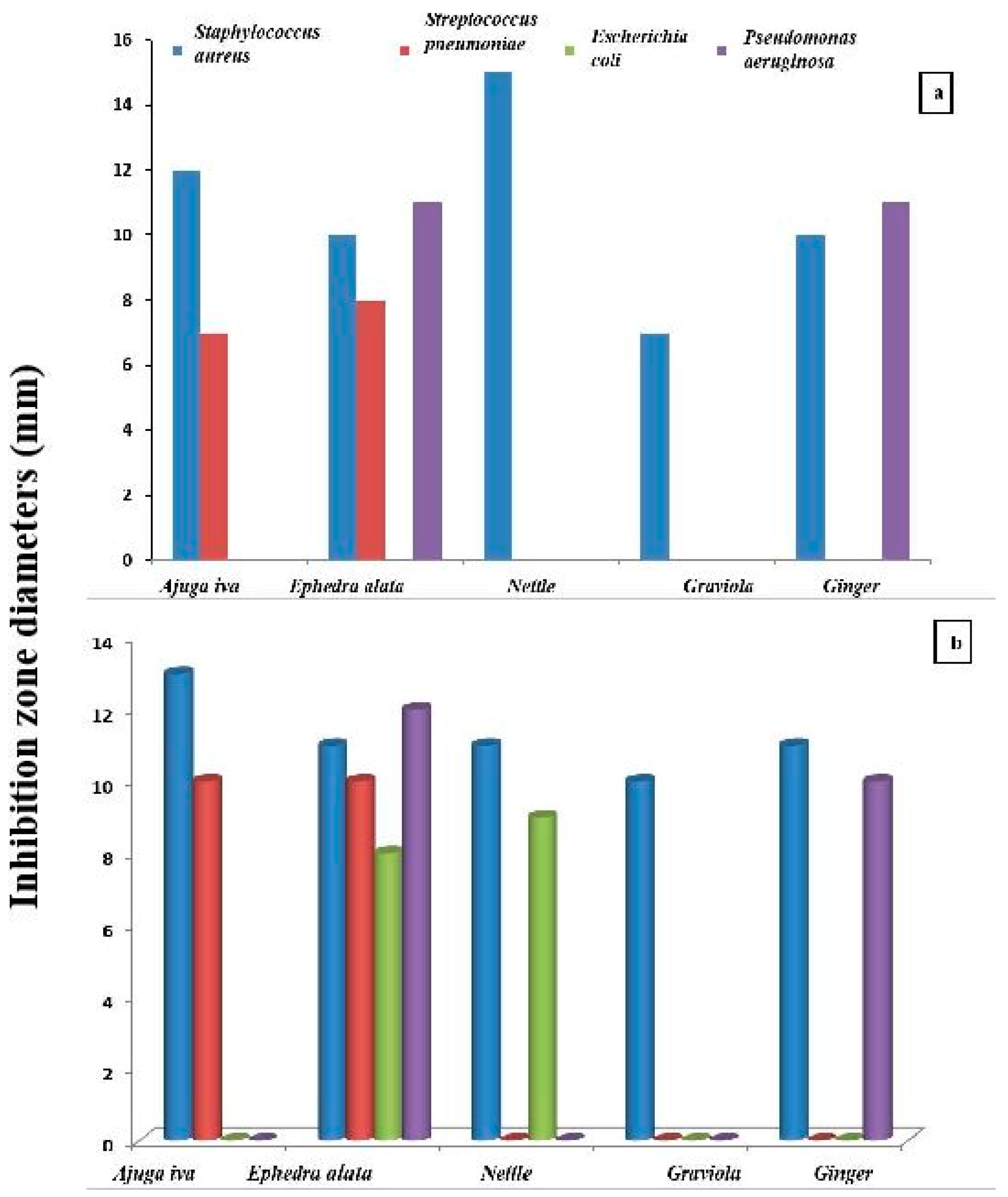
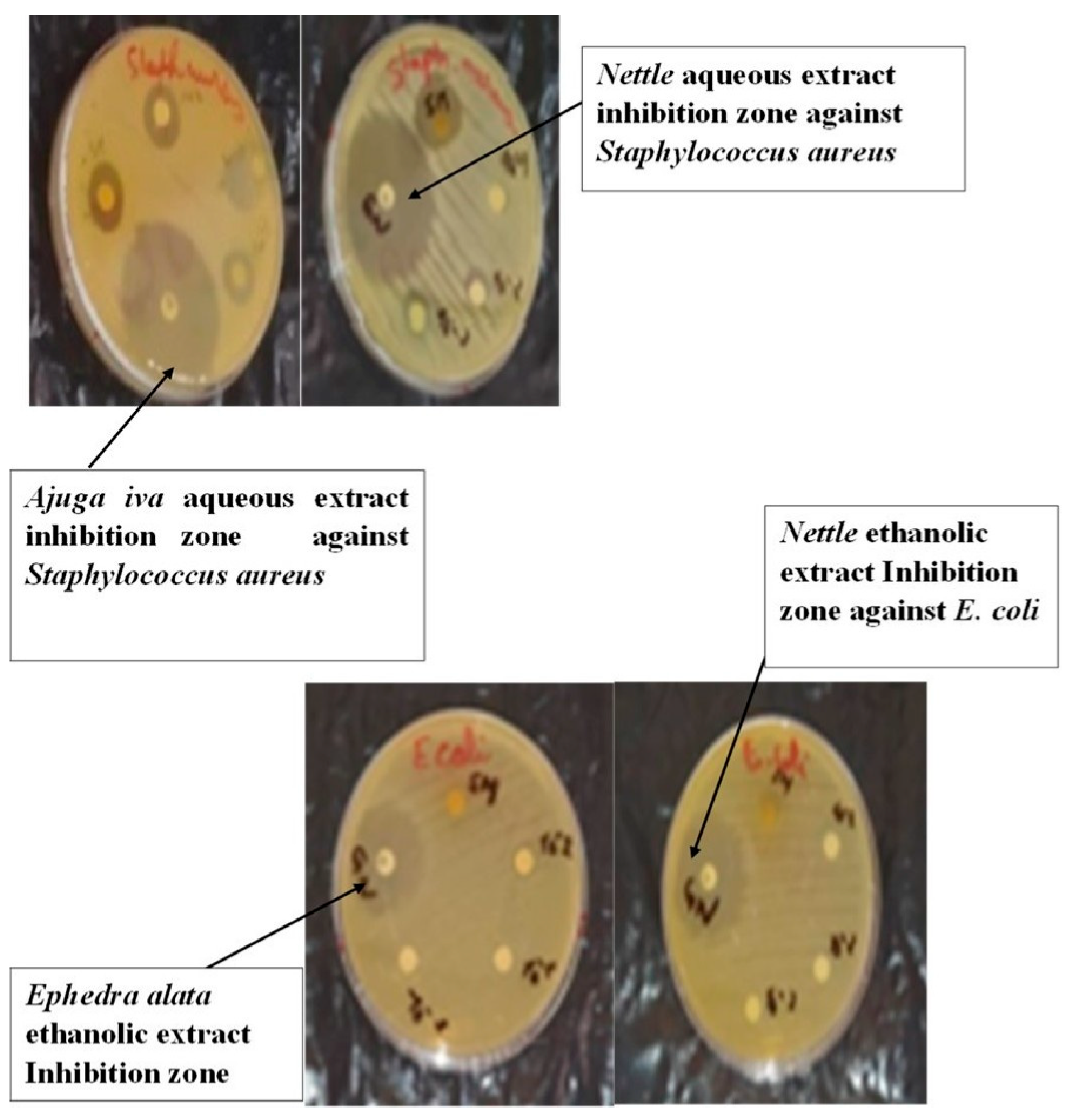
To evaluate the in vitro antibacterial potency of our extracts, the inhibition diameter was taken as an indicator. Figure 3 shows that the diameter of the inhibition zone changes with the bacteria strain type and the extract’s nature. In fact, all of the tested extracts exhibited an inhibitory effect more significantly against Staphylococcus aureus. In the case of this last strain, the aqueous extracts of ajuga iva and nettle have the highest inhibition diameters compared to other plants (Figure 4). The variations in the antimicrobial activity of samples explain variations in their chemical composition.
In previous studies, a rather concentrated solution was used to observe inhibition. As shown in the work of Albayati et al. [46] on ginger extracted through maceration with ethanol, the relationship showed an antibacterial effect on Escherichia coli for concentrations above 50 mg/mL and no inhibition for a concentration of 25 mg/mL. The diameter of inhibition increases with concentration from 15.33 mm at 50 mg/mL to 23 mm at 100 mg/mL.
According to our results, ephedra alata and ajuga iva are recorded as inhibitors of streptococcus pneumoniae bacteria, while the other plants have not demonstrated any inhibitory effects on these bacteria.
The antibacterial action is more pronounced for the ethanolic extract compared to the aqueous one, which may be due to the lack of a plant extract fraction that may contain important antibacterial and antioxidant elements.
The occurrence of flavonoids, as summarized in Figure 2, may be responsible for good antibacterial activity and is further related to the medicinal value index. This result is in agreement with the literature [46,47].
The antibacterial effect is also affected by the extraction method, as seen in the work of [47] on ginger, where larger inhibition diameters were obtained with ethanol maceration followed by decoction and cold-water extraction.

3.5. FTIR Analysis

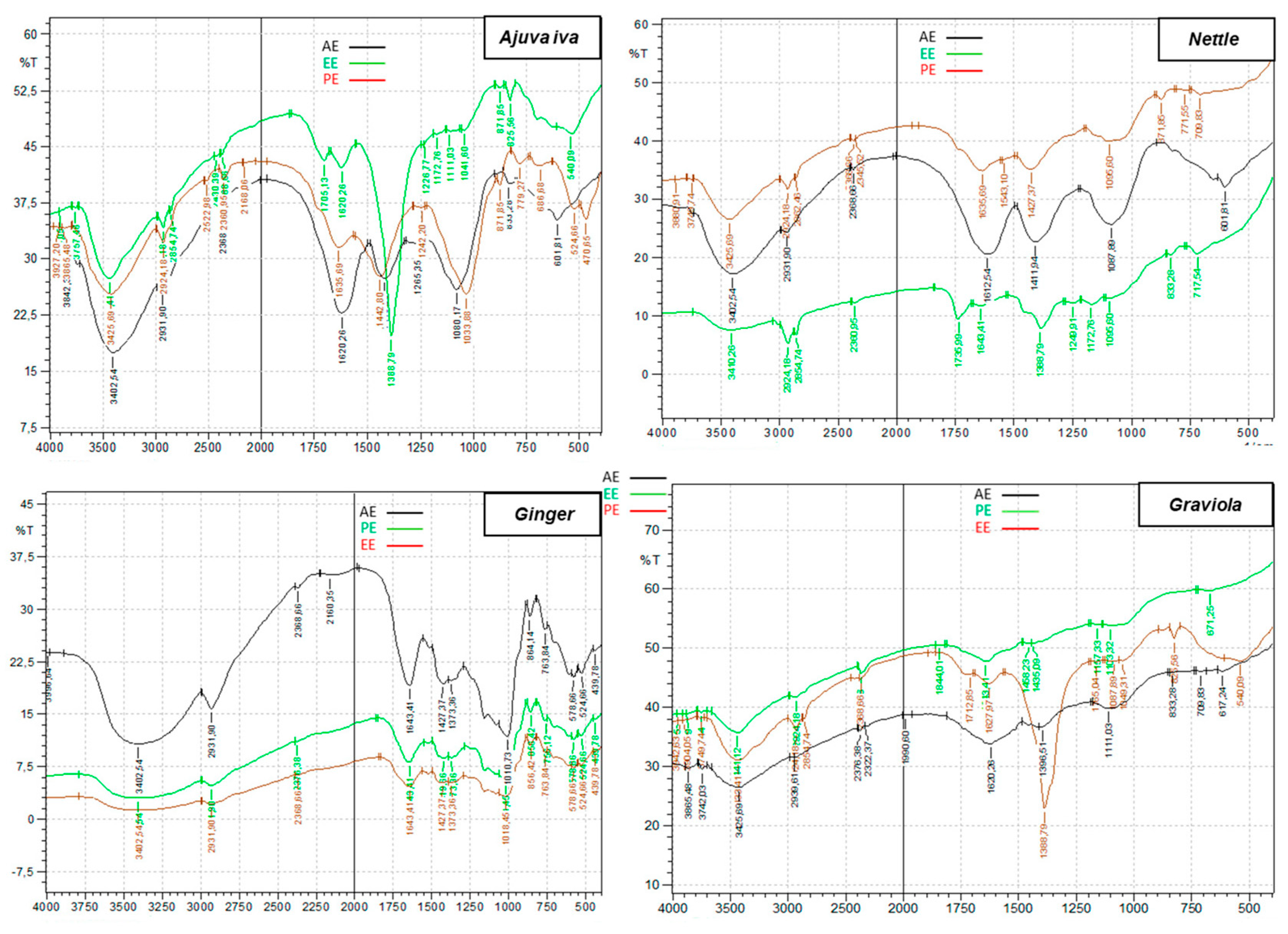
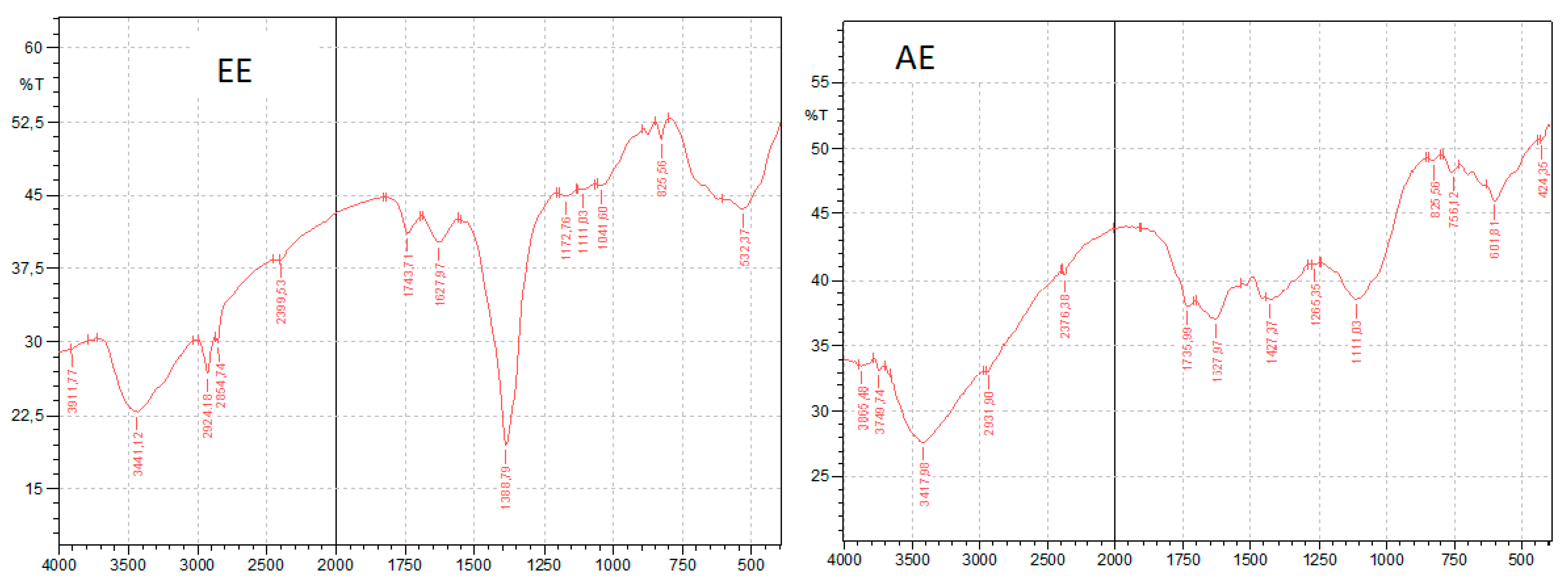
The aim of infrared spectrometry is to identify the chemical functions present in a product. From the results of infrared analysis of the various extract fractions illustrated in Figure 5 and Figure 6, it emerges that the five plants are characterized by several variable-function bonds. The broad bands around 3400 cm−1indicate O–H stretching vibrations of hydroxyl groups, suggesting the presence of alcohols and phenols [48,49], which may contribute to the bioactivity observed in our study. Considering the intensity of this peak in both extracts, it can be seen that there is a superiority of the presence of this kind of molecule in the case of ethanolic extract, and this is also confirmed by the result obtained by the dosage (Table 1). The peak at 1388 cm−1 corresponds to the in-plane deformation of the O-H bond in phenolic compounds [48,49].
The narrow bands around 2924 to 2926 cm−1 correspond to the valence vibration of the C-H bond (alkane function). Strong bands around 1630 to 1640 cm−1 characterize the double bond [47,48].

4. Conclusions

In this study, the antibacterial potency and total content of both flavonoids and polyphenols were investigated for five medicinal plants (ginger, ajuga iva, graviola, ephedra alata, and nettle) chosen on the basis of the frequency of their use by the local population for the treatment of diabetes and cancer. For both of the extraction methods used, ethanol maceration and decoction, our results showed that quantitative differences in extraction yield can be recorded as differences in the extraction method, the kind of plant, extraction solvent polarity, and the m/v ratio. All of the tested plants are rich in polyphenols with important content in ethanolic extracts, and the highest level is obtained in the case of ephedra alata. Compared to decoction, maceration using ethanol as the solvent can enhance the extraction of flavonoids.
Among all of the plants tested, ajuga iva and graviola were the richest ones in flavonoid content. All of the extracts have an inhibitory effect on Staphylococcus aureus bacteria and variable effects on other bacteria, with great improvement exerted by ethanolic extracts. Only ephedra alata and nettle can exhibit antibacterial activity on Escherichia coli, and only ephedra alata and ajuga Iva have an inhibitory effect against Streptococcus pneumoniae bacteria. For future purposes, we suggest using the green extraction method forthe alkaloids class, which are highly effective in the treatment of cancer, and evaluating the anti-cancer activity of these plants.

Author Contributions

Conceptualization, N.M., L.M., D.K., N.B., N.T., J.Z., H.T. and A.A.; data curation, N.M., J.Z. and A.A.; formal analysis, N.M., L.M., D.K., N.B., N.T., J.Z., H.T. and A.A.; investigation, N.M., L.M., D.K., N.B., N.T., J.Z., H.T. and A.A.; methodology, N.M., L.M., D.K., N.B., N.T., J.Z., H.T. and A.A.; project administration, J.Z., H.T. and A.A.; resources, L.M., N.B., N.T., J.Z. and A.A.; software, N.M. and D.K.; supervision, J.Z., H.T. and A.A.; validation, N.M., L.M., D.K., N.B., N.T., J.Z., H.T. and A.A.; visualization, N.M., L.M., D.K., N.B., N.T., J.Z. and A.A.; writing—original draft, N.M. and L.M.; writing—review and editing, N.M., L.M., D.K., N.B., N.T., J.Z., H.T. and A.A. All authors have read and agreed to the published version of the manuscript.

Funding

This research received no external funding.

Institutional Review Board Statement

Not applicable.

Informed Consent Statement

Not applicable.

Data Availability Statement

No new data were created or analyzed in this study. Data sharing is not applicable to this article.

Acknowledgments

The authors sincerely acknowledge the members of the Laboratory of Chemistry (University Yahia Fares of MEDEA) for the use of several materials.

Conflicts of Interest

The authors declare that they have no know competing financial interests or personal relationships that could have appeared to influence the work reported in this paper.

Abbreviations

m/vMass of the plant/volume of the extraction solvent
MAEMicrowave-Assisted Extraction
UAEUltrasonically Assisted Extraction
mg EGA/g DE mg equivalent of gallic acid per gram of dry extract
mg EQ/g DEmg equivalent of quercetin per gram of dry extract
PPTTotal polyphenols
E coliEscherichia coli

References

  1. Farnsworth, N.R.; Akerele, O.; Bingel, A.S.; Soejarto, D.D.; Guo, Z. Medicinal plants in therapy. Bull. World Health Organ. 1985, 63, 965–981. Available online: https://pmc.ncbi.nlm.nih.gov/articles/PMC2536466/ (accessed on 15 July 2023). [CrossRef] [PubMed]
  2. Khitri, W.; Lachgueur, N.; Tasfaout, A.; Lardjam, A.; Khalfa, A. Antilithiasic plants used in traditional medicine in Oran, Algeria: Ethnobotanical survey and phytochemical screening. J. Ethnoecol. 2016, 9, 2267–2419. [Google Scholar] [CrossRef]
  3. Quieda, F. Médecine Arabe et Ethnopharmacologie, les Plantes du Coran; CNRSL Beyrouth, Ed.; Faculty de Pharmacy, Libaneese University: Liban, 2006; Available online: https://books.openedition.org/irdeditions/7247?lang=fr (accessed on 1 June 2023).
  4. Mbaïhougadobé, S.; Ngakegni-Limbili, A.C.; Gouollaly, T.; Koane, J.N.; Ngaïssona, P.; Loumpangou, C.N.; Mahmout, Y.; Ouamba, J.-M. Evaluation de l’activité anti-oxydante de trois espèces de plantes utilisées dans le traitement de la goutte au Tchad. Rev. CAMES—Série Pharm. Méd. Trad. Afr. 2017, 18, 28–35. Available online: https://asjp.cerist.dz/en/article/43129 (accessed on 1 March 2023).
  5. Ziyyat, A.; Legssyer, A.; Mekhfi, H.; Dassouli, A.; Serhrouchni, M.; Benjelloun, W. Phytotherapy of hypertension and diabetes in oriental Moroccan. J. Ethnopharmacol. 1997, 58, 45–54. [Google Scholar] [CrossRef]
  6. Accipe, L.; Abadie, A.; Neviere, R.; Bercion, S. Antioxidant Activities of Natural Compounds from Caribbean Plants to Enhance Diabetic Wound Healing. Antioxidants 2023, 12, 1079. [Google Scholar] [CrossRef] [PubMed]
  7. Allali, H.; Benmehdi, H.; Dib, M.A.; Tabti, B.; Ghalem, S.; Benabadji, N. Phytotherapy of Diabetes in West Algeria. Asian J. Chem. 2008, 20, 2701–2710. Available online: https://pdfs.semanticscholar.org/f67c/f41460e6e416c153fbb0934f0fba99aa9266.pdf (accessed on 1 June 2024).
  8. Ghasemzadeh, A.; Jaafar, H.Z.E.; Rahmat, A. Elevated Carbon Dioxide Increases Contents of Flavonoids and Phenolic Compounds, and Antioxidant Activities in Malaysian Young Ginger (Zingiber officinale Roscoe) Varieties. Molecules 2010, 15, 7907–7922. [Google Scholar] [CrossRef] [PubMed]
  9. Ilkhanizadeh1, B.; Shirpoor, A.; Khadem Ansari, M.H.; Nemati, S.; Rasmi, Y. Protective Effects of Ginger (Zingiber officinale) Extractagainst Diabetes-Induced Heart Abnormality in Rats. Diabetes Metab. J. 2016, 40, 46–53. [Google Scholar] [CrossRef]
  10. Grzanna, R.; Lindmark, L.; Frondoza, C.G. Ginger an herbal medicinal product with broad anti-inflammatory actions. J. Med. Food 2005, 8, 125–132. [Google Scholar] [CrossRef]
  11. Zaman, S.; Mirje, M.M.; Ramabhimaiah, S. Evaluation of the Anti-Ulcerogenic Effect of Zingiber officinale (Ginger) Root in Rats. Int. J. Curr. Microbiol. App. Sci. 2014, 3, 347–354. Available online: https://api.semanticscholar.org/CorpusID:81914 (accessed on 1 November 2020).
  12. Zadeh, J.B.; Kor, M.M. Physiological and pharmaceutical effects of Ginger (Zingiber officinale Roscoe) as a valuable medicinal plant. Eur. J. Exp. Biol. 2014, 4, 87–90. [Google Scholar]
  13. Mahluji, S.; Attari, V.E.; Mobasseri, M.; Payahoo, L.; Ostadrahimi, A.; Golzari, S.E. Effect of ginger (Zingiber officinale) on plasma glucose level, HbA1c and insulin sensitivity in type 2 Diabetic patients. Int. J. Food Sci. Nutr. 2013, 64, 682–686. [Google Scholar] [CrossRef] [PubMed]
  14. Karna, P.; Chagani, S.; Gundala, S.R.; Rida, P.C.; Asif, G.; Sharma, V.; Gupta, M.V.; Aneja, R. Benefits of whole ginger extract in prostate cancer. Br. J. Nutr. 2012, 107, 473–484. [Google Scholar] [CrossRef]
  15. Jeong, C.H.; Bode, A.M.; Pugliese, A.; Cho, Y.Y.; Kim, H.G.; Shim, J.H.; Jeon, Y.J.; Li, H.; Jiang, H.; Dong, Z. Gingerol Suppresses Colon Cancer Growth by Targeting Leukotriene A4 Hydrolase. Cancer Res. 2009, 69, 5584–5591. [Google Scholar] [CrossRef] [PubMed]
  16. Sharma, C.; Ahmed, T.; Sasidharan, S.; Ahmed, M.; Hussain, A. Use of Gemcitabine and Ginger Extract Infusion May Improve the Efficiency of Cervical Cancer Treatment. Afr. J. Biotec. 2009, 8, 7087–7093. [Google Scholar]
  17. Hegazi, G.A.E.; El-Lamey, T.M. In vitro Production of Some Phenolic Compounds from Ephedra alata Decne. J. Appl. Env. Biol. Sci. 2011, 1, 158–163. Available online: https://www.researchgate.net/publication/285022703 (accessed on 20 April 2023).
  18. Derbel, S.; Touzard, B.; Triki, M.A.; Chaieb, M. Seed germination responses of the Saharan plant species Ephedra alata ssp. alenda to fungicide seed treatments in the laboratory and the field. Flora 2010, 205, 471–474. [Google Scholar] [CrossRef]
  19. AL-Qarawi, A.A.; Abd-Allah, E.F.; Abeer, H. Ephedra alata as biologically-based strategy inhibit aflatoxigenic seedborne mold. Afr. J. Microb. Res. 2011, 5, 2297–2303. [Google Scholar]
  20. El-hilaly, J.; Tahraoui, T.; Israili, Z.H.; Lyoussi, B. Acute hypoglycemic, hypocholesterolemic and hypotriglyceridemic effects of continuous intravenous infusion of a lyophilised aqueous extract of Ajuga Iva L. Schreber whole plant in streptozotocininduced diabetic rats. Pak. J. Pharm. Sci. 2007, 20, 261–268. Available online: https://pubmed.ncbi.nlm.nih.gov/17604246/ (accessed on 1 July 2022).
  21. El-hilaly, J.; Amarouch, M.Y.; Morel, N.; Lyoussi, B.; Quetin-Leclercq, J. Ajuga iva water extract antihypertensive effect on stroke-prone spontaneously hypertensive rats, vasorelaxant effects ex vivo and in vitro activity of fractions. J. Ethnopharmacol. 2021, 270, 113–791. [Google Scholar] [CrossRef]
  22. Draghi, F. L’ortiteDioique (Urticadioica L.): Etude bibliographique. Ph.D. Thesis, University of Henri Poincare, Nancy, France, 2005. Available online: https://docnum.univ-lorraine.fr/public/SCDPHA_T_2005_DRAGHI_FRANCINE.pdf (accessed on 1 June 2024).
  23. Viotti, C.; Albrecht, K.; Amaducci, S.; Bardos, P.; Bertheau, C.; Blaudez, D.; Bothe, L.; Cazaux, D.; Ferrarini, A.; Govilas, J.; et al. Nettle, a Long-Known Fiber Plant with New Perspectives. Materials 2022, 15, 4288. [Google Scholar] [CrossRef] [PubMed]
  24. Gavamukulya, Y.; Wamunyokoli, F.; El-Shemy, H.A. Annona muricata: Is the natural therapy to most disease conditions including cancer growing in our backyard? A systematic review of its research history and future prospects. Asian Pac. J. Trop Med. 2017, 10, 835–848. [Google Scholar] [CrossRef]
  25. Rady, I.; Bloch, M.B.; Chamcheu, R.C.; Banang Mbeumi, S.; Anwar, M.R.; Mohamed, H.; Babatunde, A.S.; Kuiate, J.R.; Noubissi, F.K.; El Sayed, K.A.; et al. Anticancer Properties of Graviola (Annona muricata): A Comprehensive Mechanistic Review. Oxid. Med. Cell Longev. 2018, 2018, 1826170. [Google Scholar] [CrossRef]
  26. Zine, S. Le corossol (Annona muricata L.) et ses propriétés thérapeutiques: État des lieux. Sci. Pharm. 2018, 20–23. Available online: https://dumas.ccsd.cnrs.fr/dumas-01823643 (accessed on 1 July 2021).
  27. Al-Bayar, A.H. Effect of extraction method of ginger roots on antioxidant activity. Iraqi J. Agric. Sci. 2009, 40, 101–109. Available online: https://www.researchgate.net/publication/303679590 (accessed on 1 January 2023).
  28. Ali-Rachedi, F.; Meraghni, S.; Touaibia, N.; Sabrina, M. Analyse quantitative des composés phénoliques d’une endémique algérienne Scabiosa atropurpurea. sub. Maritima L. Bull. Société R. Sci. Liège 2018, 87, 13–21. [Google Scholar] [CrossRef]
  29. Laib, I.; Karit, H.; Bouzerdouna, R.; Kettouche, F.; Barkat, M. Effet de digestion gastro-intestinale in vitro sur les composés phénoliques et l’activité antioxydante d’ail triquètre Allium triquetrum L. J. Appl. Biosci. 2022, 171, 17821–17837. [Google Scholar] [CrossRef]
  30. Leswara, D.F.; Kurniasih, K.; Sholehah, I.K. The effect of maceration duration on the total flavonoids content of kaempferia parviflora wall. ex baker ethanol extract. Media ILMU Kesehat. 2024, 13, 87–94. [Google Scholar] [CrossRef]
  31. Sharif, M.F.; Bennett, M.T. The effect of different methods and solvents on the extraction of polyphenols in Ginger (Zingiber officinale). J. Teknol. 2016, 78, 11–12. [Google Scholar] [CrossRef]
  32. Rahath Kubra, I.; Rao, L.J.M.; Kumar, D. Effect of microwave-assisted extraction on the release of polyphenols from ginger (Zingiber officinale). Int. J. Food Sci. Technol. 2013, 48, 1828–1833. [Google Scholar] [CrossRef]
  33. Belazougui, K.; Mesrouk, S.; Mohammedi, H.; Akcha, S.; Aïnouz, L.; Mecherara-Idjeri, S.F. Phytochemical analysis, mineral composition, Assessment of antioxidant properties and cytotoxic potential of Ephedra alata. subsp. Alenda secondary metabolites. Food Biosci. 2023, 53, 102657. [Google Scholar] [CrossRef]
  34. Zaater, A.; Serhoud, M.O.; Ben Amor, I.; Zeghoud, S.; Hemmami, A.; Rebiai, A.; Bouras, Y.; Laiche, A.T.; Alsalme, A.; Cornu, D.; et al. Exploring the potential of ephedra alata leaf extract: Phytochemical analysis, antioxidant activity, antibacterial properties, and green synthesis of ZnO nanoparticles for photocatalytic degradation of methylene blue. Front. Chem. 2024, 12, 1367552. [Google Scholar] [CrossRef]
  35. Muanda, F.N. Identification de Polyphénols, Evaluation de leur Activité Antioxydante et Etude de Leurs Propriétés Biologiques. Ph.D. Thesis, Paul Verlaine University, Metz, France, 2010. Available online: https://hal.univ-lorraine.fr/tel-01752680v1/file/Muanda.Francois.Nsemi.SMZ1011.pdf (accessed on 1 January 2023).
  36. Do, Q.D.; Angkawijaya, A.E.; Tran-Nguyen, P.L.; Huynh, L.H.; Soetaredjo, F.E.; Ismadji, S.; Ju, Y.H. Effect of extraction solvent on total phenol content, total flavonoid content, and antioxidant activity of Limnophila aromatic. J. Food Drug Anal. 2014, 22, 96–302. [Google Scholar] [CrossRef]
  37. Bragic, S.; Sofic, E. Chemical composition of various Ephedra species. Bosn. J. Basic Med. Sci. 2015, 15, 21. [Google Scholar] [CrossRef]
  38. AL-Rimawi, F.; Abu-Lafi, S.; Abbadi, J.; Alamarneh, A. Analysis of phenolic and flavonoids of wild Ephedra alata plant extracts by LC/PDA and LC/MS and their antioxidant activity. Afr. J. Tradit. Complement. Altern. Med. 2017, 14, 130–141. [Google Scholar] [CrossRef]
  39. Kebili, Z. Contribution à l’étude de Quelques Activités Biologiques des Extraits de Ephedra alata de la région de Ouargla. Master’s Thesis, Kasdi MERBAH University, Ouargla, Algeria, 2016. [Google Scholar]
  40. Ben Salah, I.; Smaoui, A.; Mahmoudi, H.; Ouerghi, Z. Effet du solvent et de la méthode d’extraction sur les teneurs en composés phénoliques et l’activité antioxydante chez Ephedra alata alenda. IOSR J. Agric. Vet. Sci. (IOSR-JAVS) 2021, 14, 23–30. [Google Scholar]
  41. Mahmoudi, M.; Boughalleb, F.; Maaloul, S.; Mabrouk, M.; Abdellaoui, R. Phytochemical Screening, Antioxidant Potential, and LC–ESI–MS Profiling of Ephedra alata and Ephedra altissima Seeds Naturally Growing in Tunisia. Appl. Biochem. Biotechnol. 2023, 191, 5903–5915. [Google Scholar] [CrossRef]
  42. Chouikh, A. Phytochemical profile, antioxidant, analgesic and hypolipidaemic effects of ephedra alata decne female cones extract. Farmacia 2020, 68, 6. [Google Scholar] [CrossRef]
  43. Bensam, M.; Rechreche, H.; Abdelwahab, A.E.; Abu-Serie, M.M.; Ali, S.M. The role of Algerian ephedra alata ethanolic extract in inhibiting the growth of breast cancer cells by inducing apoptosis in a p53- dependent pathway. Saudi J. Biol. Sci. 2023, 30, 103650. [Google Scholar] [CrossRef]
  44. Ramanan, M.; Sinha, S.; Sudarshan, K.; Singh Aidhen, I.; Doble, M. Inhibition of the enzymes in the leukotriene and prostaglandin pathways in inflammation by 3-aryl isocoumarins. Eur. J. Med. Chem. 2016, 124, 428–434. [Google Scholar] [CrossRef]
  45. Sudarshan, K.; Singh Aidhen, I. Convenient Synthesis of 3-Glycosylated Isocoumarins. Eur. J. Org. Chem. 2017, 2017, 34–38. [Google Scholar] [CrossRef]
  46. Albayati, M.K.; Jawad, A.M.; Khudir, M.H. Evaluation of Zingiber officinaeRosc extract against pathogenic bacteria. Iraqi J. Sci. 2010, 51, 403–409. [Google Scholar]
  47. Nader, M.I.; Ghanima, K.K.; Ali, S.A.; Azhar, D.A. Antibacterial activity of ginger extracts and its essential oil on some of pathogenic bacteria. Baghdad Sci. J. 2010, 7, 1159–1165. [Google Scholar] [CrossRef]
  48. Belboukhari, N.; Merzoug, Z.; Cheriti, A.; Sekkoum, K.; Yakoubi, M. Etude de la caractérisation et la composition qualitative des huiles essentielles de six plantes médicinales par spectroscopie IR. Phy. Chem. Bio. Sub. J. 2013, 7, 14–17. Available online: https://www.researchgate.net/publication/260391075 (accessed on 1 February 2023).
  49. Schulz, H.; Özkan, G.; Baranska, M.; Krüger, H.; Özcan, M. Characterisation of essential oil plants from Turkey by IR and Raman spectroscopy. Vib. Spectrosc. 2005, 39, 249–256. [Google Scholar] [CrossRef]
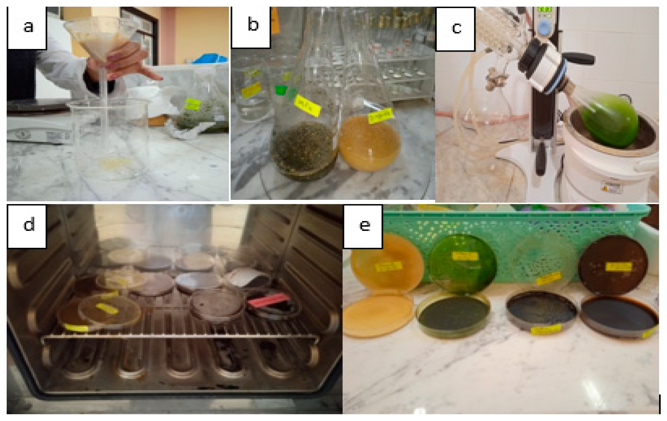
Figure 1. Plant extract preparation: (a) filtration of macerated plant. (b) Ethanolic and aqueous plant extract before solvent elimination. (c) Solvent evaporation using rotavapor. (d) Plant extract drying. (e) Dried plants’ extracts.
Microbiolres 16 00108 g001
Figure 2. Total flavonoid content in ethanolic and aqueous plant extracts in equivalent mg of quercetin per g of dried extract under two m/v ratios. (a). m/v ratio 0.1; (b) m/v ratio 0.067.
Microbiolres 16 00108 g002
Figure 3. In vitro antibacterial activity against various bacterial strains of different tested extracts. (a) Aqueous extract and (b) ethanolic extract.
Microbiolres 16 00108 g003
Figure 4. Inhibition zone of ethanolic and aqueous extracts against Staphylococcus aureus and Escherichia Coli.
Microbiolres 16 00108 g004
Figure 5. IR spectra of ajuva iva, nettle, ginger, and graviola. (AE: aqueous extract; EE: ethanolic extract; PE: plant before extraction).
Microbiolres 16 00108 g005
Figure 6. IR spectra of ephedra alata (AE: aqueous extract; EE: ethanolic extract).
Microbiolres 16 00108 g006
Table 1. Yield values obtained for various plants tested under different extraction methods.
PlantsNettleAjuga ivaGraviolaGingerEphedra Alata
Yield %Decoction m/v = 0.17.20515.2714.98525.1415.27
Decoction m/v = 0.06711.80520.99512.0225.30516.455
Maceration (2-fold)
m/v = 0.1
12.656.5653.6223.79518.22
Maceration (2-fold)
m/v = 0.067
7.929.66.6116.3926.32
Maceration (3-fold)
m/v = 0.067
6.7658.7553.0627.46521.07
Table 2. Total polyphenol content in dry extracts.
Polyphenol Concentration (mg EGA/g DE)
ExtractAqueous Extract (Decoction)Ethanolic Extract (Maceration)
m/v0.10.0670.1
(2 times)
0.067
(2 times)
0.067
(3 times)
Ginger14.73 ± 0.1731.33 ± 0.5841.10 ± 0.27101.6 ± 0.55103.92 ± 0.08
Ajuga iva39.92 ± 0.0336.14 ± 0.1657.94 ± 0.1287.15 ± 0.0688.61 ± 0.14
Graviola54.76 ± 0.7951.18 ± 0.1656.48 ± 0.5082.21 ± 0.72124.0 ± 0.27
Ephedra237.33 ± 0.13134.95 ± 0.05308.68 ± 0.59137.28 ± 0.14356.76 ± 0.57
Nettle50.41 ± 0.0841.10 ± 0.11101.6 ± 0.53133.40 ± 0.42126.41 ± 0.26
mg EGA/g DE: mg equivalent of gallic acid per gram of dry extract.
Disclaimer/Publisher’s Note: The statements, opinions and data contained in all publications are solely those of the individual author(s) and contributor(s) and not of MDPI and/or the editor(s). MDPI and/or the editor(s) disclaim responsibility for any injury to people or property resulting from any ideas, methods, instructions or products referred to in the content.