Communication

Microbiota of Punctuated Snake Eel Ophichthus remiger (Valenciennes, 1842) Reared in Recirculation System Is Dominated by Latilactobacillus

1
Laboratorio de Biotecnología de Alimentos, Instituto de Nutrición y Tecnología de los Alimentos (INTA), Universidad de Chile, Santiago 7830489, Chile
2
Laboratorio de Patobiología Acuática, Departamento de Acuicultura, Universidad Católica del Norte, Coquimbo 1780000, Chile
3
Laboratorio de Peces, Departamento de Acuicultura, Universidad Católica del Norte, Coquimbo 1780000, Chile
*
Author to whom correspondence should be addressed.
Microbiol. Res. 2025, 16(2), 38; https://doi.org/10.3390/microbiolres16020038
Submission received: 5 December 2024 / Revised: 14 January 2025 / Accepted: 3 February 2025 / Published: 5 February 2025
(This article belongs to the Special Issue Veterinary Microbiology and Diagnostics)

Abstract

:
Research on microbiota has underscored the crucial influence of microbial communities on numerous biological functions that yield positive outcomes for the host, such as digestion, nutrient metabolism, resistance against pathogen invasion, and growth performance. Concurrently, numerous variables, including the host’s diet, genetics, and physiological condition and environmental factors, influence the gut microbiota. Our study aims to characterize the bacterial community composition of the common snake eel (Ophichthus remiger), captured wild and then reared under controlled conditions. We employed a 16S rRNA gene-based approach facilitated by next-generation sequencing to conduct this analysis. The gut microbiota of the snake eel was highly dominated by bacteria from the phylum Firmicutes, comprising over 80% of the relative abundance, with Lactilactobacillus being the most important genus. The results suggest that feed-associated bacteria may influence the composition of the microbiota, contributing the most relevant bacteria within the intestinal content. This study provides the first comprehensive analysis of the gut microbiota in Ophichthus remiger, offering novel insights into the potential roles of Firmicutes and Lactilactobacillus in marine eels.

1. Introduction

In many parts of the world, fisheries are witnessing a troubling trend known as “growth overfishing”, where the size of harvested fish is declining [1]. This phenomenon occurs when fish are captured at a younger age, before they have reached their maximum size. Such intensive overfishing and unsustainable practices lead to a decrease in both the average age and size of fish within harvested populations, impacting the economic viability of fisheries. Larger individuals, which are often preferred in the marketplace, become increasingly rare [1,2].
This issue is particularly pronounced along the Chilean coast, where commonly caught species such as the Golden conger (Genypterus blacodes), Swordfish (Xiphias gladius), and Jack mackerel (Trachurus murphyi) are being harvested at smaller sizes [3,4]. As a result, there is a significant increase in bycatch—fish that are discarded because they do not meet legal size requirements for catch. This practice of discarding smaller, undersized fish accounts for an estimated 23% of bycatch in the total global catch [5], reflecting a widespread challenge in fisheries management that extends beyond local to global scales.
An innovative and supplementary approach involves repurposing bycatch as a resource for aquaculture, essentially recycling the discarded fish by raising them to a commercial size for sale. This strategy provides an opportunity to cultivate species that are traditionally acquired only through fishing. It also paves the way for advances in bio-ecological research, with the potential to achieve complete cultivation cycles for certain species. Illustrating this strategy is the common snake eel Ophichthus remiger (Valenciennes, 1842), found off the coasts of Peru and Chile [6]. This species represents the broader possibility of transforming bycatch into a viable asset for aquaculture, reflecting a shift towards more sustainable and conservation-minded fishing practices.
Studies on microbiota have highlighted the pivotal roles of microbial communities in various biological processes that benefit the host, including digestion, nutrient absorption, protection against pathogens, and immune system modulation. At the same time, factors such as diet, genetic makeup, physiological state, and environmental conditions significantly shape the composition and function of the gut microbiota [7,8,9,10]. Prior research has explored gut microbiota across a wide range of animal groups, including invertebrates, amphibians, reptiles, mammals, and birds [10,11,12,13]. In recent years, there has been growing interest in the gut microbiota of fish, particularly species of aquacultural importance. This interest stems from the recognition of the vital role that a balanced microbiota plays in supporting fish growth and development, which is essential for improving both productivity and sustainability in aquaculture systems [14]. Studies have begun to reveal how manipulations of the microbiota through diet, probiotics, and environmental modifications can promote more resilient and faster-growing aquaculture species, thus improving production efficiency and reducing reliance on antibiotics [14].
Insights into the microbiota of fish are opening new pathways to enhance nutrition and overall well-being in aquaculture species, with growing interest evident from the substantial volume of related studies, as discussed in recent reviews. This surge in research is driven by advancements in next-generation sequencing (NGS) technologies, which have broadened our understanding of the microbiota in various aquaculture-relevant species by examining factors like diet, abiotic conditions, and population density. In our study, we delve into the bacterial community composition of common snake eel Ophichthus remiger, captured wild and then reared under controlled conditions. We employ a 16S rRNA gene-based approach facilitated by next-generation sequencing to conduct this analysis.

2. Materials and Methods

2.1. Snake Eel Rearing Conditions

Juvenile common eels were captured along the coasts of Taltal (Latitude: 25°24′31″ S, Longitude: 70°29′00″ W). The selection of fishing sites was based on historical records of the presence of this resource in these areas. The recirculating aquaculture system (RAS) employed included a rotary drum filter, biofilter, fractionator-accumulator, two pumping pumps, an aeration system, and culture tanks. The total volume utilized in the system was 20 m3 with a turnover rate equivalent to 10% per day. Six circular tanks of identical specifications were installed, each with a diameter of 1.25 m, a height of 0.8 m, an effective volume of 0.5 m3, a bottom surface area of 1.23 m2, and a flow rate of 15 L/min. Following fish acclimatization, size classification was performed to ensure a maximum total length (Lt) of 42 cm in each tank. During the experiment (120 days), fish were fed a semi-moist diet designed generically for marine fish breeders due to lack of specific records for this species in culture. The diet comprised a blend of fresh fish (69% Pacific jack mackerel), fish meal (27%), fish oil (3%), and vitamin premix (1%). Food was delivered at a rate equivalent to 2% of the biomass present in each tank.

2.2. Sampling, DNA Extraction, and PCR Amplicon Sequencing

Intestinal samples were obtained as described by Romero et al. [7]. In brief, following euthanasia performed in accordance with ethical guidelines, the skin was disinfected with 70% ethanol. On a sterile surface, a longitudinal incision was made from the anus to the pectoral fins using a scalpel. The intestine was carefully located and removed by cutting at the esophagus and rectum. The extracted intestine was then separated and stored in a sterile container with a saline medium. DNA extraction was carried out using the QIAamp PowerFecal® DNA Isolation Kit (Qiagen, Venlo, The Netherlands), following the supplier’s instructions. This process included an initial lysozyme treatment (SIGMA, Darmstadt, Germany) at 0.8 mg/mL at 37 °C for 60 min, followed by proteinase K treatment (Invitrogen, Waltham, MA, USA) at 0.1 mg/mL at 37 °C for 60 min. DNA concentrations were quantified using the Qubit® dsDNA HS Assay Kit (Life Technologies, Carlsbad, CA, USA). The V4 region of the 16S rRNA gene was then amplified using primers 515F and 806R, as cited in reference [13]. The PCR mixture for each 30 μL reaction included 1.5 U of GoTaq® G2 Flexi DNA Polymerase (Promega, Madison, WI, USA), 6 μL of buffer (5×), 1.5 mM of MgCl2, 0.25 pM of each primer, 0.5 mM dNTPs, and 20 ng of DNA. PCR runs also included negative controls without DNA. All PCR procedures and amplicon purifications followed the methods described in reference [12] using the QIAquick® PCR Purification Kit (Qiagen). The concentration of each purified amplicon was determined using the Qubit® dsDNA HS Assay Kit (Life Technologies). The libraries were then sequenced on the Illumina MiSeq platform with 2 × 250 bp read lengths. Reads deposited under PRJNA1148021.

2.3. Bioinformatics Analysis

Nine NGS samples (V4 region) from the intestinal contents of eels (5) and feed (4) were analyzed. The sequences were initially evaluated in FastQC to determine the quality and length filters to be used, then the samples were processed in the R environment. The DADA2 (1.26) package was used to filter, trim, estimate error profiles, merge paired-end reads, remove chimeras, and assign taxonomy. Firstly, forward and reverse reads were truncated at 240 bp and 235 bp, respectively, using the filterAndTrim function with standard parameters (maxN = 0, truncQ = 2, and maxEE = 2). Sample inference was performed using the inferred error model, and chimeric sequences were removed using the removeBimeraDenovo function. The ASVs resulting from the previous step were assigned taxonomy by classifying them in the Silva v138.1 database using a naïve Bayesian classifier, with a minimum bootstrap confidence of 80%. The count table, taxonomy assignment results, and metadata table were compiled into a phyloseq object.
Alpha diversity was assessed using the Chao1, Shannon, and Simpson indices. The Aitchison distance was calculated as a metric of beta diversity. First, the transform function of the microbiome package was used for the centered log transformation (clr) of the count table and then, using the vegan package, the Euclidean distance was calculated. Significant differences between the bacterial communities from different groups were evaluated using PERMANOVA. Additionally, the influence of dispersion among groups was determined through betadisper analysis (the permutation test for homogeneity of multivariate dispersions).

3. Results

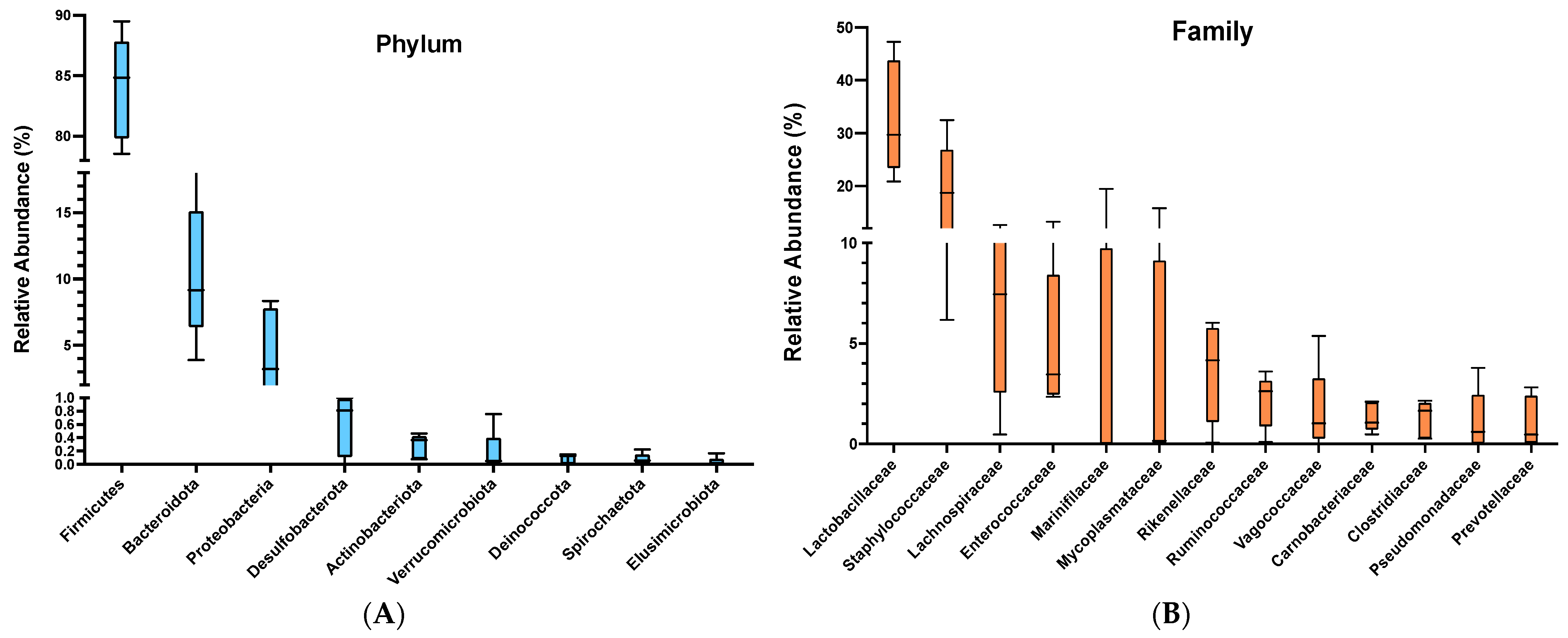
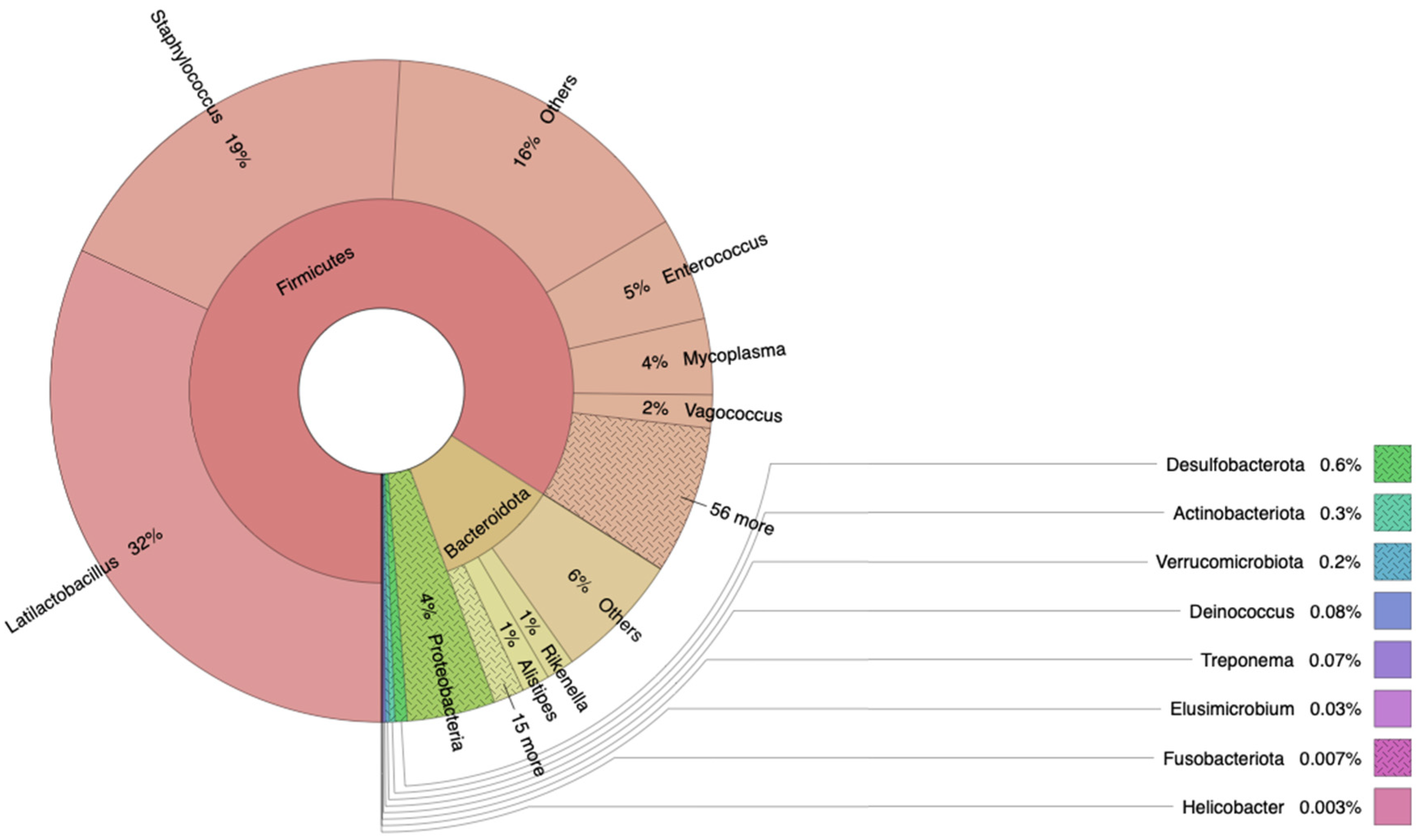
3.1. Bacterial Composition of Snake Eel Microbiota

We analyzed the bacterial communities of juvenile Ophichthus remiger using next-generation sequencing of the 16S rRNA gene. All fish sampled in this study were apparently healthy at the time of sampling. Considering the gut samples, 297,494 reads were obtained, with an average of 59,498 reads per sample. Rarefaction curves reached the saturation phase plateau (Supplementary Figure S1), indicating that the sequencing depth per sample was adequate to represent the bacterial communities.
The microbiota of snake eels was predominantly composed of bacteria belonging to the phylum Firmicutes, accounting for more than 84.03% of the relative abundance (Figure 1). Other notable phyla included Bacteroidota and Proteobacteria, with relative abundances of 10.43% and 4.30%, respectively. Minor components comprised Desulfobacterota, Actinobacteriota, and Verrucomicrobiota, with relative abundances of 0.60%, 0.29%, and 0.19%, respectively. At the family level, the most abundant taxa were Lactobacillaceae, representing 32.83% of the relative abundance, followed by Staphylococcaceae with 18.93%. Other less abundant taxa included Lachnospiraceae, Enterococcaceae, Marinifilaceae, Mycoplasmataceae, and Rikenellaceae, with relative abundances of 7.16%, 5.04%, 3.89%, 3.70%, and 3.57%, respectively. At the genus level, Latilactobacillus was the predominant genus, comprising 31.98% of the total abundance. Following Latilactobacillus, Staphylococcus accounted for 18.92%. Minor genera included Enterococcus, Mycoplasma, Vagococcus, Rikenella, Alistipes, and Pseudomonas, with relative abundances of 5.04%, 3.70%, 1.62%, 1.37%, 1.35%, and 1.10%, respectively (Figure 2).

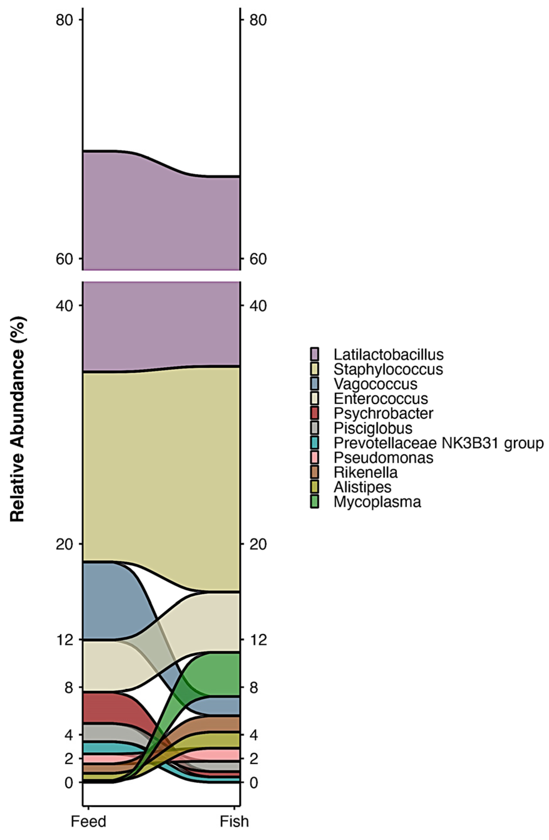
3.2. Bacterial Composition of Feed

We conducted an analysis of bacterial communities in feed, as diet is known to influence gut microbiota composition. From the feed samples, a total of 201,694 reads were obtained, averaging 50,410 reads per sample. The predominant bacterial phylum detected in the feed was Firmicutes, comprising more than 81.72% of the total reads. Additional phyla included Proteobacteria and Bacteroidota, accounting for 8.58% and 7.95% relative abundance, respectively. At the family level, Lactobacillaceae was the most abundant, representing 36.71% of the total relative abundance, followed by Staphylococcaceae with 15.98%. Minor components included Vagococcaceae, Enterococcaceae, Moraxellaceae, Lachnospiraceae, Prevotellaceae, Muribaculaceae, Rikenellaceae, and Carnobacteriaceae, with relative abundances of 6.52%, 4.37%, 3.73%, 3.19%, 2.39%, 2.11%, 2.00%, and 1.92%, respectively. At the genus level, Latilactobacillus dominated the composition, representing 34.96% of the relative abundance. Staphylococcus followed with 15.93%. Minor genera included Vagococcus, Enterococcus, Psychrobacter, Pisciglobus, and the Prevotellaceae NK3B31 group, accounting for 6.52%, 4.37%, 2.63%, 1.53%, and 1.02%, respectively. Figure 3 shows the variations in some genera between feed and gut microbiota; however, they do not show significant differences (Supplementary Materials).

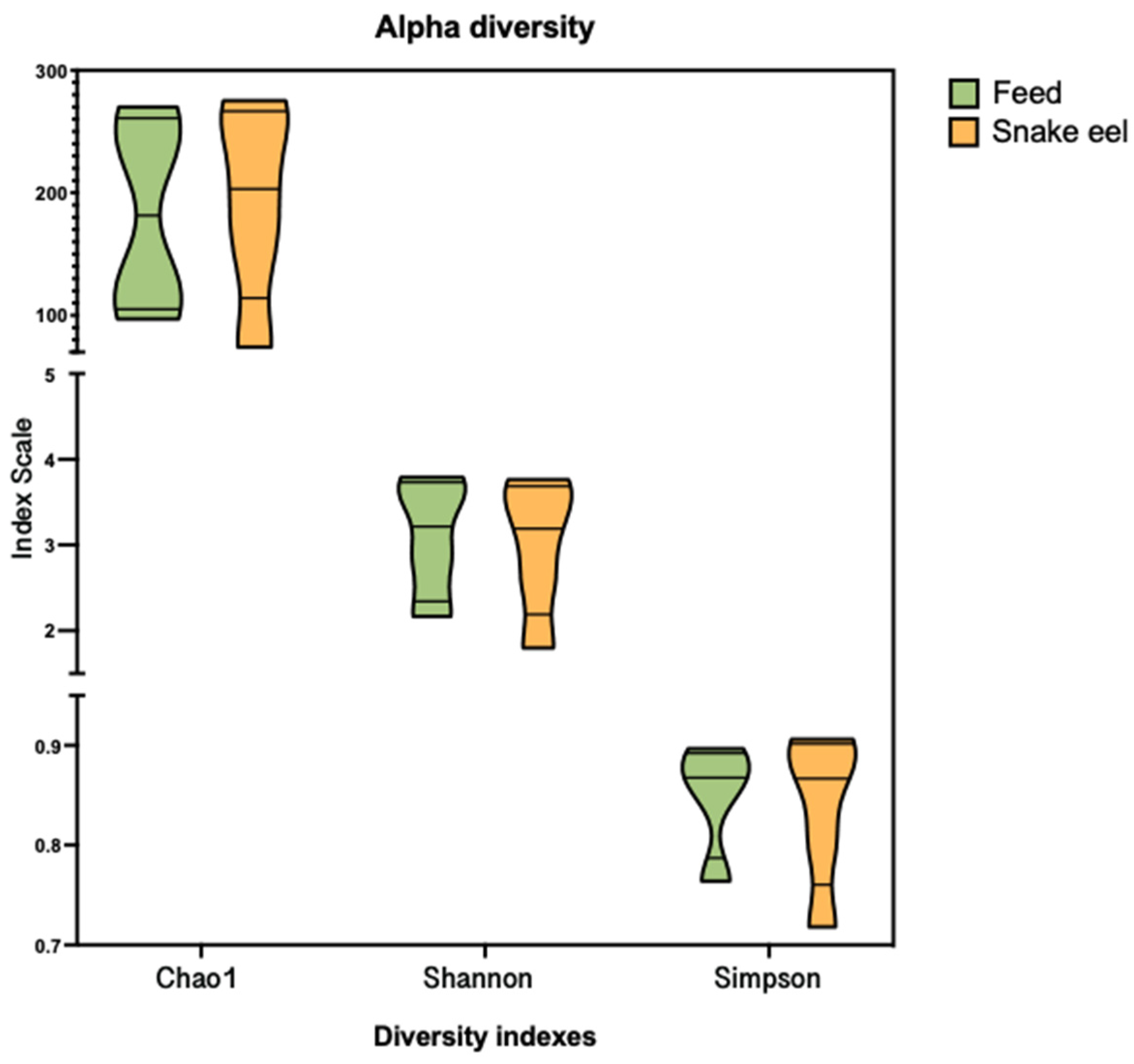
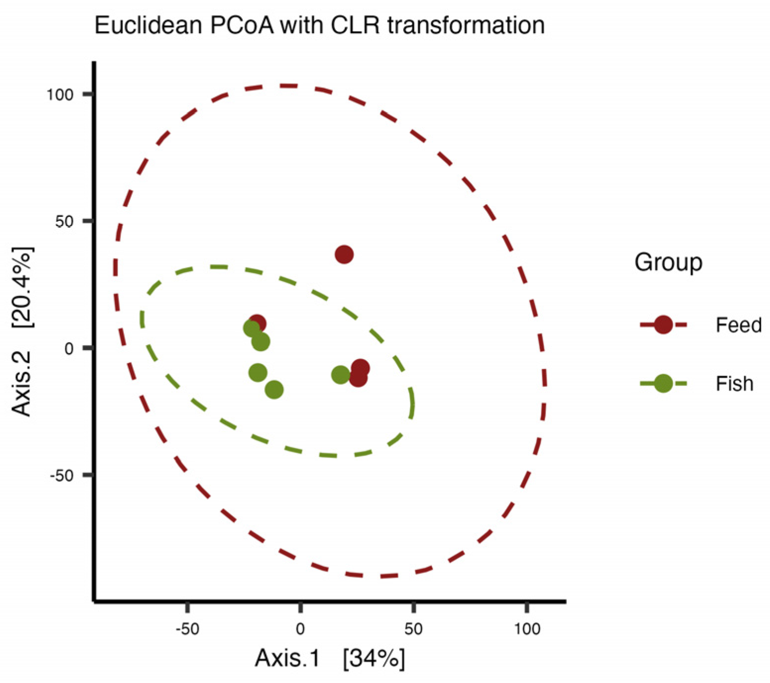
3.3. Alpha and Beta Diversity

We compared the alpha diversity indexes for feed and gut microbiota samples using metrics the richness estimator (Chao1) and the Shannon and Simpson diversity indexes (Figure 4). Statistical testing showed no differences for the Chao1, Shannon and Simpson indexes, p = 0.857; p = 0.838, p = 0.823, respectively; ANOVA).
Differences in microbial composition between samples or groups (feed versus gut microbiota) were visualized using PCoA, a powerful tool in beta diversity analysis (Figure 5). The result of PERMANOVA analysis, based on Aitchison distance, revealed no significant differences between the groups (R2 = 0.180, p value = 0.116).
In microbiota studies, Betadisper measures the homogeneity of beta diversity dispersion within different groups. It indicates the variability in sample distances to their group centroid in a dissimilarity space. In this case, groups (feed, gut microbiota) showed low dispersion, indicating more similar microbial communities within a group. The comparisons between feed and gut microbiota showed no significant differences (p value = 0.593).

4. Discussion

Approximately 18 eel species are recognized globally, but only a few have been extensively incorporated into aquaculture [15]. The most notable eel species in aquaculture are the Japanese eel (Anguilla japonica) (annual production near to 250,000 tons per year) [15], which is predominantly farmed in East Asia, particularly in Japan, China, Taiwan, and South Korea, and the European eel (Anguilla anguilla), mainly farmed in Europe [16]. These species are farmed primarily for their high economic value of around USD 15,000 per kilo [17], especially in the culinary industry. The farming techniques often involve capturing juvenile eels from the wild and growing them to market size in controlled environments. In this context, it is important to know the microbiota associated with gut and factors influencing composition to improve growth and health status.
Previous studies on other eel species have shown diverse results regarding microbiota composition. For instance, Kusumawaty [18] examined Anguilla bicolor bicolor McClelland, 1844, revealing that Proteobacteria (64.18%) and Firmicutes (33.55%) were the predominant phyla in the digestive tract of farmed eels, while Bacteroidetes (54.16%), Firmicutes (14.71%), and Fusobacteria (10.56%) were predominant in wild eels. The most prevalent genera in farmed and wild elvers were Plesiomonas (35.71%) and Cetobacterium (10.55%), respectively. Similarly, Liu [19] studied the zig-zag eel and found that Proteobacteria and Firmicutes dominated the gut microbiota of wild (accounting for 45.8% and 20.3%, respectively) and farmed (accounting for 21.4% and 75.6%, respectively) zig-zag eels. These results strongly contrast with our findings, where Firmicutes accounted for more than 80%, and Latilactobacillus was the dominant genus, representing 34.96% of the relative abundance. Interestingly, the microbiota of farmed zig-zag eels also showed a significant proportion of Lactococcus, indicating a notable presence of lactic acid bacteria in the eel microbiota.
Previous research has shown that the composition of the gut microbiota in freshwater fish varies considerably [14,20] based on their habitat and diet [21,22,23,24]. The structure of the gut microbiota is influenced by the complex interplay of numerous external factors, such as habitat differences, and internal factors, including age and diet [25]. Diet is very important when species are reared in a RAS [25]. In this context, it is interesting to mention that the study conducted by Hossain [26] examined the gut microbiome of European eels (Anguilla anguilla) from two commercial recirculating aquaculture system (RAS) farms using 16S rRNA gene sequencing. The dominant bacterial phyla in the eel gut were Proteobacteria, Bacteroidetes, Firmicutes, and Fusobacteria. The eel gut microbiome from Farm-1 had significantly higher microbial abundance and diversity compared to Farm-2, indicating that eels from different farms have distinct gut microbiota profiles. The gut bacterial community of Farm-2 eels was significantly influenced by the microbiota of the supplied feed and the surrounding tank water. Considering that, it was important to include the analysis of bacteria present in feed. The influence of diet on microbiota is profound, as it shapes the composition and diversity of microbial communities within the gut. Different diets provide various nutrients that selectively promote the growth of specific microbial populations. Additionally, in this study, the feed contributed to the bacteria observed in the intestinal content of the snake eel. For instance, carnivorous fish fed commercial or experimental diets tended to have a higher abundance of Firmicutes, a bacterial phylum associated with the digestion of proteins and fats. A high abundance of Firmicutes is frequently observed in aquaculture fish. Previous studies have identified Firmicutes as the predominant bacterial phylum in carnivorous fish fed commercial or experimental diets, such as salmonids [27]. Notably, Firmicutes was the dominant phylum in reared fish, comprising over 60% of the relative abundance [23], in other carnivorous Chilean species previously studied, including fine flounder and yellowtail [22,23,24]. This was also the case for snake eel, where the important presence of Firmicutes was noticeable (>80% relative abundance).
The snake eel is distributed in the eastern Pacific from Nicaragua to Chile [28], with Chirichigno and Vélez [29] specifying its range from Puerto Pizarro (Peru) to Valparaíso (Chile), including migrations to higher latitudes and depths during El Niño events. It primarily inhabits sandy-muddy bottoms, burying itself during the day and migrating between 50 and 800 m at night, forming size-segregated groups [29,30]. Typically found at depths between 39 and 385 m, it can withstand low oxygen levels below 1 mL/L and temperatures ranging from 10 °C to 16 °C. The snake eel is nocturnally active, with a diet consisting mainly of fish, crustaceans, and mollusks, including crabs, anchovies, polychaetes, cephalopods (octopus, squid), and eels [30,31]. Further research is needed on wild specimens to better understand the microbiota of this species in its natural environment. Based on previous experiences, it is expected that the phyla proportions will differ, as seen in the red cusk eel, where wild specimens were dominated by Tenericutes [23].

5. Conclusions

Our study reveals that the gut microbiota of the common snake eel (Ophichthus remiger) is predominantly composed of bacteria from the phylum Firmicutes, representing over 80% of the relative abundance, with Lactilactobacillus identified as the most important genus. While the results indicate that feed may influence microbiota composition by introducing relevant bacteria to the intestinal content, the absence of a control condition, such as microbiome data from wild fish, limits the statistical robustness of this conclusion. Future research should incorporate wild fish microbiota as a control and utilize larger sample sizes to better elucidate the relationship between diet and gut microbiota. Despite these limitations, this study provides valuable initial insights into the microbiota of Ophichthus remiger, which may inform future investigations into the roles of Firmicutes and Lactilactobacillus in marine fish digestion and nutrient assimilation. Additionally, it is essential to emphasize the practical significance of these findings, particularly for applications in aquaculture and fish health management.

Supplementary Materials

The following supporting information can be downloaded at https://www.mdpi.com/article/10.3390/microbiolres16020038/s1, Figure S1: Rarefraction curves; Tables S1–S4: Relative abundance at taxa level.

Author Contributions

Conceptualization, R.R. and J.R.; experimentation and methodology, C.R., E.S. and L.H.; writing—original draft preparation, C.R. and J.R.; writing—review and editing, J.R.; funding acquisition, J.R. All authors have read and agreed to the published version of the manuscript.

Funding

This research was funded by FONDECYT from ANID (Chile), grant number 1240430 (J.R.), and Anillos Regulares de Tecnología from ANID (Chile), grant number ACT240045.

Institutional Review Board Statement

Ethical code 20364-INT-UCH from the University of Chile.

Informed Consent Statement

Not applicable.

Data Availability Statement

Data will be available on request.

Acknowledgments

The authors acknowledge all staff of the Universidad Católica del Norte for providing material and assistance during fish sampling.

Conflicts of Interest

The authors declare no conflicts of interest.

References

  1. Ben-Hasan, A.; Walters, C.; Hordyk, A.; Christensen, V.; Al-Husaini, M. Alleviating Growth and Recruitment Overfishing Through Simple Management Changes: Insights from an Overexploited Long-Lived Fish. Mar. Coast. Fish. 2021, 13, 87–98. [Google Scholar] [CrossRef]
  2. Prince, J.; Hordyk, A. What to Do When You Have Almost Nothing: A Simple Quantitative Prescription for Managing Extremely Data-Poor Fisheries. Fish Fish. 2019, 20, 224–238. [Google Scholar] [CrossRef]
  3. Gozzer-Wuest, R.; Vinatea Chávez, R.A.; Olea Stranger, G.; Araya Goncalves, G.; Hiriart-Bertrand, L.; Labraña-Cornejo, R.; Alonso-Población, E. Identifying Priority Areas for Improvement in Chilean Fisheries. Front. Mar. Sci. 2023, 10, 1073397. [Google Scholar] [CrossRef]
  4. Subsecretaría de Pesca y Acuicultura. 2023. Available online: https://www.subpesca.cl/portal/618/articles-117812_recurso_1.pdf (accessed on 2 May 2024).
  5. Borges, L. The evolution of a discard policy in Europe. Fish Fish. 2015, 16, 534–540. [Google Scholar] [CrossRef]
  6. Wehrtmann, I.S.; Arana, P.M.; Barriga, E.; Gracia, A.; Pezzuto, P.R. Deep-water Shrimp Fisheries in Latin America: A Review. Lat. Am. J. Aquat. Res. 2012, 40, 497–535. [Google Scholar] [CrossRef]
  7. Romero, J.; Catalán, N.; Ramírez, C.; Miranda, C.D.; Oliva, M.; Flores, H.; Romero, M.S.; Rojas, R. High Abundance of Candidatus Arthromitus in Intestinal Microbiota of Seriolella violacea (Palm Ruff) under Reared Conditions. Fishes 2023, 8, 109. [Google Scholar] [CrossRef]
  8. McCord, A.I.; Chapman, C.A.; Weny, G.; Tumukunde, A.; Hyeroba, D.; Klotz, K.; Koblings, A.S.; Mbora, D.N.M.; Cregger, M.A.; White, B.A.; et al. Fecal microbiomes of non-human primates in Western Uganda reveal species-specific communities largely resistant to habitat perturbation. Am. Primatol. 2014, 76, 347–354. [Google Scholar] [CrossRef]
  9. Bletz, M.C.; Goedbloed, D.J.; Sanchez, E.; Reinhardt, T.; Tebbe, C.C.; Bhuju, S.; Geffers, R.; Jarek, M.; Vences, M.; Steinfartz, S. Amphibian gut microbiota shifts differentially in community structure but converges on habitat-specific predicted functions. Nat. Commun. 2016, 7, 13699. [Google Scholar] [CrossRef]
  10. Clark, R.I.; Walker, D.W. Role of gut microbiota in aging-related health decline: Insights from invertebrate models. Cell. Mol. Life Sci. 2018, 75, 93–101. [Google Scholar] [CrossRef]
  11. Grond, K.; Sandercock, B.K.; Jumpponen, A.; Zeglin, L.H. The avian gut microbiota: Community, physiology and function inwild birds. J. Avian Biol. 2018, 49, e01788. [Google Scholar] [CrossRef]
  12. Trevelline, B.K.; MacLeod, K.J.; Langkilde, T.; Kohl, K.D. Gestation alters the gut microbiota of an oviparous lizard. FEMS Microbiol. Ecol. 2019, 95, fiz086. [Google Scholar] [CrossRef]
  13. Liu, X.J.; Cao, Y.L.; Ouyang, S.; Wu, X.P. Comparative analysis of gut microbiota diversity in endangered, economical, and common freshwater mussels using 16S rRNA gene sequencing. Ecol. Evol. 2020, 21, 12015–12023. [Google Scholar] [CrossRef]
  14. Romero, J.; Ringø, E.; Merrifield, D.L. The gut microbiota of fish. In Aquaculture Nutrition: Gut Health, Probiotics and Prebiotics; Merrifield, D., Ringø, E., Eds.; Wiley-Blackwell Publishing: Oxford, UK, 2014; pp. 75–100. [Google Scholar]
  15. Hamidoghli, A.; Bae, J.; Won, S.; Lee, S.; Kim, D.J.; Bai, S.C. A Review on Japanese Eel (Anguilla japonica) Aquaculture, With Special Emphasis on Nutrition. Rev. Fish. Sci. Aquac. 2019, 27, 226–241. [Google Scholar] [CrossRef]
  16. Rasmussen, G.; Pedersen, M.I. Growth Rate and Biological Production of Yellow Eel Anguilla anguilla and Production of Silver Eel and the Number of Glass Eel to Fulfill the Danish EMP. Biol. Life Sci. 2023; preprint. [Google Scholar] [CrossRef]
  17. Available online: https://www.undercurrentnews.com/2024/04/12/glass-eel-prices-hit-all-time-high-at-15000-kg-in-japan-on-continuous-poor-harvest/ (accessed on 2 July 2024).
  18. Kusumawaty, D.; Augustine, S.M.N.; Aryani, A.; Effendi, Y.; Emran, T.B.; Tallei, T.E. Configuration of gut bacterial community profile and their potential functionality in the digestive tract of the wild and cultivated Indonesian shortfin elver-phase eels (Anguilla bicolor bicolor McClelland, 1844). 3 Biotech 2023, 13, 153. [Google Scholar] [CrossRef]
  19. Liu, X.; Fan, Y.; Mo, T.; Chen, Q.; Chen, W. Comparative Study of the Gut Microbiota Community between the Farmed and Wild Mastacembelus armatus (Zig-Zag Eel). Metabolites 2022, 12, 1193. [Google Scholar] [CrossRef]
  20. Roeselers, G.; Mittge, E.K.; Stephens, W.Z.; Parichy, D.M.; Cavanaugh, C.M.; Guillemin, K.; Rawls, J.F. Evidence for a core gut microbiota in the zebrafish. ISME J. 2011, 5, 1595–1608. [Google Scholar] [CrossRef]
  21. Villasante, A.; Ramírez, C.; Catalán, N.; Opazo, R.; Dantagnan, P.; Romero, J. Effect of dietary carbohydrate-to-protein ratio on gut microbiota in atlantic salmon (Salmo salar). Animals 2019, 9, 89. [Google Scholar] [CrossRef]
  22. Ramírez, C.; Romero, J. Fine flounder (Paralichthys adspersus) microbiome showed important differences between wild and reared specimens. Front. Microbiol. 2017, 8, 271. [Google Scholar] [CrossRef]
  23. Romero, J.; Díaz, O.; Miranda, C.D.; Rojas, R. Red Cusk-Eel (Genypterus chilensis) Gut Microbiota Description of Wild and Aquaculture Specimens. Microorganisms 2022, 10, 105. [Google Scholar] [CrossRef]
  24. Ramírez, C.; Romero, J. The microbiome of Seriola lalandi of wild and aquaculture origin reveals differences in composition and potential function. Front. Microbiol. 2017, 8, 1844. [Google Scholar] [CrossRef] [PubMed]
  25. Schmidt, V.; Amaral-Zettler, L.; Davidson, J.; Summerfelt, S.; Good, C. The influence of fishmeal-free diets on microbial communities in Atlantic salmon Salmo salar recirculation aquaculture systems. Appl. Environ. Microbiol. 2016, 82, 4470–4481. [Google Scholar] [CrossRef] [PubMed]
  26. Hossain, S. European eel (Anguilla anguilla) GI tract conserves a unique metagenomics profile in the recirculation aquaculture system (RAS). Aquac. Int. 2021, 29, 1529–1544. [Google Scholar] [CrossRef]
  27. Gajardo, K.; Jaramillo-Torres, A.; Kortner, T.; Merrifield, D.; Tinsley, J.; Bakke, A.; Krogdahl, Å. Alternative protein sources in the diet modulate microbiota and functionality in the distal intestine of Atlantic salmon (Salmo salar). Appl. Environ. Microbiol. 2017, 83, e02615-16. [Google Scholar] [CrossRef]
  28. Froese, R.; Pauly, D. (Eds.) FishBase. World Wide Web Electronic Publication. Version (10/2024). 2024. Available online: www.fishbase.org (accessed on 2 February 2024).
  29. Goicochea, C.; Mostacero, J.; Moquillaza, P. Edad y Crecimiento de Ophichthus remiger (Valenciennes) en el Norte del Mar Peruano, 2004. Inf. Inst. Mar Perú 2012, 39, 9–17. [Google Scholar]
  30. Chirichigno, N.; Vélez, J. Claves Para Identificar los Peces Marinos del Perú, 2nd ed.; Publicación Especial del Instituto del Mar del Perú: Callao, Peru, 1998. [Google Scholar]
  31. Galán Galán, J.; Carbajal, W.; Castañeda Condori, J. La Anguila Ophichthus remiger en Aguas de las Islas Lobos de Afuera, Lambayeque, Durante el 2005. Inf. Inst. Mar Perú 2007, 34, 281–294. [Google Scholar]
Figure 1. The composition of the microbiota at different taxonomic levels: phylum (A) and family (B).
Microbiolres 16 00038 g001
Figure 2. Compositions of snake eel microbiota at genus and phylum levels.
Microbiolres 16 00038 g002
Figure 3. The composition of snake eel microbiota at the genus level was compared to the bacteria found in the feed.
Microbiolres 16 00038 g003
Figure 4. The Chao, Shannon and Simpson alpha diversity indexes contrasting feed and gut microbiota.
Microbiolres 16 00038 g004
Figure 5. A PCoA of beta diversity of the gut and feed microbiota based on Aitchison distance.
Microbiolres 16 00038 g005
Disclaimer/Publisher’s Note: The statements, opinions and data contained in all publications are solely those of the individual author(s) and contributor(s) and not of MDPI and/or the editor(s). MDPI and/or the editor(s) disclaim responsibility for any injury to people or property resulting from any ideas, methods, instructions or products referred to in the content.

