Review

The Growing Antibiotic Resistance of Campylobacter Species: Is There Any Link with Climate Change?

by
Eleni V. Geladari
1,
Dimitris Kounatidis
2,
Evangelia Margellou
3,
Apostolos Evangelopoulos
4,
Edison Jahaj
5,
Andreas Adamou
6,
Vassilios Sevastianos
1,
Charalampia V. Geladari
7 and
Natalia G. Vallianou
6,*
1
Third Department of Internal Medicine, Evangelismos General Hospital, 10676 Athens, Greece
2
Diabetes Center, First Propaedeutic Department of Internal Medicine, Medical School, National and Kapodistrian University of Athens, Laiko General Hospital, 11527 Athens, Greece
3
Fifth Department of Internal Medicine, Evangelismos General Hospital, 10676 Athens, Greece
4
Medical School, National and Kapodistrian University of Athens, 11527 Athens, Greece
5
Dermatology Department, Evangelismos General Hospital, 10676 Athens, Greece
6
First Department of Internal Medicine, Sismanogleio General Hospital, 15126 Athens, Greece
7
Hellenic Society of Environmental and Climate Medicine, 17455 Athens, Greece
*
Author to whom correspondence should be addressed.
Microbiol. Res. 2025, 16(11), 226; https://doi.org/10.3390/microbiolres16110226
Submission received: 8 September 2025 / Revised: 17 October 2025 / Accepted: 19 October 2025 / Published: 22 October 2025

Abstract

Campylobacter spp. remain among the most common pathogens causing acute diarrhea worldwide. Campylobacter jejuni and Campylobacter coli are the main species that cause gastroenteritis. Campylobacteriosis is a food-borne disease, although this Gram-negative bacterium may be transmitted via water-borne outbreaks as well as direct contact with animals, emphasizing its zoonotic potential. Campylobacterisosis does not usually require hospitalization. Antimicrobials are warranted only for patients with severe disease, as well as patients who are at risk for severe disease, such as the elderly, pregnant women or immunocompromised patients. Nonetheless, the irrational use of antibiotics in human and veterinary medicine enhances antimicrobial resistance (AMR). Resistance of Campylobacter spp. to fluoroquinolones, macrolides and tetracyclines is a significant concern to the scientific community. Point mutations, horizontal gene transfer and efflux pumps are the main mechanisms for the development and transmission of AMR in Campylobacter spp. Emerging evidence suggests that climate change may indirectly contribute to the spread of AMR in Campylobacter, particularly through its influence on bacterial ecology, transmission pathways and antibiotic use patterns. Higher temperatures and extreme weather events accelerate bacterial growth, amplify the transfer of AMR genes and magnify disease transmission, including drug-resistant infections. Horizontal gene transfer, especially in the context of biofilm formation, may further perplex the situation. Excessive farming and overuse of antibiotics as growth promoters in animals may also contribute to increased AMR rates. Climate change and AMR are interconnected and pose a significant threat to global public health. Multidisciplinary strategies mitigating both phenomena are crucial in order to contain the spread of Campylobacter-related AMR. The aim of this review is to describe the molecular mechanisms that result in AMR of Campylobacter spp. and underscore the association between climate change and Campylobacteriosis. Novel methods to mitigate Campylobacter-related AMR will also be discussed.

1. Introduction

According to the World Health Organization (WHO), Campylobacter spp. are among the four main causes of diarrheal illness globally, while they are the most common cause of bacterial gastroenteritis, known as Campylobacteriosis [1]. Campylobacter spp. are ‘‘S”-shaped, Gram-negative rods belonging to the family Campylobacteraceae [2]. At the beginning of the twentieth century, it was first recognized as a cause of abortion in cattle and sheep [3]. Campylobacter spp. may reside in animal gastrointestinal (GI) tracts, especially poultry, because of poultry’s high core body temperature, which promotes survival and growth of Campylobacter spp. [4]. Many of the Campylobacter jejuni and Campylobacter coli strains may also be found in wild and domestic animals, birds and pigs as well. This microorganism may thrive in fresh or saline natural water and has the ability to grow in temperatures below 15 °C [5]. The transmission to humans is mainly achieved through animal contact, with poultry being the main reservoir [6]. When the bacterium was isolated in humans, a few decades later, it was originally called Vibrio [7]. The genus Campylobacter was identified in 1973, while the medical scientific community acknowledged the virulent effect of Campylobacter spp. in human GI tracts in the 1980s [8]. Humans get the disease by consuming contaminated animal-based foods, poultry meat not well cooked, unpasteurized milk and dairy products [9].
Seventeen species and six subspecies have been recognized as important for humans and animals, although the most common human pathogens are Campylobacter jejuni and Campylobacter coli [10]. Other species that have been isolated from patients with diarrheal disease are Campylobacter lari and Campylobacter upsaliensis, although the latter two are much less frequently encountered [11].
People who reside in different climatic zones, tropical, temperate and arctic, may suffer from Campylobacteriosis [12]. In Europe, the incidence of Campylobacter spp. infection has increased over the last decade, while in the United States there is increased prevalence of Campylobacter spp. on the West Coast [1]. The infection from Campylobacter spp. has a bimodal distribution, with the first peak being in early childhood and a marked secondary peak in young adults. There is a slight male predominance. The symptoms and disease severity are more typical and pronounced in the second peak group [13]. In countries with a temperate climate, a seasonal pattern of the disease with the highest incidence during the summer months has been reported [14].
In the UK, an epidemiological study to estimate the incidence of infection was recently conducted. It was reported that 630,000 people are being affected annually, and of those, 80,000 receive consultation from their GP [15]. Nonetheless, the mortality rate due to this microorganism is low [15]. In the United States, the mortality has been estimated to be approximately 50 to 150 patients with Campylobacteriosis, particularly those with extraintestinal disease annually [16].
The disease onset is approximately two to five days after bacterial contamination, although the incubation period may range from one to ten days [17]. Typical symptoms are diarrhea (usually bloody), fever, GI symptoms such as nausea and/or vomiting, abdominal pain and headache. The average duration of the disease is typically three to six days. Gastroenteritis due to Campylobacter spp. usually resolves spontaneously [18]. Patients at risk for severe disease are the elderly, pregnant women and immunocompromised individuals. Extraintestinal manifestations do require treatment with antibiotics such as fluoroquinolones, macrolides or aminoglycosides [19].
Complications include bacteremia, hepatitis, pancreatitis, meningitis, endocarditis, osteomyelitis, septic arthritis, neonatal sepsis and miscarriage [20]. Reactive arthritis, Guillain–Barre syndrome (GBS) and Miller Fisher syndrome are post-infectious complications; therefore, in cases of GBS, careful medical history should include the presence or absence of precedent diarrheal episodes [21,22,23].
Severe cases along with high mortality rates have been reported in children under the age of two years old in developing countries, highlighting the correlation of socio-economic status with the disease course [24]. In the era of climate change, where social inequalities are deepened due to unemployment and food insecurity, food-borne, water-borne and zoonotic infections, among them Campylobacteriosis, will increase dramatically [25]. Social discrepancies due to climate change may lead not only to enhancing cases of Campylobacteriosis, but to higher rates of complications and relapsing episodes as well [26]. On the other side, warmer temperatures and extreme weather events contribute to the spread of Campylobacter spp. [27]. The expanded use of antimicrobials in mammals and agriculture to maintain crop yields and livestock health in addition to secure food quality has resulted in the selection and dissemination of resistant strains [28]. Antimicrobial resistance (AMR) of Campylobacter spp. is a growing challenge that, together with the loss of biodiversity due to environmental stressors, may result in adverse outcomes [29]. Campylobacteriosis is increasingly recognized as a public health issue due to the rise in AMR. It is of utmost importance to invent strategies that will mitigate the phenomenon of AMR. Preventive measurements, such as biosecurity systems, carcass decontamination, water sanitation and limitations in the overuse and misuse of antibiotics could decrease disease burden [30].
The aim of this review is to acknowledge the difference between the mild and severe form of Campylobacteriosis and administer treatment only when this is indicated. Clinical management should prioritize pathogen-directed therapy over symptom-based interventions, especially in cases suggestive of systemic involvement. Moreover, we will describe the mechanisms of transmission of Campylobacter spp., as well as development of Campylobacter-related AMR. Additionally, we explore how climate change may influence the emergence and spread of AMR in Campylobacter spp., which is particularly challenging and demanding for clinicians and healthcare systems.

2. Treatment Indications

The disease is almost always symptomatic in Western countries, while in low-income and lower-middle-income countries, it is mostly asymptomatic [31]. However, although the disease may be asymptomatic in childhood, there have been cases of impaired growth in adolescents [1]. The incubation period of the disease ranges between one to seven days. Prodromal symptoms include fever, aches and delirium, and a few days later GI symptoms, such as abdominal cramping, periumbilical pain mimicking acute appendicitis and bloody diarrhea, may appear [2]. It has been reported that the presence of the plasmid pVir correlates with the development of bloody diarrhea [32]. Approximately 15% of adults may develop bloody diarrhea, while during childhood half of the patients may have bloody stool [33]. Acute infection may require supportive measures, with administration of antipyretics, intravenous fluids, correction of electrolyte disturbances and appropriate diet. It is noteworthy that antimotility agents should be avoided [30]. Antibiotics are not beneficial in the acute phase, as has been confirmed by randomized trials, which showed no significant reduction in the disease’s duration [34]. Although the diarrheal episodes resolve after a week, the abdominal cramping may persist. Patients have been reported to lose even more than 5 kg due to Campylobacteriosis [13]. While Campylobacteriosis is usually self-limiting, the increasing frequency of resistant strains and complications in vulnerable populations poses a significant therapeutic challenge. As summarized in Figure 1, antibiotic administration should be reserved for cases with invasive features such as bloody diarrhea, persistent high fever or extraintestinal manifestations [35].
It is also important to recognize the subgroups who are at risk for severe disease: the elderly, pregnant women and the immunocompromised [19]. It has been demonstrated that infections due to Campylobacter spp. may be more devastating in patients with an immunocompromised status, such as those with hypogammaglobulinemia, HIV or immune deficiencies in the context of hematological malignancies or organ transplantation [36]. Characteristically, the subpopulation of immunocompromised patients has longer disease duration and frequent relapses, as well as extraintestinal complications [36]. Cholecystitis [37], peritonitis, especially in patients undergoing peritoneal dialysis [38], rash [39], osteomyelitis [40], meningitis [41], septic pseudoaneurysm [42], pericarditis and myocarditis [43] are all known complications of Campylobacteriosis. Focal infections like septic arthritis, bursitis, osteitis, soft tissue infections and nodular skin eruptions do rarely occur [20]. In such cases, not only antibiotics, but also intravenous immunoglobulin therapy, may be administered to enhance the antibody response [44]. The role of Fecal Microbiota Transplantation (FMT) has been discussed, but more randomized trials are needed to establish this concept as a treatment option [45].
Post-infectious (PI) complications include GBS, an acute demyelinating disease of the peripheral nervous system, reactive arthritis and irritable bowel syndrome (IBS) [46]. A major complication of GBS is respiratory muscle paralysis and, in severe cases, death. The treatment is mainly supportive. Antibiotics are not given in cases of GBS triggered by Campylobacter spp. [47]. In such cases, plasma exchange or intravenous immunoglobulin (IVIG) is required. Patients who are at risk of developing severe disease should be treated with antibiotics, as they may be more susceptible to severe Campylobacter spp. infection and may benefit from antibiotic treatment [48]. If reactive arthritis (RA) develops when the Campylobacter infection is ongoing, antibiotics may be given to lessen the bacterium burden and further diminish the duration of the arthritis [49]. In cases when the infectious organism has been cleared, the medical treatment focuses on the symptoms of arthritis. First-line agents for the treatment of pain and inflammation include nonsteroidal anti-inflammatory drugs (NSAIDs), followed by corticosteroids or even disease-modifying antirheumatic drugs (DMARDs) [50]. Antibiotics are not indicated for long-lasting arthritis (lasting more than six months), even if genetic elements of Campylobacter spp. are found. Unlike GBS, early antibiotic treatment in severe cases of Campylobacteriosis does not prevent the manifestation of reactive arthritis [51]. PI-IBS is also a late complication of Campylobacteriosis, and antibiotics are not considered a first-line treatment. However, if symptoms are persistent, antibiotics should be administered with caution. The rationale of antibiotic treatment is based on the concept of reducing bacterial overgrowth [52].
Macrolides and fluoroquinolones are the main categories of antibiotics that are recommended for the treatment of Campylobacteriosis. Azithromycin treatment is recommended instead of fluoroquinolone treatment due to the increased resistance rates to quinolones worldwide [53]. The usual treatment period is three days: 500 mg orally for azithromycin, or 750 mg orally either for levofloxacin or ciprofloxacin [53]. Patients who are severely ill or have underlying comorbidities may need treatment for a longer duration, i.e., for 7 to 14 days. For those who cannot tolerate oral treatment, carbapenems and/or aminoglycosides may be administered [54]. It has been postulated that Campylobacter spp. respond to fosfomycin treatment; nonetheless, susceptibility results have not yet been evaluated thoroughly. In vitro, the bacterium has sufficient sensitivity to clindamycin, chloramphenicol and tetracyclines as well, but without clinical efficacy. The Gram-negative bacterium is intrinsically resistant to β-lactam antibiotics and trimethoprim [55].

3. AMR: Changing the World Map

Public health is threatened due to dramatic increases in AMR. As AMR has been rising constantly, in 2015 the World Health Organization (WHO) announced the Global Action Plan on Antimicrobial Resistance and indicated a Bacterial Priority Pathogen List [56]. Notably, AMR directly contributed to 1.27 million deaths globally in 2019 and was associated with an additional 4.95 million deaths [57].
Campylobacter spp. is a major pathogen and has become resistant to clinically important antimicrobials. It can be transmitted through multiple pathways; hence, it is considered a zoonotic, food-borne and water-borne disease that affects animals, mainly poultry and humans [58]. A meta-analysis conducted in China exhibited that the prevalence of Campylobacter jejuni in humans, foods, animals and the environment was 5.2% (95% CI: 4.2–6.4%), 12.5% (95% CI: 9.7–15.6%), 15.4% (95% CI: 13.2–17.6%) and 17.8% (95% CI: 9.7–27.7%), respectively. The global prevalence of multidrug-resistant (MDR) Campylobacter jejuni was 72.8% (95% CI: 62.4–82.2%), pointing towards a public health issue regarding MDR [59].
The injudicious use of antibiotics has led to AMR, even though Campylobacter spp. GI infection is a relatively benign and self-limited disease. Broad antibiotic use in animal farming is also a concern for the increasing incidence of AMR [28]. Climate change and physical disasters help in the spread of AMR species [60]. Additionally, the overuse and misuse of antibiotics in human and veterinary medicine, as well as inadequate hygiene practices in healthcare facilities and farm-to-fork systems, enable the transmission of resistant strains [61].
On a worldwide basis, Campylobacter spp. are resistant to macrolides at a rate lower than 5%. Even though macrolide resistance in specific countries, like Thailand and Ireland, has overtaken these rates, macrolides still remain the treatment of choice [29]. The highest rates of Campylobacter-related AMR have been reported with fluoroquinolones. The AMR in Southeast Asia surpasses 80%, while in European countries, like France, Hungary and Spain, as well as in some Middle East countries like Iran, the AMR to fluoroquinolones approaches 50% [62]. These very high AMR rates may be associated with increased use of antibiotics in humans and animals. Additionally, climate change, with increasing ambient temperatures and flooding, may also be implicated in the aforementioned enhanced AMR rates. Moreover, lack of policies and legislation may also contribute to higher AMR rates. A descriptive epidemiological study that was conducted in the United States between 2005 and 2018 concluded that the incidence of Campylobacter spp. infection has decreased or remained stable, whereas the AMR has dramatically increased [63]. Among factors contributing to the increase of AMR is recent international travel [64]. An enhancement on AMR to fluoroquinolones has been reported in the United States, as use of fluoroquinolones in animals has been reported since 1995. Hence, while the rate of ciprofloxacin resistance was 0% in 1989, between 2005 and 2014 it has been reported to range from 20% to 27%. Despite the fact that the use of fluoroquinolones in poultry was banned in the United States in 2005, the problem with AMR exists and is growing [65].

4. Mechanisms of Antibiotic Resistance in Campylobacter Species

Prestinaci et al. were the first to describe AMR, and defined it as a condition where the antibacterial agent is not capable of inhibiting bacterial growth, resulting in treatment failure [66]. When antibiotics are required for the treatment of Campylobacteriosis, first- and second-line agents are macrolides and fluoroquinolones, respectively [67]. As already mentioned, the irrational use of antibiotics helps bacteria gain resistance to treatment. Campylobacter spp. have generated several mechanisms of resistance; a group of Campylobacter spp. exhibits resistance to a specific class of antibacterials, while other groups acquire multidrug resistance [68]. The AMR is expanding through inappropriate hygiene practices in healthcare and food production systems. Third-line agents include tetracycline, gentamicin or phenicols, such as chloramphenicol [69].
Nonetheless, in 2013, the WHO announced an era where new antibiotics should be generated for crucial pathogens, including fluoroquinolone-resistant Campylobacter [70].
The AMR mechanisms include point mutations; gene acquisition; horizontal gene transfer through conjugation; transformation and transduction; and vertical gene transfer, where natural selection of dominant mutations occurs [71]. This section outlines the resistance mechanisms of Campylobacter spp., with particular emphasis on macrolides and fluoroquinolones, which represent the most clinically relevant drug classes in both human and veterinary settings.

4.1. Campylobacter spp. and Resistance to Macrolides

Macrolides are a class of drugs mainly used for the treatment of GI microbial diseases, Helicobacter pylori and Campylobacter spp. Infection, as well as for atypical pneumonia [68]. Antibiotics belonging in this category are also utilized in animal farming to enhance growth and treat veterinary diseases [72]. The main mechanism of action is halting bacterial protein synthesis [72]. Macrolides bind to the 50S bacterial ribosomal subunit, specifically the peptidyl transferase center formed by the 23S rRNA and block mRNA translation [73]. Macrolides may also interfere with the peptidyl transferase enzyme, which is required for the addition of amino acids to the growing protein chain. Some macrolides prevented the assembly of the 50S ribosomal subunit itself [74]. They are characterized as bacteriostatic, although at higher concentrations they may act as bactericidal. Bacteria gain resistance through various mechanisms: (i) by facilitating enzymatic inactivation of the antimicrobial agent; (ii) by altering the structure of the ribosomal subunit and/or diminishing the binding affinity of the antimicrobial agent; and (iii) by increasing efflux of the antibiotic out of the cell [75]. Enzyme-mediated methylation or point mutation in the 23S rRNA and/or L4 and L22 ribosomal proteins are the main mechanisms of ribosomal subunit mitigation [75].
Erm(B), an rRNA methylating enzyme, was recognized in both Campylobacter jejuni and Campylobacter coli from several sources: chicken, ducks, swine and humans. It has been demonstrated that the Erm(B) gene is settled within the bacterial DNA chromosome or carried by a plasmid. Erm(B) gene is linked to multidrug resistance genomic islands (MDRGIs), loci that include multiple resistance genes: [aacA-aphD, sat4, aphA-3, fosXCC, aad9 and tet(O)] [76]. The above characteristics make Campylobacter spp. highly resistant not only to macrolides but also to several other classes of antibiotics. MDRGIs have been conveyed among distinct strains of Campylobacter spp. by natural transformation under laboratory conditions [76].
The most common molecular methods that confer macrolide resistance to Campylobacter spp. are point mutations in the domain V of the 23S rRNA [77]. The exact positions in the 23S rRNA where point mutations occur are 2074 and 2075. High-level resistance to macrolides (erythromycin MIC > 128 µg/mL) is associated with the following point mutations: A2074C, A2074G and A2075G [78]. The former two-point mutations are related to decreased macrolide susceptibility, while the latter (A2075G) is the principal resistance-associated mutation. Campylobacter spp. contain three copies of 23S rRNA genes. It has been postulated that highly resistant Campylobacter spp. isolates possess the aforementioned resistance-associated mutations in all three copies [79].
Besides the ribosomal destabilization through methylation and point mutations, active efflux is another mechanism conferring macrolide resistance [80]. The CmeABC efflux system may collaborate with target mutations to confer macrolide resistance. In vitro studies have shown that inactivation of CmeABC significantly reduces the resistance to macrolide antibiotics in isolates with high-, intermediate- or low-level macrolide resistance [80]. A combination that may also lead to macrolide resistance is the synergy between the CmeABC efflux pump and mutations in the ribosomal proteins L4 (G74D) and L22 (inserted at position 86 or 98) [81].

4.2. Campylobacter spp. and Resistance to Fluoroquinolones

Quinolones, a category of antibiotics that emerged in the 1960s, are used to treat a variety of infections, from community- to hospital-acquired. They are active against both Gram-positive and Gram-negative bacteria [82]. Fluoroquinolones are among the first-line agents for the treatment of Campylobacteriosis in human and veterinary medicine. Their main mechanism of action is the inhibition of two crucial enzymes for DNA replication: DNA gyrase and topoisomerase IV [83]. Bacteria gain resistance to fluoroquinolones through mutations in the genes encoding the subunits of DNA gyrase (GyrA and GyrB) and/or topoisomerase IV (ParC and ParE). Fluoroquinolone resistance in Campylobacter spp. has been mediated by point mutations in the quinolone resistance-determining region of GyrA. Notably, mutations in GyrB, ParC and ParE genes have not been described among fluoroquinolone-resistant Campylobacter strains [84]. The susceptibility of Campylobacter spp. to fluoroquinolones is severely compromised, even if a single point mutation in the quinolone resistance-determining region of gyrA exists. The mutations that have been recognized are listed below: T86I, T86K, A70T and D90N. The most frequently detected mutation conferring fluoroquinolone resistance in Campylobacter spp. is the C257T change in the gyrA gene, which leads to the T86I substitution in DNA gyrase [85].
However, a prerequisite for resistant Campylobacter spp. to exert AMR is to also possess the functional multidrug efflux pump, CmeABC [86]. It has been reported that if fluoroquinolone-resistant mutants (carrying specific GyrA mutations) lose the cmeABC, then the resistant mutants are rendered susceptible to fluoroquinolones. Unlike macrolides, plasmid-mediated quinolone resistance determinants have not been described in Campylobacter spp. [87].

4.3. Campylobacter spp. and Resistance to Tetracyclines

Tetracyclines, discovered two decades before quinolones, have broad-spectrum activity against Gram-positive and Gram-negative bacteria, as well as Chlamydiae, Mycoplasmas, Rickettsiae and protozoan parasites. They block protein synthesis through inhibition of the attachment of aminoacyl-tRNA to the ribosomal acceptor (A) site [88]. The widespread use of tetracyclines has led to the emergence of resistant strains. There are four main mechanisms involved in resistance to tetracyclines: efflux pumps, chemical modification of tetracyclines, ribosomal protection proteins (RPPs) and mutations in rRNA [89].
The principal molecular pathways that aid in Campylobacter’s resistance to tetracyclines are the ribosomal protection protein Tet(O) and efflux pumps (CmeABC and CmeG) [90]. Tet(O) gene is responsible for the production of a ribosomal protection protein, which actively detaches tetracycline from the ribosome through GTP hydrolysis. Tet(O) is linked to an open A site on the bacterial ribosome and provokes a conformational change that releases the previously bound tetracycline molecule [91]. It has been elucidated that the tet(O) gene was acquired from a Gram-positive bacterium by horizontal gene transfer. Campylobacter jejuni and Campylobacter coli contain the tet(O) gene either in the chromosomal DNA or on a plasmid [92].
Multidrug efflux pumps, CmeABC and CmeG, contribute to tetracycline resistance in Campylobacter spp. [93]. In particular, the efflux pump CmeABC seems to act in synergy with the tet(O) protein, providing high-level resistance to tetracycline. As with fluoroquinolones, inactivation of either CmeABC or CmeG may result in enhancement of the susceptibility of Campylobacter to tetracyclines [93].

4.4. Campylobacter and Resistance to Aminoglycosides

Protein synthesis blockage is a critical step in halting bacterial growth, and aminoglycosides inhibit bacterial protein synthesis via binding to the 30S ribosomal subunit [94]. Aminoglycosides are characterized as bactericidal antibiotics and possess an aminocyclitol ring that is attached to one or more amino sugars by pseudoglycosidic bonds. They bind to the decoding site of the A site of the ribosomal subunit, thus leading to alterations in protein expression and interference with the tRNA translocation between the A-site and P-site [95]. Bacteria gain resistance against macrolides through several molecular mechanisms: (i) inhibition of the drug’s entrance into the bacterial cell either through decreased permeability or due to increased efflux of the drug [96], (ii) methylation of 16S rRNA in drug binding sites [97,98], (iii) augmentation of mutant copies in the binding sites of rRNA [97], (iv) active swarming, a characteristic adaptation mechanism of Pseudomonas aeruginosa spp. [99] and (v) enzymatic modification at the -OH or -NH2 groups of the 2-deoxystreptamine nucleus or sugar moieties of the aminoglycoside [100]. A major mechanism of resistance to aminoglycosides in Campylobacter spp. is the modification of the aminoglycoside structure by enzymes, such as aminoglycoside acetyltransferases, aminoglycoside phosphotransferases and aminoglycoside nucleotidyltransferases [101,102].
Aminoglycoside phosphotransferases are identified in Campylobacter spp. and control the phosphorylation of the 3′ hydroxyl group of the aminoglycosides. These enzymes are responsible for kanamycin and neomycin resistance [103]. Based on the resistance of specific aminoglycosides, phosphotransferases are divided into eight groups (I to VIII). It has been demonstrated that kanamycin resistance is mediated through types I, III, IV and VII [104].
Other genes that have been implicated in Campylobacter resistance are in clusters. Such genes are the aadE-sat4-aphA-3 cluster and the aacA-aphD-aac cluster that were described on the chromosome of C. coli [105]. The sat4 gene encodes a streptothricin acetyltransferase and is present either as a single gene or as part of the aadE-sat4-aphA-3 cluster in streptothricin-resistant Campylobacter spp. [106,107]. The aminoglycoside 3-adenyltransferase gene (aadA) confers resistance to streptomycin and spectinomycin, while the aminoglycoside 6-adenyltransferase gene (aadE) confers resistance to streptomycin [106,107]. The aphA-3 gene is a characteristic of Gram-positive bacteria, and its identification in Campylobacter spp. indicates the transfer of antibiotic resistance genes from Gram-positive bacteria to Gram-negative bacteria. On the contrary, the aphA-7 gene is indigenous to Campylobacter and mediates kanamycin resistance [108].
In the United States, various variants of gentamicin resistance genes, aph(2″)-Ib, Ic, If1, If3, Ih and aac(6′)-Ie/aph(2″)-If2, were isolated from Campylobacter spp. from humans and from meats found in butcher’s shops [109].

4.5. Campylobacter spp. and Resistance to Phenicols

Phenicols are divided in two main categories: nonfluorinated, such as chloramphenicol, and fluorinated, such as florfenicol [110]. Their activity aims at a plethora of Gram-positive and Gram-negative bacteria. Resistance of Campylobacter spp. to phenicols is attained by enzymatic inactivation via acetyltransferases, target site mutations in 23S rRNA, target site modification in 23S rRNA via the rRNA methyltransferase Cfr(C) or extrusion by efflux pumps [111].
It has been documented that the production of chloramphenicol acetyltransferases (CATs) leads to acetylation and drug deactivation, providing resistance to chloramphenicol, but not to florfenicol [112]. In addition, the G2073A mutation in the 23S rRNA gene of Campylobacter spp. is associated with resistance in both categories of phenicols. The cfr gene encodes an rRNA methyltransferase that methylates the adenine at position 2503 in the 23S rRNA, leading to phenicol resistance among other antimicrobials [113]. A novel study that included multidrug-resistant Campylobacter coli isolates of cattle origin demonstrated a plasmid-borne cfr-like gene, nominated as cfr(C) gene. It was concluded that cfr(C) transfers resistance to phenicols. As with other antibiotic classes, the presence of multidrug efflux pump variant RE-CmeABC makes Campylobacter resistant to phenicols [114].

4.6. Campylobacter spp. and Resistance to β-Lactams

The most widely known antimicrobial category comprises the β-lactams, which exhibit a β-lactam ring as part of their molecular structure. The main mechanism by which they exert their antimicrobial activity is the inhibition of bacterial cell wall synthesis [115]. The extensive use of antibiotics during the past decades has led to alarmingly increasing resistance to β-lactams. The mechanisms by which the bacteria exert their resistance are the following: enzymatic inactivation, reduced uptake and efflux pump. Campylobacter spp. that possess β-lactamases had higher-level resistance compared to those without β-lactamases [116]. The β-lactamase that mostly characterizes Campylobacter jejuni is OXA-61 (Cj0299), and the resistant phenotype depends on its expression level. The cation porins of Campylobacter jejuni and Campylobacter coli may exert intrinsic resistance to β-lactams by excluding those with a molecular weight greater than 360 or those that are anionic. The role of efflux pumps, CmeABC and CmeDEF, is also critical for the enhanced resistance to β-lactams [117].

4.7. Campylobacter spp. and Resistance to Fosfomycin

Fosfomycin inhibits bacterial cell wall synthesis through inactivation of MurA, an enzyme essential for the catalysis of bacterial peptidoglycan synthesis. Fosfomycin seems to be an additional option for the treatment of Campylobacteriosis, when other categories of antimicrobials have failed [118]. Clinical trials demonstrated that fosfomycin may shorten symptoms’ duration and reduce bacterial shedding in stool samples [119]. Resistance rates of Campylobacter spp. to fosfomycin have remained low. The principal mechanism of fosfomycin resistance identified in Campylobacter is the fosXCC gene, which encodes a protein that shares 63.9% identity to the fosfomycin resistance determinant FosX, documented in Listeria monocytogenes [120]. FosX inactivates fosfomycin by epoxide hydrolysis, a condition where the addition of water to the epoxide ring of the antibiotic effectively opens the ring and renders it inactive. The fosXCC gene is contained in the MDRGI in Campylobacter coli and is transferrable to Campylobacter jejuni by natural transformation [121].
The following table (Table 1) summarizes the molecular mechanisms through which Campylobacter spp. gain antimicrobial resistance.

5. The Link Between Climate Crisis and Development of Campylobacter AMR

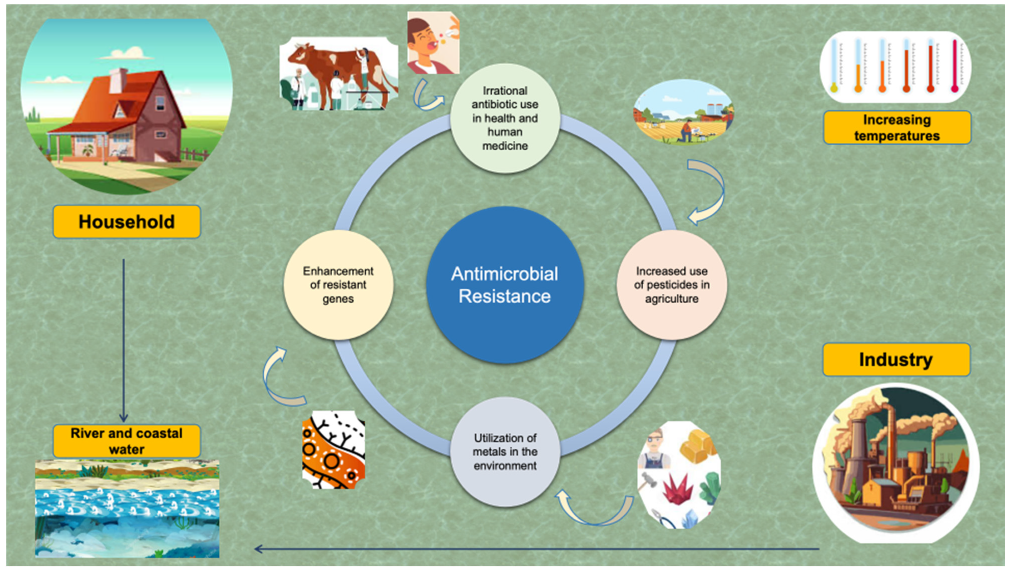
Nowadays, climate change and AMR are two major and interrelated challenges. Although they are considered as separate problems, ongoing studies provide further evidence that climate change and growing AMR are interconnected [122]. Regarding the incidence of Campylobacter spp. infections, they are expected to increase substantially and parallel to widespread antibiotic consumption (Figure 2) [123].
As global warming is already a fact, warmer winter months and more heat waves are expected. Campylobacter spp. acceleration is enhanced by escalations in temperature, thus enhancing the likelihood of food contamination and human exposure [124]. Notably, Damtew et al., in their study among 27 countries across five continents, have reported that for every 1 °C increase in ambient temperature, there was a 5% higher risk for Campylobacteriosis and Salmonellosis as well [125]. Despite the fact that the rates of infections due to Campylobacter spp. vary between different geographical zones, in the tropical and subtropical regions, sustained temperatures above 30 °C result in augmentation in the rates of Campylobacteriosis. Nevertheless, climate change is not limited to the planet’s warming. Precipitation events, such as heavy rainfalls and flooding, are expected to increase as well. These phenomena may result in the spread of Campylobacter spp. and the contamination of water sources with animal waste and sewage. Recreational water and drinking water systems may directly be affected, having a crucial impact on sanitation systems [126]. If food safety measures are not thoroughly maintained, this could compromise our privilege to fresh and clean water and food supplies. In their systematic review, Austhof et al. have documented that increased ambient temperature and precipitation events were positively related to the prevalence of Campylobacter spp. In sharp contrast, there was an inverse association between sunshine and relatively low humidity and the prevalence of Campylobacteriosis [12].
According to mathematical and meteorological models, it is assumed that Campylobacter spp. infections are expected to increase due to climate change in countries in Northern Europe. Specifically, a study by Kuhn et al. projected that Campylobacter spp. cases in Northern Europe could increase by 200% by 2100 due to climate change. This trajectory is translated into approximately 6000 more cases of Campylobacter spp. infections in the Scandinavian area attributed only to climate change. This trajectory is the result of the dual event of alarmingly increasing temperatures together with the extension of the transmission periods in summer and early autumn [124,127]. Surveillance studies point out that Campylobacter incidences are associated with increased temperatures and precipitation events in the week before Campylobacteriosis occurs. The above findings underlie the non-food transmission route of Campylobacteriosis as well [127].
However, since Campylobacteriosis is primarily a food-borne illness, it seems likely that the prevalence of the disease is expected to increase first in poultry flocks before humans. Indeed, it has been demonstrated that the prevalence of poultry Campylobacteriosis in Europe, South Korea, Wales and Canada fluctuates in response to changes in ambient temperature and humidity [128].
The epidemiology is multifactorial. Increased temperatures during late spring, summer and early autumn months support the survival of bacteria in the environment and thus animal contamination [129]. In addition, heat stress by interfering with the gut microbiota favors the acquisition, dominance and shedding of enteric pathogens, such as Campylobacter spp. [130]. Notably, heat stress has been associated with increased prevalence of pests, in particular flies, which could also be implicated in excess cases of Campylobacteriosis in humans [25,131]. During the 21st century, human activities, such as swimming in freshwater and outdoor food preparation, may account for the increase in the prevalence of the disease. While some researchers have reported a clear association between poultry consumption and human Campylobacteriosis, reductions in disease rates in the United States have been associated with the remarkable reduction in Campylobacter contamination of chicken carcasses. Nevertheless, it is noteworthy that poultry meat consumption has risen by 96% during the first two decades of the twentieth century, while it is expected to continue increasing by approximately 18% in the third decade [132,133]. This remarkable increase in poultry meat consumption, together with climate change, may account for the expected higher rates of Campylobacteriosis in the near future.
It is noteworthy that some species of Campylobacter, especially Campylobacter jejuni and Campylobacter coli, have the propensity to form biofilms. In biofilms, communities of microorganisms thrive and exhibit sustainability by adhering to an extracellular matrix. This extracellular matrix may impede antibiotic penetration, thus rendering the bacteria less susceptible to antibiotics and disinfectants. Additionally, within the biofilm, the high density and proximity between the bacteria may account for the acquisition of antibiotic resistance genes by horizontal gene transfer. Bundurus et al. have explored the molecular and genetic characteristics of Campylobacter jejuni’s ability to form biofilms. They have associated climate change with the endurance of Campylobacter spp. to extremes of pH, temperature and even desiccation. In addition, apart from alterations in genes involved in the motility of Campylobacter spp., they have proposed that changes in animal gut microbiota with probiotics may reduce the ability of the bacteria to form biofilms [134]. Overall, horizontal gene transfer mainly by conjugation, through the formation of biofilms, may be responsible for multidrug resistance in Campylobacter jejuni by efflux pumps.
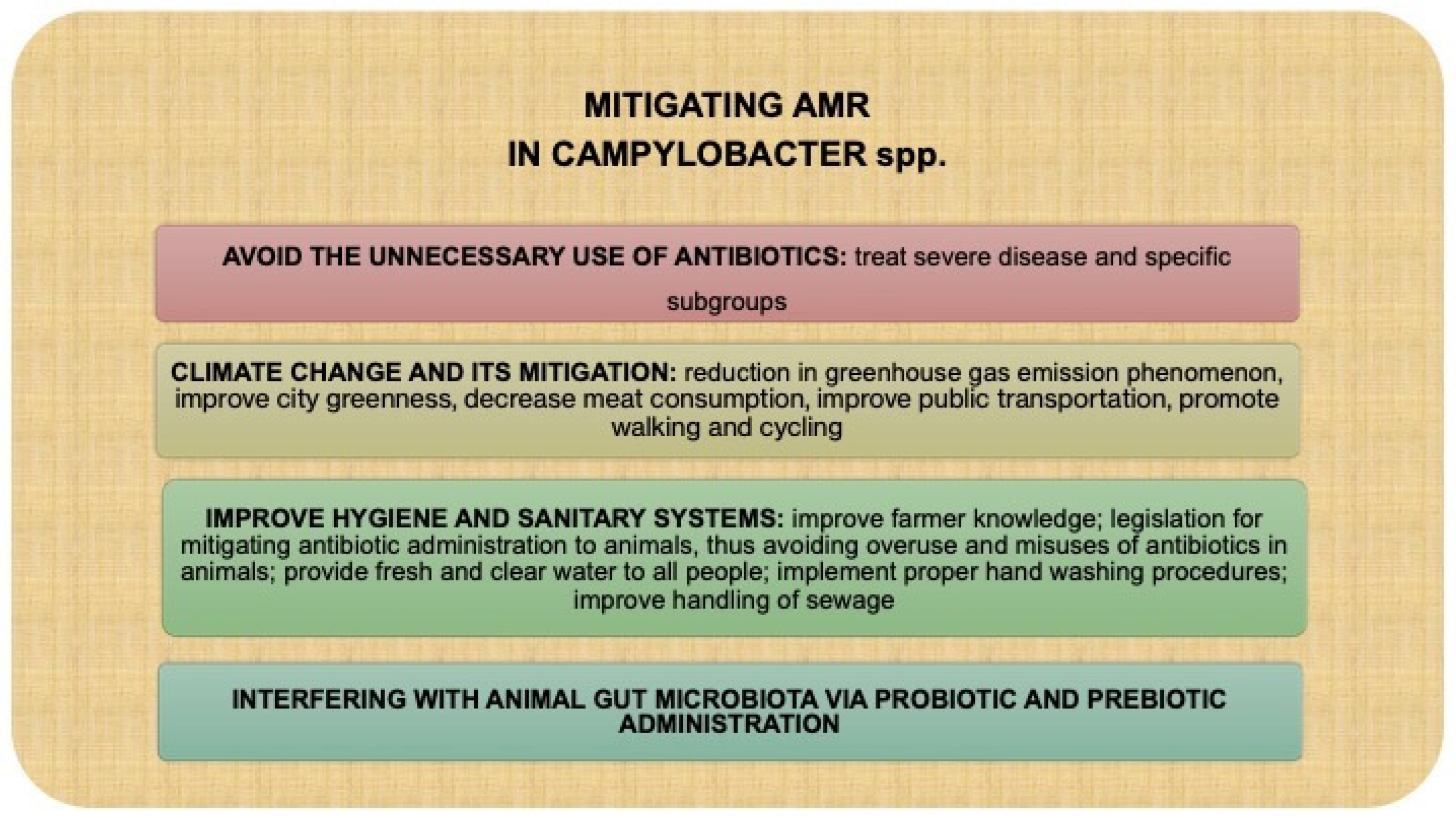
The healthcare system and public health resources will suffer serious problems from the increasing incidence of Campylobacter spp. infections. Due to antimicrobial use and the development of resistant strains, morbidity and mortality rates are forecasted to expand during the next decades [30]. Overall, climate change has been associated with increasing rates of Campylobacteriosis and Campylobacter-related AMR [27]. Thus, it is crucial to implement timely public health management and adaptation strategies to mitigate the impact of climate change on Campylobacter spp. transmission (Figure 3) [135].
Measures for controlling Campylobacter spp. are essential. Since consumption of poultry is linked to Campylobacteriosis at rates as high as 50% to 80%, pre- and post-harvest intervention strategies are mandatory to lessen the burden of disease in poultry [136]. Pre-harvest measures include the application of biosecurity protocols, such as limiting farm access, instituting single-species poultry cages and maintaining sanitary living conditions [137]. Moreover, the development of vaccines that will inhibit bacterial colonization in chicken seems highly promising as well. Prebiotics, probiotics and bacteriophages assist in maintaining a healthy poultry gut microbiome [138]. Regarding post-harvest intervention strategies, carcass decontamination is significant. Physical, chemical and biological methods are being developed to lower the burden of bacteria in poultry and reach consumer shelves in a safe manner [139].

6. Conclusions

Climate change, food insecurity, social discrepancies and the widespread use of antibiotics all contribute to the development of antibiotic-resistant Campylobacteriosis. This review synthesizes emerging data at the intersection between climate dynamics and AMR in Campylobacter spp., underscoring the need for integrated surveillance systems and climate-responsive intervention strategies within a One Health framework [140]. Molecular studies that will address emerging genes responsible for Campylobacter spp.-resistant strains are needed [141]. Policies regarding the limited use of antimicrobials in humans and mammals are of utmost importance. Applying pre- and post-harvest measurements in poultry farms is crucial to limit this AMR burden [142]. Multidisciplinary scientific teams are needed to educate the public about how infectious diseases and climate change are interconnected. The scientific community should realize the current threat and propose solutions to ensure an appropriate quality of life for all people worldwide [143].

Author Contributions

E.V.G. conceived the idea and has written major parts of this manuscript. D.K., E.M., A.E. and E.J. were responsible for the literature search and data curation; A.A. and C.V.G. wrote minor parts and were responsible for the table and the figures; V.S. and N.G.V. wrote minor parts of the manuscript, conducted the final editing and supervised the whole work. All authors have read and agreed to the published version of the manuscript.

Funding

The authors declare that no funding was received for the preparation of this manuscript.

Institutional Review Board Statement

Not applicable.

Data Availability Statement

No new data were created or analyzed in this study.

Conflicts of Interest

The authors declare no conflict of interest.

Abbreviations

AMR: antimicrobial resistance; DMARDs: disease-modifying antirheumatic drugs; FMT: Fecal Microbiota Transplantation; GBS: Guillain–Barre syndrome; GI: gastrointestinal; IBD: Inflammatory Bowel Disease; IVIG: intravenous immunoglobulin; NSAIDs: nonsteroidal anti-inflammatory drugs; WHO: World Health Organization.

References

  1. Kaakoush, N.O.; Castaño-Rodríguez, N.; Mitchell, H.M.; Man, S.M. Global Epidemiology of Campylobacter Infection. Clin. Microbiol. Rev. 2015, 28, 687–720. [Google Scholar] [CrossRef]
  2. Man, S.M. The clinical importance of emerging Campylobacter species. Nat. Rev. Gastroenterol. Hepatol. 2011, 8, 669–685. [Google Scholar] [CrossRef]
  3. Silva, J.; Leite, D.; Fernandes, M.; Mena, C.; Gibbs, P.A.; Teixeira, P. Campylobacter spp. as a Foodborne Pathogen: A Review. Front. Microbiol. 2011, 2, 200. [Google Scholar] [CrossRef]
  4. Hakeem, M.J.; Lu, X. Survival and Control of Campylobacter in Poultry Production Environment. Front. Cell Infect. Microbiol. 2021, 10, 615049. [Google Scholar] [CrossRef]
  5. Ahmed, N.A.; Gulhan, T. Campylobacter in Wild Birds: Is It an Animal and Public Health Concern? Front. Microbiol. 2022, 12, 812591. [Google Scholar] [CrossRef]
  6. Al Hakeem, W.G.; Fathima, S.; Shanmugasundaram, R.; Selvaraj, R.K. Campylobacter jejuni in Poultry: Pathogenesis and Control Strategies. Microorganisms 2022, 10, 2134. [Google Scholar] [CrossRef] [PubMed]
  7. Vinzent, R.; Dumas, J.; Picard, N. Septicemie grave au cours de la grossesse, due a un vibrian. Avortement consecutif. Bull. Acad. Nat. Med. 1947, 131, 90–92. [Google Scholar]
  8. Costa, D.; Iraola, G. Pathogenomics of Emerging Campylobacter Species. Clin. Microbiol. Rev. 2019, 32, e00072-18. [Google Scholar] [CrossRef]
  9. Khairullah, A.R.; Yanestria, S.M.; Effendi, M.H.; Moses, I.; Kusala, M.; Fauzia, K.; Ayuti, S.; Fauziah, I.; Silaen, O.; Riwu, K.; et al. Campylobacteriosis: A rising threat in foodborne illnesses. Open Vet. J. 2024, 14, 1733–1750. [Google Scholar] [CrossRef] [PubMed]
  10. Sierra-Arguello, Y.M.; Perdoncini, G.; Rodrigues, L.B.; dos Santos, L.R.; Borges, K.A.; Furian, T.Q.; Salle, C.T.P.; Moraes, H.L.d.S.; Gomes, M.J.P.; Nascimento, V.P.D. Identification of pathogenic genes in Campylobacter jejuni isolated from broiler carcasses and broiler slaughterhouses. Sci. Rep. 2021, 11, 4588. [Google Scholar] [CrossRef]
  11. Veronese, P.; Dodi, I. Campylobacter jejuni/coli Infection: Is It Still a Concern? Microorganisms 2024, 12, 2669. [Google Scholar] [CrossRef] [PubMed]
  12. Austhof, E.; Warner, S.; Helfrich, K.; Pogreba-Brown, K.; Brown, H.E.; Klimentidis, Y.C.; Walter, E.S.; Jervis, R.H.; White, A. Exploring the association of weather variability on Campylobacter—A systematic review. Environ. Res. 2024, 252, 118796. [Google Scholar] [CrossRef] [PubMed]
  13. Igwaran, A.; Okoh, A.I. Human campylobacteriosis: A public health concern of global importance. Heliyon 2019, 5, e02814. [Google Scholar] [CrossRef]
  14. Tam, C.C.; Rodrigues, L.C.; O’Brien, S.J.; Hajat, S. Temperature dependence of reported Campylobacter infection in England, 1989-1999. Epidemiol Infect. 2006, 134, 119–125. [Google Scholar] [CrossRef]
  15. Tam, C.C.; Rodrigues, L.C.; Viviani, L.; Dodds, J.P.; Evans, M.R.; Hunter, P.R.; Gray, J.J.; Letley, L.H.; Rait, G.; Tompkins, D.S.; et al. Longitudinal study of infectious intestinal disease in the UK (IID2 study): Incidence in the community and presenting to general practice. Gut 2012, 61, 69–77. [Google Scholar] [CrossRef]
  16. Goddard, M.R.; O’Brien, S.; Williams, N.; Guitian, J.; Grant, A.; Cody, A.; Colles, F.; Buffet, J.-C.; Adlen, E.; Stephens, A.; et al. A restatement of the natural science evidence based regarding the source, spread and control of Campylobacter species causing human disease. Proc. Biol. Sci. 2022, 289, 20220400. [Google Scholar] [CrossRef]
  17. Awofisayo-Okuyelu, A.; Hall, I.; Adak, G.; Hawker, J.I.; Abbott, S.; McCARTHY, N. A systematic review and meta-analysis on the incubation period of Campylobacteriosis. Epidemiol. Infect. 2017, 145, 2241–2253. [Google Scholar] [CrossRef]
  18. Guo, Y.T.; Hsiung, C.A.; Wu, F.T.; Chi, H.; Huang, Y.-C.; Liu, C.-C.; Huang, Y.-C.; Lin, H.-C.; Shih, S.-M.; Huang, C.-Y.; et al. Clinical manifestations and risk factors of Campylobacter gastroenteritis in children in Taiwan. Biomed. J. 2023, 46, 100590. [Google Scholar] [CrossRef]
  19. White, A.E.; Ciampa, N.; Chen, Y.; Kirk, M.; Nesbitt, A.; Bruce, B.B.; Walter, E.S. Characteristics of Campylobacter and Salmonella Infections and Acute Gastroenteritis in Older Adults in Australia, Canada, and the United States. Clin. Infect. Dis. 2019, 69, 1545–1552. [Google Scholar] [CrossRef] [PubMed]
  20. Roberts, S.C.; Stone, S.M.; Sutton, S.H.; Flaherty, J.P. Cutaneous manifestations of Campylobacter jejuni infection: A case report and review of the literature. Infect. Dis. Clin. Pract. 2020, 28, 61–63. [Google Scholar] [CrossRef]
  21. Hannu, T.; Kauppi, M.; Tuomala, M.; Laaksonen, I.; Klemets, P.; Kuusi, M. Reactive arthritis following an outbreak of Campylobacter jejuni infection. J. Rheumatol. 2004, 31, 528–530. [Google Scholar] [PubMed]
  22. Nachamkin, I.; Allos, B.M.; Ho, T. Campylobacter species and Guillain-Barré syndrome. Clin. Microbiol. Rev. 1998, 11, 555–567. [Google Scholar] [CrossRef]
  23. Jacobs, B.C.; Hazenberg, M.P.; van Doorn, P.A.; Endtz, H.P.; van der Meché, F.G. Cross-reactive antibodies against gangliosides and Campylobacter jejuni lipopolysaccharides in patients with Guillain-Barré or Miller Fisher syndrome. J. Infect. Dis. 1997, 175, 729–733. [Google Scholar] [CrossRef]
  24. Rosenberg, A.; Weinberger, M.; Paz, S.; Valinsky, L.; Agmon, V.; Peretz, C. Ambient temperature and age-related notified Campylobacter infection in Israel: A 12-year time series study. Environ. Res. 2018, 164, 539–545. [Google Scholar] [CrossRef]
  25. Queenan, K.; Häsler, B. Climate change and campylobacteriosis from chicken meat: The changing risk factors and their importance. Food Control. 2025, 173, 111193. [Google Scholar] [CrossRef]
  26. Rushton, S.P.; Sanderson, R.A.; Diggle, P.J.; Shirley, M.D.F.; Blain, A.P.; Lake, I.; Maas, J.A.; Reid, W.D.K.; Hardstaff, J.; Williams, N.; et al. Climate, human behaviour or environment: Individual-based modelling of Campylobacter seasonality and strategies to reduce disease burden. J. Transl. Med. 2019, 17, 34. [Google Scholar] [CrossRef]
  27. Dietrich, J.; Hammerl, J.A.; Johne, A.; Kappenstein, O.; Loeffler, C.; Nöckler, K.; Rosner, B.; Spielmeyer, A.; Szabo, I.; Richter, M.H. Impact of climate change on foodborne infections and intoxications. J. Health Monit. 2023, 8 (Suppl. S3), 78–92. [Google Scholar] [CrossRef]
  28. Manyi-Loh, C.; Mamphweli, S.; Meyer, E.; Okoh, A. Antibiotic Use in Agriculture and Its Consequential Resistance in Environmental Sources: Potential Public Health Implications. Molecules 2018, 23, 795. [Google Scholar] [CrossRef] [PubMed]
  29. Portes, A.B.; Panzenhagen, P.; Pereira Dos Santos, A.M.; Junior, C.A.C. Antibiotic Resistance in Campylobacter: A Systematic Review of South American Isolates. Antibiotics 2023, 12, 548. [Google Scholar] [CrossRef] [PubMed]
  30. Bukari, Z.; Emmanuel, T.; Woodward, J.; Ferguson, R.; Ezughara, M.; Darga, N.; Lopes, B.S. The Global Challenge of Campylobacter: Antimicrobial Resistance and Emerging Intervention Strategies. Trop. Med. Infect. Dis. 2025, 10, 25. [Google Scholar] [CrossRef]
  31. Ahmed, A.O.; Ameh, J.A.; Abubakar, M.B.; Raufu, I.A. Campylobacteriosis in Africa: A neglected demon. Microbe 2025, 7, 100409. [Google Scholar] [CrossRef]
  32. Tracz, D.M.; Keelan, M.; Ahmed-Bentley, J.; Gibreel, A.; Kowalewska-Grochowska, K.; Taylor, D.E. pVir and bloody diarrhea in Campylobacter jejuni enteritis. Emerg. Infect. Dis. 2005, 11, 838–843. [Google Scholar] [CrossRef]
  33. Grzybowska-Chlebowczyk, U.; Kalita, B.; Flak-Wancerz, A.; Jasielska, M.; Więcek, S.; Wojcieszyn, M.; Horowska-Ziaja, S.; Chlebowczyk, W.; Woś, H. Clinical course of Campylobacter infections in children. Pediatria Polska. 2013, 88, 329–334. [Google Scholar] [CrossRef]
  34. Ternhag, A.; Asikainen, T.; Giesecke, J.; Ekdahl, K. A meta-analysis on the effects of antibiotic treatment on duration of symptoms caused by infection with Campylobacter species. Clin. Infect. Dis. 2007, 44, 696–700. [Google Scholar] [CrossRef] [PubMed]
  35. Ruiz-Palacios, G.M. The health burden of Campylobacter infection and the impact of antimicrobial resistance: Playing chicken. Clin. Infect. Dis. 2007, 44, 701–703. [Google Scholar] [CrossRef]
  36. Najjar, I.; Paluca, F.; Loukidis, K.; Tarr, P.E. Recurrent Campylobacter Enteritis in Patients with Hypogammaglobulinemia: Review of the Literature. J. Clin. Med. 2020, 9, 553. [Google Scholar] [CrossRef]
  37. Gerritsen van der Hoop, A.; Veringa, E.M. Cholecystitis caused by Campylobacter jejuni. Clin. Infect. Dis. 1993, 17, 133. [Google Scholar] [CrossRef]
  38. Lang, C.L.; Chiang, C.K.; Hung, K.Y.; Wu, K.D. Campylobacter jejuni peritonitis and bacteremia in a patient undergoing continuous ambulatory peritoneal dialysis. Clin. Nephrol. 2009, 71, 96–98. [Google Scholar] [CrossRef] [PubMed]
  39. Monselise, A.; Blickstein, D.; Ostfeld, I.; Segal, R.; Weinberger, M. A case of cellulitis complicating Campylobacter jejuni subspecies bacteremia and review of the literature. Eur. J. Clin. Microbiol. Infect. Dis. 2004, 23, 718–721. [Google Scholar] [CrossRef] [PubMed]
  40. Pisipati, S.; Zafar, A.; Zafar, Y. Campylobacter coli bacteraemia: How common is it? BMJ Case Rep. 2020, 13, e236634. [Google Scholar] [CrossRef]
  41. Kusulja, M.; Santini, M.; Margetić, K.; Guzvinec, M.; Šoprek, S.; Butić, I.; Andrašević, A.T. Meningitis caused by Campylobacter jejuni: A case presentation and literature review. Acta Clin. 2021, 76, 318–323. [Google Scholar] [CrossRef] [PubMed]
  42. Roan, J.N.; Ko, W.C.; Luo, C.Y. Abdominal septic aortic pseudoaneurysm caused by Campylobacter jejuni infection: Report of a case. Surg. Today 2009, 39, 137–140. [Google Scholar] [CrossRef]
  43. Hannu, T.; Mattila, L.; Rautelin, H.; Siitonen, A.; Leirisalo-Repo, M. Three cases of cardiac complications associated with Campylobacter jejuni infection and review of the literature. Eur. J. Clin. Microbiol. Infect. Dis. 2005, 24, 619–622. [Google Scholar] [CrossRef]
  44. Roa-Bautista, A.; Brown, L.K.; Tadros, S.; Burns, S.O.; Godbole, G.; Lowe, D.M. Clinical Features, Immunological Characteristics, and Treatment Outcomes of Campylobacter spp. Infections in Patients with Common Variable Immunodeficiency. J. Allergy Clin. Immunol. Pract. 2023, 11, 3493–3501.e4. [Google Scholar] [CrossRef]
  45. Pang, J.; Beyi, A.F.; Looft, T.; Zhang, Q.; Sahin, O. Fecal Microbiota Transplantation Reduces Campylobacter jejuni Colonization in Young Broiler Chickens Challenged by Oral Gavage but Not by Seeder Birds. Antibiotics 2023, 12, 1503. [Google Scholar] [CrossRef]
  46. de la Torre, R.G.; Morís, G.; Martínez, D.P.; Montes, I.C. Guillain-Barré syndrome, tuberculosis and inflammatory bowel disease: A multiple association. Int. Arch. Med. 2010, 3, 15. [Google Scholar] [CrossRef]
  47. Shang, P.; Feng, J.; Wu, W.; Zhang, H.L. Intensive Care and Treatment of Severe Guillain-Barré Syndrome. Front. Pharmacol. 2021, 12, 608130. [Google Scholar] [CrossRef] [PubMed]
  48. Zaki, H.A.; Iftikhar, H.; Najam, M.; Masood, M.; Al-Marri, N.D.R.; Elgassim, M.A.M.; Fayed, M.; Shaban, E.E. Plasma exchange (PE) versus intravenous immunoglobulin (IVIG) for the treatment of Guillain-Barré syndrome (GBS) in patients with severe symptoms: A systematic review and meta-analysis. eNeurologicalSci 2023, 31, 100468. [Google Scholar] [CrossRef]
  49. Pope, J.E.; Krizova, A.; Garg, A.X.; Thiessen-Philbrook, H.; Ouimet, J.M. Campylobacter reactive arthritis: A systematic review. Semin. Arthritis Rheum. 2007, 37, 48–55. [Google Scholar] [CrossRef]
  50. Janssen, R.; Krogfelt, K.A.; Cawthraw, S.A.; van Pelt, W.; Wagenaar, J.A.; Owen, R.J. Host-pathogen interactions in Campylobacter infections: The host perspective. Clin. Microbiol. Rev. 2008, 21, 505–518. [Google Scholar] [CrossRef] [PubMed]
  51. Shafiee, D.; Salpynov, Z.; Gusmanov, A.; Khuanbai, Y.; Mukhatayev, Z.; Kunz, J. Enteric Infection-Associated Reactive Arthritis: A Systematic Review and Meta-Analysis. J. Clin. Med. 2024, 13, 3433. [Google Scholar] [CrossRef]
  52. Berumen, A.; Lennon, R.; Breen-Lyles, M.; Griffith, J.; Patel, R.; Boxrud, D.; Decuir, M.; Farrugia, G.; Smith, K.; Grover, M. Characteristics and Risk Factors of Post-Infection Irritable Bowel Syndrome After Campylobacter Enteritis. Clin. Gastroenterol. Hepatol. 2021, 19, 1855–1863.e1. [Google Scholar] [CrossRef] [PubMed]
  53. Wieczorek, K.; Osek, J. Antimicrobial resistance mechanisms among Campylobacter. Biomed. Res. Int. 2013, 2013, 340605. [Google Scholar] [CrossRef] [PubMed]
  54. Wei, B.; Kang, M. In vitro activity of fosfomycin against Campylobacter isolates from poultry and wild birds. PLoS ONE 2018, 13, e0200853. [Google Scholar] [CrossRef] [PubMed]
  55. Salam, M.A.; Al-Amin, M.Y.; Salam, M.T.; Pawar, J.S.; Akhter, N.; Rabaan, A.A.; Alqumber, M.A.A. Antimicrobial Resistance: A Growing Serious Threat for Global Public Health. Healthcare 2023, 11, 1946. [Google Scholar] [CrossRef]
  56. Coque, T.M.; Cantón, R.; Pérez-Cobas, A.E.; Fernández-de-Bobadilla, M.D.; Baquero, F. Antimicrobial Resistance in the Global Health Network: Known Unknowns and Challenges for Efficient Responses in the 21st Century. Microorganisms 2023, 11, 1050. [Google Scholar] [CrossRef]
  57. Ferraz, M.P. Antimicrobial Resistance: The Impact from and on Society According to One Health Approach. Societies 2024, 14, 187. [Google Scholar] [CrossRef]
  58. Facciolà, A.; Riso, R.; Avventuroso, E.; Visalli, G.; Delia, S.A.; Laganà, P. Campylobacter: From microbiology to prevention. J. Prev. Med. Hyg. 2017, 58, E79–E92. [Google Scholar]
  59. Li, Z.; Cai, H.; Xu, B.; Dong, Q.; Jia, K.; Lin, Z.; Wang, X.; Liu, Y.; Qin, X. Prevalence, antibiotic resistance, resistance and virulence determinants of Campylobacter jejuni in China: A systematic review and meta-analysis. One Health. 2025, 20, 100990. [Google Scholar] [CrossRef]
  60. Furlan, J.P.R.; Sellera, F.P.; Lincopan, N.; Debone, D.; Miraglia, S.G.E.K.; Tavella, R.A. Catastrophic floods and antimicrobial resistance: Interconnected threats with wide-ranging impacts. One Health 2024, 19, 100891. [Google Scholar] [CrossRef]
  61. Fernández Salgueiro, M.; Cernuda Martínez, J.A.; Gan, R.K.; Arcos González, P. Climate change and antibiotic resistance: A scoping review. Environ. Microbiol. Rep. 2024, 16, e70008. [Google Scholar] [CrossRef]
  62. Dalhoff, A. Global fluoroquinolone resistance epidemiology and implications for clinical use. Interdiscip. Perspect. Infect. Dis. 2012, 2012, 976273. [Google Scholar] [CrossRef] [PubMed]
  63. Ford, L.; Healy, J.M.; Cui, Z.; Ahart, L.; Medalla, F.; Ray, L.C.; Reynolds, J.; E Laughlin, M.; Vugia, D.J.; Hanna, S.; et al. Epidemiology and Antimicrobial Resistance of Campylobacter Infections in the United States, 2005–2018. Open Forum Infect. Dis. 2023, 10, ofad378. [Google Scholar] [CrossRef] [PubMed]
  64. Sodagari, H.R.; Agrawal, I.; Sohail, M.N.; Yudhanto, S.; Varga, C. Monitoring antimicrobial resistance in Campylobacter isolates of chickens and turkeys at the slaughter establishment level across the United States, 2013–2021. Epidemiol. Infect. 2024, 152, e41. [Google Scholar] [CrossRef]
  65. Kenyon, C. Positive Association between the Use of Quinolones in Food Animals and the Prevalence of Fluoroquinolone Resistance in E. coli and K. pneumoniae, A. baumannii and P. aeruginosa: A Global Ecological Analysis. Antibiotics 2021, 10, 1193. [Google Scholar] [CrossRef]
  66. Prestinaci, F.; Pezzotti, P.; Pantosti, A. Antimicrobial resistance: A global multifaceted phenomenon. Pathog. Glob. Health 2015, 109, 309–318. [Google Scholar] [CrossRef] [PubMed]
  67. Dai, L.; Sahin, O.; Grover, M.; Zhang, Q. New and alternative strategies for the prevention, control, and treatment of antibiotic-resistant Campylobacter. Transl. Res. 2020, 223, 76–88. [Google Scholar] [CrossRef]
  68. Shen, Z.; Wang, Y.; Zhang, Q.; Shen, J. Antimicrobial Resistance in Campylobacter spp. Microbiol. Spectr. 2018, 6, 10–1128. [Google Scholar] [CrossRef]
  69. Kiskó, G.; Bajramović, B.; Elzhraa, F.; Erdei-Tombor, P.; Dobó, V.; Mohácsi-Farkas, C.; Taczman-Brückner, A.; Belák, Á. The Invisible Threat of Antibiotic Resistance in Food. Antibiotics 2025, 14, 250. [Google Scholar] [CrossRef] [PubMed]
  70. Murugaiyan, J.; Kumar, P.A.; Rao, G.S.; Iskandar, K.; Hawser, S.; Hays, J.P.; Mohsen, Y.; Adukkadukkam, S.; Awuah, W.A.; Jose, R.A.M.; et al. Progress in Alternative Strategies to Combat Antimicrobial Resistance: Focus on Antibiotics. Antibiotics 2022, 11, 200. [Google Scholar] [CrossRef]
  71. Burmeister, A.R. Horizontal Gene Transfer. Evol. Med. Public. Health 2015, 2015, 193–194. [Google Scholar] [CrossRef] [PubMed]
  72. Paul, D.; Chawla, M.; Ahrodia, T.; Narendrakumar, L.; Das, B. Antibiotic Potentiation as a Promising Strategy to Combat Macrolide Resistance in Bacterial Pathogens. Antibiotics 2023, 12, 1715. [Google Scholar] [CrossRef] [PubMed]
  73. Krawczyk, S.J.; Leśniczak-Staszak, M.; Gowin, E.; Szaflarski, W. Mechanistic Insights into Clinically Relevant Ribosome-Targeting Antibiotics. Biomolecules 2024, 14, 1263. [Google Scholar] [CrossRef]
  74. Vázquez-Laslop, N.; Mankin, A.S. How Macrolide Antibiotics Work. Trends Biochem. Sci. 2018, 43, 668–684. [Google Scholar] [CrossRef]
  75. Zhang, F.; Cheng, W. The Mechanism of Bacterial Resistance and Potential Bacteriostatic Strategies. Antibiotics 2022, 11, 1215. [Google Scholar] [CrossRef]
  76. Jehanne, Q.; Bénéjat, L.; Ducournau, A.; Aptel, J.; Pivard, M.; Gillet, L.; Jauvain, M.; Lehours, P. Increasing rates of erm(B) and erm(N) in human Campylobacter coli and Campylobacter jejuni erythromycin-resistant isolates between 2018 and 2023 in France. Antimicrob Agents Chemother. 2025, 69, e0166824. [Google Scholar] [CrossRef]
  77. Wei, B.; Kang, M. Molecular Basis of Macrolide Resistance in Campylobacter Strains Isolated from Poultry in South Korea. Biomed. Res. Int. 2018, 2018, 4526576. [Google Scholar] [CrossRef] [PubMed]
  78. Bolinger, H.; Kathariou, S. The Current State of Macrolide Resistance in Campylobacter spp.: Trends and Impacts of Resistance Mechanisms. Appl. Environ. Microbiol. 2017, 83, e00416-17. [Google Scholar] [CrossRef]
  79. Gao, F.; Tu, L.; Chen, M.; Chen, H.; Zhang, X.; Zhuang, Y.; Luo, J.; Chen, M. Erythromycin resistance of clinical Campylobacter jejuni and Campylobacter coli in Shanghai, China. Front. Microbiol. 2023, 14, 1145581. [Google Scholar] [CrossRef]
  80. Gibreel, A.; Wetsch, N.M.; Taylor, D.E. Contribution of the CmeABC efflux pump to macrolide and tetracycline resistance in Campylobacter jejuni. Antimicrob. Agents Chemother. 2007, 51, 3212–3216. [Google Scholar] [CrossRef] [PubMed]
  81. Cagliero, C.; Mouline, C.; Payot, S.; Cloeckaert, A. Involvement of the CmeABC efflux pump in the macrolide resistance of Campylobacter coli. J. Antimicrob. Chemother. 2005, 56, 948–950. [Google Scholar] [CrossRef]
  82. Pham, T.D.M.; Ziora, Z.M.; Blaskovich, M.A.T. Quinolone antibiotics. Medchemcomm 2019, 10, 1719–1739. [Google Scholar] [CrossRef]
  83. Hooper, D.C.; Jacoby, G.A. Topoisomerase Inhibitors: Fluoroquinolone Mechanisms of Action and Resistance. Cold Spring Harb. Perspect. Med. 2016, 6, a025320. [Google Scholar] [CrossRef] [PubMed]
  84. Shariati, A.; Arshadi, M.; Khosrojerdi, M.A.; Abedinzadeh, M.; Ganjalishahi, M.; Maleki, A.; Heidary, M.; Khoshnood, S. The resistance mechanisms of bacteria against ciprofloxacin and new approaches for enhancing the efficacy of this antibiotic. Front. Public Health 2022, 10, 1025633. [Google Scholar] [CrossRef]
  85. Luangtongkum, T.; Jeon, B.; Han, J.; Plummer, P.; Logue, C.M.; Zhang, Q. Antibiotic resistance in Campylobacter: Emergence, transmission and persistence. Future Microbiol. 2009, 4, 189–200. [Google Scholar] [CrossRef] [PubMed]
  86. Yao, H.; Shen, Z.; Wang, Y.; Deng, F.; Liu, D.; Naren, G.; Dai, L.; Su, C.-C.; Wang, B.; Wang, S.; et al. Emergence of a Potent Multidrug Efflux Pump Variant That Enhances Campylobacter Resistance to Multiple Antibiotics. mBio 2016, 7, e01543-16. [Google Scholar] [CrossRef]
  87. Luo, N.; Sahin, O.; Lin, J.; Michel, L.O.; Zhang, Q. In vivo selection of Campylobacter isolates with high levels of fluoroquinolone resistance associated with gyrA mutations and the function of the CmeABC efflux pump. Antimicrob. Agents Chemother. 2003, 47, 390–394. [Google Scholar] [CrossRef] [PubMed]
  88. Chopra, I.; Roberts, M. Tetracycline antibiotics: Mode of action, applications, molecular biology, and epidemiology of bacterial resistance. Microbiol. Mol. Biol. Rev. 2001, 65, 232–260. [Google Scholar] [CrossRef]
  89. Grossman, T.H. Tetracycline Antibiotics and Resistance. Cold Spring Harb. Perspect. Med. 2016, 6, a025387. [Google Scholar] [CrossRef]
  90. Li, W.; Atkinson, G.C.; Thakor, N.S.; Allas, Ü.; Lu, C.-C.; Chan, K.-Y.; Tenson, T.; Schulten, K.; Wilson, K.S.; Hauryliuk, V.; et al. Mechanism of tetracycline resistance by ribosomal protection protein Tet(O). Nat. Commun. 2013, 4, 1477. [Google Scholar] [CrossRef]
  91. Connell, S.R.; Tracz, D.M.; Nierhaus, K.H.; Taylor, D.E. Ribosomal protection proteins and their mechanism of tetracycline resistance. Antimicrob. Agents Chemother. 2003, 47, 3675–3681. [Google Scholar] [CrossRef]
  92. Lynch, C.T.; Lynch, H.; Burke, S.; Hawkins, K.; Buttimer, C.; Mc Carthy, C.; Egan, J.; Whyte, P.; Bolton, D.; Coffey, A.; et al. Antimicrobial Resistance Determinants Circulating among Thermophilic Campylobacter Isolates Recovered from Broilers in Ireland Over a One-Year Period. Antibiotics 2020, 9, 308. [Google Scholar] [CrossRef] [PubMed]
  93. Pacífico, C.; Wösten, M.M.S.M.; Hilbert, F. Low-Level Tetracycline Resistance Gene tet(O)_3 in Campylobacter jejuni. Antibiotics 2023, 12, 426. [Google Scholar] [CrossRef]
  94. Krause, K.M.; Serio, A.W.; Kane, T.R.; Connolly, L.E. Aminoglycosides: An Overview. Cold Spring Harb. Perspect. Med. 2016, 6, a027029. [Google Scholar] [CrossRef]
  95. Shiraliyev, R.; Orman, M.A. Metabolic disruption impairs ribosomal protein levels, resulting in enhanced aminoglycoside tolerance. Elife 2024, 13, RP94903. [Google Scholar] [CrossRef]
  96. Jana, S.; Deb, J.K. Molecular understanding of aminoglycoside action and resistance. Appl. Microbiol. Biotechnol. 2006, 70, 140–150. [Google Scholar] [CrossRef] [PubMed]
  97. Magnet, S.; Blanchard, J.S. Molecular insights into aminoglycoside action and resistance. Chem. Rev. 2005, 105, 477–498. [Google Scholar] [CrossRef]
  98. Doi, Y.; Arakawa, Y. 16S ribosomal RNA methylation: Emerging resistance mechanism against aminoglycosides. Clin. Infect. Dis. 2007, 45, 88–94. [Google Scholar] [CrossRef] [PubMed]
  99. Lambert, T.; Gerbaud, G.; Trieu-Cuot, P.; Courvalin, P. Structural relationship between the genes encoding 3′-aminoglycoside phosphotransferases in Campylobacter and in gram-positive cocci. Ann. Inst. Pasteur Microbiol. 1985, 136B, 135–150. [Google Scholar] [CrossRef]
  100. Ramirez, M.S.; Tolmasky, M.E. Aminoglycoside modifying enzymes. Drug Resist. Update 2010, 13, 151–171. [Google Scholar] [CrossRef]
  101. Garneau-Tsodikova, S.; Labby, K.J. Mechanisms of Resistance to Aminoglycoside Antibiotics: Overview and Perspectives. Medchemcomm 2016, 7, 11–27. [Google Scholar] [CrossRef]
  102. Zhang, X.; Zhou, Q.; Tang, M.; Pu, J.; Zhang, J.; Lu, J.; Zhang, Y.; Gao, Y. Aminoglycoside Resistance and Possible Mechanisms in Campylobacter Spp. Isolated from Chicken and Swine in Jiangsu, China. Front. Microbiol. 2021, 12, 716185. [Google Scholar] [CrossRef]
  103. Zárate, S.G.; De la Cruz Claure, M.L.; Benito-Arenas, R.; Revuelta, J.; Santana, A.G.; Bastida, A. Overcoming Aminoglycoside Enzymatic Resistance: Design of Novel Antibiotics and Inhibitors. Molecules 2018, 23, 284. [Google Scholar] [CrossRef]
  104. Guirado, P.; Miró, E.; Iglesias-Torrens, Y.; Navarro, F.; Campoy, S.; Alioto, T.S.; Gómez-Garrido, J.; Madrid, C.; Balsalobre, C. A New Variant of the aadE-sat4-aphA-3 Gene Cluster Found in a Conjugative Plasmid from an MDR Campylobacter jejuni Isolate. Antibiotics 2022, 11, 466. [Google Scholar] [CrossRef]
  105. Qin, X.; Zerr, D.M.; McNutt, M.A.; Berry, J.E.; Burns, J.L.; Kapur, R.P. Pseudomonas aeruginosa syntrophy in chronically colonized airways of cystic fibrosis patients. Antimicrob. Agents Chemother. 2012, 56, 5971–5981. [Google Scholar] [CrossRef]
  106. Nirdnoy, W.; Mason, C.J.; Guerry, P. Mosaic structure of a multiple-drug-resistant, conjugative plasmid from Campylobacter jejuni. Antimicrob. Agents Chemother. 2005, 49, 2454–2459. [Google Scholar] [CrossRef]
  107. Werner, G.; Hildebrandt, B.; Witte, W. Aminoglycoside-streptothricin resistance gene cluster aadE-sat4-aphA-3 disseminated among multiresistant isolates of Enterococcus faecium. Antimicrob. Agents Chemother. 2001, 45, 3267–3269. [Google Scholar] [CrossRef]
  108. Gharbi, M.; Kamoun, S.; Hkimi, C.; Ghedira, K.; Béjaoui, A.; Maaroufi, A. Relationships between Virulence Genes and Antibiotic Resistance Phenotypes/Genotypes in Campylobacter spp. Isolated from Layer Hens and Eggs in the North of Tunisia: Statistical and Computational Insights. Foods 2022, 11, 3554. [Google Scholar] [CrossRef] [PubMed]
  109. Papadopoulos, D.; Petridou, E.; Papageorgiou, K.; Giantsis, I.A.; Delis, G.; Economou, V.; Frydas, I.; Papadopoulos, G.; Hatzistylianou, M.; Kritas, S.K. Phenotypic and Molecular Patterns of Resistance among Campylobacter coli and Campylobacter jejuni Isolates, from Pig Farms. Animals 2021, 11, 2394. [Google Scholar] [CrossRef] [PubMed]
  110. Lysitsas, M.; Triantafillou, E.; Spyrou, V.; Billinis, C.; Valiakos, G. Phenotypic Investigation of Florfenicol Resistance and Molecular Detection of floR Gene in Canine and Feline MDR Enterobacterales. Vet. Sci. 2024, 11, 71. [Google Scholar] [CrossRef] [PubMed]
  111. Long, K.S.; Poehlsgaard, J.; Kehrenberg, C.; Schwarz, S.; Vester, B. The Cfr rRNA methyltransferase confers resistance to Phenicols, Lincosamides, Oxazolidinones, Pleuromutilins, and Streptogramin A antibiotics. Antimicrob. Agents Chemother. 2006, 50, 2500–2505. [Google Scholar] [CrossRef]
  112. Biswas, T.; Houghton, J.L.; Garneau-Tsodikova, S.; Tsodikov, O.V. The structural basis for substrate versatility of chloramphenicol acetyltransferase CATI. Protein Sci. 2012, 21, 520–530. [Google Scholar] [CrossRef]
  113. Gharbi, M.; Tiss, R.; Hamdi, C.; Hamrouni, S.; Maaroufi, A. Occurrence of Florfenicol and Linezolid Resistance and Emergence of optrA Gene in Campylobacter coli Isolates from Tunisian Avian Farms. Int. J. Microbiol. 2024, 2024, 1694745. [Google Scholar] [CrossRef]
  114. Tang, Y.; Dai, L.; Sahin, O.; Wu, Z.; Liu, M.; Zhang, Q. Emergence of a plasmid-borne multidrug resistance gene cfr(C) in foodborne pathogen Campylobacter. J. Antimicrob. Chemother. 2017, 72, 1581–1588. [Google Scholar] [CrossRef] [PubMed]
  115. De Rosa, M.; Verdino, A.; Soriente, A.; Marabotti, A. The Odd Couple(s): An Overview of Beta-Lactam Antibiotics Bearing More Than One Pharmacophoric Group. Int. J. Mol. Sci. 2021, 22, 617. [Google Scholar] [CrossRef] [PubMed]
  116. Reygaert, W.C. An overview of the antimicrobial resistance mechanisms of bacteria. AIMS Microbiol. 2018, 4, 482–501. [Google Scholar] [CrossRef] [PubMed]
  117. Griggs, D.J.; Peake, L.; Johnson, M.M.; Ghori, S.; Mott, A.; Piddock, L.J. Beta-lactamase-mediated beta-lactam resistance in Campylobacter species: Prevalence of Cj0299 (bla OXA-61) and evidence for a novel beta-Lactamase in C. jejuni. Antimicrob. Agents Chemother. 2009, 53, 3357–3364. [Google Scholar] [CrossRef]
  118. Castañeda-García, A.; Blázquez, J.; Rodríguez-Rojas, A. Molecular Mechanisms and Clinical Impact of Acquired and Intrinsic Fosfomycin Resistance. Antibiotics 2013, 2, 217–236. [Google Scholar] [CrossRef]
  119. Dijkmans, A.C.; Zacarías, N.V.O.; Burggraaf, J.; Mouton, J.W.; Wilms, E.B.; Van Nieuwkoop, C.; Touw, D.J.; Stevens, J.; Kamerling, I.M.C. Fosfomycin: Pharmacological, Clinical and Future Perspectives. Antibiotics 2017, 6, 24. [Google Scholar] [CrossRef]
  120. Mattioni Marchetti, V.; Hrabak, J.; Bitar, I. Fosfomycin resistance mechanisms in Enterobacterales: An increasing threat. Front. Cell Infect. Microbiol. 2023, 13, 1178547. [Google Scholar] [CrossRef]
  121. Arca, P.; Rico, M.; Braña, A.F.; Villar, C.J.; Hardisson, C.; Suárez, J.E. Formation of an adduct between fosfomycin and glutathione: A new mechanism of antibiotic resistance in bacteria. Antimicrob. Agents Chemother. 1988, 32, 1552–1556. [Google Scholar] [CrossRef]
  122. Magnano San Lio, R.; Favara, G.; Maugeri, A.; Barchitta, M.; Agodi, A. How Antimicrobial Resistance Is Linked to Climate Change: An Overview of Two Intertwined Global Challenges. Int. J. Environ. Res. Public Health 2023, 20, 1681. [Google Scholar] [CrossRef] [PubMed]
  123. Sweileh, W.M. Global research publications on irrational use of antimicrobials: Call for more research to contain antimicrobial resistance. Glob. Health 2021, 17, 94. [Google Scholar] [CrossRef]
  124. Kuhn, K.G.; Nygård, K.M.; Guzman-Herrador, B.; Sunde, L.S.; Rimhanen-Finne, R.; Trönnberg, L.; Jepsen, M.R.; Ruuhela, R.; Wong, W.K.; Ethelberg, S. Campylobacter infections expected to increase due to climate change in Northern Europe. Sci. Rep. 2020, 10, 13874. [Google Scholar] [CrossRef]
  125. Damtew, Y.T.; Tong, M.; Varghese, B.M.; Anikeeva, O.; Hansen, A.; Dear, K.; Driscoll, T.; Zhang, Y.; Capon, T.; Bi, P. The impact of temperature on non-typhoidal Salmonella and Campylobacter infections: An updated systematic review and meta-analysis of epidemiological evidence. EBioMedicine 2024, 109, 105393. [Google Scholar] [CrossRef] [PubMed]
  126. Jung, Y.-J.; Khant, N.A.; Kim, H.; Namkoong, S. Impact of Climate Change on Waterborne Diseases: Directions towards Sustainability. Water 2023, 15, 1298. [Google Scholar] [CrossRef]
  127. Semenza, J.C. Climate Change and Contagion: The Circuitous Impacts from Infectious Diseases. J. Infect. Dis. 2024, 229, 928–930. [Google Scholar] [CrossRef]
  128. Skarp, C.P.A.; Hänninen, M.L.; Rautelin, H.I.K. Campylobacteriosis: The role of poultry meat. Clin. Microbiol. Infect. 2016, 22, 103–109. [Google Scholar] [CrossRef]
  129. Strakova, N.; Shagieva, E.; Ovesna, P.; Korena, K.; Michova, H.; Demnerova, K.; Kolackova, I.; Karpiskova, R. The effect of environmental conditions on the occurrence of Campylobacter jejuni and Campylobacter coli in wastewater and surface waters. J. Appl. Microbiol. 2022, 132, 725–735. [Google Scholar] [CrossRef]
  130. Jiang, S.; Yan, F.F.; Hu, J.Y.; Mohammed, A.; Cheng, H.W. Bacillus subtilis-based probiotic improves skeletal health and immunity in broiler chickens exposed to heat stress. Animals 2021, 11, 1494. [Google Scholar] [CrossRef]
  131. Balta, I.; Lemon, J.; Murnane, C.; Pet, I.; Vintila, T.; McCleery, D.; Callaway, T.; Douglas, A.; Stef, L.; Corcionivoschi, N. The One Health aspect of climate events with impact on foodborne pathogens transmission. One Health 2024, 19, 100926. [Google Scholar] [CrossRef]
  132. USDA. Global Market Analysis. United States Department of Agriculture. Livestock and Poultry: World Markets and Trade; Foreign Agricultural Service: Washington, DC, USA, 2023. [Google Scholar]
  133. OECD; FAO. Agricultural Outlook 2021–2030. (Chapter 6): Meat; OECD Publishing: Paris, France, 2021. [Google Scholar]
  134. Bundurus, I.A.; Balta, I.; Pet, I.; Stef, L.; Popescu, C.A.; McCleery, D.; Lemon, J.; Callaway, T.; Douglas, A.; Corcionivoschi, N. Mechanistic concepts involved in biofilm associated processes of Campylobacter jejuni: Persistence and inhibition in poultry environments. Poult. Sci. 2024, 10, 104328. [Google Scholar] [CrossRef]
  135. Awad, D.A.; Masoud, H.A.; Hamad, A. Climate changes and food-borne pathogens: The impact on human health and mitigation strategy. Clim. Change 2024, 177, 92. [Google Scholar] [CrossRef]
  136. Taha-Abdelaziz, K.; Singh, M.; Sharif, S.; Sharma, S.; Kulkarni, R.R.; Alizadeh, M.; Yitbarek, A.; Helmy, Y.A. Intervention Strategies to Control Campylobacter at Different Stages of the Food Chain. Microorganisms 2023, 11, 113. [Google Scholar] [CrossRef]
  137. Neelawala, R.N.; Edison, L.K.; Kariyawasam, S. Pre-Harvest Non-Typhoidal Salmonella Control Strategies in Commercial Layer Chickens. Animals 2024, 14, 3578. [Google Scholar] [CrossRef]
  138. Naeem, M.; Bourassa, D. Probiotics in Poultry: Unlocking Productivity Through Microbiome Modulation and Gut Health. Microorganisms 2025, 13, 257. [Google Scholar] [CrossRef]
  139. Abbas, R.Z.; Alsayeqh, A.F.; Aqib, A.I. Role of Bacteriophages for Optimized Health and Production of Poultry. Animals 2022, 12, 3378. [Google Scholar] [CrossRef] [PubMed]
  140. Baars, C.; Barbir, J.; Paulino Pires Eustachio, J.H. How Can Climate Change Impact Human Health via Food Security? A Bibliometric Analysis. Environments 2023, 10, 196. [Google Scholar] [CrossRef]
  141. Mohamed, M.I.; Khalifa, H.O.; Habib, I. The role of virulence genes in Campylobacter pathogenicity: A perspective from the Gulf Cooperation Council countries. Front. Cell Infect. Microbiol. 2025, 15, 1584835. [Google Scholar] [CrossRef]
  142. Oliveira, M.; Antunes, W.; Mota, S.; Madureira-Carvalho, Á.; Dinis-Oliveira, R.J.; Dias da Silva, D. An Overview of the Recent Advances in Antimicrobial Resistance. Microorganisms 2024, 12, 1920. [Google Scholar] [CrossRef] [PubMed]
  143. Danasekaran, R. One Health: A Holistic Approach to Tackling Global Health Issues. Indian. J. Community Med. 2024, 49, 260–263. [Google Scholar] [CrossRef] [PubMed]
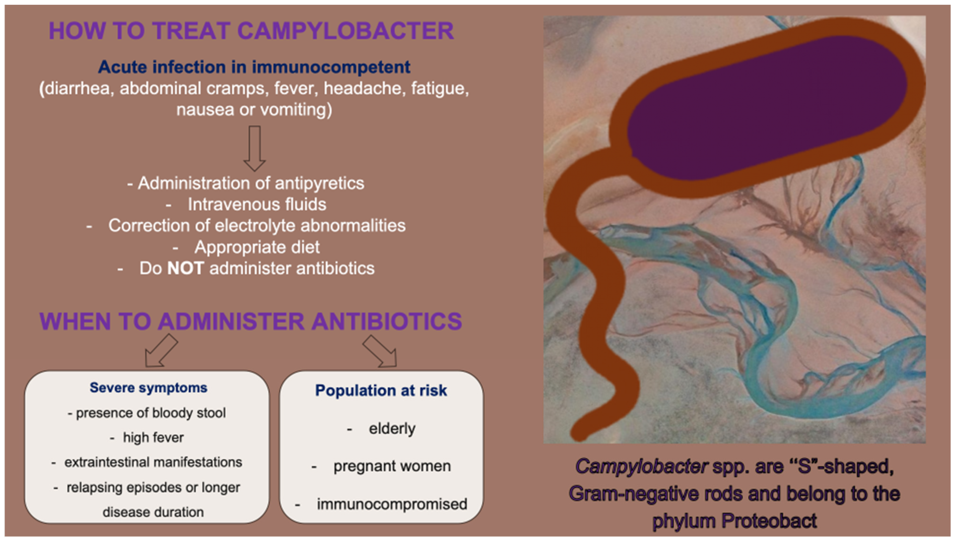
Figure 1. When antibiotics are not always the treatment: indications for antibiotics’ administration.
Microbiolres 16 00226 g001
Figure 2. Increase of AMR in the era of climate crisis and industrial development.
Microbiolres 16 00226 g002
Figure 3. Mitigating AMR in Campylobacter spp.
Microbiolres 16 00226 g003
Table 1. Mechanisms of Campylobacter species resistance to antibiotics.
Resistance GenesPoint
Mutations
Active EffluxOther MechanismsStudy [Ref.]
MacrolidesErm(B),
MDRGIs loci; aacA-aphD, sat4, aphA-3, fosXCC, aad9 and tet(O)
domain V of the 23S rRNA;
A2074C, A2074G, A2075G
CmeABC; synergy with point mutations in the ribosomal proteins L4 (G74D) and L22 (inserted at position 86 or 98) Zhang et al. [75]
Jehanne et al. [76]
Bolinger et al. [78]
Gibreel et al. [80]
Cagliero et al. [81]
FluoroquinolonesGyrA; fluoroquinolone resistance in Campylobacter spp.T86I, T86K, A70T and D90NCmeABC; synergy with point mutations in GyrA genes Shariati et al. [84]
Luangtongkum et al. [85]
Yao et al. [86]
TetracyclinesTet(O)-CmeABC
CmeG; synergy with mutations in Tet(O) gene
Grossman et al. [89]
Li et al. [90]
Connell et al. [91]
AminoglycosidesaadE-sat4-aphA-3 cluster
aacA-aphD-aac cluster
sat4 gene
aph(2″)-Ib, Ic, If1, If3, Ih
aac(6′)-Ie/aph(2″)-If2
binding sites of rRNA-modification of the aminoglycoside structure by enzymes such as aminoglycoside acetyltransferases, aminoglycoside phosphotransferases and aminoglycoside nucleotidyltransferasesGuirado et al. [104]
Werner et al. [107]
Gharbi et al. [100]
Papadopoulos et al. [101]
Phenicols23S rRNA
cfr gene
cfr(C) gene; Campylobacter coli isolates of cattle origin
G2073A mutation
Methylation of the adenine at position 2503 in the 23S rRNA
variant RE-CmeABC; synergy with point mutations Long et al. [103]
Gharbi et al. [108]
Tang et al. [114]
β-Lactams--CmeABC
CmeDEF; synergy with β-lactamase OXA-61
OXA-61 (Cj0299); β-lactamase present in C. jejuni and coliReygaert et al. [116]
Griggs et al. [117]
FosfomycinfosXCC gene; part of the MDRGI loci-- Castaneda–Garcia et al. [118]
Mattioni Marchetti et al. [120]
Disclaimer/Publisher’s Note: The statements, opinions and data contained in all publications are solely those of the individual author(s) and contributor(s) and not of MDPI and/or the editor(s). MDPI and/or the editor(s) disclaim responsibility for any injury to people or property resulting from any ideas, methods, instructions or products referred to in the content.