Article

Isolation/Characterization of Colletotrichum gloeosporioides from Tea and MeJA-Induced Antioxidant Defenses

by
Chunju Peng
1,2,
Yu Wang
2,
Xuan Zhou
2,
Shifu Ma
3,
Zhiguo Shan
4,
Shuai Wan
2,
Zekun Xue
2,
Huiling Mei
2,
Yan Tang
1,
Shujing Liu
2,
Rui Han
2,
Xinghui Li
2,5,* and
Guanghui Zeng
1,*
1
Wenzhou Key Laboratory of Early Sprouting Tea Plant Breeding, Wenzhou Academy of Agricultural Sciences, Wenzhou 325006, China
2
Tea Research Institute, Nanjing Agricultural University, Nanjing 210095, China
3
Tea Industry Development Service Center of Wenxian, Longnan 746400, China
4
School of Tea and Coffee, Pu’er University, Pu’er 665000, China
5
Cangnan Industrial Research Institute of Modern Agriculture, Wenzhou 325800, China
*
Authors to whom correspondence should be addressed.
Microbiol. Res. 2025, 16(10), 220; https://doi.org/10.3390/microbiolres16100220
Submission received: 9 September 2025 / Revised: 3 October 2025 / Accepted: 6 October 2025 / Published: 8 October 2025

Abstract

Anthracnose is a leaf fungal disease caused by multiple Colletotrichum species. Currently, the predominant deployment of chemical agents for anthracnose control increases ecological pollution risks and potential food safety concerns. The comprehension of the pathogenic mechanism and physicochemical properties of anthracnose is, therefore, essential for effective prevention and control. In this study, the pathogenic strain (pathogen) was isolated from the infected tea plant (Camellia sinensis) leaves and was identified as Colletotrichum gloeosporioides based on microscope observations and gene sequences. This fungus exhibited optimal growth at 28 °C and a pH of 6, with a lethal temperature threshold of 53 °C on PDA plate medium. The 80% tea saponin and 10% polymycin B could effectively inhibit its mycelium growth. Notably, the 10% polyoxin B exhibits a stronger inhibitory effect with an EC50 value of 1.07 mg mL−1. Following infection with Colletotrichum gloeosporioides, the resistant cultivar ‘Zhongcha 108’ exhibited higher levels of H2O2 and O2 than the susceptible ‘Longjing 43’, with later symptom onset and slower disease progression. Although the exogenous treatment of methyl jasmonate (MeJA) did not inhibit C. gloeosporioides directly, it significantly reduced lesion areas in ‘Longjing 43’ leaves caused by C. gloeosporioides. This treatment increased peroxidase and superoxide dismutase activities, but limited malondialdehyde content, thereby enhancing ‘Longjing 43’ resistance to the pathogen. The findings provide scientific guidance for the anthracnose prevention and control in tea gardens.

1. Introduction

Tea plant anthracnose is a leaf fungal disease caused by Colletotrichum and occurs across major tea-producing regions worldwide [1]. Anthracnose causes damage to important tea-producing areas worldwide. As one of the most serious diseases of tea leaves, anthracnose was found in all southern provinces suitable for tea planting in China [2]. In warm tea-producing areas, such as southwestern Japan, anthracnose outbreaks commonly recur from summer to autumn; prevention and control typically involve more than four fungicide applications per year [3,4]. Among about 14–50% crop losses caused annually by pests and plant pathogens in Assam State tea-producing areas of India, tea anthracnose is one of the most serious leaf diseases [5]. Anthracnose is also a major leaf disease in tea plantations off the coast of South Korea [6].
The clarification of pathogenic characteristics is crucial for determining the etiology of the disease, comprehending its occurrence patterns, and formulating effective prevention and control strategies. The morphological and molecular biological methods were used for anthracnose identification. Anthracnose is caused by various pathogens, each with distinct characteristics when affecting tea trees [7]. It is generally believed that there are a total of 20 species of anthracnose fungi on tea trees, of which three are compound species, namely C. gloeosporioides (including C. siamense, C. aenigma, and C. fructicola), C. acutatum (including C. fioriniae) and C. boninense (including C. karstii), and the fourth category does not contain the compound species, which are C. truncatum, C. cliviae, C. crassipes, C. camelliae, C. jiangxiense, C. wuxiense, C. henanense, C. endophytica, C. carveri, C. majus, C. chongqingense, and C. alienum, and individual unnamed ones are not counted [8,9,10,11,12,13,14,15,16].
To manage these outbreaks, integrated programs combine cultural practices and fungicide applications; several management measures have been proposed. Therefore, fungicide treatment is the most effective measure to protect plants from Colletotrichum infection. Concretely, chlorothalonil, carbendazim, and difenoconazole all have inhibitory effects on the development of anthracnose [17]. Extended or improper chemical control may drive resistance, disrupt benefits, and increase residue and environmental burdens, underscoring the need for integrated management. Therefore, it is imperative to adopt more optimal measures for anthracnose control. A variety of synthesized secondary metabolites in tea plants, including caffeine, tea polyphenols, and tea saponins, exhibit significant antibacterial properties. For instance, exogenous caffeine can stimulate the elevation of endogenous jasmonic acid levels in tea plants, thereby augmenting the disease resistance of tea plants and inhibiting the invasion of anthracnose [18]. In vitro antifungal tests demonstrated that caffeine exhibited a more potent inhibitory effect on the growth of Colletotrichum compared to tea polyphenols and catechins [19]. The effective utilization of tea plant metabolites for the control of fungal diseases is both intriguing and promising.
Here, a tea-derived C. gloeosporioides isolate was recovered from symptomatic leaves, identified by morphological traits and molecular markers, and characterized to establish in vitro benchmarks for growth and antifungal sensitivity (tea saponin, polyoxin B). In parallel, the potential of methyl jasmonate (MeJA) to prime leaf-level resistance in tea was assessed by comparing disease progression between the resistant ‘Zhongcha 108’ and the susceptible ‘Longjing 43’, examining the direct in vitro activity of MeJA across practical concentrations, and quantifying the effects of MeJA pretreatment on lesion development together with peroxidase (POD) and superoxide dismutase (SOD) activities. Collectively, these pathogen- and host-focused assessments provide parameters to inform anthracnose prevention and control in tea production and a basis for the breeding and deployment of anthracnose-resistant cultivars.

2. Materials and Methods

2.1. Isolation and Identification of Pathogen

The diseased leaves of ‘Longjing 43’ that exhibit typical symptoms of anthracnose were collected. According to established research protocols, a pathogenic fungus was isolated by using the conventional tissue isolation method to obtain pure culture strains through subsequent single-spore isolation [7,20].
The isolated and purified strains were cultured in PDA medium for approximately 7 days to collect desiccated mycelium. The pathogenic DNA was extracted by Ezup column fungal genomic DNA extraction kit based on the manufacturer’s instructions. The ribosomal intergenic spacer sequence (rDNA-ITS), Glutamine synthetase (GS), and glyceraldehyde-3-phosphate dehydrogenase (GAPDH) were amplified by using universal primers ITS1 (TCCGTAGGTGAACCTGCGG)/ITS4 (TCCTCCGCTTATTGATATGC), GS (ATGGCCGAGTACATCTGG/GAACCGTCGAAGTTCCAC) and GAPDH (GCCGTCAACGACCCCTTCATTGA/GGGTGGAGTCGTACTTGAGCATGT) [21,22,23]. The PCR products were analyzed and sequenced for molecular biological identification.

2.2. Characteristics of C. gloeosporioides

After culturing on a PDA plate medium for 5 days, a 6 mm fungal disk was carefully transferred to the center of a fresh PDA plate medium for morphological identification and characteristic observation of relevant pathogens. The fungus was then incubated at 28 °C to observe and record its growth condition every 24 h. The spore suspension was collected using established methods, followed by microscopic examination to observe and measure the morphology and dimensions of conidia [24].
The 6 mm fungal disks were cultured in a constant temperature incubator under dark conditions at temperatures of 5 °C, 10 °C, 15 °C, 20 °C, 25 °C, 28 °C, 30 °C, 35 °C, and 40 °C. After five days’ culture, the colony diameter was measured to calculate the growth rate at different temperatures, and sporulation was evaluated [25,26]. The fungus disks were placed into a sterile 10 mL centrifuge tube and overlaid with sterile water. The tube was incubated in a water bath at 45, 47, 49, 51, 53, or 55 °C for 10 min. After cooling to room temperature, the fungus disks were aseptically transferred to the center of the PDA plate and incubated at 28 °C for 5 days. Then, the growth and development of hyphae were carefully observed and recorded. Each temperature was set to 3 repeats.
The 6 mm fungal disks were transferred on PDA plate medium with varying pH (i.e., 4, 5, 6, 7, 8, 9, 10, and 11) and cultured in darkness at 28 °C in the incubator [25]. After 5 days’ culture, the colony diameter was measured to calculate the growth rate, as well as the conidia yield and the germination rate on the PDA plate medium with different pH values. The pH value of each PDA medium was set to 3 replicates.

2.3. The In Vitro Antifungal Activity Tests

The antifungal activity of the 80% tea saponin and 10% polyoxin B formulations against C. gloeosporioides was assessed by measuring inhibition of mycelial growth. For tea saponin, 4.00 g of the 80% formulation was dissolved in sterile water to a final volume of 25 mL to obtain a 160 mg mL−1 stock, which was diluted to working solutions of 80, 40, 20, and 10 mg mL−1. For polyoxin B, 500 mg of the 10% formulation was dissolved in sterile water to 25 mL to obtain a 20 mg mL−1 stock, which was diluted to 10, 12.5, 15, and 17.5 mg mL−1. Then, 5 mL of each working solution was added to 50 mL of molten PDA to prepare drug-amended media containing 80% tea saponin at final concentrations of 16, 8, 4, 2, and 1 mg mL−1 and 10% polyoxin B at 1, 1.25, 1.50, 1.75, and 2 mg mL−1. PDA supplemented with sterile water only served as the control. The 6 mm fungal disks were placed in the center of the drug-containing and control mediums, respectively, and then invertedly incubated at 28 °C in darkness for 5 days to measure and record the diameter of colonies [27]. Each group was treated with 3 replicates.

2.4. Infection and Histochemical Staining of Tea Plant Leaves

Two contrasting cultivars were selected to anchor the assay pipeline: ‘Zhongcha 108’ (resistant) and ‘Longjing 43’ (susceptible). This pairing provided a high-contrast reference under standardized conditions (uniform leaf age, identical inoculation, and MeJA pretreatment). After disinfection with a 75% (v/v) ethanol solution, the inoculation method employed was based on previous research [24]. The inoculated leaves were cultured in a controlled chamber set at 25 °C, with a relative humidity of 95% and a light–dark cycle of 12 h. The symptoms of the leaves were observed and measured through DAB and NBT staining methods as described in a previous study [28]. The leaves of ‘Longjing 43’ were sampled at 2, 4, 12, and 24 h after inoculation with C. gloeosporioides to investigate its infection process using trypan blue and aniline blue staining methods [29]. Each group was treated with 3 replicates.

2.5. Exogenous Methyl Jasmonate Treatment

The exogenous methyl jasmonate (MeJA) solution was dissolved in PDA medium following the established method to obtain PDA medium with varying concentrations of MeJA, aiming to gain a preliminary understanding of the direct impact of MeJA on C. gloeosporioides [19]. The inhibitory effect of MeJA on the mycelial growth of C. gloeosporioides was assessed using the mycelial growth rate method. Each group was treated with 3 replicates.
Subsequently, the MeJA effect on the resistance of tea plant leaves to anthracnose was studied through gradient concentrations (i.e., 0.01, 0.05, 0.1, 0.5, 1 mmol L−1). The leaves of ‘Longjing 43’ were treated with 20 μL of MeJA solution at various concentrations for 6 h, while sterile water was used as the control treatment. Following the 6 h treatment period, a spore suspension with a concentration of about 1.0 × 10⁵ CFU mL−1 was introduced to the stab wound site. The inoculated leaves were cultured in a controlled chamber at 25 °C, with a relative humidity of 95% and a light–dark cycle of 12 h. Each group was treated with 3 replicates.

2.6. Enzyme Activity

The ‘Longjing 43’ leaves were treated with an exogenous hormone solution containing 0.1 mmol L−1 MeJA, while sterile water was used as the control treatment. The samples were collected at 0, 2, 4, 6, and 8 days after inoculation with C. gloeosporioides for the determination of resistance physiological indices. The peroxidase (POD), superoxide dismutase (SOD), and malondialdehyde (MDA) enzyme activities were determined in accordance with the instructions provided by the manufacturer (BC0095, BC0170, BC0025, Solarbio, Beijing, China).

2.7. Data Statistical Analysis

All assays were performed with a single tea-derived C. gloeosporioides isolate to standardize growth and sensitivity measurements across experiments. GraphPad Prism 8.0.2 software was used for statistical analysis of the data. Calculations followed the manufacturer’s instructions, and statistical analyses were performed using Student’s t-test (* 0.01 < p < 0.05, ** p < 0.01).

3. Results

3.1. Characteristics of C. gloeosporioides Strains

The pathogenic strain was isolated from the anthracnose-infected leaves collected from Zhongshanling tea plantations in Nanjing, Jiangsu Province (Figure 1A). The multilocus phylogenetic analysis of the concatenated sequences of ITS, GS, and GAPDH genes placed the isolates described as C. gloeosporioides through DNAMAN 7 BLAST homology analysis (Figure S1), and identified C. gloeosporioides with a 97.50% for the ITS, 94.12% for the GAPDH, and 93.1% for the GS, supporting the isolate’s identification as C. gloeosporioides.
This strain was purified and cultured to observe its morphological characteristics. Along with the growth on the PDA plate medium, the colony gradually turned into gray from the initial white after 3–4 days of continuous culture. The aerial hyphae appeared dense and grayish-white with yellowish-white on the opposite side of the colony after culture for 7 days (Figure 1B). The hyphae exhibit a curved morphology without any branching. The conidium has a colorless single-celled, rod-shaped structure and measures 11.34 ± 1.31 × 4.35 ± 0.51 μm in size (Figure 1B). Therefore, combined with morphological characteristics and microscopic structure, the isolated fungus was identified as C. gloeosporioides.
The purified pathogenic fungus was inoculated onto the ‘Longjing43’ leaves to observe the subsequent development of the infection process on the tea tree leaves using aniline blue staining. The results demonstrated the presence of rod-shaped spores at 4 hpi. Spore germination tubes were observed at 8 hpi, followed by the appearance of hyphae after 12 hpi. The formation of appressorium was detected at 24 hpi (Figure 1C).

3.2. Effects of Environment and Fungicides on C. gloeosporioides Growth

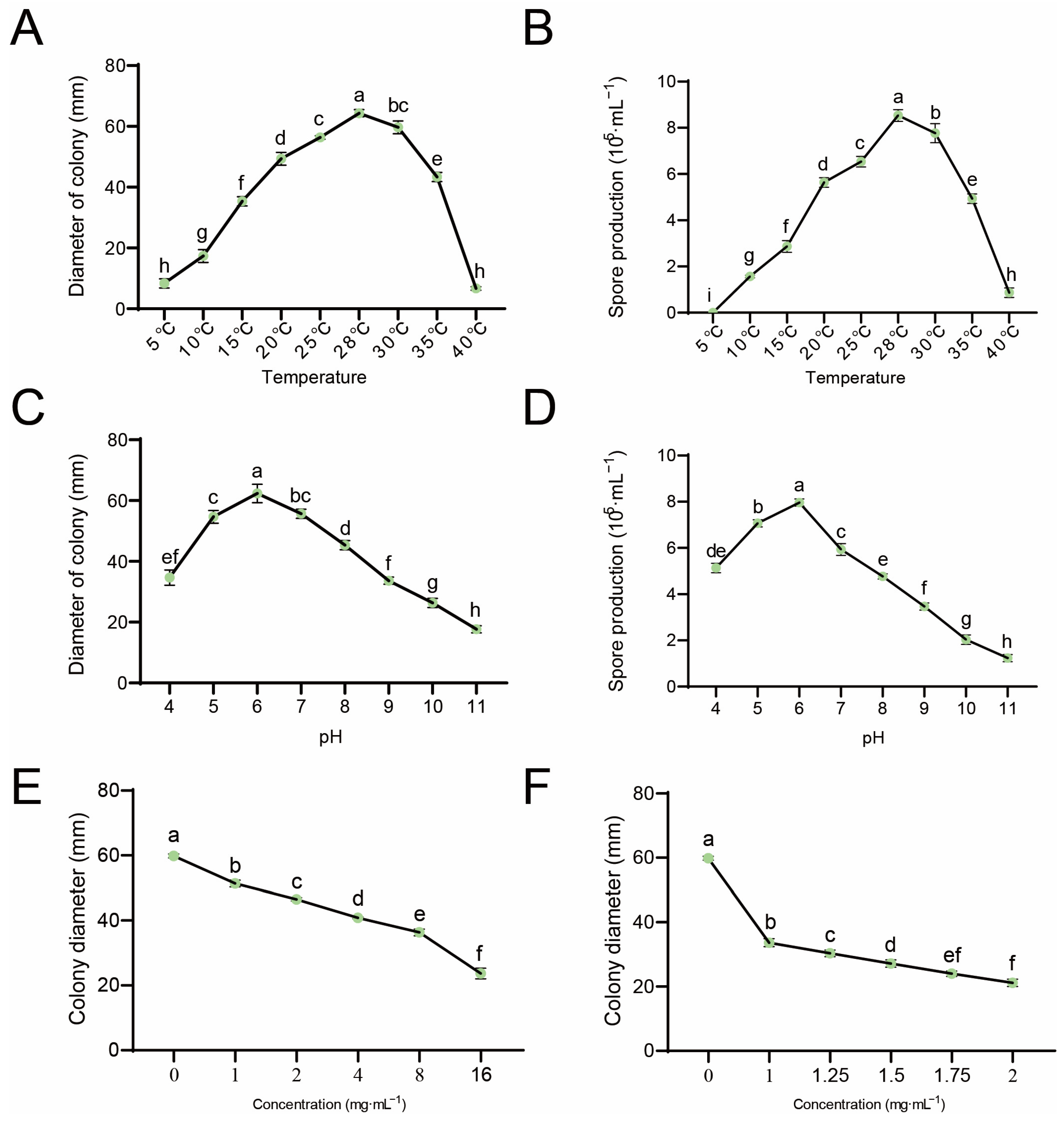
Using this isolate as a reference, in vitro growth responses, lethal temperature, and EC50 values were determined under harmonized conditions. Culture temperature effect on C. gloeosporioides was investigated, which demonstrated the normal growth of mycelia within a temperature range of 5–40 °C. Among these, the optimal growth was observed at 28 °C, with a colony diameter reaching 64.3 mm, an average growth rate of 12.86 mm d−1, and the maximum spore production of about 8.53 × 10⁵ mL−1. However, the temperatures of 5 °C and 40 °C both significantly impair the mycelial activity and growth, resulting in the slowest rates of colony expansion. After culturing for 5 days, the respective colony diameters measured 8.33 mm and 6.67 mm, with average growth rates recorded as 1.67 mm d−1 and 1.33 mm d−1, respectively. Furthermore, when cultured at 5 °C, the strain exhibited no spore production, while at 40 °C, spore production was found to be minimal and was only about 8.7 × 10⁴ mL−1. The culture temperature below 20 °C or above 35 °C had a significant inhibitory effect on strain sporulation. Sporulation showed an increase between 5 and 28 °C, but decreased between 28 and 40 °C (Figure 2A,B). The water bath at 53 °C and 55 °C for 10 min impaired growth and development of the pathogen, which indicated that temperatures of 53 °C and above possess lethal effects on C. gloeosporioides.
The effect of pH value on C. gloeosporioides was investigated. The results demonstrated that the mycelia exhibited normal growth within a pH range of 5–9, particularly with the most rapid growth at pH 6. Its colony diameter reached 62.33 mm with an average growth rate of about 12.47 mm d−1. However, the mycelial activity was found to be poor, and the growth rate was slow under pH 10 and pH 11. After 5 days, the colony diameters measured 26.33 mm and 17.67 mm, with average growth rates of 5.27 mm d−1 and 3.53 mm d−1, respectively (Figure 2C). The maximum spore yield reached 7.97 × 10⁵ CFU mL−1 at pH 6, while the sporulation of the strain significantly decreased after pH 9, with a minimum sporulation of only 1.23 × 10⁵ CFU mL−1 (Figure 2D).
Supplementary tests in medium with varying concentrations of 80% tea saponin and 10% polyoxin B revealed a positive correlation between the agent concentration and the growth of C. gloeosporioides mycelium (Figure 2E,F). The relative antifungal rate reached 15.79% at 1 mg mL−1 of 80% tea saponin. Meanwhile, at a concentration of 2 mg mL−1 for 10% polyoxin B, the relative antifungal rate significantly increased to 71.83%. The virulence analysis revealed that the EC50 values for 80% tea saponin and 10% polyoxin B against C. gloeosporioides were determined to be 8.21 mg mL−1 (y = 1.1356x + 3.9615, R2 = 0.9695) and 1.07 mg mL−1 (y = 2.1338x + 4.9389, R2 0.9796), respectively, indicating that 10% polyoxin B exhibited a superior direct antifungal effect.

3.3. Response of Different Resistant Tea Leaves to C. gloeosporioides

The C. gloeosporioides were inoculated onto ‘Longjing 43’ and ‘Zhongcha 108’ leaves, aiming to investigate the response of different tea plant varieties with varying resistance levels towards C. gloeosporioides.
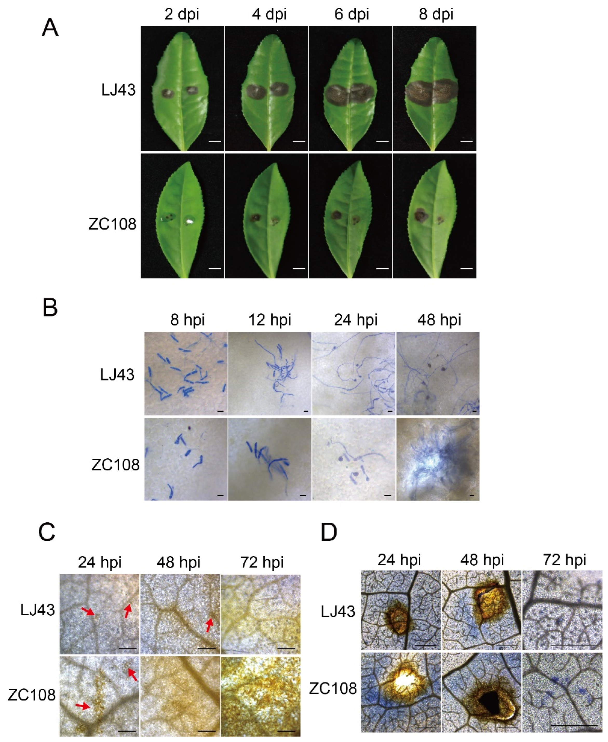
‘Longjing 43’ leaves exhibited conspicuous lesions at 2 dpi (day post-inoculation). The lesion diameter of ‘Longjing 43’ was significantly greater than that of ‘Zhongcha 108’ at 4 dpi. At 6–8 dpi, differences between the two cultivars were more pronounced. The leaves of ‘Longjing 43’ showed large and concave lesions, resulting in lesions with circular edges. The symptoms of ‘Zhongcha 108’ showed small spots and did not deteriorate seriously (Figure 3A). Overall, ‘Zhongcha 108’ exhibited a higher level of resistance to C. gloeosporioides infection compared to ‘Longjing 43’.
The growth and development of C. gloeosporioides in leaves of different resistant tea plants were observed using aniline blue staining. The staining results revealed that the development of C. gloeosporioides conidia on the leaves of ‘Longjing 43’ exhibited a higher rate compared to that on ‘Zhongcha 108’. C. gloeosporioides initiated germination on the leaves of both tea cultivars and developed germ tubes at 8 hpi; however, a lower number of conidia germinated on the leaves of ‘Zhongcha 108’. The ‘Longjing 43’ leaves exhibited hyphal growth at 12 hpi, whereas ‘Zhongcha 108’ continued to develop germ tubes. The hyphae on ‘Longjing 43’ leaves exhibited robust growth and developed appressoria, whereas the hyphae of ‘Zhongcha 108’ showed less proliferation with some still in a germination state, indicating potential hindrance at 24 to 48 hpi (Figure 3B).
The production and accumulation of H2O2 were observed by DAB staining. The results demonstrated less H2O2 production in ‘Longjing 43’ upon attachment of C. gloeosporioides at 24 hpi. In contrast, ‘Zhongcha 108’ exhibited evident oxidation as indicated by DAB staining and displayed a yellowish-brown coloration. The ‘Longjing 43’ exhibited localized DAB staining at 48 hpi, whereas ‘Zhongcha 108’ displayed a substantial area of yellowish-brown discoloration. By 72 hpi, ‘Longjing 43’ demonstrated a higher accumulation of H2O2 (Figure 3C).
The production and accumulation of O2 were observed by NBT staining. The results demonstrated that a substantial blue-purple lesion developed adjacent to the inoculation site of ‘Zhongcha 108’, while only a limited light blue-purple discoloration appeared on ‘Longjing 43’ leaves after 24 h of the inoculation. After 48 h of inoculation, the intensity of blue-purple discoloration surrounding the inoculation wounds in both varieties exhibited a decrease. A greater number of blue-purple sites were observed outside the inoculation site of ‘Zhongcha 108’, while fewer were found on the leaves of ‘Longjing 43’ at 72 hpi (Figure 3D).

3.4. Effect of Exogenous Hormone Treatment on C. gloeosporioides

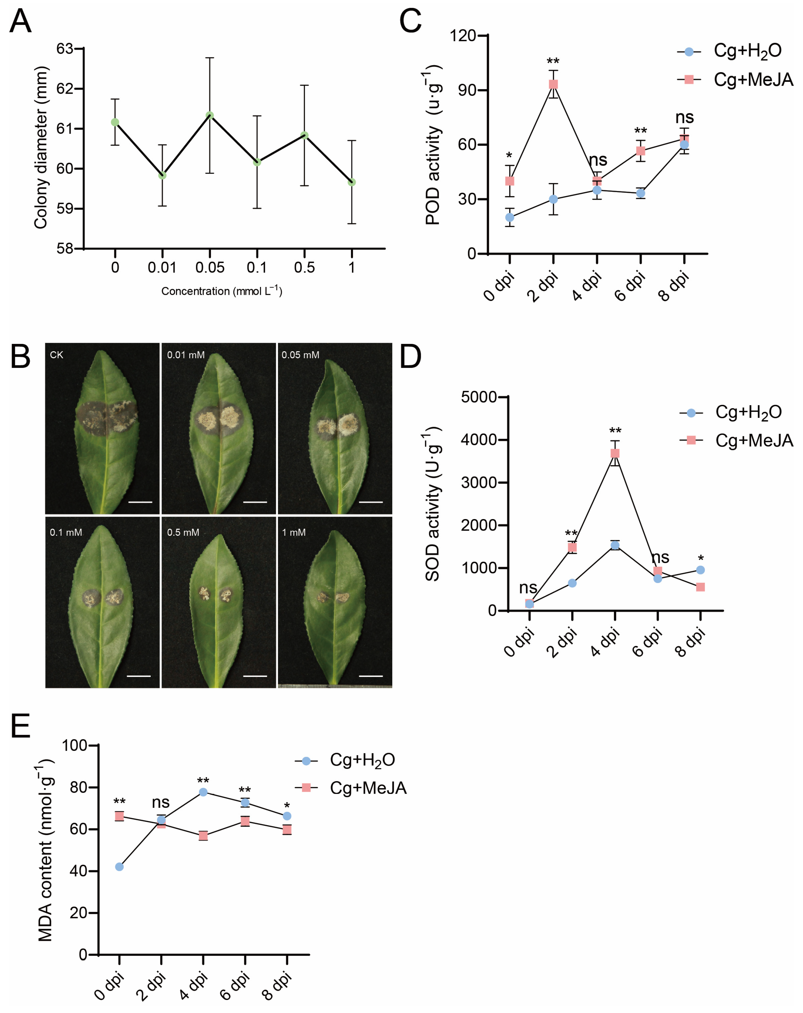
The application of MeJA significantly suppressed the spore germination, germ tube elongation, and mycelial growth of C. acutatum [30]. Therefore, the direct effect of exogenous MeJA on C. gloeosporioides was evaluated through the inoculation of 6 mm C. gloeosporioides fungal disks on the PDA plate medium with different concentrations of MeJA. After incubation at 28 °C for 5 days, the colony diameter in the control group reached 61.17 mm. Compared to the control group, no significant difference in the colony diameter was observed in the exogenous treatment group and among various MeJA concentrations (Figure 4A) on PDA plate medium treated with different concentrations of MeJA, indicating that exogenous MeJA treatment did not exert direct inhibitory effects on the growth of C. gloeosporioides.
To investigate the impact of exogenous hormones on tea plant leaf defense against C. gloeosporioides, various concentrations of MeJA were applied for the exogenous treatment of leaves 6 h before the infection. The lesion area expanded to the leaf margins along with the leaf decay in the control at 5 dpi with C. gloeosporioides. The exogenous application of MeJA significantly reduced the area of leaf lesions. Notably, a concentration of 1 mM MeJA effectively inhibited pathogen expansion without virtual leaf rot (Figure 4B), which indicated that exogenous MeJA induced resistance of ‘Longjing 43’ against C. gloeosporioides and inhibited the infection spread on leaves. The positive correlation was observed with increasing hormone concentrations. The inhibitory effect was most pronounced at a concentration of 1 mM MeJA, while it was least significant at a concentration of 0.01 mM (Figure 4B).

3.5. Changes in Antioxidant Enzyme Activity in Tea Plant Leaves Under Exogenous MeJA Treatment

A concentration of 0.1 mM MeJA was chosen for the treatment of ‘Longjing 43’ leaves [31]. After inoculation with C. gloeosporioides, the POD activity was measured. The POD activity in the exogenous treatment group of 0.1 mM MeJA exhibited a sharp increase, reaching its peak at 2 dpi. Subsequently, there was a significant decline in enzyme activity at 4 dpi, returning to levels similar to those observed at 0 hpi. However, it remained consistently high at both 6 dpi and 8 dpi. The POD activity in the control exhibited a relatively gradual change, with a slight increase observed at 2 dpi. Subsequently, almost no significant alterations in POD activity were found at both 4 dpi and 6 dpi. However, after 6 dpi, a notable upward trend in POD activity was observed (Figure 4C).
The SOD activity treated with 0.1 mM MeJA exhibited a similar trend to that of the control at 0 dpi, but it displayed a rapid increase at 2 dpi and subsequently surged to its peak at 4 dpi. Then, the enzyme activity declined to the same level as the control at 6 dpi. At 8 dpi, the SOD activity decreased to a level lower than that of the control. The change in SOD activity in the control was relatively gradual, with a slight increase observed at 2 dpi. Subsequently, there was a continuous rise in SOD enzyme activity, reaching its peak at 4 dpi. However, at 6 dpi, there was a decline in SOD activity followed by another increase until 8 dpi, when it surpassed that of the 0.1 mM MeJA treatment group (Figure 4D).
The MDA content in the 0.1 mM MeJA group exhibited minimal changes following inoculation, with the highest level observed at 0 hpi, which was subsequently maintained at a stable level from 2 dpi to 8 dpi. A slight increase was observed at 6 dpi, followed by a subsequent decline. The MDA content in the control exhibited an initial increase followed by a subsequent decrease. Following inoculation, the MDA content was consistently higher compared to the 0.1 mM MeJA treatment at almost all time points. At 2 dpi and 8 dpi, both groups reached a similar level (Figure 4E).

4. Discussion

C. gloeosporioides strain was obtained from tea plant leaves infected with anthracnose through molecular biological and morphological identification of pathogens. Its colony characteristics observed on the PDA plate medium were consistent with previous studies [12,15].
According to histopathological observation, although Colletotrichum has a variety of pathogens causing tea anthracnose, its conidia germination and infection on tea leaves are roughly the same. Firstly, when the mycelia or conidia fall on the lower epidermis of leaves, they adhere to the trichome of tea leaves and then germinate to form germ tubes [32]. During germination, conidia produce one or more germ tubes. The top of the germ tube contacts the epidermal cell, thickening the cell wall [33,34]. Colletotrichum can invade directly through the cuticle on the leaf surface regardless of the appressorium. After the appressorium matures, the infection nail penetrates the host stratum corneum to complete the colonization [33]. In this study, we observed a similar development process in tea plant leaves after infection with C. gloeosporioides (Figure 1C).
Temperature is another important factor affecting fungi’s growth; for instance, unacceptable temperatures decrease or stop their growth [2]. The conidia germinate in distilled water after 48 h with a rate of about 45.4% at 22 °C and 44.1% at 27 °C, respectively. The low temperatures at −2 to 4 °C hinder germination ability only with a 1.3% germination rate in 5 days [35]. Various studies have demonstrated that spores exhibit germination within the temperature range of 12–40 °C, with an optimal temperature for germination at 24~28 °C. However, certain regions also display a germination rate exceeding 50% at temperatures as low as 20 °C and as high as 32 °C. Furthermore, it has been observed that the mycelium exhibits its most rapid growth within the temperature range of 25~28 °C [26,36]. Different Colletotrichum species show various tolerances to temperature. For example, C. fruticola grows in a wide temperature range of 15 °C to 30 °C, with a significant decrease in growth rate at 35 °C and growth stopping at 40 °C. On the other hand, C. camelliae stops growing when the temperature exceeds 35 °C, and its optimal temperature is 25 °C [26]. For C. siamense, C. fioriniae, and C. Karstii, the optimal temperature range is 26 °C to 28 °C [37]. Our study indicated that C. gloeosporioides exhibited normal growth across a temperature range of 5–40 °C, with the optimal growth temperature being 28 °C. The temperature below 5 °C or above 40 °C significantly restricts the spore growth and production. The lethal temperature of C. gloeosporioides was 53 °C, which effectively eliminated the pathogen after a 10 min treatment in a water bath (Figure 2A,B).
The pH value is another critical factor in fungal–host interactions as it can change the activities of polygalacturonase (PG) and pectinase (PL), which alter pathogen virulence [38]. Both C. camelliae and C. fructicola grow in acidic to weakly alkaline environments, with no significant difference in growth between pH 5 and pH 8. However, C. camelliae showed a downward trend at pH 9 [26]. The initial pH of the medium was reported to be alkaline, and after 4 days of culture, the mycelium exhibited a slight shift towards neutrality [25]. In this study, C. gloeosporioides exhibits a broad adaptability to variations in culture pH, can grow and develop optimally within a pH range of 4 to 11. The mycelium demonstrates its highest growth rate and spore production at pH 6; however, the high pH levels at 10 and 11 diminish the mycelium’s activity, thus slowing growth rates and reducing spore production (Figure 2C,D).
Fungicide treatment is the most effective measure to protect plants from Colletotrichum infection. Chlorothalonil, carbendazim, and difenoconazole have inhibitory effects on anthracnose [17]. Screening and toxicity tests of nine chemical fungicides found that 40% prochloraz water emulsion at a concentration of 0.1 μg mL−1 is the most effective method to control anthracnose [16]. A total of 80% tea saponin, a plant-derived saponin preparation that has shown antifungal effects in vitro (including against Colletotrichum), has been used in some systems as a biobased antifungal or adjuvant [39]; and 10% polyoxin B is a member of the polyoxin group used in crop disease control and is reported to provide preventive activity in other host–pathogen systems [40]. Reports from fruit, vegetables, and ornamental crops indicate that tea saponin can reduce mycelial growth and/or sporulation and may improve spray performance, whereas polyoxin products can limit lesion development and sporulation when applied at appropriate doses and timings; however, performance is context-dependent across species and environments [41,42]. The mycelium growth rate method confirmed that both 80% tea saponin and 10% polyoxin B exhibited significant inhibition against the growth of C. gloeosporioides mycelium (Figure 2E,F). These observations justified our in vitro concentration-response comparison as a necessary first step before detached-leaf and greenhouse inoculation trials in the tea and before considering integration into Integrated Pest Management (IPM) programs.
The resistance to anthracnose of different tea plant cultivars was evaluated for breeding. Many tea cultivars have varying degrees of resistance to anthracnose pathogens. A major green tea cultivar, ‘Yabukita’, is susceptible to anthracnose in more than 70% of tea fields in Japan [3]. Most Assam varieties are resistant or highly resistant to anthracnose in Japan [43,44,45]. Green tea cultivars introduced from China, such as ‘Chin Shin daah Pan’ and ‘Ying Jy Horong Shin’, show moderate resistance to this disease [3]. Additionally, ‘Yun Kang 10’ is resistant to anthracnose [46], while ‘Zhenong 139’ and Taiwan ‘Chin-shin Oolong Tea’ are susceptible [47,48]. Anthocyanin-rich tea cultivar ‘Sunrouge’ is screened from the natural cross population of ‘Cha Chuukanbohon Nou 6’ (C. taliensis × C. sinensis), with slight resistance to anthracnose [49,50]. ‘Zhongcha 108’ (ZC108) is a disease-resistant tea cultivar developed from ‘Longjing 43’ (LJ43) by radiation-induced mutagenesis, whereas LJ43 is susceptible to anthracnose [19,51,52]. Compared to ‘Zhongcha 108’ leaves, a more rapid and earlier progression of anthracnose was observed on ‘Longjing 43’ leaves in our study (Figure 3A,B).
Reactive oxygen species (ROS)-mediated defense against fungal pathogens is an important component of the plant immune system. Overexpression of grape VqWRKY56 led to increased ROS accumulation and enhanced resistance against E. necator [53]. Previous studies have demonstrated that ROS play a pivotal role in conferring resistance to P. theae in tea plants [54]. As one of the most valuable and stable ROS, the compound H2O2 exerts inhibitory effects on the germination of various pathogenic fungal spores, and also actively participates in the oxidative cross-linking process of cell wall glycoproteins to reinforce their structure, which leads to the induction of allergic necrosis reactions, ultimately enhancing disease resistance in plants [55,56,57,58]. The conversion of O2 into H2O2 and O2 serves as a protective mechanism against oxidative damage in cells [59]. According to Wang et al. (2018), the marked accumulation of H2O2 at infection sites in the resistant cultivar ‘Zhongcha 108’ implicates H2O2 in defense against C. fructicola [58]. In line with previous studies, the levels of H2O2 and O2 in the leaves of ‘Zhongcha 108’ were significantly higher than those observed in ‘Longjing 43’ at 24–72 hpi following C. gloeosporioides inoculation (Figure 3C,D). These findings confirm that ROS play a pivotal role in determining the variations in resistance to anthracnose among different tea plant cultivars.
The inhibitory effects of MeJA on spore germination and mycelial growth of Alternaria alternata have been documented in previous studies [60]. In vitro antifungal experiments showed that MeJA treatment significantly inhibited spore germination, germ tube elongation, and hyphal growth of C. acutatum [30]. Reported in vitro effective concentrations vary with pathogen and assay format: for Alternaria alternata, MeJA at 100–500 μL L−1 (and as low as 50–150 μL L−1 in another study) inhibited mycelial growth and spore germination [61]; for Colletotrichum (including C. acutatum), 1–10 mM MeJA reduced spore germination and hyphal growth [62]; and for Aspergillus flavus, 10−3–10−8 M MeJA suppressed aflatoxin production and altered fungal development [63]. However, this study revealed no significant disparity in C. gloeosporioides mycelium growth on PDA plate medium containing varying concentrations of MeJA compared to the control (Figure 4A), which indicates that MeJA itself has no toxic effect on C. gloeosporioides and does not affect the normal growth of mycelium.
A plethora of studies have demonstrated that exogenous application of MeJA can elicit disease resistance in plants. For instance, the application of MeJA through spraying on banana (Musa nana Lour.) fruits has been shown to significantly reduce lesion diameter and disease index, thereby activating the endogenous defense system of bananas [64]. Treatment with 0.1 mmol·L−1 MeJA could improve the susceptibility of avocado (Persea americana) fruit to anthracnose and reduce the incidence rate [31]. The application of MeJA can advantageously enhance post-harvest disease resistance in kiwifruit (Actinidia chinensis), significantly reduce lesion diameter following inoculation with Botryosphaeria dothidea, and control the occurrence of soft rot [65]. The present study revealed that the exogenous application of MeJA on the leaves of ‘Longjing 43’ effectively suppressed the growth and dissemination of C. gloeosporioides, with obvious dose relations, thereby enhancing the concentration-dependent disease resistance in ‘Longjing 43’ (Figure 4B). Additionally, studies have reported that MeJA exhibits no in vitro antifungal activity against Botrytis cinerea but demonstrates the ability to mitigate disease severity and impede lesion development caused by Botrytis cinerea infection in freesia (Freesia hybrida) through exogenous treatment [66]. The findings of this study are in line with the obtained results that MeJA effectively inhibits pathogen infection, spreads to leaves, and alleviates associated symptoms. Moreover, this study also found that the degree of symptom alleviation is positively correlated with the concentration of exogenously applied MeJA within a specific range. These observations suggest that MeJA induces defense responses in ‘Longjing 43’, thereby enhancing its resistance against C. gloeosporioides.
The peroxidase (POD), superoxide dismutase (SOD), and catalase (CAT) are crucial antioxidant enzymes responsible for scavenging or utilizing reactive oxygen species (ROS). As pivotal components of plant defense, alterations in the activity of ROS-scavenging enzymes have been demonstrated to respond to a diverse array of environmental stresses [59]. The malondialdehyde (MDA) generated from the peroxidation of membrane lipids due to ROS in plants has been commonly employed as an indicator for assessing plant stress resistance. The increase in MDA content signifies severe damage to the plant’s membranes [67]. The exogenous treatment of MeJA enhanced the activities of defense-related enzymes POD, CAT, and PPO in banana fruits following inoculation with the pathogen C. musae [64]. The treatment of kiwifruit with 0.1 mmol L−1 MeJA significantly augmented the activities of relevant antioxidant enzymes, such as CAT, POD, SOD, PPO, etc., while concurrently diminishing the extent of membrane lipid peroxidation [65]. This study revealed that exogenous treatment with 0.1 mM MeJA significantly augmented the activities of POD and SOD enzymes in the leaves of ‘Longjing 43’ compared to the control, while concurrently restraining the elevation in MDA content to a certain extent (Figure 4C–E). The application of MeJA enhances the anthracnose resistance of ‘Longjing 43’ by effectively reducing the excessive accumulation of ROS in this cultivar. The application of exogenous MeJA can thus serve as an environmentally friendly, safe, and efficacious control strategy for safeguarding tea plants against C. gloeosporioides. The antioxidant/oxidative damage panel (POD, SOD, and MDA) was chosen to provide robust, comparable readouts under MeJA pretreatment; it does not encompass all defense pathways. Future studies will extend the panel to catalase (CAT), PR genes, and structural/chemical defenses (lignin, callose, and phytoalexins) to better resolve resistance mechanisms.
Findings represent reference benchmarks for this isolate under defined conditions and should not be over-generalized to the species complex; broader representativeness will require the same standardized pipeline applied across multiple tea-derived isolates from different regions.

5. Conclusions

In this study, C. gloeosporioides was isolated and identified from the infected tea plant leaves with anthracnose, and its biological characteristics and infection process were clarified. The responses of different tea varieties with resistance to anthracnose were investigated after inoculations into ‘Longjing 43’ and ‘Zhongcha 108’ leaves to elucidate the crucial role of ROS accumulation in the tea tree’s defense against anthracnose. The present study also determined the antifungal effects of 80% tea saponin and 10% polyoxin B against C. gloeosporioides, highlighting their potential for further utilization in field-based prevention and control strategies against tea tree anthracnose. It is recommended to develop rational and effective measures for prevention and control. The application of exogenous hormone MeJA additionally enhanced the resistance of ‘Longjing 43’ leaves to anthracnose, thereby providing significant support for anthracnose prevention and control.

Supplementary Materials

The following supporting information can be downloaded at https://www.mdpi.com/article/10.3390/microbiolres16100220/s1, Figure S1. Consistency alignment of fungal DNA sequences from ITS, GAPDH, and GS.

Author Contributions

C.P.: Writing—original draft, visualization, data curation, conceptualization, and funding acquisition. Y.W.: Methodology, investigation, and formal analysis. X.Z.: Investigation and formal analysis. S.M.: Methodology and project administration. Z.S.: Investigation and formal analysis. S.W.: Investigation and formal analysis. Z.X.: Methodology, investigation, and formal analysis. H.M.: Methodology and investigation. Y.T.: Data curation and validation. S.L.: Writing—review and editing. R.H.: Conceptualization, validation, and writing—review and editing. X.L.: Conceptualization and funding acquisition. G.Z.: Validation, resources, funding, investigation, and project administration. All authors have read and agreed to the published version of the manuscript.

Funding

This research was funded by Wenxian Science and Technology Plan Project (2024CX001), Modern Agriculture Industry Enhancement Project of Cangnan County (2024CNYJY07), National Program for SRT (X2025103070018), Nanjing Agricultural Major Technology Collaborative Promotion Plan Project (2025NJXTTG 07), Science and Technology Plan Program of Wen County (2023--X.QKJ--02), Wenzhou Agricultural New Variety Breeding Collaborative Project (ZX2024004-2), Key Research and Development Program of Jiangsu (BE2023364), National Key Research and Development Program of China (2022YFD1200505).

Institutional Review Board Statement

Not applicable.

Informed Consent Statement

Not applicable.

Data Availability Statement

The original contributions presented in this study are included in the article/Supplementary Material. Further inquiries can be directed to the corresponding authors.

Acknowledgments

We extend our thanks to Yuehua Ma (Central laboratory of College of Horticulture, Nanjing Agricultural University) for using the microscope (Olympus IX73, Japan); ZEISS LSM 800, Germany; Princeton PIXIS 1024B, USA, and Quantitative real-time PCR (CFX96, Bio-rad, USA).

Conflicts of Interest

The authors declare no conflicts of interest.

References

  1. Crous, P.W.; Gams, W.; Stalpers, J.A.; Robert, V.; Stegehuis, G. MycoBank: An online initiative to launch mycology into the 21st century. Stud. Mycol. 2004, 50, 19–22. [Google Scholar]
  2. Wei, L.; Ding, Y.; Peng, Y.; Zihao, W.; Guiyi, G. Research Progress on Anthracnose of Tea Plant. Chin. J. Trop. Agric. 2016, 36, 20–26. [Google Scholar]
  3. Yoshida, K.; Takeda, Y. Evaluation of anthracnose resistance among tea genetic resources by wound-inoculation assay. Jpn. Agric. Res. Q. 2006, 40, 379–386. [Google Scholar] [CrossRef]
  4. Moriwaki, J.; Sato, T. A new combination for the causal agent of tea anthracnose: Discula theae-sinensis (I. Miyake) Moriwaki & Toy. Sato, comb. nov. J. Gen. Plant Pathol. 2009, 75, 359–361. [Google Scholar] [CrossRef]
  5. Rabha, A.J.; Naglot, A.; Sharma, G.D.; Gogoi, H.K.; Veer, V. In vitro evaluation of antagonism of endophytic Colletotrichum gloeosporioides against potent fungal pathogens of Camellia sinensis. Indian J. Microbiol. 2014, 54, 302–309. [Google Scholar] [CrossRef] [PubMed]
  6. Kim, G.-H.; Lim, M.-T.; Hur, J.-S.; Yum, K.-J.; Koh, Y.-J. Biological control of tea anthracnose using an antagonistic bacterium of Bacillus subtilis isolated from tea leaves. Plant Pathol. J. 2009, 25, 99–102. [Google Scholar] [CrossRef]
  7. Wang, Y.-C.; Hao, X.-Y.; Wang, L.; Xiao, B.; Wang, X.-C.; Yang, Y.-J. Diverse Colletotrichum species cause anthracnose of tea plants (Camellia sinensis (L.) O. Kuntze) in China. Sci. Rep. 2016, 6, 35287. [Google Scholar] [CrossRef] [PubMed]
  8. Weir, B.; Johnston, P.; Damm, U. The Colletotrichum gloeosporioides species complex. Stud. Mycol. 2012, 73, 115–180. [Google Scholar] [CrossRef] [PubMed]
  9. Hyde, K.; Cai, L.; Cannon, P.; Crouch, J.; Crous, P.; Damm, U.; Goodwin, P.; Chen, H.; Johnston, P.; Jones, E. Colletotrichum—Names in current use. Fungal Divers. 2009, 39, 147–182. [Google Scholar]
  10. Damm, U.; Cannon, P.F.; Woudenberg, J.; Johnston, P.; Weir, B.; Tan, Y.; Shivas, R.; Crous, P. The Colletotrichum boninense species complex. Stud. Mycol. 2012, 73, 1–36. [Google Scholar] [CrossRef]
  11. Damm, U.; Cannon, P.; Woudenberg, J.; Crous, P. The Colletotrichum acutatum species complex. Stud. Mycol. 2012, 73, 37–113. [Google Scholar] [CrossRef] [PubMed]
  12. Liu, F.; Weir, B.; Damm, U.; Crous, P.W.; Wang, Y.; Liu, B.; Wang, M.; Zhang, M.; Cai, L. Unravelling Colletotrichum species associated with Camellia: Employing ApMat and GS loci to resolve species in the C. gloeosporioides complex. Persoonia—Mol. Phylogeny Evol. Fungi 2015, 35, 63–86. [Google Scholar] [CrossRef]
  13. Wan, Y.; Zou, L.; Zeng, L.; Tong, H.; Chen, Y. A new Colletotrichum species associated with brown blight disease on Camellia sinensis. Plant Dis. 2021, 105, 1474–1481. [Google Scholar] [CrossRef]
  14. Dickens, J.; Cook, R. Glomerella cingulata on Camellia. Plant Pathol. 1989, 38, 75–85. [Google Scholar] [CrossRef]
  15. Guo, M.; Pan, Y.; Dai, Y.; Gao, Z. First report of brown blight disease caused by Colletotrichum gloeosporioides on Camellia sinensis in Anhui Province, China. Plant Dis. 2014, 98, 284. [Google Scholar] [CrossRef]
  16. Chen, M.; Zhong, L.; Zhang, Z.; Peng, C.; Ke, D.; Gan, P.; Wang, Z.; Wei, R.; Liu, W.; Yang, J. Isolation and identification of Colletotrichum as fungal pathogen from tea and preliminary fungicide screening. Qual. Assur. Saf. Crops Foods 2022, 14, 92–101. [Google Scholar] [CrossRef]
  17. Wang, J.; Liu, F.-M.; Wen, J.-J.; Bian, X.-L. Study on residue of difenoconazole in tea and its transfer from made tea to infusion. Chin. J. Pestic. Sci. 2010, 12, 299–302. [Google Scholar]
  18. Li, X.; Ahammed, G.J.; Li, Z.; Tang, M.; Yan, P.; Han, W. Decreased biosynthesis of jasmonic acid via lipoxygenase pathway compromised caffeine-induced resistance to Colletotrichum gloeosporioides under elevated CO2 in tea seedlings. Phytopathology 2016, 106, 1270–1277. [Google Scholar] [CrossRef]
  19. Wang, Y.-C.; Qian, W.-J.; Li, N.-N.; Hao, X.-Y.; Wang, L.; Xiao, B.; Wang, X.-C.; Yang, Y.-J. Metabolic changes of caffeine in tea plant (Camellia sinensis (L.) O. Kuntze) as defense response to Colletotrichum fructicola. J. Agric. Food Chem. 2016, 64, 6685–6693. [Google Scholar] [CrossRef] [PubMed]
  20. Cai, L.; Hyde, K.; Taylor, P.; Weir, B.; Waller, J.; Abang, M.; Zhang, J.; Yang, Y.; Phoulivong, S.; Liu, Z. A polyphasic approach for studying Colletotrichum. Fungal Divers. 2009, 39, 183–204. [Google Scholar]
  21. Templeton, M.D.; Rikkerink, E.H.; Solon, S.L.; Crowhurst, R.N. Cloning and molecular characterization of the glyceraldehyde-3-phosphate dehydrogenase-encoding gene and cDNA from the plant pathogenic fungus Glomerella cingulata. Gene 1992, 122, 225–230. [Google Scholar] [CrossRef] [PubMed]
  22. Glass, N.L.; Donaldson, G.C. Development of primer sets designed for use with the PCR to amplify conserved genes from filamentous ascomycetes. Appl. Environ. Microbiol. 1995, 61, 1323–1330. [Google Scholar] [CrossRef] [PubMed]
  23. Guerber, J.C.; Liu, B.; Correll, J.C. Characterization of diversity in Colletotrichum acutatum sensu lato by mating compatibility, mtDNA and intron RFLPs, and sequence analysis of two gene introns. Mycologia 2003, 95, 872–895. [Google Scholar] [CrossRef]
  24. Jeyaraj, A.; Elango, T.; Yu, Y.; Chen, X.; Zou, Z.; Ding, Z.; Zhao, Z.; Chen, X.; Li, X.; Chen, L. Impact of exogenous caffeine on regulatory networks of microRNAs in response to Colletotrichum gloeosporioides in tea plant. Sci. Hortic. 2021, 279, 109914. [Google Scholar] [CrossRef]
  25. Abe, T.; Kono, M. Studies on the anthracnose of Tea bush. II. Physiological characters and pathogenicities of anthracnose fungi isolated from apparently healthy leaves. Sci. Rep. Fac. Agric. Saikyo Univ. 1956, 8, 81–88. [Google Scholar]
  26. Lu, Q.; Wang, Y.; Li, N.; Ni, D.; Yang, Y.; Wang, X. Differences in the characteristics and pathogenicity of Colletotrichum camelliae and C. fructicola isolated from the tea plant [Camellia sinensis (L.) O. Kuntze]. Front. Microbiol. 2018, 9, 3060. [Google Scholar] [CrossRef]
  27. Mao, G.; Tian, Y.; Shi, J.; Liao, C.; Huang, W.; Wu, Y.; Wen, Z.; Yu, L.; Zhu, X.; Li, J. Design, synthesis, antibacterial, and antifungal evaluation of phenylthiazole derivatives containing a 1, 3, 4-thiadiazole thione moiety. Molecules 2024, 29, 285. [Google Scholar] [CrossRef]
  28. Wang, C.-F.; Huang, L.-L.; Buchenauer, H.; Han, Q.-M.; Zhang, H.-C.; Kang, Z.-S. Histochemical studies on the accumulation of reactive oxygen species (O2 and H2O2) in the incompatible and compatible interaction of wheat—Puccinia striiformis f. sp. tritici. Physiol. Mol. Plant Pathol. 2007, 71, 230–239. [Google Scholar] [CrossRef]
  29. Han, R.; Yin, W.; Ahmad, B.; Gao, P.; Li, Z.; Wang, X. Pathogenesis and immune response in resistant and susceptible cultivars of grapevine (Vitis spp.) against Elsinoe ampelina infection. Phytopathology 2021, 111, 799–807. [Google Scholar] [CrossRef]
  30. Cao, S.; Zheng, Y.; Yang, Z.; Tang, S.; Jin, P.; Wang, K.; Wang, X. Effect of methyl jasmonate on the inhibition of Colletotrichum acutatum infection in loquat fruit and the possible mechanisms. Postharvest Biol. Technol. 2008, 49, 301–307. [Google Scholar] [CrossRef]
  31. Glowacz, M.; Roets, N.; Sivakumar, D. Control of anthracnose disease via increased activity of defence related enzymes in ‘Hass’ avocado fruit treated with methyl jasmonate and methyl salicylate. Food Chem. 2017, 234, 163–167. [Google Scholar] [CrossRef]
  32. Yamada, K.; Sonoda, R. A fluorescence microscopic study of the infection process of Discula theae-sinensis in tea. Jpn. Agric. Res. Q. 2014, 48, 399–402. [Google Scholar] [CrossRef]
  33. Wang, Y.; Hao, X.; Huang, Y.; Yue, C.; Wang, B.; Cao, H.; Wang, L.; Wang, X.; Yang, Y.; Xiao, B. Phylogenetic study of Colletotrichum species associated with Camellia sinensis from the major tea areas in China. Sci. Agric. Sin. 2015, 48, 4924–4935. [Google Scholar]
  34. Li, X.-l.; Luo, L.-b.; Hu, X.-q.; Lou, B.-g.; He, Y. Revealing the chemical changes of tea cell wall induced by anthracnose with confocal Raman microscopy. Spectrosc. Spectr. Anal. 2014, 34, 1571–1576. [Google Scholar]
  35. Abe, T.; Kono, M. Studies on the anthracnose of Tea bush. IV. On the sporulation, germination, and infection of conidia of the anthracnose fungi. Sci. Rep. Kyoto Univ. Agric. 1959, 11, 38–43. [Google Scholar]
  36. He, S.; An, T.; Liu, S. Validation of reliable reference genes for RT-qPCR studies of target gene expression in Colletotrichum camelliae during spore germination and mycelial growth and interaction with host plants. Front. Microbiol. 2019, 10, 2055. [Google Scholar] [CrossRef]
  37. Liu, W. Anthracnose Pathogens Identification and the Genetic Diversity of Tea Plant; Fujian Agriculture and Forestry University: Fuzhou, China, 2013. [Google Scholar]
  38. MacKenzie, D.; Jeenes, D.; Belshaw, N.; Archer, D. Regulation of secreted protein production by filamentous fungi: Recent developments and perspectives. Microbiology 1993, 139, 2295–2307. [Google Scholar] [CrossRef] [PubMed]
  39. Li, Z.Y.; Sun, Q.; Ma, N.; Zhang, F.J.; Zhang, S.; Zhang, Z.Q.; Wang, X.-F.; Sun, P.; You, C.-X.; Zhang, Z. Inhibitory effect of tea saponin on major apple-disease-inducing fungi. Phytopathology 2023, 113, 1853–1866. [Google Scholar] [CrossRef] [PubMed]
  40. Endo, A.; Kakiki, K.; Misato, T. Mechanism of action of the antifugal agent polyoxin D. J. Bacteriol. 1970, 104, 189–196. [Google Scholar] [CrossRef]
  41. Meng, X.; Li, J.; Bi, F.; Zhu, L.; Ma, Z. Antifungal activities of crude extractum from Camellia semiserrata Chi (Nanshancha) seed cake against Colletotrichum musae, Colletotrichum gloeosporioides and Penicillium italicum in vitro and in vivo fruit test. Plant Pathol. J. 2015, 31, 414. [Google Scholar] [CrossRef]
  42. Suda, K.; Kochi, S.I. Biological efficacy of polyoxin D in crop protection. J. Pestic. Sci. 2025, 50, 19–30. [Google Scholar] [CrossRef] [PubMed]
  43. Hamaya, E. Susceptibility of Japanese and Assam tea cultivars to the tea anthracnose fungus. Ann. Phytopathol. Soc. Jpn. 1981, 47, 406. [Google Scholar]
  44. Takeda, Y. Studies on variations in genetic resources of tea in Japan and application to tea [Camellia sinensis] breeding. Bull. Natl. Inst. Veg. Tea Sci. 2002, 1, 97–180. [Google Scholar]
  45. Yoshida, K.; Takeda, Y. Assay method for screening of tea plants with resistance to anthracnose caused by Colletotrichum theae-sinensis by use of novel wound-inoculation. Bull. Natl. Inst. Veg. Tea Sci. 2004, 137–146. [Google Scholar] [CrossRef]
  46. Wang, Y.; Lu, Q.; Xiong, F.; Hao, X.; Wang, L.; Zheng, M.; Li, N.; Ding, C.; Wang, X.; Yang, Y. Genome-wide identification, characterization, and expression analysis of nucleotide-binding leucine-rich repeats gene family under environmental stresses in tea (Camellia sinensis). Genomics 2020, 112, 1351–1362. [Google Scholar] [CrossRef]
  47. Shi, Y.L.; Sheng, Y.Y.; Cai, Z.Y.; Yang, R.; Zheng, X.Q. Involvement of Salicylic Acid in Anthracnose Infection in Tea Plants Revealed by Transcriptome Profiling. Int. J. Mol. Sci. 2019, 20, 2439. [Google Scholar] [CrossRef]
  48. Chen, J.S.; Thseng, F.M.; Ko, W.H. Improvement of Survival and Subsequent Growth of Tea Cuttings. HortScience 1990, 25, 305–306. [Google Scholar] [CrossRef]
  49. Takeda, Y. Cross compatibility of tea (Camellia sinensis) and its allied species in the genus Camellia. Jpn. Agric. Res. Q. 1990, 24, 111–116. [Google Scholar]
  50. Nesumi, A.; Ogino, A.; Yoshida, K.; Taniguchi, F.; Murakami, A. ‘Sunrouge’, a New Tea Cultivar with High Anthocyanin. Jpn. Agric. Res. Q. 2012, 46, 321–328. [Google Scholar] [CrossRef]
  51. Yang, Y.; Yang, S.; Yang, Y.; Zeng, J. Investigation on the breeding of new tea cultivar, Zhongcha 108, with early-sprouting, superior-quality and suitable for manufacturing high-qulity green tea. China Tea 2003, 25, 12–14. [Google Scholar]
  52. Wang, L.; Wang, Y.; Cao, H.; Hao, X.; Zeng, J.; Yang, Y.; Wang, X. Transcriptome analysis of an anthracnose-resistant tea plant cultivar reveals genes associated with resistance to Colletotrichum camelliae. PLoS ONE 2016, 11, e0148535. [Google Scholar] [CrossRef] [PubMed]
  53. Wang, Y.; Wang, X.; Fang, J.; Yin, W.; Yan, X.; Tu, M.; Liu, H.; Zhang, Z.; Li, Z.; Gao, M. VqWRKY56 interacts with VqbZIPC22 in grapevine to promote proanthocyanidin biosynthesis and increase resistance to powdery mildew. New Phytol. 2023, 237, 1856–1875. [Google Scholar] [CrossRef]
  54. Senthilkumar, P.; Thirugnanasambantham, K.; Mandal, A.K.A. Suppressive subtractive hybridization approach revealed differential expression of hypersensitive response and reactive oxygen species production genes in tea (Camellia sinensis (L.) O. Kuntze) leaves during Pestalotiopsis thea infection. Appl. Biochem. Biotechnol. 2012, 168, 1917–1927. [Google Scholar] [CrossRef]
  55. Rashid, A. Defense responses of plant cell wall non-catalytic proteins against pathogens. Physiol. Mol. Plant Pathol. 2016, 94, 38–46. [Google Scholar] [CrossRef]
  56. Shetty, N.P.; Jrgensen, H.J.L.; Jensen, J.D.; Collinge, D.B.; Shetty, H.S. Roles of reactive oxygen species in interactions between plants and pathogens. Eur. J. Plant Pathol. 2008, 121, 267–280. [Google Scholar] [CrossRef]
  57. Li, A.; Wang, M.; Zhou, R.; Kong, X.; Huo, N.; Wang, W.S.; Jia, J. Comparative analysis of early H2O2 accumulation in compatible and incompatible wheat–powdery mildew interactions. Plant Pathol. 2005, 54, 308–316. [Google Scholar] [CrossRef]
  58. Wang, Y.; Hao, X.; Lu, Q.; Wang, L.; Qian, W.; Li, N.; Ding, C.; Wang, X.; Yang, Y. Transcriptional analysis and histochemistry reveal that hypersensitive cell death and H2O2 have crucial roles in the resistance of tea plant (Camellia sinensis (L.) O. Kuntze) to anthracnose. Hortic. Res. 2018, 5, 18. [Google Scholar] [CrossRef]
  59. Li, G.; Zhang, X.; Zhang, S. The relationship between active oxygen metabolism and resistance to late blight in potato. Potato Res. 2018, 61, 365–373. [Google Scholar] [CrossRef]
  60. Kępczyńska, E.; Kępczyński, J. Inhibitory effect of methyl jasmonate on development of phytopathogen Alternaria alternata (Fr.) Keissl. and its reversal by ethephon and ACC. Acta Physiol. Plant. 2005, 27, 491–496. [Google Scholar] [CrossRef]
  61. Chen, J.; Zou, X.; Liu, Q.; Wang, F.; Feng, W.; Wan, N. Combination effect of chitosan and methyl jasmonate on controlling Alternaria alternata and enhancing activity of cherry tomato fruit defense mechanisms. Crop Prot. 2014, 56, 31–36. [Google Scholar] [CrossRef]
  62. Quiróz-López, E.P.; Rentería-Martínez, M.E.; Ramírez-Bustos, I.I.; Moreno-Salazar, S.F.; Martínez-Ruíz, F.E.; Villar-Luna, E.; Fernández-Herrera, E. Efecto del ácido salicílico y metil jasmonato sobre Colletotrichum sp. en frutos de mango. Trop. Subtrop. Agroecosyst. 2021, 24, 44. [Google Scholar] [CrossRef]
  63. Goodrich-Tanrikulu, M.; Mahoney, N.E.; Rodriguez, S.B. The plant growth regulator methyl jasmonate inhibits aflatoxin production by Aspergillus flavus. Microbiology 1995, 141, 2831–2837. [Google Scholar] [CrossRef] [PubMed]
  64. Zhu, S.; Ma, B. Benzothiadiazole-or methyl jasmonate-induced resistance to Colletotrichum musae in harvested banana fruit is related to elevated defense enzyme activities. J. Hortic. Sci. Biotechnol. 2007, 82, 500–506. [Google Scholar] [CrossRef]
  65. Pan, L.; Zhao, X.; Chen, M.; Fu, Y.; Xiang, M.; Chen, J. Effect of exogenous methyl jasmonate treatment on disease resistance of postharvest kiwifruit. Food Chem. 2020, 305, 125483. [Google Scholar] [CrossRef]
  66. Darras, A.I.; Terry, L.A.; Joyce, D.C. Methyl jasmonate vapour treatment suppresses specking caused by Botrytis cinerea on cut Freesia hybrida L. flowers. Postharvest Biol. Technol. 2005, 38, 175–182. [Google Scholar] [CrossRef]
  67. Ali, E.; Hussain, N.; Shamsi, I.H.; Jabeen, Z.; Siddiqui, M.H.; Jiang, L.-x. Role of jasmonic acid in improving tolerance of rapeseed (Brassica napus L.) to Cd toxicity. J. Zhejiang Univ. Sci. B 2018, 19, 130. [Google Scholar] [CrossRef]
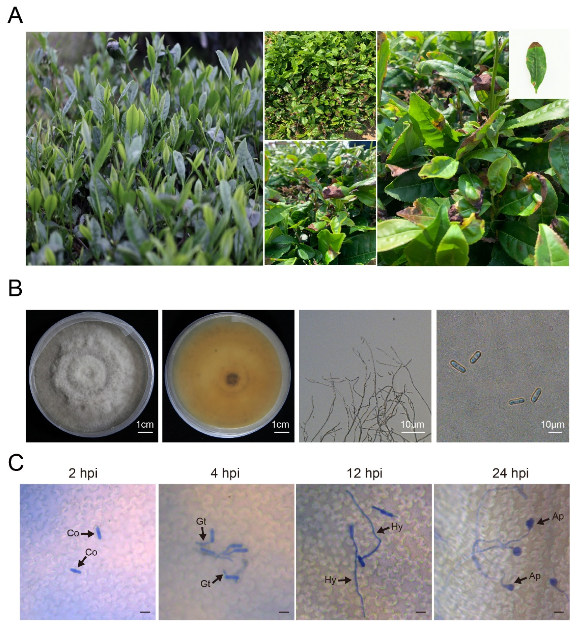
Figure 1. Characteristics of Colletotrichum gloeosporioides strains. (A) Healthy and anthracnose-infected tea plants. (B) Colony, upper and reverse, hypha, and conidia. (C) The development progress of Colletotrichum gloeosporioides in tea plant leaves. The arrows indicate the representation of the following: C, conidium; GT, germ tubes; H, hyphae; and A, appressorium. Scar bar = 10 μm.
Microbiolres 16 00220 g001
Figure 2. Effect of culture conditions on Colletotrichum gloeosporioides. (A) Effect of different temperatures on mycelial growth of Colletotrichum gloeosporioides. (B) Effect of different temperatures on spore production of Colletotrichum gloeosporioides. (C) Effect of different pH on mycelial growth of Colletotrichum gloeosporioides. (D) Effect of different pH on spore production of Colletotrichum gloeosporioides. (E) Inhibition of mycelial growth of Colletotrichum gloeosporioides by 80% tea saponin. (F) Inhibition of mycelial growth of Colletotrichum gloeosporioides by 10% polyoxin B. Note: Data with three biological replicates were introduced as mean values ± standard deviation (p < 0.05).
Microbiolres 16 00220 g002
Figure 3. Response of different resistant tea leaves to Colletotrichum gloeosporioides. (A) Disease incidence on leaves of the susceptible variety ‘Longjing 43’ and the resistant variety ‘Zhongcha 108’ at 2, 4, 6, and 8 dpi. Scar bar = 1 cm. (B) Growth and development of Colletotrichum gloeosporioides conidia on different resistant tea plant varieties. Scale bar = 10 μm. (C) H2O2 accumulation in tea leaves intercropped with Colletotrichum gloeosporioides. Scale bar = 500 μm. The red arrows indicate the accumulation sites. (D) O2 accumulation in tea plant leaves intercropped with Colletotrichum gloeosporioides. Scale bar = 500 μm.
Microbiolres 16 00220 g003
Figure 4. Effect of exogenous hormone treatment on Colletotrichum gloeosporioides. (A) Effect of exogenous MeJA on mycelial growth of Colletotrichum gloeosporioides. (B) Leaf phenotypes at 5 d exogenous MeJA treatment followed by inoculation with Colletotrichum gloeosporioides. (C) Peroxidase (POD) activity of ‘Longjing 43’ leaves after inoculation with Colletotrichum gloeosporioides. (D) Superoxide dismutase (SOD) activity of ‘Longjing 43’ leaves after inoculation with Colletotrichum gloeosporioides. (E) Malondialdehyde (MDA) content of ‘Longjing 43’ leaves after inoculation with Colletotrichum gloeosporioides. Note: Data with three biological replicates were introduced as mean values ± standard deviation (*, p < 0.05; **, p < 0.01), “ns” means no difference.
Microbiolres 16 00220 g004
Disclaimer/Publisher’s Note: The statements, opinions and data contained in all publications are solely those of the individual author(s) and contributor(s) and not of MDPI and/or the editor(s). MDPI and/or the editor(s) disclaim responsibility for any injury to people or property resulting from any ideas, methods, instructions or products referred to in the content.