Article

Phylogenetic Incongruence of Cyclic di-GMP-Activated Glycosyltransferase nfrB with 16S rRNA Gene Tree Reflects In Silico-Predicted Protein Structural Divergence in Diaphorobacter nitroreducens Isolated from Estero de Paco, Manila, Philippines

by
Ram Julius L. Marababol
and
Windell L. Rivera
*
Pathogen-Host-Environment Interactions Research Laboratory, Institute of Biology, College of Science, University of the Philippines Diliman, Quezon City 1101, Philippines
*
Author to whom correspondence should be addressed.
Microbiol. Res. 2025, 16(10), 212; https://doi.org/10.3390/microbiolres16100212
Submission received: 1 August 2025 / Revised: 14 September 2025 / Accepted: 23 September 2025 / Published: 26 September 2025

Abstract

Diaphorobacter nitroreducens is a Gram-negative bacterium ubiquitously found in wastewater, recognized for its ecological adaptability and potential applications in environmental, biomedical, and industrial processes. Central to its adaptability is the nfrB gene, which encodes a cyclic di-3′,5′-guanylate (c-di-GMP)-activated glycosyltransferase. This enzyme facilitates the secretion of biofilm-associated extracellular polymeric substances (EPS), essential for its survival and functionality in diverse environments. Using complete EMJH media as a selective medium, D. nitroreducens was successfully isolated from soil and water samples from Estero de Paco, Manila, Philippines, enabling downstream analyses of its nfrB gene. Phylogenetic analyses revealed that the nfrB gene tree deviates significantly from the canonical 16S rRNA gene tree, with D. nitroreducens clustering alongside members of the Enterobacteriaceae family. This deviation suggests the potential influence of horizontal gene transfer, adaptive evolution, or lineage-specific pressures on nfrB evolution. Structural analysis of NfrB through Alphafold 3 prediction demonstrated a conserved N-terminal region across taxa, except for the outgroup Zymomonas mobilis. Conversely, the C-terminal region, housing the catalytic domain, showed considerable diversity, reflecting adaptive modifications across bacterial lineages. Despite this variability, the putative binding site for cyclic di-3′,5′-guanylate remained conserved, indicating a balance between functional conservation and adaptive diversification. These findings not only deepen the existing understanding of bacterial signaling and glycosylation mechanisms but also provide insights into the evolutionary dynamics of glycosyltransferases. Furthermore, the study underscores the potential of NfrB as a target for innovative applications, including the design of novel biocatalysts and the development of informed strategies for bacterial management in environmental, industrial, and biotechnological contexts.

1. Introduction

Diaphorobacter nitroreducens, a Gram-negative bacillus commonly found in wastewater environments, has emerged as a promising microorganism with distinct ecological and biotechnological potential [1]. Renowned for its involvement in nitrogen cycling, pollutant degradation, and the utilization of complex organic matter, D. nitroreducens plays a pivotal role in maintaining ecosystem stability, driving biogeochemical cycles, and offering innovative solutions to environmental, industrial, and biomedical challenges [2,3]. Despite its promise, our understanding of the genetic and molecular mechanisms underlying its ecological resilience and functional capabilities remains sparse, limiting the ability to fully exploit this microorganism for applied purposes [2,4,5,6].
One of the key genetic elements contributing to the ecological adaptability of D. nitroreducens is the nfrB gene, which encodes a cyclic di-3′,5′-guanylate (c-di-GMP)-activated glycosyltransferase. This enzyme mediates the secretion of biofilm-associated extracellular polymeric substances (EPS), which are integral to microbial survival and functionality in diverse environments [7]. EPS facilitates pollutant degradation, including hydrocarbons, through increasing absorption ability, enhancing stress tolerance, and coordinating community-level behaviors, making it indispensable for environmental [8]. Understanding the regulatory and structural dynamics of this glycosyltransferase opens pathways for harnessing D. nitroreducens in applications such as biofilm engineering, pollutant remediation, and bioenergy production [9].
While the functional characteristics of nfrB have been studied with phage interactions and operon-level dynamics within Enterobacteriaceae, such as it being part of a multicomponent glycan synthase complex and how it facilitates N4 phage infection, its phylogenetic diversity and functional variations outside this phylum remain [7,10]. Expanding the phylogenetic understanding of nfrB is crucial for several reasons. First, it provides insights into the evolutionary trajectory of glycosyltransferases and their adaptations across bacterial lineages [11]. Second, it enhances our comprehension of microbial community dynamics and the ecological roles of non-model organisms like D. nitroreducens [12]. Finally, it allows for the identification of novel functionalities and biotechnological applications of glycosyltransferases beyond model systems, potentially broadening the scope of their utility [13].
This study aimed to bridge the gap in understanding by integrating phylogenetic and structural analyses of the nfrB gene in D. nitroreducens. By elucidating its evolutionary history and structural adaptations, a foundation for understanding its role in bacterial ecology and its potential applications in biotechnology will be provided. The findings aim to highlight the importance of studying non-model microorganisms to uncover novel biochemical pathways and evolutionary strategies that address critical challenges in environmental sustainability and industrial innovation.

2. Materials and Methods

2.1. Sample Collection

Sample collection was conducted at three distinct locations within Estero de Paco, Manila, Philippines, with the following coordinates: Sampling Site 1: 14.57842, 120.993883, Sampling Site 2: 14.578055, 120.993925, Sampling Site 3: 14.577889, 120.993932. At each site, approximately 25 g of soil and 50 milliliters of water were collected from proximate areas. The chosen sampling points were strategically selected close to densely populated informal settlements and significant commercial hubs, such as the Paco Market. Importantly, these sites are located upstream from the confluence with the Pasig River, a characteristic that contributes to the notably sluggish water flow during periods of low precipitation. These sampling sites were chosen because wastewater-impacted environments, such as those near densely populated informal settlements and busy commercial areas, provide favorable conditions for D. nitroreducens, which are commonly associated with sludge and nutrient-rich effluents. The upstream location and reduced flow further promote the accumulation of organic matter, making these areas representative hotspots for detecting the said bacteria [1,2].

2.2. Putative D. nitroreducens Isolation

To initiate the isolation process, soil samples were combined with an equal volume of sterile water. Subsequently, the mixture was allowed to undergo sedimentation, permitting the solid particles to settle within both the soil-water mixture and the water samples alone. The supernatant from these suspensions was then carefully filtered through a 2.2-micrometer pore size filter to remove large particulates and debris while allowing bacterial cells of interest to pass through. This filtrate was subsequently inoculated into a liquid culture medium composed of 2X EMJH base medium, supplemented with 0.2% agar, and 10X STAFF (Sulfamethoxazole, Trimethoprim, Amphotericin B, Fosfomycin, and 5-Fluorouracil). Although traditionally formulated for Leptospira, this was employed for the isolation of D. nitroreducens because of its minimal nutrient composition that supports the survival of oligotrophic bacteria. D. nitroreducens, frequently found in nutrient-limited environments such as wastewater sludge, can thrive in media with simple carbon and nitrogen sources. The presence of long-chain fatty acids (as carbon sources), ammonium salts (as nitrogen sources), and essential mineral salts in EMJH provides a balanced yet minimalistic environment that reduces competition from fast-growing heterotrophs, thereby favoring the growth and recovery of D. nitroreducens. The inoculated cultures were incubated at a controlled temperature of 30 °C for a duration of two to three weeks. The extended incubation time was necessary because D. nitroreducens is inherently a slow-growing organism, adapted to nutrient-limited and stable environments such as activated sludge. Furthermore, as a denitrifier with a narrow range of utilizable carbon sources and given the use of minimal medium, its growth required a longer period to reach culturable levels [2,14]. Throughout this incubation period, the cultures were monitored under a microscope to assess the presence of microbial growth.
Cultures exhibiting characteristics indicative of putative Diaphorobacter growth, specifically the presence of bacilli based on microscopic analysis, were selected for further investigation. These cultures were then transferred to a solid culture medium consisting of 2X EMJH base medium, with 10% sodium pyruvate, and 0.8% agar, with the addition of 10X STAFF. The inoculated plates were incubated at 30 °C for a period of two to three weeks. Colonies displaying translucent, transparent, or white appearances were carefully picked and subsequently reinoculated into the previously described liquid medium for subsequent analysis (n = 27).

2.3. DNA Extraction

Genomic DNA was extracted from the putative Diaphorobacter isolates using a modified boil lysis method as previously described by Veloso et al. (2000) [15]. Specifically, liquid cultures, incubated for a period of two to three weeks, were washed twice with PBS before being mixed with 1X TE buffer. The resulting mixture was then subjected to a high temperature of 100 °C for a duration of 15 min to facilitate cell lysis and DNA release. To precipitate the liberated DNA, the lysate was incubated under cold conditions in the presence of cold absolute ethanol. The precipitated DNA was subsequently recovered through centrifugation and resuspended in a sterile 1X TE buffer solution for optimal preservation. The purity and concentration of the DNA extracts were assessed using a Multiskan SkyHigh Spectrophotometer to ensure suitability for downstream applications. To preserve integrity and minimize degradation, the DNA samples were stored at −20 °C until further analysis.

2.4. Molecular Identification and Sequencing of Putative D. nitroreducens

The extracted DNA was subjected to polymerase chain reaction (PCR) amplification to confirm the identity of D. nitroreducens. Targeting the nfrB gene, the amplification process was initiated using nfrB-F (TTACCGCTCGAGGTGCTTTCGGTGGTCTGC) and nfrB-R (TGTTAACCCGGGTTACTTAGTCGCGTCAGA). The primers were designed as described by Green & Sambrook (2012) [16]. The PCR mixtures were prepared using GoTaq Green Master Mix (Promega, Madison, WI, USA) and subjected to thermal cycling in a T100™ Thermal Cycler (BioRad, Hercules, CA, USA). The amplification protocol consisted of an initial denaturation step at 94 °C for 5 min, followed by 35 cycles of denaturation at 94 °C for 30 s, annealing at 56 °C for 30 s, and extension at 72 °C for 30 s. A final extension step was performed at 72 °C for 10 min to complete the amplification process.
To corroborate the identity of the putative D. nitroreducens, the 16S rRNA gene was amplified using fD1 (CCGAATTCGTCGACAACAGAGTTTGATCCTGGCTCAG) and rD1 (CCCGGGATCCAAGCTTAAGGAGGTGATCCAGC) primers [17]. PCR conditions consisted of initial denaturation at 94 °C for 5 min, followed by 30 cycles of denaturation at 94 °C for 30 s, annealing at 55 °C for 30 s, extension at 72 °C for 2 min, and a final extension at 72 °C for 5 min. The amplified PCR products from both the nfrB and 16S rRNA gene amplifications were subjected to electrophoresis on a 1.5% agarose gel at 280 volts for 45 min to visualize and assess the amplicon sizes. The PCR products were sent to Macrogen, South Korea, for sequencing. All sequences obtained in this study were submitted to the NCBI GenBank database.

2.5. Phylogenetic and In Silico Protein Structure Analyses

Sequence alignment was performed using the ClustalW function in Bioedit v5.0.9 [18]. Model testing was performed using Jmodel v.2.1.10. The most optimal model in the Akaike Information Criterion (AIC) test was utilized for the subsequent analyses [19]. A qualitative test for oversaturation was performed using PAUP* v.4.0a to compute the corrected and uncorrected distances as well as the transition and transversion values [20]. Quantitative test for oversaturation was performed using DAMBE v.5.0.25 using its Xia Test function [21]. Construction of neighbor-joining and maximum parsimony trees was performed using PAUP* v.4.0a [19]. The Maximum Likelihood tree was constructed using PhyML v.3.1, while the Bayesian Inference tree was performed using MrBayes v.3.2.7a [22,23]. The tree was edited and visualized using iTOL v.7.0 [24]. The g1 test for detecting phylogenetic signal was performed using PAUP* v.4.0a, as well as the hypothesis testing using the Shimodaira-Hasegawa Test [20].
Protein structures were predicted using AlphaFold 3, and model quality was assessed based on predicted Template Modeling (pTM) and predicted Local Distance Difference Test (pLDDT) scores [25]. Predicted models with pTM values > 0.70 and pLDDT scores > 90 were selected for downstream analyses. The protein structures were visualized and aligned using PyMOL v.3.1.3, and models with root mean square deviation (r.m.s.d.) values < 2 were considered successfully superimposed [26].

3. Results

3.1. EMJH Media Supports D. nitroreducens Growth

Water and soil sample supernatants were incubated in complete liquid EMJH media to cultivate potential D. nitroreducens isolates. Initial microscopic observations revealed the presence of rod-shaped bacteria, which were subsequently plated on semi-solid EMJH media. Colonies exhibiting distinct morphologies, namely opaque white, transparent, and translucent appearances, were isolated for further examination. While colony morphology provides a practical basis for the preliminary selection of isolates, it is inherently non-specific and may not reliably reflect taxonomic identity or functional traits. Therefore, these observations were used only as an initial guide, with molecular analyses required to achieve accurate characterization.
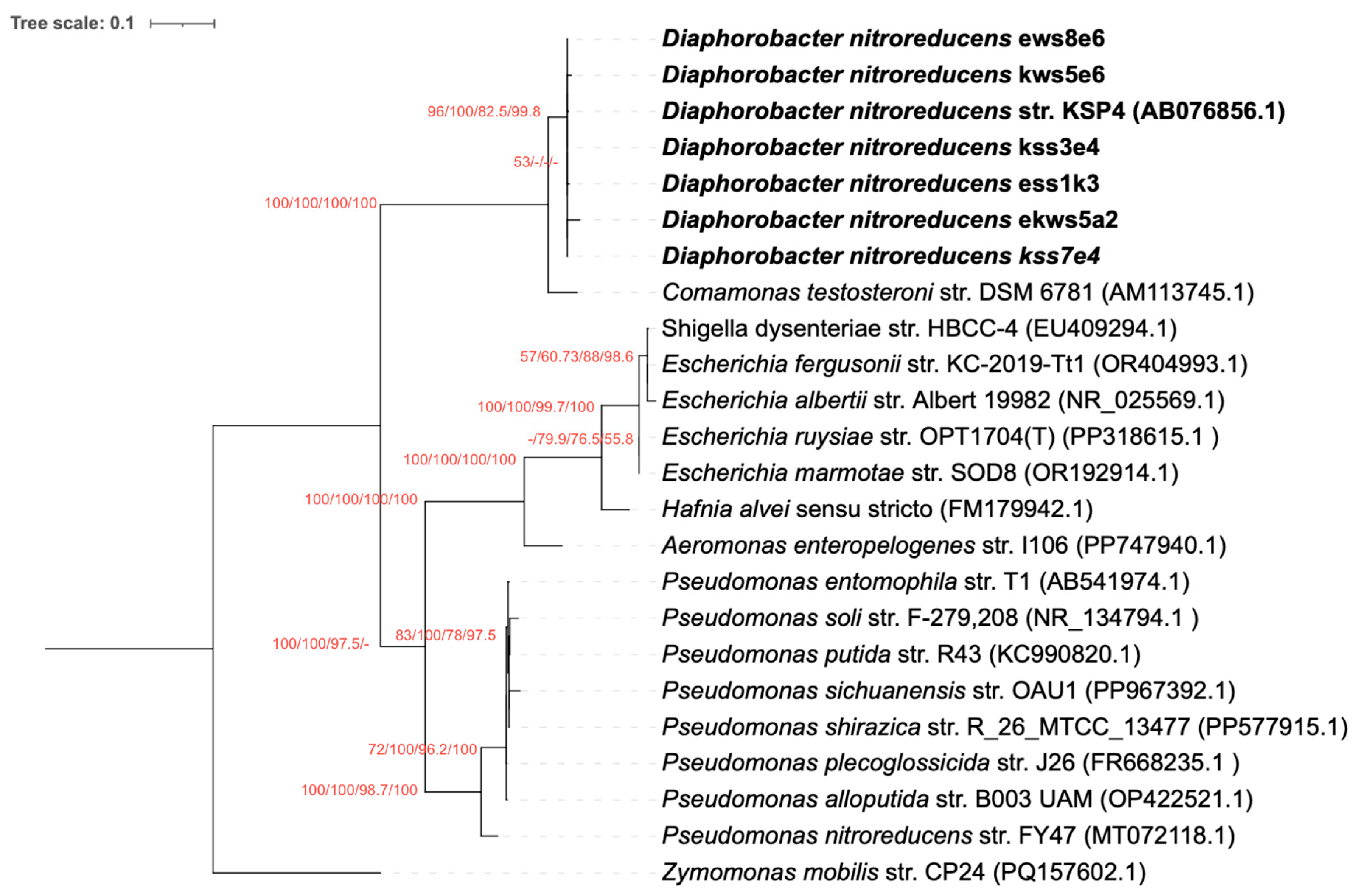
To confirm the identity of the isolates, PCR amplification of the nfrB gene, which encodes cyclic di-3′,5′-guanylate-activated glycosyltransferase, was performed. Of the 27 isolates obtained, 22 (81.48%) produced positive results in agarose gel electrophoresis. Sequencing of the PCR products, followed by BLAST+ v.2.16.0 analysis, showed high sequence identity (99–97%) to D. nitroreducens. Phylogenetic analysis was conducted using the nfrB gene sequences with four different methods: neighbor-joining, maximum parsimony, maximum likelihood, and Bayesian inference. The resulting phylogenetic trees displayed highly consistent topologies as reflected by their high bootstrap and posterior probability values (Figure 1), with all 22 positive isolates forming a monophyletic cluster alongside the D. nitroreducens reference sequence from the NCBI GenBank. High bootstrap values across all methods provided strong support for this clustering, confirming the isolates as phylogenetically closely related and belonging to the same species.

3.2. The nfrB Gene Trees Are Incongruent with the Canonical 16S rRNA Gene Phylogeny

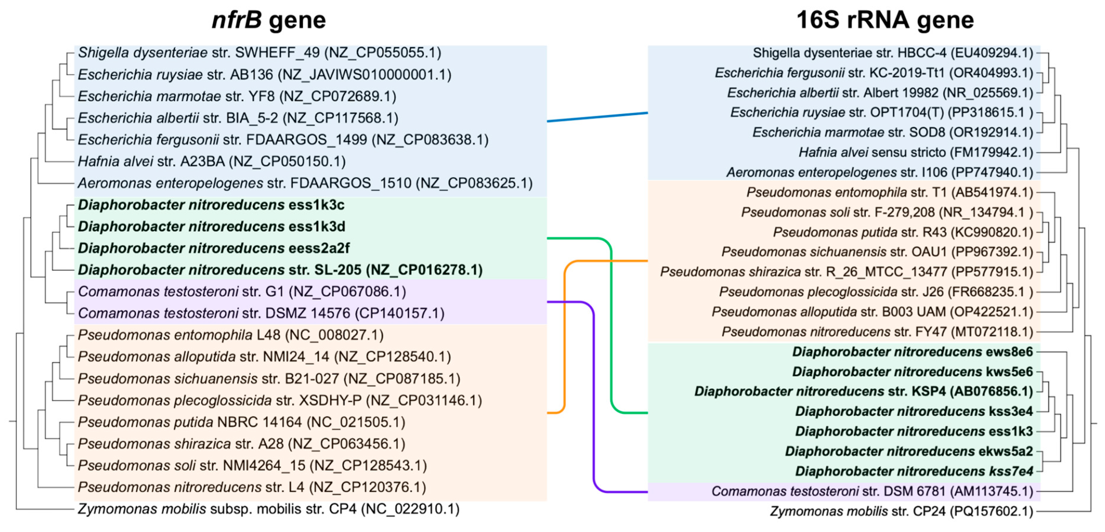
To assess the reliability of the nfrB phylogenetic tree (Figure 1), the 16S rRNA gene was amplified via PCR and sequenced for comparison. BLAST analysis of the 16S rRNA gene sequences revealed a high similarity (97–99%) to D. nitroreducens, consistent with the identification of the isolates. Phylogenetic trees based on the 16S rRNA gene were constructed using neighbor-joining, maximum parsimony, maximum likelihood, and Bayesian inference methods. These trees showed consistent topologies, with the isolates forming monophyletic clusters with D. nitroreducens reference sequences from the NCBI GenBank, supported by strong bootstrap values across all methods (Figure 2). Comparison of the 16S rRNA gene tree and the nfrB gene tree revealed notable differences in phylogenetic clustering, as shown in Figure 3. In the nfrB tree, D. nitroreducens isolates clustered distinctly with members of the Enterobacteriaceae family, forming a phylogenetic relationship not observed in the 16S rRNA gene tree. Conversely, the 16S rRNA gene phylogeny positioned Enterobacteriaceae closer to Pseudomonas, reflecting the canonical relationships based on overall bacterial phylogeny.
To determine the extent of the incongruence, the Shimodaira-Hasegawa (SH) test using RELL bootstrap (one-tailed test) was used to compare the topology of the optimal nfrB gene tree to the topology of the optimal 16S rRNA gene tree. This was performed by constraining the nfrB gene tree to the topology of the optimal 16S rRNA gene tree. Results showed that the constrained tree has statistically significantly lower log-likelihood as seen in Table 1. This indicates that the nfrB sequence data is a significantly poorer fit under the 16S rRNA-based constraint than under the optimal nfrB topology. This statistical outcome demonstrates that the nfrB gene tree is not merely a stochastic variant of the 16S rRNA tree but instead reflects a fundamentally different evolutionary signal. These differences suggest that the nfrB gene phylogeny may be influenced by factors beyond species-level evolutionary divergence, potentially including horizontal gene transfer or selective pressures unique to this gene. The deviation from 16S rRNA gene-based phylogeny underscores the potential functional or ecological adaptations shaping nfrB gene evolution, distinguishing it from the broader genomic context represented by the 16S rRNA gene.

3.3. Overall Structure of Cyclic di-3′,5′-Guanylate-Activated Glycosyltransferase NfrB of D. nitroreducens

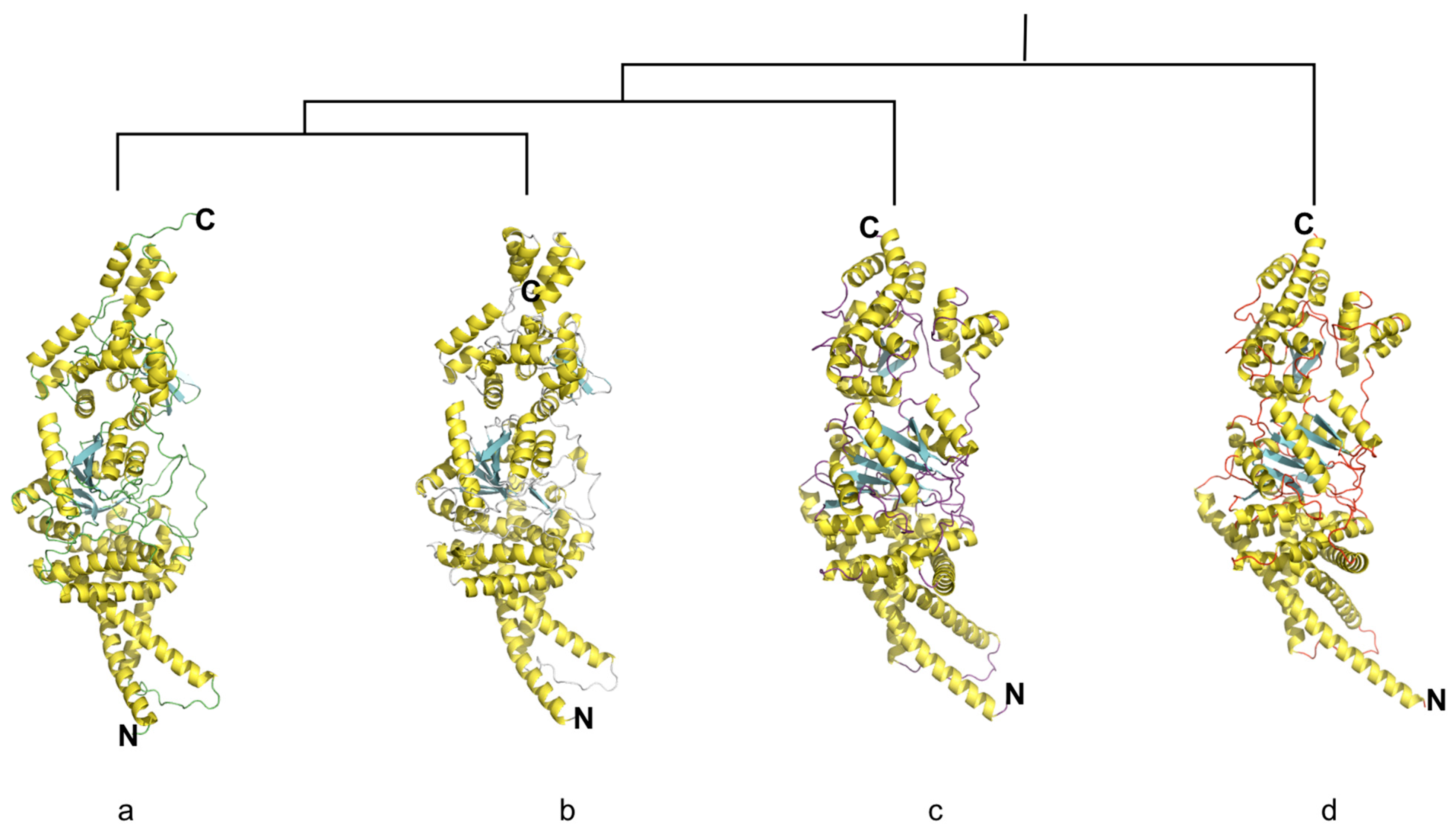
The structure of cyclic di-3′,5′-guanylate-activated glycosyltransferase NfrB was predicted using AlphaFold 3. Representative sequences from four clades: D. nitroreducens str. SL-205 (NZ_CP016278.1), E. ruysiae str. AB136 (NZ_JAVIWS010000001.1), P. sichuanensis str. NMI24_14 (NZ_CP087185.1), and Z. mobilis subsp. mobilis str. CP4 (NC_022910.1) were analyzed. The predicted structures exhibited high confidence, with pTM scores of 0.88, 0.87, 0.88, and 0.90, respectively. The majority of the predicted models had confident pIDDT values, indicating reliable structural predictions (Figure 4).
The structure of NfrB in D. nitroreducens was characterized in detail. It comprises 11 β-strands, 29 α-helices, and 40 310 helices, as determined through secondary structure analysis using PyMOL. The topology of the NfrB protein is consistent with that of other representatives from different clades, suggesting conserved structural features across homologs. The predicted surface area of NfrB in D. nitroreducens is 76,818.234 Å2, with approximately 50.26% of the surface area being solvent-accessible based on calculations using PyMOL. The explicit molecular mass of the protein is 77,412.0671 Da, with a formal charge of −2. These findings indicate a highly stable and accessible protein structure, consistent with its enzymatic role in cyclic di-3′,5′-guanylate-mediated glycosyltransferase activity.

3.4. The Structure of Putative Binding Site Domains of NfrB Is Conserved Among Phyla

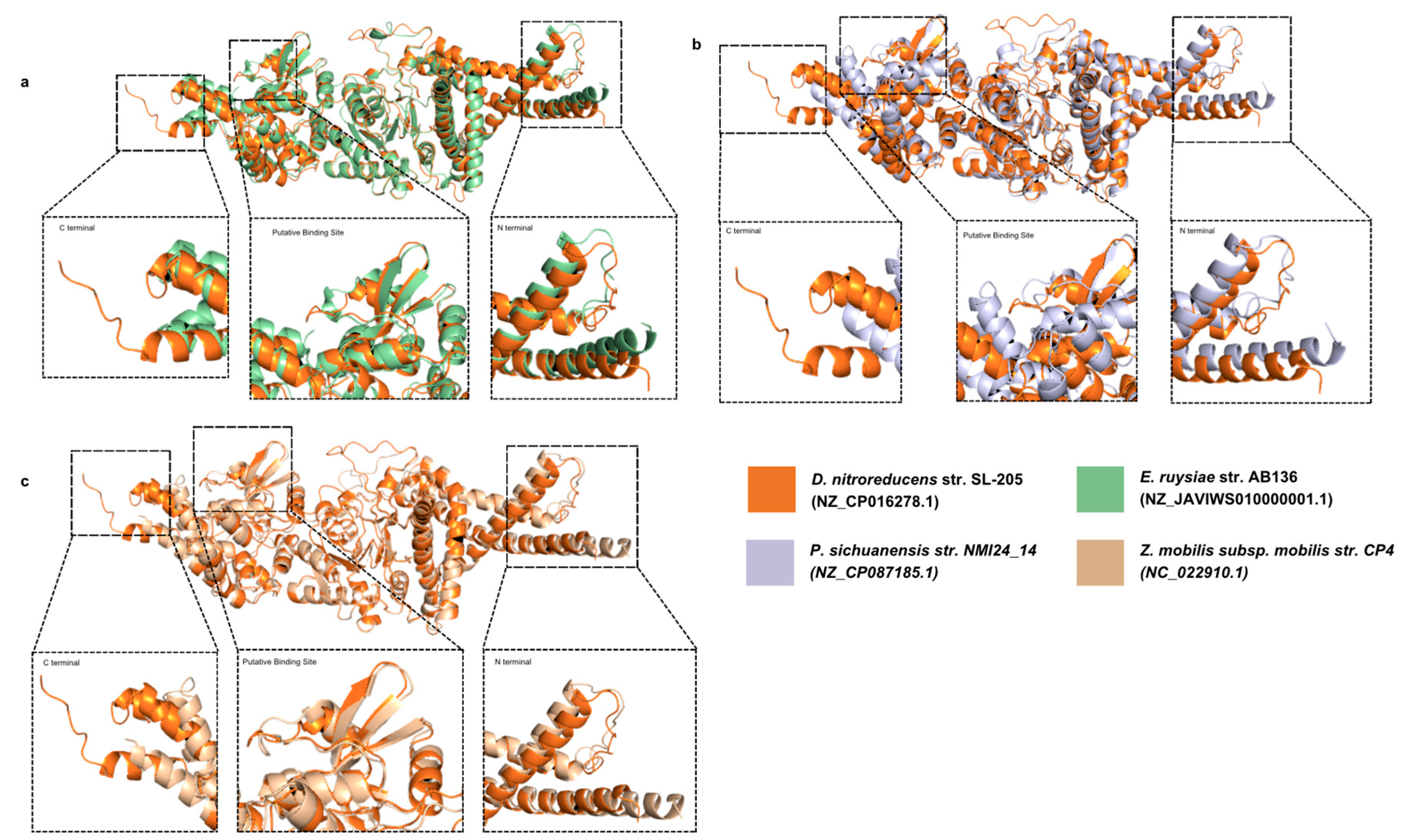
Structural analysis of the NfrB glycosyltransferase of D. nitroreducens revealed significant conservation across representative sequences from three other phylogenetic clades: E. ruysiae, P. sichuanensis, and Z. mobilis. Superimposition of the DnGT structure with these sequences yielded root mean square deviation (r.m.s.d.) values of 0.911 Å (4142 Cα atoms), 1.473 Å (3931 Cα atoms), and 0.589 Å (3505 Cα atoms), respectively. These r.m.s.d. values underscore a high degree of structural similarity, although variations were observed, primarily in the N- and C-terminal regions and the overall core domain conformation (Figure 5). Despite these differences, the putative binding site of the glycosyltransferase domain is conserved across the sequences, as evidenced by the alignment of secondary structures within these residues. The conserved binding site underscores the functional importance of NfrB in bacterial physiology.
The N-terminal region exhibited notable divergence, particularly between D. nitroreducens and the outgroup Z. mobilis. The N-terminal of Z. mobilis was approximately six residues longer than that of D. nitroreducens, suggesting species-specific structural adaptations. The C-terminal regions displayed even greater variability among the analyzed sequences. The NfrB structure of E. ruysiae included an additional α-helix (residues 731–742) at the C-terminal region, absent in the other sequences. Conversely, the C-terminal regions of P. sichuanensis and Z. mobilis were truncated compared to D. nitroreducens. Specifically, the last α-helix of P. sichuanensis (residues 698–722) aligned with two α-helices (residues 706–719 and 723–732) and a 310 helix (residues 720–722) in D. nitroreducens. Surprisingly, the final α-helix of Z. mobilis (residues 703–727) superimposed with a single α-helix in D. nitroreducens.
The core glycosyltransferase domain further highlighted structural distinctions. The solvent-accessible surface area ratios showed that D. nitroreducens had a more exposed and accessible glycosyltransferase domain compared to the other sequences, reflecting differences in its conformation. Surface potential charge analysis further corroborated these findings, demonstrating that the core glycosyltransferase domain in D. nitroreducens had a larger exposed surface area relative to its counterparts. These structural insights emphasize the conservation of the glycosyltransferase domain across diverse bacterial taxa while highlighting subtle species-specific adaptations that may reflect evolutionary strategies to optimize substrate binding and catalytic efficiency under distinct environmental conditions.

4. Discussion

D. nitroreducens is an increasingly recognized Gram-negative bacterium with significant potential in environmental, industrial, and biotechnological applications [3,27]. Known for its metabolic versatility, including nitrite reduction and complex substrate utilization, this organism is poised to address challenges in bioremediation and sustainable industrial processes [2]. This study represents a critical step forward in elucidating the genetic, structural, and functional characteristics of the nfrB gene, encoding cyclic di-3′,5′-guanylate-activated glycosyltransferase, which plays a pivotal role in bacterial signaling and glycosylation processes. These findings expand our understanding of D. nitroreducens and offer insights into leveraging its potential for scientific and industrial advancements.
The ability of D. nitroreducens to grow robustly in complete EMJH media highlights its metabolic flexibility and adaptability to a defined nutrient environment, offering a significant advance in its isolation and study. Complete EMJH media, originally formulated for culturing Leptospira species, provides a unique blend of nutrients that mimic natural environmental conditions, supporting the growth of bacteria [28]. This development addresses a long-standing challenge in microbiological studies, where general-purpose media often fail to provide the selective pressures or specific nutrients required to cultivate and isolate niche-adapted microorganisms. Previous studies relied heavily on general media formulations, such as LB broth, which are optimized for rapidly growing and metabolically versatile microbes [1]. However, these media do not selectively suppress competing organisms, which may result in low isolation success rates and contamination by faster-growing species [29]. This lack of specificity not only complicates the study of D. nitroreducens but also limits the reproducibility of experiments and the ability to obtain pure cultures for in-depth analysis. By utilizing complete EMJH media, this study demonstrates a practical and reproducible method for isolating D. nitroreducens, allowing for more targeted investigations of its physiology, metabolic pathways, and ecological roles. This medium supports its growth while selectively favoring its isolation over less adaptable species, creating a controlled environment that enhances research efficiency [28,30]. Furthermore, the robust growth observed in EMJH media reflects the bacterium’s capacity to thrive in nutrient-rich and defined environments, suggesting potential applications in industrial or environmental settings where tailored media formulations can be optimized for specific processes involving D. nitroreducens [31].
The use of nfrB was recently found to encode an initial receptor for phage N4 infection, and a c-di-GMP-controlled glycosyltransferase that synthesizes EPS, which impedes motility [7]. However, its use for systematic identification of microorganisms has not been explored. The use of nfrB amplification to identify isolates and its consistent clustering with D. nitroreducens sequences from databases in phylogenetic analyses highlights its genetic stability and specificity. These results validate nfrB as a reliable marker for studying strain diversity and provide a solid foundation for future genetic and functional studies. Furthermore, the observed discrepancies between nfrB- and 16S rRNA gene-based phylogenies offer critical insights into microbial evolution. While the 16S rRNA gene remains the gold standard for taxonomic classification, the distinct evolutionary trajectory of nfrB underscores the limitations of single-marker phylogenies. The deviations suggest that nfrB evolution may be influenced by horizontal gene transfer, adaptive evolution, or other lineage-specific pressures, which is consistent with nfrB association with bacteriophage infection, which likely confers horizontal gene transfer [10]. This highlights the need for multi-gene phylogenetic approaches to more accurately capture the complexity of microbial relationships, as single-gene trees may not fully represent species history. Indeed, multi-locus sequence analysis (MLSA) and other concatenated-gene approaches are now widely accepted as more robust frameworks for bacterial phylogeny [27,32,33].
The structural characterization of NfrB using AlphaFold 3 and comparative analyses with representative sequences from other clades further emphasize its evolutionary significance. The conserved core glycosyltransferase domain, coupled with variations in the N- and C-terminal regions and core domain, reflects a balance between functional conservation and adaptive diversification. The N-terminal having a more consistent conformation among phyla, except for the outgroup, is consistent with how the N-terminal region of glycosyltransferases is generally immobilized [34]. Furthermore, the diversity within the C-terminals is attributed to its inclusion of a catalytic domain, which makes it very sensitive to modification [35,36]. These structural adaptations, in addition to the unique conformation of core glycosyltransferase in D. nitroreducens, may enhance substrate interaction and catalytic efficiency, which are crucial for the organism’s ecological and functional roles. Such features could be harnessed for biotechnological applications, including biofilm engineering, enzymatic biocatalysis, and synthetic biology [37]. Cyclic di-3′,5′-guanylate, as a second messenger, regulates vital bacterial processes, including biofilm formation, motility, and exopolysaccharide production [7]. The functional insights into NfrB underscore its potential as a target for modulating these processes in D. nitroreducens. For example, controlled manipulation of glycosylation pathways could optimize exopolysaccharide production, enhancing biofilm stability or biodegradability for environmental and industrial purposes [38]. Additionally, the structural differences observed in NfrB among representative clades could inform future efforts to develop engineered strains tailored to specific applications, such as pollutant degradation or specialized metabolite production. Further experimental validation will be required to determine whether these structural features can be leveraged for such purposes.
The findings of this study have broader implications for microbial ecology and biotechnology. Understanding the functional and structural diversity of NfrB not only deepens our knowledge of bacterial signaling and glycosylation but also opens avenues for innovative applications. For instance, the conserved glycosyltransferase domain may serve as a model for studying enzyme–substrate interactions in other bacterial systems, while the unique features of D. nitroreducens NfrB could inspire the design of novel biocatalysts [36]. Furthermore, the ecological role of NfrB in biofilm formation and environmental adaptability could inform strategies for microbiome management in agriculture, wastewater treatment, and industrial [13]. Importantly, this study contributes uniquely by providing a comparative structural perspective on NfrB across diverse taxa using in silico predictions. Nevertheless, experimental validation is needed to confirm the efficiency of NfrB activity in different lineages, to elucidate the mechanistic basis of its evolution, and to assess the functional relevance of the observed structural differences.

5. Conclusions

This study sheds light on the evolutionary and structural nuances of the cyclic di-3′,5′-guanylate-activated glycosyltransferase nfrB in D. nitroreducens, a bacterium of growing biotechnological and ecological importance. By juxtaposing the nfrB phylogeny against the canonical 16S rRNA gene tree, we uncovered distinct evolutionary trajectories suggestive of functional specialization, potentially shaped by horizontal gene transfer and adaptive evolution. Importantly, statistical support from the Shimodaira–Hasegawa (SH) test confirmed that the nfrB phylogeny is significantly incongruent with the 16S rRNA gene tree, demonstrating that these deviations are not merely descriptive but quantitatively validated. These findings underscore the limitations of single-marker phylogenetic analyses and advocate for multi-gene approaches to unravel the complexities of microbial evolution.
The in silico structural elucidation of NfrB, based on sequence, revealed a conserved glycosyltransferase binding site domain alongside species-specific adaptations in the N- and C-terminal regions. These structural insights, paired with functional attributes, highlight the role of NfrB in mediating biofilm formation and ecological resilience. Such features are not only pivotal for bacterial survival in diverse environments but may also offer future applications in biofilm engineering, pollutant degradation, and synthetic biology. Beyond expanding knowledge of D. nitroreducens, this study establishes a framework for exploring the genetic underpinnings of glycosyltransferases across bacterial lineages. This integrated approach paves the way for leveraging non-model organisms to address pressing challenges in environmental sustainability and industrial innovation, reaffirming the need for comprehensive studies at the intersection of phylogenetics, structural biology, and applied microbiology.

Author Contributions

Conceptualization, R.J.L.M. and W.L.R.; Methodology, R.J.L.M.; Software, R.J.L.M.; Validation, W.L.R.; Formal Analysis, R.J.L.M.; Investigation, R.J.L.M.; Resources, W.L.R.; Data Curation, R.J.L.M.; Writing—Original Draft Preparation, R.J.L.M. and W.L.R.; Writing—Review and Editing, W.L.R.; Visualization, R.J.L.M.; Supervision, W.L.R.; Project Administration, W.L.R.; Funding Acquisition, W.L.R. All authors have read and agreed to the published version of the manuscript.

Funding

This study was generously funded by the Department of Science and Technology, Philippines (Project No. 9258).

Institutional Review Board Statement

Not applicable.

Data Availability Statement

The original contributions presented in this study are included in the article. Further inquiries can be directed to the corresponding author.

Acknowledgments

We extend our sincere thanks to the research staff of the Pathogen-Host-Environment Interactions Research Laboratory (PHEIRL), particularly Chembie A. Almazar and Yvette B. Montala, for their valuable technical assistance. We also gratefully acknowledge the technical support provided by Ian Kendrich C. Fontanilla.

Conflicts of Interest

The authors declare no conflicts of interest.

References

  1. Khan, S.T.; Hiraishi, A. Diaphorobacter nitroreducens gen. nov., sp. nov., a poly(3-hydroxybutyrate)-degrading denitrifying bacterium isolated from activated sludge. J. Gen. Appl. Microbiol. 2002, 48, 299–308. [Google Scholar] [CrossRef]
  2. Khan, S.T.; Nagao, Y.; Hiraishi, A. Nitrate removal performance of Diaphorobacter nitroreducens using biodegradable plastics as the source of reducing power. AIP Conf. Proc. 2015, 1649, 71–78. [Google Scholar] [CrossRef]
  3. Ni, Y.; Li, R.; Shen, X.; Yi, D.; Ren, Y.; Wang, F.; Geng, Y.; You, Q. Diaphorobacter nitroreducens synergize with oxaliplatin to reduce tumor burden in mice with lung adenocarcinoma. mSystems 2024, 9, e0132323. [Google Scholar] [CrossRef]
  4. Khardenavis, A.A.; Kapley, A.; Purohit, H.J. Simultaneous nitrification and denitrification by diverse Diaphorobacter sp. Appl. Microbiol. Biotechnol. 2007, 77, 403–409. [Google Scholar] [CrossRef]
  5. Singh, D.; Ramanathan, G. Catechol 2,3-dioxygenase isoenzymes from Diaphorabacter sp. strain DS2 demonstrate different substrate preferences. Biochem. Biophys. Res. Commun. 2025, 782, 152591. [Google Scholar] [CrossRef] [PubMed]
  6. Li, J.; Hong, M.; Lv, J.; Tang, R.; Wang, R.; Yang, Y.; Liu, N. Enhancement on migration and biodegradation of Diaphorobacter sp. LW2 mediated by Pythium ultimum in soil with different particle sizes. Front. Microbiol. 2024, 15, 1391553. [Google Scholar] [CrossRef]
  7. Junkermeier, E.H.; Hengge, R. A Novel locally c-di-GMP-controlled exopolysaccharide synthase required for bacteriophage N4 infection of Escherichia coli. mBio 2021, 12, e03249-21. [Google Scholar] [CrossRef]
  8. Huang, L.; Jin, Y.; Zhou, D.; Liu, L.; Huang, S.; Zhao, Y.; Chen, Y. A review of the role of extracellular polymeric substances (EPS) in wastewater treatment systems. Int. J. Environ. Res. Public Health 2022, 19, 12191. [Google Scholar] [CrossRef] [PubMed]
  9. Wen, L.; Huang, Y.; Wang, W.; Zhang, L.; Xu, J.; Li, Z.; Xu, P.; Tang, H. A novel Diaphorobacter sp. strain isolated from saponification wastewater shows highly efficient phenanthrene degradation. Environ. Res. 2022, 214, 114047. [Google Scholar] [CrossRef]
  10. Sellner, B.; Prakapaitė, R.; Van Berkum, M.; Heinemann, M.; Harms, A.; Jenal, U. A new sugar for an old phage: A c-di-GMP-dependent polysaccharide pathway sensitizes Escherichia coli for bacteriophage infection. mBio 2021, 12, e03246-21. [Google Scholar] [CrossRef] [PubMed]
  11. Zhu, F.; Wu, R.; Zhang, H.; Wu, H. Structural and biochemical analysis of a bacterial glycosyltransferase. In Methods in Molecular Biology; Humana Press: Totowa, NJ, USA, 2013; pp. 29–39. [Google Scholar] [CrossRef]
  12. Yakovlieva, L.; Walvoort, M.T.C. Processivity in bacterial glycosyltransferases. ACS Chem. Biol. 2019, 15, 3–16. [Google Scholar] [CrossRef]
  13. Schmid, J.; Heider, D.; Wendel, N.J.; Sperl, N.; Sieber, V. Bacterial glycosyltransferases: Challenges and opportunities of a highly diverse enzyme class toward tailoring natural products. Front. Microbiol. 2016, 7, 182. [Google Scholar] [CrossRef]
  14. Rezvani, F.; Sarrafzadeh, M.-H.; Ebrahimi, S.; Oh, H.-M. Nitrate removal from drinking water with a focus on biological methods: A review. Environ. Sci. Pollut. Res. 2017, 26, 1124–1141. [Google Scholar] [CrossRef]
  15. Veloso, I.; Lopes, M.; Salas, C.; Moreira, E. A comparison of three DNA extractive procedures with Leptospira for polymerase chain reaction analysis. Mem. Inst. Oswaldo Cruz 2000, 95, 339–343. [Google Scholar] [CrossRef] [PubMed]
  16. Green, M.R.; Sambrook, J. Molecular Cloning: A Laboratory Manual, 4th ed.; Cold Spring Harbor Laboratory Press: New York, NY, USA, 2012. [Google Scholar]
  17. Morey, R.E.; Galloway, R.L.; Bragg, S.L.; Steigerwalt, A.G.; Mayer, L.W.; Levett, P.N. Species-specific identification of Leptospiraceae by 16S RRNA gene sequencing. J. Clin. Microbiol. 2006, 44, 3510–3516. [Google Scholar] [CrossRef] [PubMed]
  18. Hall, T.A. Bioedit: A User-friendly biological sequence alignment editor and analysis program for Windows 95/98/NT. Nucleic Acids Symp. Ser. 1999, 41, 95–98. [Google Scholar]
  19. Darriba, D.; Taboada, G.L.; Doallo, R.; Posada, D. jModelTest 2: More models, new heuristics and parallel computing. Nat. Methods 2012, 9, 772. [Google Scholar] [CrossRef]
  20. Cummings, M.P. PAUP * (Phylogenetic analysis using parsimony (and other methods)). In Dictionary of Bioinformatics and Computational Biology; John Wiley & Sons: Hoboken, NJ, USA, 2004. [Google Scholar] [CrossRef]
  21. Xia, X. DAMBE7: New and improved tools for data analysis in molecular biology and evolution. Mol. Biol. Evol. 2018, 35, 1550–1552. [Google Scholar] [CrossRef]
  22. Guindon, S.; Dufayard, J.-F.; Lefort, V.; Anisimova, M.; Hordijk, W.; Gascuel, O. New algorithms and methods to estimate maximum-likelihood phylogenies: Assessing the performance of PHYML 3.0. Syst. Biol. 2010, 59, 307–321. [Google Scholar] [CrossRef]
  23. Ronquist, F.; Teslenko, M.; Van Der Mark, P.; Ayres, D.L.; Darling, A.; Höhna, S.; Larget, B.; Liu, L.; Suchard, M.A.; Huelsenbeck, J.P. MrBayes 3.2: Efficient Bayesian phylogenetic inference and model choice across a large model space. Syst. Biol. 2012, 61, 539–542. [Google Scholar] [CrossRef]
  24. Letunic, I.; Bork, P. Interactive Tree of Life (iTOL) v6: Recent updates to the phylogenetic tree display and annotation tool. Nucleic Acids Res. 2024, 52, W78–W82. [Google Scholar] [CrossRef]
  25. Abramson, J.; Adler, J.; Dunger, J.; Evans, R.; Green, T.; Pritzel, A.; Ronneberger, O.; Willmore, L.; Ballard, A.J.; Bambrick, J.; et al. Accurate structure prediction of biomolecular interactions with AlphaFold 3. Nature 2024, 630, 493–500. [Google Scholar] [CrossRef]
  26. Delano, W.L. The PyMOL Molecular Graphics System. 2002. Available online: https://ci.nii.ac.jp/naid/10020095229/ (accessed on 14 December 2024).
  27. Wang, J.; Chu, L. Biological nitrate removal from water and wastewater by solid-phase denitrification process. Biotechnol. Adv. 2016, 34, 1103–1112. [Google Scholar] [CrossRef]
  28. Guedes, I.B.; De Souza, G.O.; De Paula Castro, J.F.; Cavalini, M.B.; De Souza Filho, A.F.; Aizawa, J.; Cortez, A.; Heinemann, M.B. Improvement of the enrichment used in the EMJH medium (Ellinghausen–McCullough–Johnson–Harris) for the cultivation of Leptospira spp. Rev. Argent. Microbiol. 2021, 54, 95–99. [Google Scholar] [CrossRef]
  29. Heylen, K.; Vanparys, B.; Wittebolle, L.; Verstraete, W.; Boon, N.; De Vos, P. Cultivation of denitrifying bacteria: Optimization of isolation conditions and diversity study. Appl. Environ. Microbiol. 2006, 72, 2637–2643. [Google Scholar] [CrossRef]
  30. Loureiro, A.P.; Martins, G.; Pinto, P.; Narduche, L.; Teixeira, R.C.; Lilenbaum, W. Usage of a selective media (EMJH-STAFF) in primary culturing of pathogenic leptospires from bovine clinical samples. Lett. Appl. Microbiol. 2015, 61, 603–606. [Google Scholar] [CrossRef]
  31. Erşan, Y.Ç.; De Belie, N.; Boon, N. Microbially induced CaCO3 precipitation through denitrification: An optimization study in minimal nutrient environment. Biochem. Eng. J. 2015, 101, 108–118. [Google Scholar] [CrossRef]
  32. Maiden, M.C.J.; Van Rensburg, M.J.J.; Bray, J.E.; Earle, S.G.; Ford, S.A.; Jolley, K.A.; McCarthy, N.D. MLST revisited: The gene-by-gene approach to bacterial genomics. Nat. Rev. Microbiol. 2013, 11, 728–736. [Google Scholar] [CrossRef]
  33. Teichman, S.; Lee, M.D.; Willis, A.D. Analyzing microbial evolution through gene and genome phylogenies. Biostatistics 2023, 25, 786–800. [Google Scholar] [CrossRef]
  34. Shimma, Y.-I.; Saito, F.; Oosawa, F.; Jigami, Y. Construction of a library of human glycosyltransferases immobilized in the cell wall of Saccharomyces cerevisiae. Appl. Environ. Microbiol. 2006, 72, 7003–7012. [Google Scholar] [CrossRef]
  35. Mollicone, R.; Gibaud, A.; François, A.; Ratcliffe, M.; Oriol, R. Acceptor specificity and tissue distribution of three human α-3-fucosyltransferases. Eur. J. Biochem. 1990, 191, 169–176. [Google Scholar] [CrossRef] [PubMed]
  36. Bencúrová, M.; Rendić, D.; Fabini, G.; Kopecky, E.-M.; Altmann, F.; Wilson, I.B.H. Expression of eukaryotic glycosyltransferases in the yeast Pichia Pastoris. Biochimie 2003, 85, 413–422. [Google Scholar] [CrossRef] [PubMed]
  37. Hong, S.; Shi, Y.; Wu, N.C.; Grande, G.; Douthit, L.; Wang, H.; Zhou, W.; Sharpless, K.B.; Wilson, I.A.; Xie, J.; et al. Bacterial glycosyltransferase-mediated cell-surface chemoenzymatic glycan modification. Nat. Commun. 2019, 10, 1799. [Google Scholar] [CrossRef] [PubMed]
  38. Anyaogu, D.C.; Mortensen, U.H. Manipulating the glycosylation pathway in bacterial and lower eukaryotes for production of therapeutic proteins. Curr. Opin. Biotechnol. 2015, 36, 122–128. [Google Scholar] [CrossRef] [PubMed]
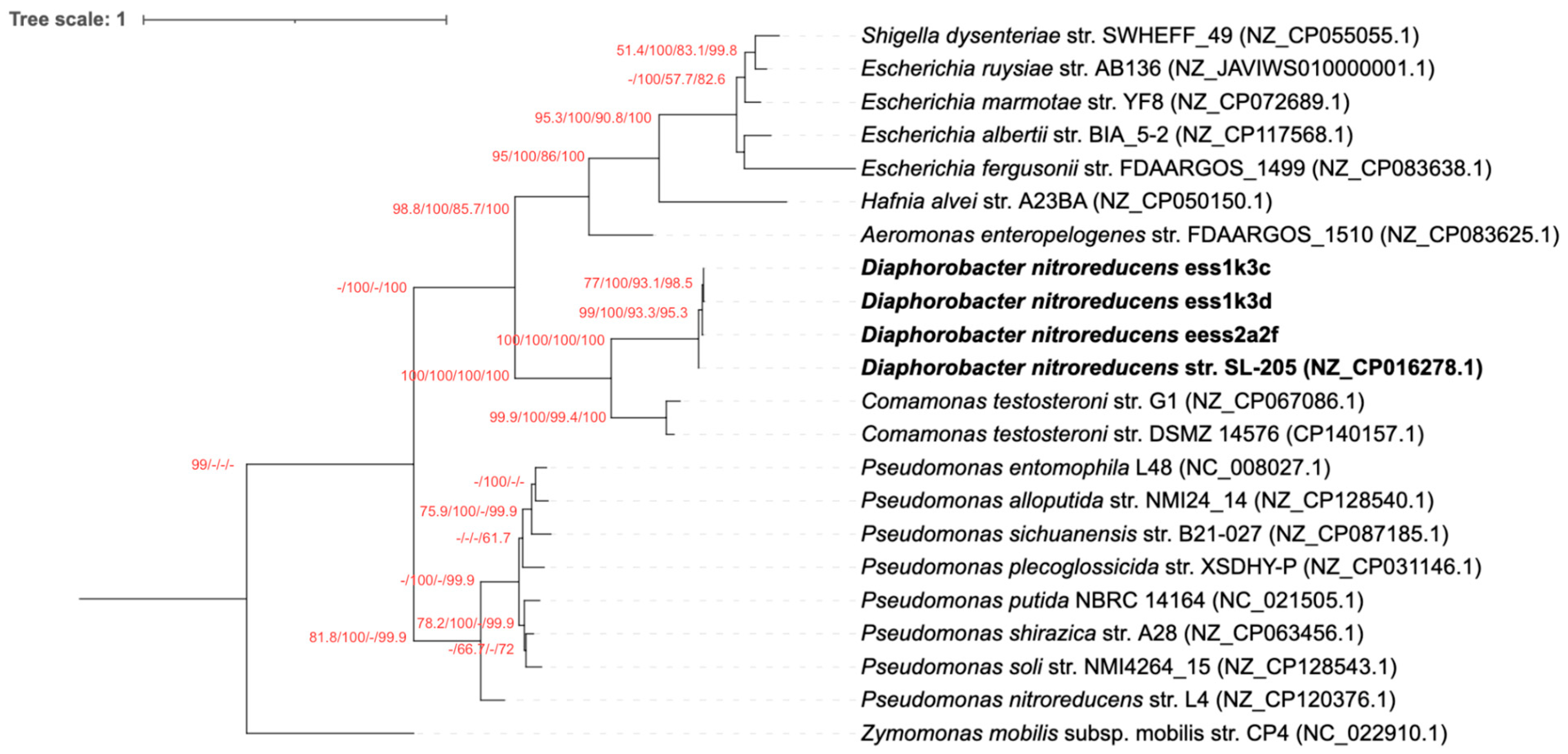
Figure 1. Maximum likelihood tree of nfrB genes. The tree is based on 699 nucleotides of the bacterial cyclic di-3′,5′-guanylate-activated glycosyltransferase nfrB gene and uses the TIM3+I+G as the model of DNA substitution. The tree is rooted in Zymomonas mobilis subsp. mobilis strain CP4 (NC_022910.1). Identical sequences are represented with only one sequence. Values on nodes represent percentage out of 1000 bootstrap samples in neighbor joining, maximum parsimony, maximum likelihood, and Bayesian inference trees, respectively (values less than 50% are not shown). The scale bar represents one nucleotide substitution for every nucleotide.
Microbiolres 16 00212 g001
Figure 2. Maximum likelihood tree of 16S rRNA genes. The tree is based on 1178 nucleotides of the bacterial 16S rRNA gene and uses the GTR+I+G as its model of DNA substitution. The tree is rooted on Zymomonas mobilis strain CP4 (NC_022910.1). Identical sequences are represented with only one sequence. Values on nodes represent percentage out of 1000 bootstrap samples in neighbor-joining, maximum parsimony, maximum likelihood, and Bayesian inference trees, respectively (values less than 50% are not shown). The scale bar represents one nucleotide substitution every ten nucleotides.
Microbiolres 16 00212 g002
Figure 3. Comparison of the maximum likelihood tree topology of nfrB and 16S rRNA gene. Branch lengths are ignored. The tree is divided into four clusters: the Enterobacteriaceae (blue), Pseudomonas (orange), Diaphorobacter (green), and Comamonas (violet). Identical sequences are represented with only one sequence.
Microbiolres 16 00212 g003
Figure 4. Cartoon representation of secondary structural elements of the cyclic di-3′,5′-guanylate-activated glycosyltransferase NfrB of (a) D. nitroreducens str. SL-205 (NZ_CP016278.1) (b) E. ruysiae str. AB136 (NZ_JAVIWS010000001.1) (c) P. sichuanensis str. NMI24_14 (NZ_CP087185.1) (d) Z. mobilis subsp. mobilis str. CP4 (NC_022910.1). N- and C-termini of the proteins are labeled.
Microbiolres 16 00212 g004
Figure 5. Structural superposition of NfrB proteins from three representative bacterial sequences with D. nitroreducens strain SL-205. (a) superposition with E. ruysiae strain AB136, (b) superposition with P. sichuanensis strain NMI24_14, and (c) superposition with Z. mobilis subsp. mobilis strain CP4. In each comparison, N- and C-termini and putative binding sites are highlighted in enlarged views.
Microbiolres 16 00212 g005
Table 1. Shimodaira–Hasegawa test results using RELL bootstrap (one-tailed test) for the nfrB gene phylogeny. Tree 1 represents the topology constrained to group Enterobacteriaceae with Pseudomonas, following the optimal 16S rRNA gene tree shown in Figure 2. Tree 2 corresponds to the optimal nfrB gene tree shown in Figure 1. * p < 0.05 indicates significant rejection of the tested topology.
Tree−ln LΔ −ln LSHWtd-SH
26377.75631(best)
16641.07196263.315650.0000 *0.0000 *
Disclaimer/Publisher’s Note: The statements, opinions and data contained in all publications are solely those of the individual author(s) and contributor(s) and not of MDPI and/or the editor(s). MDPI and/or the editor(s) disclaim responsibility for any injury to people or property resulting from any ideas, methods, instructions or products referred to in the content.