Article

Genomic Characterization of Laodelphax striatellus Permutotetra-like Virus and Self-Cleavage Function of Viral Capsid Protein

1
College of Life Science, Liaoning Normal University, Dalian 116081, China
2
Institute of Plant Protection, Jiangsu Academy of Agricultural Sciences, Nanjing 210014, China
*
Author to whom correspondence should be addressed.
These authors contributed equally to this work.
Microbiol. Res. 2025, 16(1), 9; https://doi.org/10.3390/microbiolres16010009
Submission received: 2 December 2024 / Revised: 23 December 2024 / Accepted: 31 December 2024 / Published: 2 January 2025
(This article belongs to the Special Issue Veterinary Microbiology and Diagnostics)

Abstract

:
Laodelphax striatellus permutotetra-like virus (LsPLV) is a novel insect virus identified via small RNA deep sequencing. At present, there is a lack of awareness of LsPLV, restricting research on its utilization in biocontrol. In this paper, the full-length genome of LsPLV was cloned and analyzed, then viral capsid protein (CP) was expressed and prepared as an antibody, and CP property was tested. It was found that the LsPLV genome was 4667 nt in length, encoding two proteins, RNA-dependent RNA polymerase (RdRP) and CP, and the palm subdomain conserved region in RdRp was arranged in a “C–A–B” permutation pattern, exhibiting the typical characteristics of permutotetra-like viruses. Phylogenetic analysis suggested that LsPLV shared the highest homology (excluding LsPLV1) with a Nodaviridae virus (QLI47702.1), and their nucleotide identities of RdRP and CP were 55.4% and 59.2%, respectively. After expression, purified CP exhibited two bands of 60 kDa and 47 kDa, suggesting a potential cleavage in the protein. LsPLV CP in L. striatellus was detected by Western blot, and except for the complete CP band, the specific bands with molecular weights lower than CP were also detected, indicating that CP underwent cleavage. Detection of purified CP in vitro showed that the cleavage could occur independent of any protease, confirming that CP has self-cleavage characteristics.

1. Introduction

Insect viruses are defined as the viruses with insects as hosts. More specifically, insect viruses are referred to as insect-specific viruses (ISVs), which exclusively infect and propagate in specific insect hosts. They primarily belong to Baculoviridae, Entomopoxvirinae, Dicistroviridae, Iflaviridae, Densovirinae, etc. These viruses have several advantages as insecticides, including environmental safety, prolonged efficacy, and a reduced likelihood of target pests developing resistance, thereby making them valuable resources for the development of insect virus insecticides [1,2]. To date, over 600 species of insect pathogenic viruses from 15 families have been identified [3]. Broadly, insect viruses also encompass those that can proliferate in insects, as well as in vertebrates or higher plants [4,5], which primarily include the members of Bunyavirales, Flaviviridae, Togaviridae, Reoviridae, and Rhabdoviridae, etc. Those viruses are arthropod-borne and affect both animals and plants, having potential severe impacts on human health, agricultural production, and ecosystem stability, so they are critical targets for human prevention and control efforts. For vector insects, the interactions between them and ISVs in their bodies are highly complex and diverse, in which ISVs are no longer solely pathogenic factors but also can coexist symbiotically with insect hosts, regulating host behaviors, such as feeding, growth and development, reproduction, and even virus transmission [6,7,8]. Therefore, investigating ISVs in vector insects can uncover biocontrol resources and provide significant insights into the interactions between vectors and arthropod-borne viruses. Knowledge has important value for controlling viral transmission and mitigating its associated damages.
The small brown planthopper (Laodelphax striatellus Fallén, SBPH) (Hemiptera: Delphacidae) is a significant agricultural pest affecting cereal crops, which feeds on the phloem sap of crops using its piercing-sucking mouthparts. In addition to causing direct damage through feeding, SBPH acts as a crucial vector for various plant viruses, including rice stripe virus (RSV), rice black-streaked dwarf virus, barley yellow striate mosaic virus, wheat rosette stunt virus, etc., thereby posing a serious threat to the production of grain crops such as rice, wheat, and corn [9,10,11,12]. As an important vector insect, SBPH has been the subject of extensive research from multiple perspectives. With regard to its ISVs, more than 20 endogenous viruses have been identified in SBPH [13,14,15,16,17]. In previous studies, 12 ISVs were detected from SBPH using small RNA deep sequencing technology by the communicating author’s laboratory, and a novel permutotetra-like virus, provisionally named Laodelphax striatellus permutotetra-like virus (LsPLV), was discovered [13]. This discovery marks the first identification of a permutotetra-like virus in Hemiptera insects.
Tetraviruses are a class of viruses classified based on their capsid structure (spherical icosahedron, T = 4) [18], which belong to the positive-sense single-stranded RNA viruses, and were initially isolated from lepidopteran insects (moths and butterflies) [19,20], with subsequent identification in other insects such as fruit flies and mosquitoes [21]. Initially classified under the Tetraviridae family, tetraviruses were later reclassified by the International Committee on Taxonomy of Viruses (ICTV) into three families: Alphatetraviridae, Carmotetraviridae, and Permutotetraviridae. The family Permutotetraviridae is characterized by a rearrangement of conserved regions within the palm subdomain of RNA-dependent RNA polymerase (RdRP), in which the GDD box (C motif) is located upstream of the A and B motifs, forming a “C–A–B” arrangement [22], which contrasts with the “A–B–C” arrangement found in the conserved regions of the RdRP palm subdomain in most RNA viruses [23]. To further investigate the biological functional characteristics of LsPLV, the full-length genomic sequence of LsPLV was cloned, and viral capsid protein (CP) was expressed using a prokaryotic expression system and prepared polyclonal antibody. Additionally, the self-cleavage property of CP was analyzed, which provided basic support for rapid virus detection, the biocontrol development utilizing LsPLV, and the investigation of LsPLV-SBPH interactions.

2. Materials and Methods

2.1. Insect Populations and Virus

The SBPH populations used in this study were collected from Haian, Jiangsu Province, China (32.57° N, 120.45° E with an elevation of 5 m a.s.l.). Since the SBPH populations can efficiently transmit RSV, they have been maintained as stock in the laboratory to study RSV-SBPH interactions for 17 years. After a small RNA deep sequencing, LsPLV was discovered in our SBPH populations [13].

2.2. Cloning of Viral Genomic Sequences

It was previously discovered that LsPLV shares high homology with Drosophila A virus (DAV) [13]. Therefore, the tBLASTn program was employed to search homologous contigs with the RdRP and CP amino acid sequences of DAV in the SBPH transcriptome database. Based on the contig sequence information obtained from this search, six pairs of specific primers targeting the different regions of the LsPLV full-length genome were designed (Table 1). Overlapping sequences were retained between adjacent regions to ensure accurate assembly.
Total RNA was extracted from SBPH following the standard protocol of TRIzol reagent (Invitrogen, Carlsbad, CA, USA). The concentration and quality of RNA were determined with a NanoDrop 2000C spectrophotometer (Thermo Scientific, Wilmington, NC, USA). First-strand cDNA was synthesized with random hexamers by using the PrimeScript™ 1st strand cDNA Synthesis Kit (TaKaRa, Dalian, China). Six specific primers were used in RT-PCR for high-fidelity amplifying viral genomic fragments. The PCR products were cloned into the pMD18-T vector (TaKaRa) and sequenced.
Based on the obtained viral sequences, the 5′ and 3′ terminals of LsPLV genomic RNA were amplified by rapid amplification of cDNA ends (RACE) technology following the kit instructions. The primers used were listed in Table 1. In the 5′ RACE procedure (kit from Sangon Biotech, Shanghai, China), the specific primer PLV530-R was used to synthesize 5′-first-strand cDNA in the presence of Reverse Transcriptase Mix (RNase H-), and 10–15 dC residues were added under the action of terminal deoxynucleotidyl transferase (TdT). The 5′-Adaptor primer containing dG oligonucleotides was paired with PLV530-R for the primary PCR reaction, followed by a secondary nested PCR reaction using the primers 5′ Race outer and PLV463-R to amplify the viral 5′ end fragment. In the 3′ RACE procedure (kit from TaKaRa), the 3′ end of the viral RNA was added to a poly(A) tail using poly(A) polymerase, and then the 3′-first-strand cDNA was synthesized with PrimeScript™ RTase and the 3′ RACE adaptor containing dT oligonucleotides. The primary and secondary PCR amplifications were performed using the pairs of 3′ RACE outer/PLV-VP-F931 and 3′ RACE inner/PLV-VP-F1004, respectively. The PCR products were cloned and sequenced to obtain the sequences of the viral 5′ and 3′ ends.
Based on the sequencing results, the six segment sequences and two end sequences were assembled using the overlaps between adjacent regions. After redundant sequences were removed, the full-length genome of LsPLV was determined.

2.3. Analysis of the Whole Genome of LsPLV

The genomic structural features, sequence homology, and phylogeny of the LsPLV whole genome were analyzed using bioinformatics software, including DNAstar Lasergene (version 7.10), BioEdit (version 7.0.5), as well as the NCBI BLAST program (http://www.ncbi.nlm.nih.gov/BLAST/Blast.cgi) (accessed on 30 December 2021). The schematic diagram of the LsPLV genome was created and mapped after determining the viral open reading frame (ORF). Phylogenetic analysis of the LsPLV full-length sequence was conducted by using MEGA-X software (version 1.0) [24]. The phylogenetic trees of LsPLV and representative permutotetra-like virus isolates were reconstructed using the Neighbor-Joining method [25], with branch lengths in the same scales of the evolutionary distances. The evolutionary distances were calculated using the Kimura 2-parameter model, and the reliability was validated with 1000 bootstrap replicates.

2.4. Gene Cloning, Prokaryotic Expression, and Purification of LsPLV Capsid Protein

Based on the determined CP gene location in the LsPLV genome, specific primers targeting CP were designed (Table 1). After a PCR with SBPH cDNA as a template, the amplification products were cloned into the pMD18-T vector, and then CP was subcloned into the prokaryotic expression vector pET-32a (Novagen, Madison, WI, USA) using restriction enzymes (Sac I/Hind III) to obtain the recombinant plasmid pET-CP, which was confirmed to be free of frameshift mutations via sequencing. The pET-CP plasmid was transformed into Escherichia coli strain BL21 (DE3) pLys S cells, followed by IPTG induction (0.4 mmol/L, 28 °C for 4 h). The E. coli cells were collected from 2 L of inducing bacterial culture. After sonication, the supernatant was passed through a Ni2+-NTA agarose column (Sangon Biotech) for affinity chromatography, and the purified CP was obtained via elution. The expression products and purified CP were analyzed by 12% SDS-PAGE, followed by Western blot analysis with anti-His monoclonal antibody.

2.5. Preparation of Polyclonal Antibody Against CP

Following the method described by Piao et al. [26], the purified CP fusion protein was used to immunize rabbits, and rabbit serum was collected as CP antiserum. The purified CP (antigen) was coupled to CNBr-Sepharose 4B (GE HealthCare, Little Chalfont, UK), and high-purity CP antibody was obtained from CP antiserum using immunoaffinity purification methods according to the instructions. The antigen was diluted to 50 μg/mL for coating, and after serial dilution, the titer of CP antibody was determined using an indirect enzyme-linked immunosorbent assay (ELISA) as described by Jiang et al. [27], with pre-immune rabbit serum as a negative control. The OD450 mean value of the negative control was set as N; when the OD450 value of the test sample was P ≥ 2 × N, it was considered positive.

2.6. Detection of Capsid Protein

The purified CP and LsPLV in SBPH were detected by Western blot using the prepared CP antibody with a dilution of 1:50,000. Thirty 4th-instar SBPH nymphs were selected from the LsPLV-infected SBPH population to extract total proteins, as described previously [15]. Total SBPH proteins separated by 12% SDS-PAGE were blotted onto nitrocellulose membrane and probed with CP antibody and subsequent horseradish peroxidase-conjugated IgG, with LsPLV-uninfected SBPH proteins as a control. To analyze the self-cleavage properties of CP, the purified CP was incubated in phosphate-buffered saline (PBS) at 25 °C and 4 °C for 24 h, followed by Western blot.

3. Results

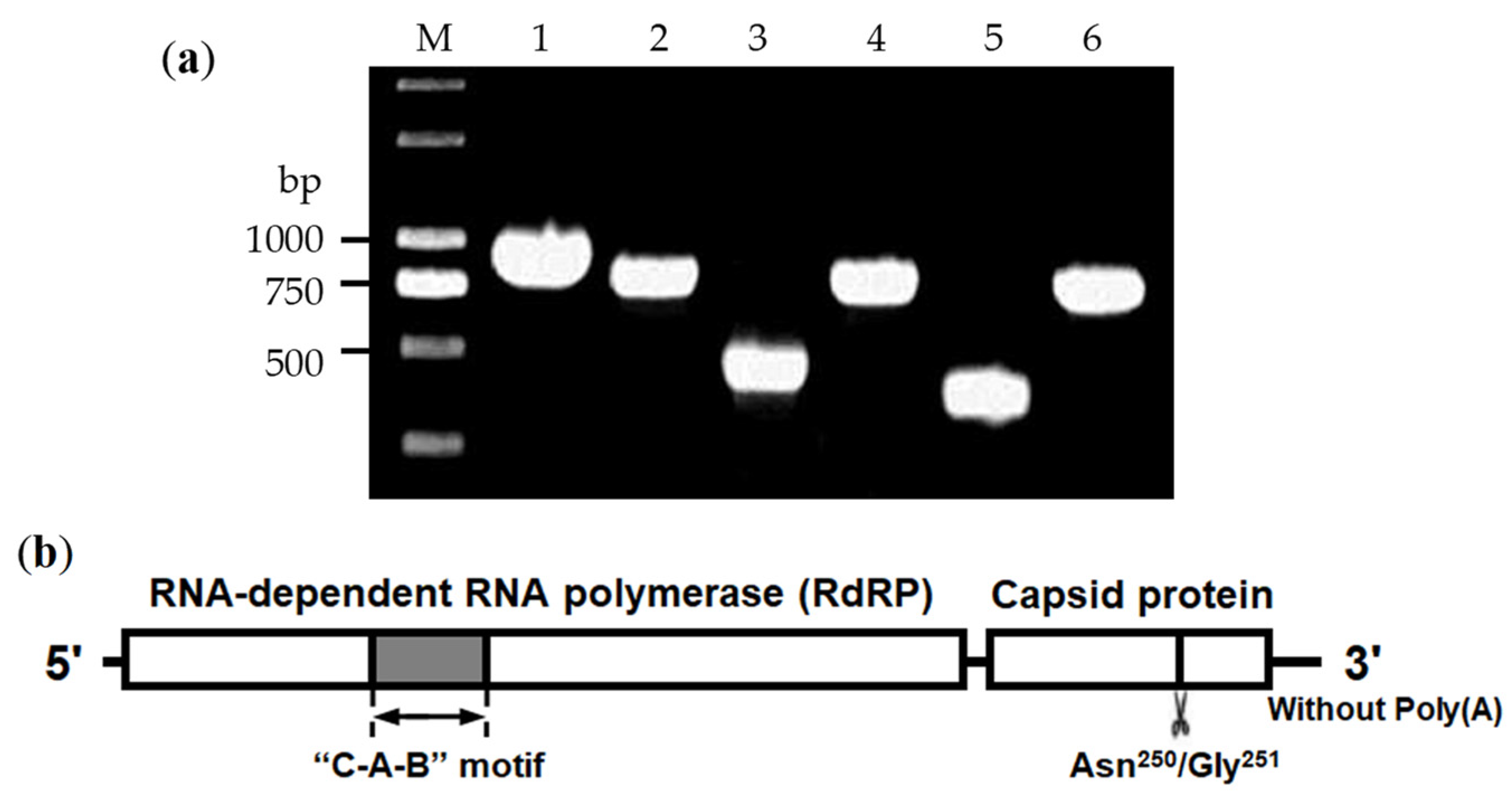
3.1. Cloning of LsPLV Genome

A strategy of “segment amplification plus end cloning” was employed to clone the whole genome of LsPLV. After PCR amplification, all six specific segments were successfully amplified, cloned, and sequenced with their corresponding primers (Figure 1a). Overlapping sequences were found from adjacent segments and used to assemble the main sequence of LsPLV genomic RNA, and then the viral 5′ and 3′ end fragments were also cloned by 5′ RACE and 3′ RACE. Ultimately, the whole genomic sequence of LsPLV was obtained through eight segments of sequence assembly and was submitted to the NCBI database (GenBank No. PQ441957).

3.2. Analysis of LsPLV Genome Features

Analyses revealed that the LsPLV genome is 4667 nt in length and contains two large ORFs encoding the functional proteins RdRP and CP (Figure 1b). The genome includes a 5′ non-coding region from 1 to 87 nt, an RdRP gene (88-3324 nt), an intergenic region (3325-3367 nt), a CP gene (3368-4480 nt), and a 3′ non-coding region from 4481 to 4667 nt without a poly(A) tail at the 3′ end. The RdRP protein consists of 1079 amino acids with a relative molecular weight of approximately 118.9 kDa, and CP consists of 371 amino acids with a relative molecular weight of approximately 40.4 kDa. Amino acid sequence alignment indicated that the RdRP sequence was 100% identical to previously reported partial sequences of LsPLV RdRP [13]. The conserved region (328-472 AA) of the RdRP palm subdomain also exhibits a “C–A–B” arrangement, which is a significant characteristic of permutotetra-like viruses.

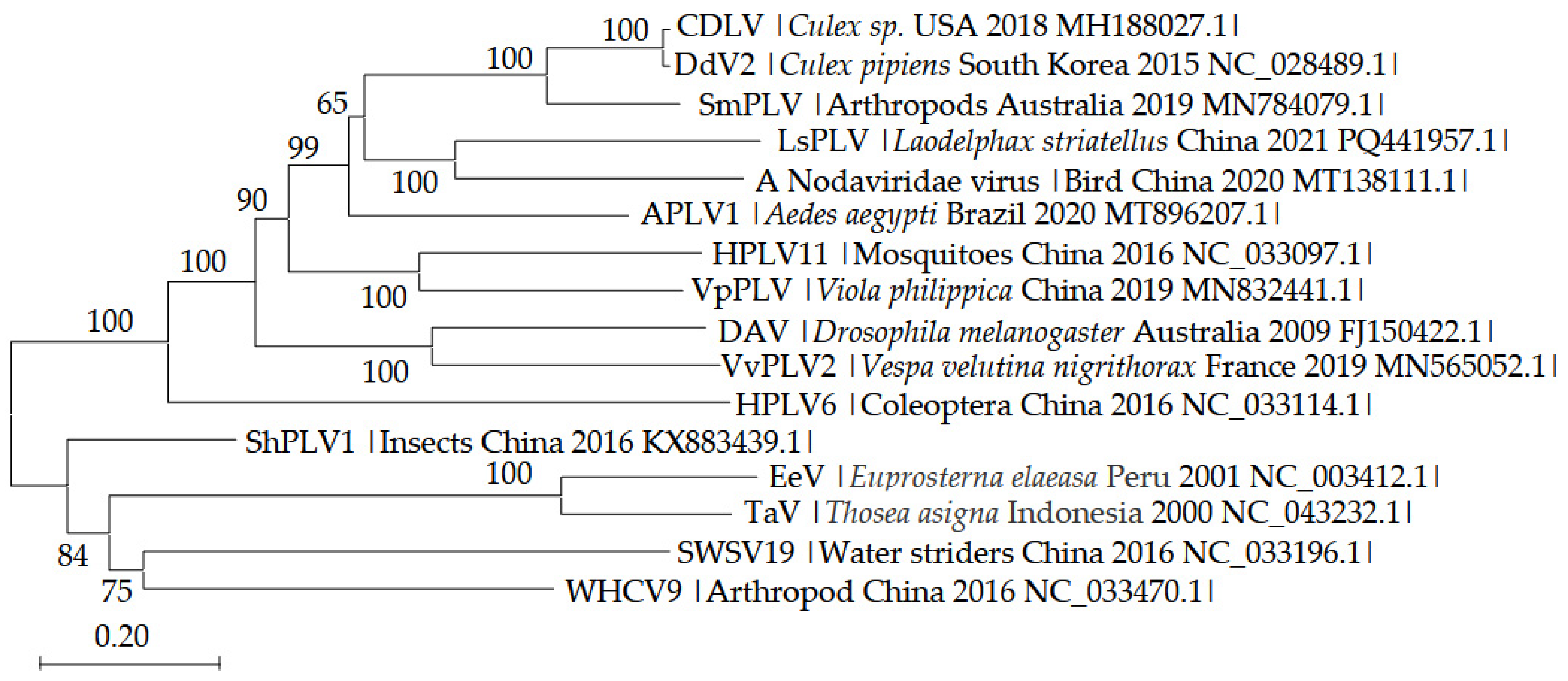
3.3. Phylogenetic Analysis of LsPLV Genome Sequence

After a Blastn alignment of the LsPLV genome sequence against the NCBI database, there was only one significant match, Laodelphax striatellus permutotetra-like virus 1 (LsPLV1, BK068171.1) [28], which was from Illumina sequencing and just released on 19 November 2024 in NCBI. The nucleotide sequence identity of the two virus genomes was 88.1%, and the nucleotide and amino acid sequence identities of their RdRPs were 87.6% and 94.9%, suggesting the two viruses are closely related. However, the hypothetical CP (729 nt) in LsPLV1 was different from the CP (1113 nt) of LsPLV identified in this study. Few significant matches of LsPLV in NCBI suggested substantial nucleotide sequence differences among LsPLV and other permutotetra-like viruses. Excluding LsPLV1, the Blastp alignment of viral two functional proteins indicated the virus with the highest similarity (50.4%) in RdRP amino acid sequence was DAV (AKH67341.1), while a Nodaviridae virus (QLI47702.1) shared the highest similarity (56.3%) with the CP amino acid sequence of LsPLV. Based on the above Blast alignment, 15 representative virus isolates with homology to LsPLV were selected for phylogenetic analysis of the LsPLV genome. The results showed that the 16 virus isolates clustered into two major branches (Figure 2), in which LsPLV and a Nodaviridae virus clustered, confirming the BLASTp results.

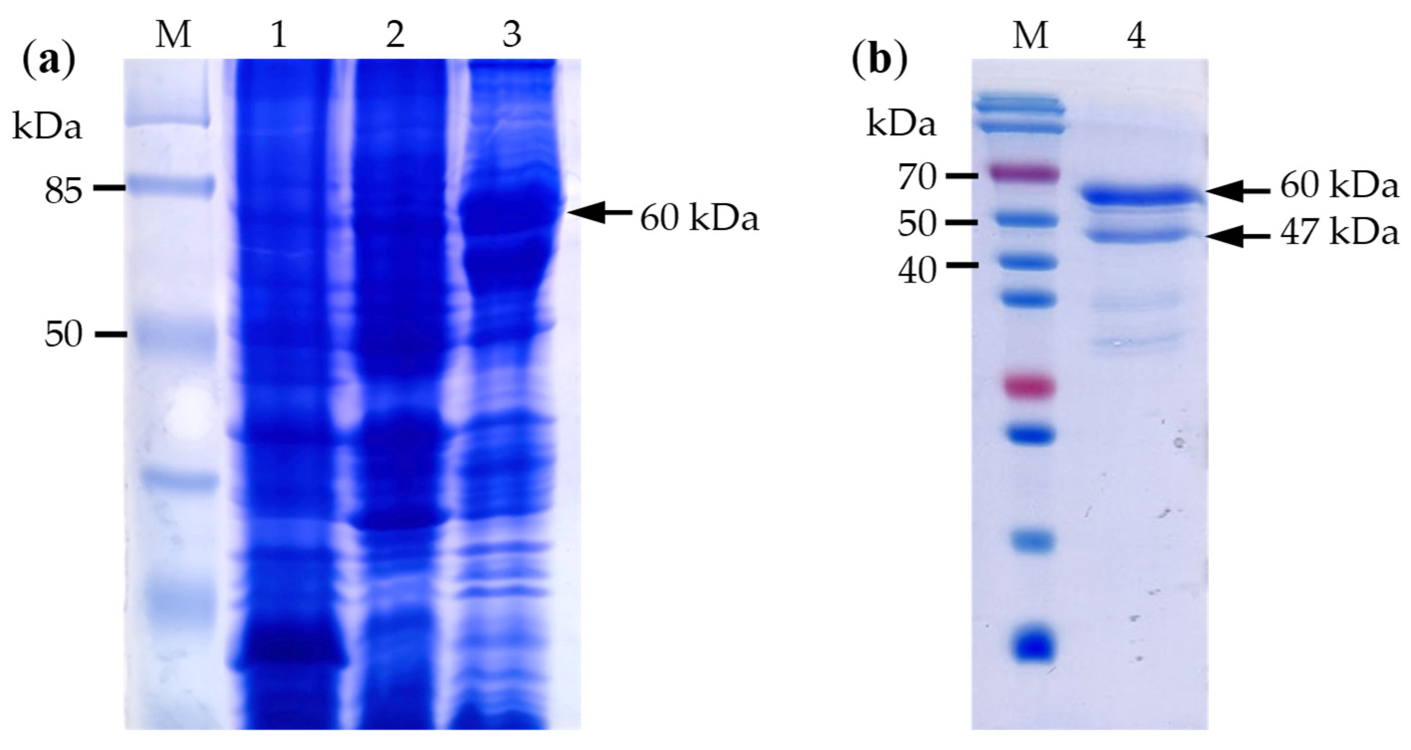
3.4. Cloning and Protein Expression of LsPLV CP Gene

After the CP gene in the viral genome was identified, a single PCR product of CP was amplified, cloned, and sequenced. The result indicated that the sequence of CP was 1113 nt, which was identical to the predicted CP gene in the LsPLV genome. To prepare CP antibody, CP was expressed in E. coli BL21 cells, and the results demonstrated that total proteins from expressing strains exhibited thick specific bands at the expected size of about 60 kDa (Figure 3a), suggesting successful prokaryotic expression of CP in E. coli. After expression products were purified, the electrophoretic analysis revealed there was a high-concentration-specific band at 60 kDa and another specific band at 47 kDa (Figure 3b). Anti-His antibody could react specifically with the two bands, suggesting they were both expression products from the pET-CP vector rather than E. coli strain proteins. The result corresponded with the electrophoretic separation results of the total expression products, indicating successful purification of LsPLV CP. The appearance of two bands in the purified CP also suggested there was a potential cleavage in the CP.

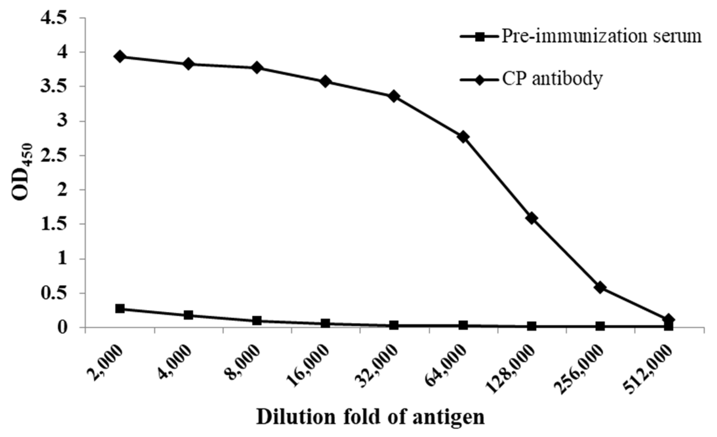
3.5. Potency and Specificity of CP Antibody

The high-purity CP antibody was obtained from CP antiserum via immunoaffinity chromatography, and the potency was assessed by an indirect ELISA. The results showed that a positive reaction (P ≥ 2 × N) was still observed even when the antibody was diluted 256,000 times (Figure 4), indicating that the potency of the prepared LsPLV CP antibody was 1:256,000. In Western blot analysis of the purified CP, the two specific bands (60 kDa and 47 kDa) were detected using CP antibody (Figure 5a), further suggesting the cleavage of CP.

3.6. Detection of LsPLV in SBPH and Self-Cleavage Property of CP

Western blot of SBPH total proteins showed that three protein bands were detected from LsPLV-infected SBPH (Figure 5b), while no band was observed in LsPLV-uninfected SBPH, which indicated that there was no cross-reactivity between the CP antibody and SBPH other proteins, demonstrating strong specificity of the antibody. The detected three bands were approximately 41 kDa, 28 kDa, and 13 kDa. The 41 kDa band represented the full-length CP of LsPLV, which was consistent with the theoretical value size. The presence of the 28 kDa and 13 kDa bands indicated that the viral CP was cleaved in SBPH, and these bands represented cleavage products of CP, which was basically consistent with the cleavage observed in the purified CP.
To analyze the self-cleavage property of the CP, purified CP incubating in PBS for 24 h was assessed. The results indicated that two bands were still detectable; however, the concentration of the 60 kDa band significantly decreased, no longer being the predominant band, while the 47 kDa band increased significantly, becoming the main band (Figure 5c). It was suggested that the full-length CP (60 kDa) can self-cleave a 13 kDa peptide independent of any protease, resulting in a 47 kDa product, thereby confirming the self-cleavage characteristic of LsPLV CP. The self-cleavage characteristic is not strongly dependent on temperature, as the self-cleavage activity was found to be similar at both 25 °C and 4 °C (Figure 5c).

4. Discussion

Tetraviruses are a class of spherical, icosahedral (T = 4), positive-sense, single-stranded RNA viruses [18], which is known to infect only insects, with reports of their presence worldwide [19,20,21,29]. However, compared to other types of insect viruses, the number of reported cases of tetraviruses is relatively low, and research on their utilization in biological control is limited. Dendrolimus punctatus tetravirus (DpTV) is the earliest discovered tetravirus in China, which is an isometric virion with two single-stranded RNAs belonging to the genus Omegatetravirus of the family Tetraviridae [30]. Previously, the communicating author’s laboratory first identified a unique tetravirus, LsPLV, in the Hemiptera insect (SBPH) using high-throughput sequencing technology [13]. LsPLV was characterized by the arrangement of its RdRP palm subdomain conserved region in a “C–A–B” pattern. To further investigate this virus, we identified the full-length genome of LsPLV, expressed its coat protein (CP), and prepared antibody for rapid detection of LsPLV. Additionally, we confirmed the self-cleavage characteristic of CP, laying the groundwork for research on its utilization in biological control and its interactions with SBPH.
Almost simultaneously with the submission of this article, LsPLV1, a virus very similar to LsPLV, was publicly released in the NCBI database [28]. The two viruses have a common host (SBPH) and share 88.1% genome sequence identity and 94.9% amino acid sequence identity of RdRP. Differently, the predicted CP gene (729 nt) in LsPLV1 is shorter than the CP gene (1113 nt) of LsPLV; in spite of this, the two CPs have 87.5% and 85.2% identities in nucleotide and amino acid sequences, respectively. Notably, if 1–123 nucleotides at the N-terminus of LsPLV1-CP are excluded, the truncated LsPLV1-CP shares up to 97.0% amino acid sequence identity with LsPLV-CP. The LsPLV genome was obtained through Sanger sequencing, while the LsPLV1 genome was from Illumina sequencing with limited accuracy, so the sequence difference between the two viruses may be caused by Illumina sequencing deviation. In addition, the host SBPH populations of the two viruses also share a common ancestor population from Haian, Jiangsu, China. According to the above-mentioned, it was considered that LsPLV and LsPLV1 were probably two different isolates of the same virus. Because of this, LsPLV1 was not selected for phylogenetic analysis.
Excluding LsPLV1, phylogenetic analysis of the full genomic sequence revealed that LsPLV was most closely related to a Nodaviridae virus, with nucleotide sequence identities of 55.4% and 59.2% and amino acid sequence identities of 45.3% and 56.3% for RdRP and CP, respectively, indicating significant genetic differences among permutotetra-like viruses from different host origins. Notably, a Nodaviridae virus is derived from metagenomic sequencing samples of avian gastrointestinal tracts, whereas this type of virus only infects insects. It is therefore inferred that the virus is carried by insects consumed by birds. Viola philippica permutotetra-like virus (VpPLV) (MN832441.1) from the plant V. philippica is likely also in a similar situation (“sample contamination”), which derives probably from virus residues from insect excrement on the surface of plant leaves. It is suggested that such viruses commonly infect insect hosts in nature, and their particle structure is relatively stable and resistant to degradation, allowing them to survive for a certain period of time in natural environments, which demonstrates their potential for biological control applications.
During the process of CP expression and purification, it was discovered that the LsPLV CP might possess self-cleavage functionality. Using the prepared antibody to detect CP, we confirmed its self-cleavage characteristics under both in vivo and in vitro conditions, demonstrating that this self-cleavage does not require the involvement of proteases. In addition to detecting a full-length CP of 41 kDa in SBPH, two additional bands of approximately 28 kDa and 13 kDa were also observed, which represented cleavage products of CP. It is certain that CP can remove a 13 kDa peptide, although the exact location of the self-cleavage remains undetermined. Coincidentally, the results of the in vitro analysis provided evidence for the location of the self-cleavage site. In vitro analysis, the molecular weights of the two CP bands (60 kDa and 47 kDa) also differed by approximately 13 kDa. Since the CP used in vitro analysis was an expressed fusion protein with a 19 kDa Trx-His-tag at the N-terminus, only when the self-cleavage site is located at the C-terminus can two proteins of 47 kDa and 13 kDa be produced and explained. Therefore, it can be concluded that the self-cleavage site of CP is located at the C-terminus of the CP.
The self-cleavage of CP precursor during the maturation stage of virus assembly is a common characteristic of tetraviruses, which is a necessary step for the viral particle maturation [31,32,33,34]. This study confirmed that LsPLV CP exhibited self-cleavage functionality, further validating that LsPLV is a tetravirus. The CPs of known tetraviruses all have a self-cleavage site at the C-terminus, and site sequence is relatively conserved. The cleavage sites of viruses such as Thosea asigna virus, Nudaurelia ω virus, Helicoverpa armigera stunt virus, Providence virus, and DpTV are Asn/Phe [30,31,33,35,36], whereas the cleavage site of Nudaurelia β virus is Asn/Gly [37]. Analysis of LsPLV CP amino acid sequence revealed that the C-terminus also contains the Asn250/Gly251 site. Upon cleavage at this site, the theoretical sizes of the resulting two peptide segments are 27.5 kDa and 13.0 kDa, which are basically consistent with the protein sizes detected in SBPH. Therefore, it was hypothesized that the self-cleavage site of the LsPLV CP was Asn250/Gly251 (Figure 1b). The actual cleavage site still needs to be identified through subsequent mass spectrometry analysis of the protein terminal sequence. The processing signals flanking this site also require further investigation. This self-cleavage sequence can serve as a functional element for future applications in protein engineering and “cell factory” research.

Author Contributions

Conceptualization, S.L. and J.P. (Jun Piao); methodology, S.L.; software, S.L. and J.Z.; validation, formal analysis and investigation, S.L., J.P. (Jun Piao), J.Z., L.Z., J.P. (Jingai Piao), H.W. and Y.X.; resources, S.L.; data curation, S.L.; writing—original draft preparation, S.L.; writing—review and editing, S.L.; visualization, S.L. and L.Z.; supervision, S.L. and J.P. (Jun Piao); project administration, S.L.; funding acquisition, S.L. All authors have read and agreed to the published version of the manuscript.

Funding

This research was funded by the National Key Research and Development Program of China, grant number 2023YFD1400300.

Institutional Review Board Statement

Not applicable.

Informed Consent Statement

Not applicable.

Data Availability Statement

The data presented in this study are available on request from the corresponding author.

Conflicts of Interest

The authors declare no conflicts of interest.

References

  1. Tinsley, T.W. The potential of insect pathogenic viruses as pesticidal agents. Annu. Rev. Entomol. 1979, 24, 63–87. [Google Scholar] [CrossRef] [PubMed]
  2. Johnson, R.M.; Rasgon, J.L. Densonucleosis viruses (‘densoviruses’) for mosquito and pathogen control. Curr. Opin. Insect Sci. 2018, 28, 90–97. [Google Scholar] [CrossRef] [PubMed]
  3. Lefkowitz, E.J.; Dempsey, D.M.; Hendrickson, R.C.; Orton, R.J.; Siddell, S.G.; Smith, D.B. Virus taxonomy: The database of the international committee on taxonomy of viruses (ICTV). Nucleic Acids Res. 2018, 46, D708–D717. [Google Scholar] [CrossRef]
  4. Yu, X.Z.; Jia, D.S.; Wang, Z.; Li, G.J.; Chen, M.N.; Liang, Q.F.; Zhou, Y.Y.; Liu, H.; Xiao, M.; Li, S.T.; et al. A plant reovirus hijacks endoplasmic reticulum-associated degradation machinery to promote efficient viral transmission by its planthopper vector under high temperature conditions. PLoS Pathog. 2021, 17, e1009347. [Google Scholar] [CrossRef]
  5. Whitfield, A.E.; Huot, O.B.; Martin, K.M.; Kondo, H.; Dietzgen, R.G. Plant rhabdoviruses-their origins and vector interactions. Curr. Opin. Virol. 2019, 33, 198–207. [Google Scholar] [CrossRef]
  6. Ban, L.; Didon, A.; Jonsson, L.M.; Glinwood, R.; Delp, G. An improved detection method for the Rhopalosiphum padi virus (RhPV) allows monitoring of its presence in aphids and movement within plants. J. Virol. Methods 2007, 142, 136–142. [Google Scholar] [CrossRef]
  7. Ryabov, E.V.; Keane, G.; Naish, N.; Evered, C.; Winstanley, D. Densovirus induces winged morphs in asexual clones of the rosy apple aphid, Dysaphis plantaginea. Proc. Natl. Acad. Sci. USA 2009, 106, 8465–8470. [Google Scholar] [CrossRef]
  8. Ban, L.P.; Ahmed, E.; Ninkovic, V.; Delp, G.; Glinwood, R. Infection with an insect virus affects olfactory behaviour and interactions with host plant and natural enemies in an aphid. Entomol. Exp. Appl. 2008, 127, 108–117. [Google Scholar] [CrossRef]
  9. Li, S.; Wang, S.J.; Wang, X.; Li, X.L.; Zi, J.Y.; Ge, S.S.; Cheng, Z.B.; Zhou, T.; Ji, Y.H.; Deng, J.H.; et al. Rice stripe virus affects the viability of its vector offspring by changing developmental gene expression in embryos. Sci. Rep. 2015, 5, 7883. [Google Scholar] [CrossRef]
  10. Fang, S.; Yu, J.; Feng, J.; Han, C.; Li, D.; Liu, Y. Identification of rice black-streaked dwarf fijivirus in maize with rough dwarf disease in China. Arch. Virol. 2001, 146, 167–170. [Google Scholar] [CrossRef]
  11. Di, D.P.; Zhang, Y.L.; Zhang, A.H.; Yan, C.; Yang, F.; Lu, Y.G.; Tian, L.Z.; Wang, X.B.; Miao, H.Q. Identification of a virus transmitted by small brown planthopper in wheat. Acta Phytopathol. Sin. 2016, 46, 453–460. [Google Scholar] [CrossRef]
  12. Gong, Z.X.; Zheng, Q.X.; Peng, H.; Cao, T.Q.; Shikata, E. Study on relationship between WRSV in China and NCMV in Japan. Chin. J. Virol. 1985, 1, 257–261. [Google Scholar] [CrossRef]
  13. Piao, J.; Zhang, L.J.; Piao, J.A.; Zhou, Y.J.; Li, S. Discovery of viruses from small brown planthopper by small RNA deep sequencing. Biotechnol. Bull. 2022, 38, 281–288. [Google Scholar] [CrossRef]
  14. Toriyama, S.; Guy, P.L.; Fuji, S.; Takahashi, M. Characterization of a new picorna-like virus, himetobi P virus, in planthoppers. J. Gen. Virol. 1992, 73, 1021–1023. [Google Scholar] [CrossRef] [PubMed]
  15. Li, S.; Ge, S.S.; Wang, X.; Sun, L.J.; Liu, Z.W.; Zhou, Y.J. Facilitation of rice stripe virus accumulation in the insect vector by himetobi P virus VP1. Viruses 2015, 7, 1492–1504. [Google Scholar] [CrossRef]
  16. Wu, N.; Zhang, P.P.; Liu, W.W.; Cao, M.J.; Massart, S.; Wang, X.F. Complete genome sequence and characterization of a new iflavirus from the small brown planthopper (Laodelphax striatellus). Virus Res. 2019, 272, 197651. [Google Scholar] [CrossRef]
  17. Yang, Q.K.; Zhang, Y.; Andika, I.B.; Liao, Z.F.; Kondo, H.; Lu, Y.H.; Cheng, Y.; Li, L.Y.; He, Y.Q.; He, Y.J. Horizontal transfer of a retrotransposon from the rice planthopper to the genome of an insect DNA virus. J. Virol. 2019, 93, e01516-18. [Google Scholar] [CrossRef]
  18. Hanzlik, T.N.; Gordon, K. The Tetraviridae. Ada. Virus Res. 1997, 48, 101–168. [Google Scholar] [CrossRef]
  19. Reinganum, C.; Robertson, J.S.; Tinsley, T.W. A new group of RNA viruses from insects. J. Gen. Virol. 1978, 40, 195–202. [Google Scholar] [CrossRef]
  20. Juckes, I.R.M. Comparison of some biophysical properties of the Nudaurelia β and ε viruses. J. Gen. Virol. 1979, 42, 89–94. [Google Scholar] [CrossRef]
  21. Ambrose, R.L.; Lander, G.C.; Maaty, W.S.; Bothner, B.; Johnson, J.E.; Johnson, K.N. Drosophila A virus is an unusual RNA virus with a T=3 icosahedral core and permuted RNA-dependent RNA polymerase. J. Gen. Virol. 2009, 90, 2191–2200. [Google Scholar] [CrossRef] [PubMed]
  22. Gorbalenya, A.E.; Pringle, F.M.; Zeddam, J.L.; Luke, B.T.; Ward, V.K. The palm subdomain-based active site is internally permuted in viral RNA-dependent RNA polymerases of an ancient lineage. J. Mol. Biol. 2002, 324, 47–62. [Google Scholar] [CrossRef] [PubMed]
  23. Poch, O.; Sauvaget, I.; Delarue, M.; Tordo, N. Identification of four conserved motifs among the RNA-dependent polymerase encoding elements. EMBO J. 1989, 8, 3867–3874. [Google Scholar] [CrossRef] [PubMed]
  24. Kumar, S.; Stecher, G.; Li, M.; Knyaz, C.; Tamura, K. MEGA X: Molecular evolutionary genetics analysis across computing platforms. Mol. Biol. Evol. 2018, 35, 1547–1549. [Google Scholar] [CrossRef] [PubMed]
  25. Saitou, N.; Nei, M. The neighbor-joining method: A new method for reconstructing phylogenetic trees. Mol. Biol. Evol. 1987, 4, 406–425. [Google Scholar] [CrossRef]
  26. Piao, J.; Xu, C.L.; Piao, J.A.; Zhou, Y.J.; Li, S. Preparation of the polyclonal antibody of capsid protein VP1 of Himetobi P virus in the small brown planthopper, Laodelphax striatellus (Hemiptera: Delphacidae) and its application in the virus detection. Acta Entomol. Sin. 2019, 62, 823–829. [Google Scholar] [CrossRef]
  27. Jiang, J.X.; Chen, Z.X.; Zhou, X.P. Production of a monoclonal antibody to sugarcane mosaic virus and its application for virus detection in China. J. Phytopathol. 2010, 151, 361–364. [Google Scholar] [CrossRef]
  28. Mao, Q.Z.; Ye, Z.X.; Yuan, J.N.; Ning, C.; Chen, M.N.; Xu, Z.T.; Qi, Y.H.; Zhang, Y.; Li, T.; He, Y.J.; et al. Diversity and transmissibility of RNA viruses in the small brown planthopper, Laodelphax striatellus. J. Virol. 2024, 98, e0019124. [Google Scholar] [CrossRef]
  29. Yi, F.M.; Zhang, J.M.; Liu, C.F.; Hu, Y.Y. Advances in tetravirus research. Acta Entomol. Sin. 2005, 48, 792–798. [Google Scholar] [CrossRef]
  30. Yi, F.M.; Zhang, J.M.; Yu, H.Y.; Liu, C.F.; Wang, J.P.; Hu, Y.Y. Isolation and identification of a new tetravirus from Dendrolimus punctatus larvae collected from Yunnan Province, China. J. Gen. Virol. 2005, 86, 789–796. [Google Scholar] [CrossRef]
  31. Agrawal, D.; Johnson, J. Assembly of the T = 4 Nudaurelia capensis ω virus capsid protein, post-translational cleavage, and specific encapsidation of its mRNA in a baculovirus expression system. Virology 1995, 207, 89–97. [Google Scholar] [CrossRef] [PubMed]
  32. Canady, M.A.; Tihova, M.; Hanzlik, T.N.; Johnson, J.E.; Yeager, M. Large conformational changes in the maturation of a simple RNA virus, Nudaurelia capensis ω virus (NωV). J. Mol. Biol. 2000, 299, 573–584. [Google Scholar] [CrossRef] [PubMed]
  33. Pringle, F.M.; Kalmakoff, J.; Ward, V.K. Analysis of the capsid processing strategy of Thosea asigna virus using baculovirus expression of virus-like particles. J. Gen. Virol. 2001, 82, 259–266. [Google Scholar] [CrossRef]
  34. Taylor, D.J.; Krishna, N.K.; Canady, M.A.; Schneemann, A.; Johnson, J.E. Large-scale, pH-dependent quaternary structure changes in an RNA virus capsid are reversible in the absence of subunit autoproteolysis. J. Virol. 2002, 76, 9972–9980. [Google Scholar] [CrossRef]
  35. Hanzlik, T.N.; Dorrian, S.J.; Johnson, K.N.; Brooks, E.M.; Gordon, K.H.J. Sequence of RNA2 of the Helicoverpa armigera stunt virus (Tetraviridae) and bacterial expression of its genes. J. Gen. Virol. 1995, 76, 799–811. [Google Scholar] [CrossRef]
  36. Pringle, F.M.; Johnson, K.N.; Goodman, C.L.; McIntosh, A.H.; Ball, L.A. Providence virus: A new member of the Tetraviridae that infects cultured insect cells. Virology 2003, 306, 359–370. [Google Scholar] [CrossRef]
  37. Gordon, K.; Williams, M.R.; Hendry, D.A.; Hanzlik, T.N. Sequence of the genomic RNA of nudaurelia beta virus (Tetraviridae) defines a novel virus genome organization. Virology 1999, 258, 42–53. [Google Scholar] [CrossRef]
Figure 1. Clone and schematic diagram of LsPLV genome. (a) RT-PCR amplification of LsPLV genome gene segments. Lane M: DNA Marker 5000, lane 1–6: Six gene segments of LsPLV. (b) Genome organization of LsPLV.
Microbiolres 16 00009 g001
Figure 2. Phylogenetic tree based on the complete genomic sequences of different permutotetra-like viruses. The phylogenetic trees were inferred using the Neighbor-Joining method in MEGA-X. The percentages of replicate trees in which the associated taxa clustered together in the bootstrap test (1000 replicates) are shown next to the branches. The trees were drawn to scale with the evolutionary distances as branch lengths, in which the evolutionary distances are in the units of the number of base substitutions per site. Viral name information used in the tree is as follows: CDLV: Culex Daeseongdong-like virus, DdV2: Daeseongdong virus 2, SmPLV: Smithfield permutotetra-like virus, APLV1: Aedes permutotetra-like virus 1, HPLV11: Hubei permutotetra-like virus 11, VpPLV: Viola philippica permutotetra-like virus, DAV: Drosophila A virus, VvPLV2: Vespa velutina-associated permutotetra-like virus 2, HPLV6: Hubei permutotetra-like virus 6, ShPLV1: Shuangao permutotetra-like virus 1, EeV: Euprosterna elaeasa virus, TaV: Thosea asigna virus, SWSV19: Sanxia water strider virus 19, WHCV9: Wuhan house centipede virus 9.
Microbiolres 16 00009 g002
Figure 3. Protein expression and purification of LsPLV CP. (a) SDS-PAGE of the expression products of the LsPLV CP gene, (b) SDS-PAGE of purified CP. Lane M: Protein molecular weight marker; lane 1: Escherichia coli with pET-32a vector induced by IPTG; lane 2: non-induced E. coli strain with pET-CP; lane 3: E. coli with pET-CP induced by IPTG; lane 4: purified CP fusion protein.
Microbiolres 16 00009 g003
Figure 4. Titers of polyclonal antibody against CP by ELISA.
Microbiolres 16 00009 g004
Figure 5. Verification of CP self-cleavage. (a) Western blot detection of purified CP using polyclonal antibody, (b) detection of LsPLV in SBPH, (c) detection of CP after static incubation. Lane M: Protein molecular weight marker; lane 1: purified CP; lane 2: total protein from LsPLV-uninfected SBPH; lane 3: total protein from LsPLV-infected SBPH; lane 4: CP incubated at 25 °C for 24 h; lane 5: CP incubated at 4 °C for 24 h.
Microbiolres 16 00009 g005
Table 1. Primers used in this study.
Primer and PurposeSequence (5′→3′)LsPLV Genome Location (nt)
Amplification of gene segments
LsPLV-F1ACAACGACAATGGACGCAAGCAACCC16-1182
LsPLV-R1TGATCCGTCCATCTGTTTGAAATCTGG
LsPLV-F2CCCAGATTTCAAACAGATGGACGG1155-1956
LsPLV-R2GTAGAGGGCCAGGCAGAACTCC
LsPLV-F3TAGACTACCCCGACTCCTCCGGAT1901-2495
LsPLV-R3GTGGGGAGCGGGGTGACAAGGGG
LsPLV-F4CCAACCCCCTGGACATAGTCTCCCTC2444-3305
LsPLV-R4GCCTGGGCCAATCTTCTTTTCCGCCTCT
LsPLV-F5AGGAAGCTGAACCGGAAGACGCT3253-3641
LsPLV-R5CGTTGATGTTGAAGGACTGAAGAAGGG
LsPLV-F6TCCCTTCTTCAGTCCTTCAACAT3614-4394
LsPLV-R6GGTTTCCCTGACCCACGTACTTGC
5′RACE
5′Adaptor primerGCTGTCAACGATACGCTACGTAACGGCAT
GACAGTGGGGGGGGGGGGGG
5′Race outerGCTGTCAACGATACGCTACGTAAC
LsPLV463-RCCGTGTAGACCTTGGACACGCACAT
LsPLV530-RGCCCAGGACACTGTCGTTCTTCCCAT
3′RACE
3′Race AdaptorTACCGTCGTTCCACTAGTGATTTCACTATG
ACGTTTTTTTTTTTTTTTT
3′Race outerTACCGTCGTTCCACTAGTGATTTC
3′Race innerAGTGATTTCACTATGACG
LsPLV-CP-F1004GCAAGTACGTGGGTCAGGGAAACC
LsPLV-CP-F931GGAGACACCATCAACTCCACGTCCTG
Construction of expression vector
LsPLV-CP-FGAGCTCATGAACAACCACACGTGTAAGAT3368-4480
LsPLV-CP-RAAGCTTCACGGACTGTGAAGAGAGTCG
Bold font indicates the purpose of primers, and underlining indicates a restriction enzyme site.
Disclaimer/Publisher’s Note: The statements, opinions and data contained in all publications are solely those of the individual author(s) and contributor(s) and not of MDPI and/or the editor(s). MDPI and/or the editor(s) disclaim responsibility for any injury to people or property resulting from any ideas, methods, instructions or products referred to in the content.

