Article

Probiotic Enterococcus faecium CRL 183 Inhibits Candida albicans Biofilm In Vitro

by
Virgínia Barreto Lordello
1,
Sarah Raquel de Annunzio
1,
Eliane Vale da Silva
1,
Maria Pía Taranto
2,
Carla Raquel Fontana
1 and
Daniela Cardoso Umbelino Cavallini
1,*
1
School of Pharmaceutical Sciences, Sao Paulo State University (UNESP), Rodovia Araraquara Jaú, Km 01—s/n, Campus Ville, Araraquara 14800-903, Brazil
2
Reference Center for Lactobacilli (CERELA-CONICET), Chacabuco 145, San Miguel de Tucumán 4000, Argentina
*
Author to whom correspondence should be addressed.
Microbiol. Res. 2024, 15(4), 2102-2113; https://doi.org/10.3390/microbiolres15040141
Submission received: 30 August 2024 / Revised: 23 September 2024 / Accepted: 30 September 2024 / Published: 13 October 2024

Abstract

:
Candida albicans is the most prevalent fungal microorganism of human microbiota and one of the few fungi capable of causing diseases in humans, depending on the host’s immune defense capacity. The similarity between fungal and host cells promotes several adverse effects during antifungal pharmacotherapy, and antimicrobial resistance increase is a major concern. Therefore, the search for alternative treatments and prevention strategies is urgent. In this context, probiotic bacteria, such as the strain Enterococcus faecium CRL 183, seem to be a viable alternative with its benefits to the immune system, activity against pathogens, and safety use well-documented through in vitro, in vivo, and clinical studies. Thus, this study aimed to evaluate if this probiotic strain prevents C. albicans ATCC 90028 biofilm colonization in vitro. To test the anti-Candida activity of the probiotic strain E. faecium CRL 183, we combined polymicrobial biofilms (C. albicans + E. faecium) with different proportions of fungi: a probiotic was formed (1:1, 1:10, 1:100) during the formation (24 h) and maturation (48 h) periods of the biofilm. The results show that E. faecium established itself with C. albicans in polymicrobial biofilms without losing its cellular viability. The probiotic strain significantly antagonized (p < 0.0001) C. albicans biofilm formation (up to 99.9% reduction in 24 h) and maturation (up to 99.43% reduction in 48 h). According to these results, E. faecium CRL183 may be a promising resource to prevent the formation of fungal biofilms.

1. Introduction

Candida albicans is the most prevalent fungal species of human microbiota that innocuously colonizes the human skin and mucosa, as well as the gastrointestinal and reproductive tract [1]. It is also one of the few fungi capable of causing diseases in humans, commonly characterized as superficial infections such as oral or vaginal candidiasis, affecting 75% of women at least once in their lifetime [1,2].
In certain patient groups, superficial infections may become invasive, reaching the bloodstream (candidemia) or vital organs such as the kidneys, liver, and heart [2,3,4]. It can occur when C. albicans overgrowth because of an imbalance in the host microbiota due to changes in local pH and nutritional availability or the impairment of the host immune defenses [1]. Highly associated with nosocomial infections, it is the fourth most isolated microorganism in the bloodstream, with mortality rates of up to 40% [1,4] Among its main virulence factors, we can highlight the polymorphism, production of toxins, hydrolytic enzymes, and biofilm formation [3,5,6,7,8].
Like most infections caused by biofilms, C. albicans infections are long-lasting, difficult to eliminate, and highly resistant to pharmacotherapy, such as amphotericin B, fluconazole, voriconazole, and nystatin [9]. Furthermore, fungi and humans are eukaryotes, and their cells are similar at the molecular level, making it more difficult to find or design drugs that target fungi without affecting human cells [10]. In this way, the search for alternatives for treating and preventing diseases, especially the ones caused by fungal biofilms, is urgent. Among the current alternatives, researchers have been investigating the action of probiotics, which are defined as “live microorganisms, which, when administered in adequate amounts, confer a health benefit on the host” [11]. Such microorganisms should preferably be of human origin, among which stand out the genera Lactobacillus, Bifidobacterium, and, to a lesser extent, Enterococcus, Streptococcus, and Saccharomyces [12].
It is well established that a great variety of probiotic strains could impair C. albicans biofilm formation and maturation [5,13,14] and inhibit the fungal polymorphism, adhesion, and invasion of host tissues [14,15,16]. Probiotics are also able to downregulate several genes associated with virulence factors of C. albicans, such as ALS3, HWP1, BCR1, and CPH1 [14].
Among these microorganisms, the interaction between the genera Enterococcus spp. and Candida spp. seems particularly interesting. Both are commonly co-isolated from the same niches, either as commensal members of the microbiota, colonizing the gut, or in polymicrobial infections, losing in prevalence in those cases only to S. aureus [17]. The literature also shows that the fungus favors this bacterium [18,19], whereas Enterococcus spp. significantly attenuates fungus virulence in polymicrobial infections through the secretion of proteases, bacteriocins, and lactic acid [10,17,20,21,22]. These findings show that several Enterococcus spp. metabolites can considerably reduce polymorphism, the total biofilm biomass [20,21,22], epithelial invasion, inflammation, and fungal load in a murine model of oropharyngeal candidiasis through an increase in macrophages antifungal activity [22]. Another anti-Candida protein (ACP) characterized by Shekh and Roy [10] showed a broad-spectrum activity against eight strains of multidrug-resistant C. albicans. It is important to highlight that the bacterial strain used against Candida spp. was described as innocuous in none of these studies, much less having a probiotic claim [10,17,20,21,22].
There is a lot of concern about Enterococcus spp. due to its ability to become resistant to antibiotics and transfer resistance genes, especially vancomycin resistance, to other pathogens [23]. Because of that, the anti-Candida activity of Enterococcus spp. metabolites has been evaluated, whereas the cell–cell interaction was not considered suitable. However, the direct competition for adhesion sites and nutrients may be a potentiator of the anti-Candida effect promoted by bacteria metabolites [5,13,14,16,24].
Enterococcus faecium CRL 183 is a probiotic strain isolated from traditional Argentinian cheese with no virulence factors, pathogenicity, antimicrobial resistance, or adverse effects identified [25]. Several beneficial systemic effects promoted by this probiotic strain are documented, i.e., the modulation of the immune system and intestinal microbiota, reductions in symptoms related to colitis [26], the regulation of the lipid profile, the inhibition of atherosclerotic lesions [27,28], breast adenocarcinoma [29], chemically induced colon cancer development [30], and body weight gain [31] in different murine models. Furthermore, the regular intake of a soy product fermented with the same strain was related to a positive modulation of the lipid profile in moderately hypercholesterolemic patients [32]. It is essential to highlight that in some of those studies, E. faecium CRL183 was used as a starter culture of a fermented soy-based product, either combined or not with other probiotic strains [28,29,31,32].
E. faecium CRL183 also inhibited S. mutans growth in an agar well diffusion assay, and it was resistant to human saliva, showing the ability to grow (up to 3.64 Log10 CFU/mL) in this environment after 24 h. These results indicate its potential to colonize the oral cavity and act as an alternative approach to local infection control [33]. Therefore, this study aimed to evaluate if E. faecium CRL 183 could prevent C. albicans biofilm formation and maturation through direct competition, as the antifungal activity of this strain has never been tested.

2. Materials and Methods

The probiotic strain E. faecium CRL 183 was obtained from the Reference Center for Lactobacilli—CERELA/CONICET (San Miguel de Tucumán, Tucumán, Argentina) and C. albicans ATCC 90,028 were obtained from the American Type Culture Collection (Manassas, VA, USA). The microorganisms were kept frozen at −80 °C in a cryogenic tube containing proper culture media with the addition of 20% v/v glycerol up to the time of its use.
Before their use, the strains were thawed and subcultured in specific culture media: E. faecium in Bile Esculin Agar (Acumedia, Lansing, MI, USA) and C. albicans in Sabouraud Dextrose Agar supplemented with chloramphenicol (0.00005 g/mL) (SDA—Acumedia, Lansing, MI, USA) and incubated at 37 °C for 48 h.

2.1. Assurance of Probiotic Strain Safety

As stated before, E. faecium CRL 183 has its safety consumption attested through many in vivo and clinical studies. It does not show any antibiotic resistance genes or virulence factors [25,26]. The E. faecium CRL 183 was submitted to the antimicrobial susceptibility test by disc diffusion performed according to the Clinical and Laboratory Standards Institute (CLSI) [34].
A standard inoculum (turbidity equivalent to a McFarland 0.5 scale) was seeded with a sterile swab in 140 × 15 mm Petri dishes (Corning®, Corning, New York, NY, USA) containing Mueller Hinton Agar (Difco, Detroit, MI, USA). Discs containing antimicrobials ciprofloxacin 5 mg; chloramphenicol 30 mg; erythromycin 15 mg; nitrofurantoin 300 mg; norfloxacin 10 mg; tetracycline 30 mg; and vancomycin 30 mg (SENSIFAR—Cefar Diagnostica, São Paulo, São Paulo, Brazil) were placed on the inoculated agar with the aid of sterile forceps. The plates were incubated at 35 ± 2 °C, with an incubation time of 16–18 h for most antimicrobials, except for vancomycin, which required 24 h of incubation. After this period, the diameter of inhibition growth halos caused by the antimicrobial discs was assessed. The results were interpreted according to the criteria recommended by the Clinical and Laboratory Standards Institute (CLSI) [34].

2.2. Biofilm Formation and pH Evaluation

The biofilm assays were carried out following the methods described by Fontana et al. [35] and Zago et al. [8], with modifications. The microorganisms freshly cultivated as described in growth conditions were inoculated with a sterile loop in 2 mL of enriched broth containing 0.026 g/mL of brain–heart infusion (BHI—Kasvi, Curitiba, Brazil), 0.01 g/mL of yeast extract (YE—Acumedia, MI, USA), 0.02 g/mL of trypticase soy broth (TSB—Acumedia, MI, USA), and were supplemented with 20% g/mL sucrose (Synth, Diadema, Brazil) to obtain yeast and probiotic bacteria inoculum [8,35]. The suspensions obtained were standardized by spectrophotometry reading on a Synergy H1M microplate reader (Biotek, Winooski, VT, USA). The two standardized microbial suspensions were then diluted to obtain different inoculums of yeast (106 or 108 CFU/mL) and probiotics (107 or 108 CFU/mL).
Monospecies biofilm assays were conducted on 96-well microplates with each well containing 75 μL of yeast or probiotic suspension plus 75 μL of enriched broth, i.e., 150 μL per well. Polymicrobial biofilm assays were also performed in 96-well microplates, with each well containing the suspension of each microorganism in the following proportions: 75 μL C. albicans 108 CFU/mL + 75 μL E. faecium 108 CFU/mL (1:1), 75 μL C. albicans 106 CFU/mL + 75 μL E. faecium 107 CFU/mL (1:10), and 75 μL C. albicans 106 CFU/mL + 75 μL E. faecium 108 CFU/mL (1:100). The microplates were incubated at 35 ± 2 °C for 24 h to evaluate the interaction of microorganisms in biofilm adhesion and formation for 48 h to evaluate it in the biofilm maturation.
In the case of 48 h biofilms, they were split into two groups (48A and 48B) to evaluate if the metabolites produced by E. faecium within the first 24 h played or did not play a role in the destabilization of C. albicans biofilms. In group 48A, after the first 24 h, the supernatant from each well was carefully replaced with 150 μL of fresh enriched broth, and the plates returned to the incubator. Therefore, the metabolites produced were removed, and new nutrients were added to the microenvironment. In group 48B, no intervention was made during the 48 h of incubation.
After the formation (24 h) and maturation (48 h) times of the biofilms, all the supernatants from the wells were aspirated carefully to avoid the removal of adhered biofilms, and the pH of those supernatants was measured with pH-fix test strips (Neumann-Neander, Düren, Germany). After this procedure, the supernatant was discarded.

2.3. Quantification of Viable Cells and Determination of the Anti-Candida Activity of E. faecium CRL 183

The adhered biofilms were scraped off the wells and resuspended vigorously in 150 μL of enriched broth with a sterile pipette tip for 30 s. Serial decimal dilutions of those suspensions were made using the culture medium itself as a diluent. Monospecies biofilms were quantified by plating the obtained dilutions in SDA supplemented with chloramphenicol (for C. albicans) and Bile Esculin Agar (for E. faecium). The polymicrobial biofilms were plated onto both culture media. The plates were incubated for 24 h at 35 ± 2 °C, and the number of fungal and probiotic cells present in the biofilms was determined by counting colony-forming units (CFU). The experiments were performed in four replicates and repeated in three independent assays.
The decimal reduction (DR) of Candida albicans cell viability in the presence of probiotic strain is determined using Equation (1):
DR = Log10 (Cam) − Log10 (Cap)
where Cam is the colony-forming unit (CFU/mL) values of C. albicans present in the monospecies biofilms (positive control) and Cap is the colony-forming unit (CFU/mL) values of C. albicans present in the polymicrobial biofilms.
Then, the percentual reduction (PR) in fungal population in the polymicrobial biofilms after 24 or 48 h can be calculated with Equation (2):
PR = (1 − 10−DR) × 100
where DR is the decimal reduction obtained by Equation (1).

2.4. Scanning Electron Microscopy (SEM)

This analysis was performed through FEG-SEM (Model JSM-7500F, JEOL, Tokyo, Japan) to visualize the interactions between probiotics and yeasts within the biofilms. The experimental conditions were as follows: polymicrobial biofilm at 1:100 pathogen: probiotic ratio and monospecies biofilms: C. albicans (initial inoculum of yeast: 106 CFU/mL) and E. faecium (initial inoculum of bacteria 108 CFU/mL). Biofilms were formed as described above in a sterile coverslip placed at the bottom of 6-well microliter plates.
After the periods of incubation (24 h or 48 h), the biofilm supernatants were gently removed, and the coverslips were washed with PBS. The biofilms were fixed with a 2.5% v/v glutaraldehyde solution (Merck, Darmstadt, Germany) and then dehydrated with an ethanol series (30% v/v, 50% v/v, 70% v/v, 85% v/v, and 95% v/v ethanol solution for 15 min each; two washes with 100% v/v ethanol for 15 min). The samples were dried at 37 °C and kept in a vacuum desiccator until the analysis day.
The coverslips containing adhered biofilms were then attached to the stub surfaces with double-sided adhesive tape, coated with a layer of carbon, and observed through FEG-SEM (JEOL JSM-7500F). The photomicrographs were taken at 1000×, 3000×, and 5000× magnifications.

2.5. Statistical Analysis

To verify the statistical significance, the results were submitted to one-way ANOVA followed by Tukey’s multiple comparisons test (parametric data). This was performed using GraphPad Prism version 7.00 for Windows (GraphPad Software, La Jolla, CA, USA) with a minimum significance level of 5% (p ≤ 0.05).

3. Results

3.1. Assurance of Probiotic Strain Safety

The antimicrobial susceptibility test results (Table 1) were interpreted according to the standard table for the interpretation of inhibition halos present in the diagnostic test package insert following CLSI recommendations [34]. The probiotic strain was sensitive to the antimicrobials tested, including vancomycin. Regarding erythromycin (15 mg), E. faecium CRL 183 exhibited an inhibition zone diameter of 22 mm, showing intermediate sensitivity to this antibiotic (14–22 mm: intermediate; 23 mm: sensitive).

3.2. Biofilm pH Evaluation

For C. albicans monospecies biofilms in formation, the supernatant pH was 7.0 at all concentrations tested. After 48 h, with or without culture medium renewal, the pH was reduced to 5.0. There was no difference between the pH of E. faecium monospecies biofilm supernatants and the polymicrobial biofilm supernatants in any experimental conditions (initial concentration, incubation time, and culture medium exchange), which remained in the order of 5.0.

3.3. Anti-Candida Activity of E. faecium CRL 183

The interaction between the fungus and probiotics was analyzed in polymicrobial biofilms. The survival of E. faecium in an environment co-colonized by C. albicans was evaluated by quantifying its colonies and comparing it with the results obtained in the E. faecium monospecies biofilm (control). It was observed that this probiotic could establish itself and survive in the microenvironment, competing for space and nutrients with C. albicans. There was a significant statistical difference with the control group only in the 1:1 ratio of fungus/probiotic (Figure 1). The renewal of the culture medium in 48 h biofilms (48A) did not result in a statistically significant difference with the non-renewal group (48B) in the ratios 1:10 and 1:100 (Figure 1B). In the ratio 1:1, the renewal of the culture medium (48A) impaired E. faecium survival in the presence of C. albicans if compared with group 48B. Presumably, metabolites produced within the first 24 h helped E. faecium survive in the microenvironment, where there was a high concentration of C. albicans.
Despite this positive effect that C. albicans exerts concerning Enterococcus spp. the association between these microorganisms cannot be considered mutually beneficial. This is especially true in the context of the present study since the presence of the fungus impaired the viability of E. faecium only in the 1:1 ratio (Figure 1), while the probiotic negatively affected the cellular viability of C. albicans in all experimental conditions (Figure 2). The reduction in C. albicans within the 24 h biofilms ranged from 1.57 to 3.08 Log10 CFU/mL; these results indicate that, during the formation of biofilms, the fungal reduction depends on the probiotic concentration. However, in terms of the percentage of viable cell reduction, ranging from 97.12% to 99.92% for 24 h biofilms, there was no statistical difference between 1:1, 1:10, and 1:100.
In mature polymicrobial biofilms (48 h) C. albicans reduction ranged from 2.00 to 2.30 Log10 CFU/mL (Figure 2) corresponding to the PR of 99 to 99.43% for this group where the removal of metabolites and renewal of nutrients took place (48A), which was not significantly different from the 48B group where a reduction from 1.46 to 2.15 Log10 CFU/mL was observed (Figure 2) or, in percentage, 96.54 to 99.30%. The 48 h results indicate that the fungal reduction was not dependent on the initial probiotic dose for mature biofilms since there was no significant statistical difference between the groups (1:1, 1:10, and 1:100).

3.4. Scanning Electron Microscopy (SEM)

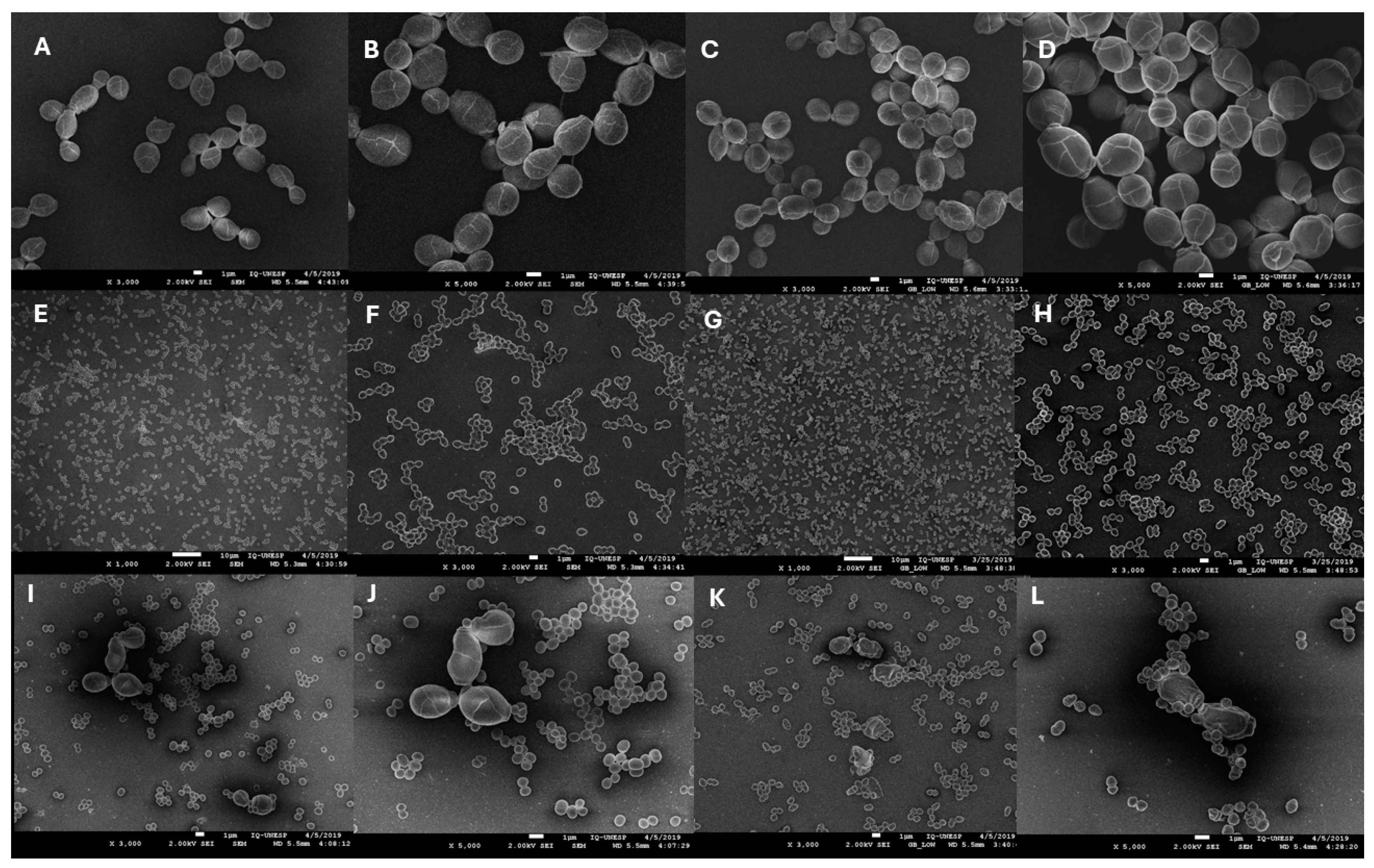
The SEM analysis is shown in Figure 3 and allows us to illustrate the results obtained by counting colony-forming units. In monospecies biofilms or controls, it was possible to observe cellular aggregates surrounded by EPS and scattered throughout the coverslips. A lower number of fungal cells (Figure 3A–D) was observed compared to bacteria (Figure 3E–H), which corroborates the cell counting results obtained for monospecies biofilms. In polymicrobial biofilms (Figure 3I–L), it was possible to observe a considerable reduction in the number of C. albicans cells compared to the control biofilms (Figure 3A–D) of this species. This did not occur with the number of E. faecium seen in the polymicrobial biofilm, which was equal to that found in the monospecies biofilm. Furthermore, the images of the polymicrobial biofilms (Figure 3I–L) permitted the evaluation that there is a physical interaction between the fungus and the probiotic cells.

4. Discussion

While adverse effects due to the administration of probiotic bacteria are uncommon, ensuring its susceptibility to antimicrobials is mandatory. It is known that antibiotic resistance can be intrinsic or acquired due to a chromosomal mutation or by horizontal gene transfer [36]. There is a particular concern about the spread of Vancomycin-Resistant Enterococci (VRE) since some genus species may become a nosocomial pathogen and a reservoir for resistance genes, leading to clinical isolates resistant to all antibiotics [23]. The strain E. faecium CRL 183 has been documented for its absence of antimicrobial resistance genes [25] and was susceptible to all classes of antimicrobials tested; therefore, it can be considered safe.
Factors such as nutrient availability, temperature, and pH are determinants for the survival and behavior of microorganisms, especially in polymicrobial environments. It is a consensus that probiotic strains can produce lactic acid and other organic acids, which, in turn, can cross the plasma membrane of yeast cells, increasing the activity of H+-ATPase. This mechanism promotes the energy exhaustion of yeast cells, inhibiting their growth and promoting cell death [37]. Furthermore, neutral to alkaline is the optimal extracellular pH for Candida spp. because it aids hyphal morphogenesis, among other fungal virulence strategies [38]. E. faecium is a lactic acid bacterium (LAB), and acid production could be one of the key mechanisms by which this probiotic impairs C. albicans viability [23]. A study that evaluated inhibition on several Candida species promoted by Limosilactobacillus reuteri (formerly Lactobacillus reuteri) showed that almost complete inhibition was achieved in pH 3.6, which is a very acidic level. However, extensive acidification may be concern with its application in the oral cavity due to the role of acid in caries lesions [37]. In our study, the biofilm supernatants’ pH remained within the range of healthy human saliva pH, which is 5.3 to 7.8, depending on the stimulation state [39].
Similar to the results shown in Figure 1, Mason et al. [18] and Mason et al. [19] demonstrated that C. albicans SC5314 promoted the gastric and enteric colonization of Enterococcus spp. while antagonizing other lactic bacteria, such as Lactobacillus spp., during the recovery of gut microbiota following treatment with the antibiotic cefoperazone. Although the mechanism involved was not elucidated, the authors suggest that it was similar to the one C. albicans exerts on bacteria from phylum Bacteroidetes. The fungus could aid in its adhesion and fixation to the cecum; yeast glucans could be a potential source of nutrients for these bacteria, or else, C. albicans could suppress other bacteria that antagonize Enterococcus spp. [18].
The reduction in C. albicans viable cells during biofilm formation was affected by the fungal/probiotic ratio. Cruz et al. [20] demonstrated that Caenorhabditis elegans, a study model of polymicrobial infections, presented less mortality caused by C. albicans when it was co-inoculated by Enterococcus spp. The survival rate of the nematode was dose-dependent: the higher the number of colonies of Enterococcus spp. inoculated, the greater the protection against C. albicans pathogenicity. This study further determined that the pathogenicity of both microorganisms was attenuated during polymicrobial infection in C. elegans [20].
The phases of yeast biofilm formation include cell adhesion to a surface, cell differentiation, the maturation phase, and the dispersion stage, in which mature biofilms release cells to spread colonization. The maturation of the C. albicans biofilm usually takes around 38–72 h and includes the development of a thick layer of EPS where yeasts and hyphae are present, forming a dense structure [40,41]. A biofilm with 48 h of maturation has a higher density, and the structure becomes more compact and organized. After 72 h, the biofilm is well established, with a thick layer of interwoven cells and a robust extracellular matrix. At this stage, the biofilm is more resistant to antifungal agents due to its complex organization. A maturation time of 24 to 48 h has been adopted in different studies to evaluate the inhibitory effect of drugs and probiotic strains on Candida spp. biofilms. [5,42,43]. For biofilm maturation, the microorganism ratio did not influence the results. Matsubara et al. [5] also did not observe a significant relationship between inoculated probiotic dose and biofilm destabilization.
Among the Enterococcus spp. mechanism of action to downregulate C. albicans pathogenicity in the literature majorly suggests the action of proteases, peptides, and bacteriocins secreted by bacteria [10,20,21,22]. For example, researchers identified the action of two proteases (gelatinase biosynthesis activating cluster peptide—GBAP) that activate the virulence regulating system of Enterococcus spp., which, when secreted by this bacterium, prevent C. albicans polymorphism and, consequently, fungal pathogenesis [20]. The same effect was observed when C. elegans, previously infected with C. albicans, was treated with the supernatant (filtered and sterilized) of an Enterococcus faecalis inoculum. Thus, Cruz et al. [20] determined that, in their study, the inhibitory activity was correlated with the metabolites secreted rather than a direct cellular competition between the bacteria and the fungus.
In more recent research, the action of the EntV, an enterocin encoded by the Ef1097 gene, which is present in all strains of E. faecalis sequenced to date, was described [22]. This enterocin alone, as well as its synthetic version, was able to prevent C. albicans polymorphism and biofilm formation on different substrates and experimental conditions. Pre-formed C. albicans biofilms (24 h/~30 μm thick) were treated with EntV and underwent a significant perturbation with a reduction in their thickness to ~15 μm. Biofilms that were not treated with EntV increased their thickness to >50 μm. The peptide also protected macrophages by enhancing their antifungal activity and reducing epithelial invasion, inflammation, and fungal load in a murine model of oropharyngeal candidiasis.
Bachtiar et al. [21] observed that E. faecalis cps2 (a non-encapsulated clinical isolate) and E. faecalis ATCC 29212, as well as their metabolites, did not inhibit C. albicans biofilm formation. However, both strains and their secreted metabolites significantly reduced (>50%) the maturation of these biofilms. These results were confirmed by the downregulation promoted on ALS1 and ALS3 (adhesion-related genes) and on EFB1, a gene used to quantify the harmful effects of antifungal agents against mature Candida biofilms.
In our study, it was not possible to determine if the anti-Candida activity of E. faecium CRL 183 was modulated by its metabolites, as suggested in the literature cited above. However, as can be seen in Figure 2B, the renewal of the culture medium (48A) did not significantly influence C. albicans survival compared to the group in which there was no intervention (48B). Therefore, removing E. faecium metabolites secreted during the first 24 h and the new nutrient supply did not favor the cellular viability of C. albicans. In addition, the E. faecium population was approximately 11 Log10 CFU/mL in the polymicrobial biofilms, and the C. albicans population was approximately 7 Log10 CFU/mL under all experimental conditions. This means that the probiotic growth was much higher than the fungal growth within polymicrobial biofilms. This may suggest that, in our study model, the inhibitory action of C. albicans may not be only mediated by metabolites produced by E. faecium but most likely due to the direct cell competition for nutrients and space. This mechanism of direct competition and limitation of environmental nutrients can provoke a metabolic reprogramming on C. albicans, reducing its virulence. Mailänder-Sánchez et al. [15] suggest that this is the mechanism by which the probiotic strain Lacticaseibacillus rhamnosus GG (formerly Lactobacillus rhamnosus GG) prevents adhesion, invasion, the formation of hyphae (preventing epithelial damage), glucose depletion and the ergosterol synthesis of C. albicans. Another inhibitory mechanism may have been the culture medium acidification in polymicrobial biofilms promoted by E. faecium, which is an LAB. This reduction in pH inhibits the growth of yeast cells and prevents C. albicans filamentation [37,38]. Hypha formation is one of the main virulence factors of C. albicans and is directly related to its pathogenicity and biofilm stability [1,2].
Vilela et al. [13] suggested this exact mechanism in the suppression of C. albicans polymorphism when it was stimulated with cultures of Lactobacillus acidophilus ATCC 4356 or filtered supernatants from this probiotic culture. They also reported a reduction of up to 57.5% in the CFU counting of C. albicans biofilms stimulated with the probiotic cell culture and a 45.10% reduction in biofilms stimulated with probiotic culture-filtered supernatants. Galleria mellonella was used as an experimental candidiasis model, and either treatment or prophylaxis with L. acidophilus cells or their metabolites greatly increased the survival of this insect.
Ribeiro et al. [14] observed a similar effect on the interaction of C. albicans with Lacticaseibacillus rhamnosus (formerly Lactobacillus rhamnosus) cells: a 52.2% reduction in biofilm quantification in CFU/mL and a 48% reduction in metabolic activity. The stimulation with only the filtered supernatant of L. rhamnosus decreased, to a lesser extent, C. albicans growth (39.4%) and metabolic activity (61%).
Matsubara et al. [5] observed that Lacticaseibacillus rhamnosus (formerly Lactobacillus rhamnosus), Lacticaseibacillus casei (formerly Lactobacillus casei), and Lactobacillus acidophilus cell suspensions significantly reduced C. albicans SC5314 biofilm formation in 32, 59, and 56.3%, respectively. When 48 h biofilms were stimulated with the same probiotic suspensions, the C. albicans reduction was 61.8% (L. rhamnosus), 54.7% (L. casei), and 34.2% (L. acidophilus). By stimulating C. albicans biofilms with the filtered supernatant of these lactobacillus cultures, the reduction was less effective in the formation, and there was no reduction in biofilm maturation. This result demonstrates that the inhibitory action on C. albicans was also not mediated only by probiotic-secreted metabolites.
Therefore, our results seemed to be very promising once E. faecium CRL183 presented an ability to impair C. albicans biofilm formation by up to 99.92% and its biofilm maturation by 99.43%, which are much higher rates than those presented by other Enterococcus spp. [10,20,21,22] or probiotic strains [5,13,14].
The images obtained through SEM allowed us to visually confirm the hypothesis that in the present study, the direct competition and limitation of environmental nutrients played an important role. It is exciting to observe that C. albicans not only appears at a lesser amount but also with its morphology very damaged (indicated by the arrow) in 48 h polymicrobial biofilms (Figure 3K,L) in comparison with C. albicans monospecies biofilms in the same period (Figure 3C,D), which could be caused by the lactic acid crossing the yeast membrane [37]. It was expected to see hyphal formation in C. albicans biofilms in our SEM images, which did not occur. This may be explained by the lack of molecular factors favoring C. albicans polymorphism in the culture medium used in the experimental protocol since it is known that the culture medium plays a fundamental role in the growth, morphology, and biofilm formation of C. albicans [38,44]. Another reason could be that several washes with ethanol removed hyphae and pseudohyphae since these structures developed in the outermost layers of biofilms, while yeasts initially inoculated were found in the basal layers adhered to the biotic or abiotic substrate [45]. Yet, our results remain promising in the context of prevention, as yeasts are the infecting forms that initiate biofilms and are responsible for the spread to non-infected sites [45,46].

5. Conclusions

In conclusion, according to the methodologies applied in this in vitro study, E. faecium CRL 183 significantly inhibited the formation and maturation of fungal biofilms in all evaluated pathogen/probiotic ratios. These promising results demonstrate the need for in-depth studies to investigate the mechanisms by which E. faecium CRL183 acts against C. albicans. In addition, local release forms of probiotics in regions susceptible to candidiasis also need to be evaluated.

Author Contributions

D.C.U.C., V.B.L., C.R.F. and S.R.d.A.: Conceptualization and Methodology. V.B.L.: Data curation, Writing—Original draft preparation. V.B.L. and S.R.d.A.: Visualization, Investigation. D.C.U.C., C.R.F. and M.P.T.: Supervision. V.B.L.: Software, Validation. V.B.L., D.C.U.C., C.R.F., S.R.d.A. and E.V.d.S.: Writing—Reviewing and Editing. All authors have read and agreed to the published version of the manuscript.

Funding

This work was partially supported by Coordenação de Aperfeiçoamento de Pessoal de Nível Superior—Brasil (CAPES)—Finance Code 001. Virginia Barreto Lordello was funded by a Masters scholarship granted by CNPq (Conselho Nacional de Desenvolvimento Científico e Tecnológico). Daniela Cardoso Umbelino Cavallini – CNPq - PQ - 2 - Processo: 308612/2021-3. PROPG – UNESP.

Institutional Review Board Statement

Not applicable for studies not involving humans or animals.

Informed Consent Statement

Not applicable.

Data Availability Statement

All data generated or analyzed during this study are included in this published paper.

Conflicts of Interest

The authors declare no conflicts of interest.

References

  1. Nobile, C.J.; Johnson, A.D. Candida albicans biofilms and human disease. Annu. Rev. Microbiol. 2015, 69, 71–92. [Google Scholar] [CrossRef] [PubMed]
  2. Mayer, F.L.; Wilson, D.; Hube, B. Candida albicans pathogenicity mechanisms. Virulence 2013, 4, 119–128. [Google Scholar] [CrossRef] [PubMed]
  3. Han, K.H.; Park, S.J.; Choi, S.J.; Park, J.Y.; Lee, K.H. Immunological features of macrophages induced by various morphological structures of Candida albicans. J. Microbiol. Biotechnol. 2013, 23, 1031–1040. [Google Scholar] [CrossRef] [PubMed]
  4. Mattei, A.S.; Alves, S.H.; Severo, C.B.; Da Silva Guazzelli, L.; De Mattos Oliveira, F.; Severo, L.C. Determination of germ tube, phospholipase, and proteinase production by bloodstream isolates of Candida albicans. Rev. Soc. Bras. Med. Trop. 2013, 46, 340–342. [Google Scholar] [CrossRef] [PubMed]
  5. Matsubara, V.H.; Wang, Y.; Bandara, H.M.H.N.; Mayer, M.P.A.; Samaranayake, L.P. Probiotic lactobacilli inhibit early stages of Candida albicans biofilm development by reducing their growth, cell adhesion, and filamentation. Appl. Microbiol. Biotechnol. 2016, 100, 6415–6426. [Google Scholar] [CrossRef]
  6. Andes, D.; Nett, J.; Oschel, P.; Albrecht, R.; Marchillo, K.; Pitula, A. Development and characterization of an in vivo central venous catheter Candida albicans Biofilm Model. Infect. Immun. 2004, 72, 6023–6031. [Google Scholar] [CrossRef]
  7. Niewerth, M.; Korting, H.C. Phospholipases of Candida albicans. Phospholipasen von Candida albicans. Mycoses 2001, 367, 361–367. [Google Scholar] [CrossRef]
  8. Zago, C.E.; Silva, S.; Sanitá, P.V.; Barbugli, P.A.; Dias, C.M.I.; Lordello, V.B.; Vergani, C.E. Dynamics of biofilm formation and the Interaction between Candida albicans and methicillin-susceptible (MSSA) and -resistant Staphylococcus aureus (MRSA). PLoS ONE 2015, 10, e0123206. [Google Scholar] [CrossRef]
  9. Fanning, S.; Mitchell, A.P. Fungal biofilms. PLoS Pathog. 2012, 8, e1002585. [Google Scholar] [CrossRef]
  10. Shekh, R.M.; Roy, U. Biochemical characterization of an anti-Candida factor produced by Enterococcus faecalis. BMC Microbiol. 2012, 12, 132. [Google Scholar] [CrossRef]
  11. Hill, C.; Guarner, F.; Reid, G.; Gibson, G.R.; Merenstein, D.J.; Pot, B.; Morelli, L.; Canani, R.B.; Flint, H.J.; Salminen, S.; et al. Expert consensus document: The international scientific association for probiotics and prebiotics consensus statement on the scope and appropriate use of the term probiotic. Nat. Rev. Gastroenterol. Hepatol. 2014, 11, 506–514. [Google Scholar] [CrossRef] [PubMed]
  12. Saarela, M.; Mogensen, G.; Fonden, R.; Matto, J.; Mattila-Sandholm, T. Probiotic bacteria: Safety, functional and technological properties. J. Biotechnol. 2000, 84, 197–215. [Google Scholar] [CrossRef]
  13. Vilela, S.F.G.; Barbosa, J.O.; Rossoni, R.D.; Santos, J.D.; Prata, M.C.A.; Anbinder, A.L.; Jorge, A.O.C.; Junqueira, J.C. Lactobacillus acidophilus ATCC 4356 inhibits biofilm formation by C. albicans and attenuates the experimental candidiasis in Galleria mellonella. Virulence 2015, 6, 29–39. [Google Scholar] [CrossRef]
  14. Ribeiro, F.C.; de Barros, P.P.; Rossoni, R.D.; Junqueira, J.C.; Jorge, A.O.C. Lactobacillus rhamnosus inhibits Candida albicans virulence factors in vitro and modulates immune system in Galleria mellonella. J. Appl. Microbiol. 2017, 122, 201–211. [Google Scholar] [CrossRef] [PubMed]
  15. Mailänder-Sánchez, D.; Braunsdorf, C.; Grumaz, C.; Müller, C.; Lorenz, S.; Stevens, P.; Wagener, J.; Hebecker, B.; Hube, B.; Bracher, F.; et al. Antifungal defense of probiotic Lactobacillus rhamnosus GG is mediated by blocking adhesion and nutrient depletion. PLoS ONE 2017, 12, e0184438. [Google Scholar] [CrossRef] [PubMed]
  16. Ishikawa, K.H.; Mayer, M.P.A.; Miyazima, T.Y.; Matsubara, V.H.; Silva, E.G.; Paula, C.R.; Campos, T.T.; Nakamae, A.E.M. A multispecies probiotic reduces oral Candida colonization in denture wearers. J. Prosthodont. 2015, 24, 194–199. [Google Scholar] [CrossRef]
  17. Garsin, D.A.; Lorenz, M.C. Candida albicans and Enterococcus faecalis in the gut: Synergy in commensalism? Gut Microbes 2013, 4, 409–415. [Google Scholar] [CrossRef]
  18. Mason, K.L.; Downward, J.R.E.; Mason, K.D.; Falkowski, N.R.; Eaton, K.A.; Kao, J.Y.; Young, V.B.; Huffnagle, G.B. Candida albicans and bacterial microbiota interactions in the cecum during recolonization following broad-spectrum antibiotic therapy. Infect. Immun. 2012, 10, 3371–3380. [Google Scholar] [CrossRef]
  19. Mason, K.L.; Erb Downward, J.R.; Falkowski, N.R.; Young, V.B.; Kao, J.Y.; Huffnagle, G.B. Interplay between the gastric bacterial microbiota and Candida albicans during postantibiotic recolonization and gastritis. Infect. Immun. 2012, 80, 150–158. [Google Scholar] [CrossRef]
  20. Cruz, M.R.; Graham, C.E.; Gagliano, B.C.; Lorenz, M.C.; Garsin, D.A. Enterococcus faecalis inhibits hyphal morphogenesis and virulence of Candida albicans. Infect. Immun. 2013, 81, 189–200. [Google Scholar] [CrossRef]
  21. Bachtiar, E.W.; Dewiyani, S.; Surono Akbar, S.M.; Bachtiar, B.M. Inhibition of Candida albicans biofilm development by unencapsulated Enterococcus faecalis cps2. J. Dent. Sci. 2016, 11, 323–330. [Google Scholar] [CrossRef] [PubMed]
  22. Graham, C.E.; Cruz, M.R.; Garsin, D.A.; Lorenz, M.C. Enterococcus faecalis bacteriocin EntV inhibits hyphal morphogenesis, biofilm formation, and virulence of Candida albicans. Proc. Natl. Acad. Sci. USA 2017, 114, 4507–4512. [Google Scholar] [CrossRef] [PubMed]
  23. Hanchi, H.; Mottawea, W.; Sebei, K.; Hammami, R. The Genus Enterococcus: Between probiotic potential and safety concerns-an update. Front. Microbiol. 2018, 9, 1791. [Google Scholar] [CrossRef]
  24. Pujia, A.M.; Costacurta, M.; Fortunato, L.; Merra, G.; Cascapera, S.; Calvani, M.; Gratteri, S. The probiotics in dentistry: A narrative review. Eur. Rev. Med. Pharmacol. Sci. 2017, 21, 1405–1412. [Google Scholar]
  25. Saavedra, L.; Taranto, M.P.; Sesma, F.; De Valdez, G.F. Homemade traditional cheeses for the isolation of probiotic Enterococcus faecium strains. Int. J. Food Microbiol. 2003, 88, 241–245. [Google Scholar] [CrossRef] [PubMed]
  26. Celiberto, L.S.; Bedani, R.; Dejani, N.N.; Ivo De Medeiros, A.; Zuanon, J.A.S.; Spolidorio, L.C.; Adorno, M.A.T.; Varesche, M.B.A.; Galvão, F.C.; Valentini, S.R.; et al. Effect of a probiotic beverage consumption (Enterococcus faecium CRL 183 and Bifidobacterium longum ATCC 15707) in rats with chemically induced colitis. PLoS ONE 2017, 12, e0175935. [Google Scholar] [CrossRef]
  27. Cavallini, D.C.; Bedani, R.; Bomdespacho, L.Q.; Vendramini, R.C.; Rossi, E.A. Effects of probiotic bacteria, isoflavones and simvastatin on lipid profile and atherosclerosis in cholesterol-fed rabbits: A randomized double-blind study. Lipids Health Dis. 2009, 8, 1. [Google Scholar] [CrossRef]
  28. Cavallini, D.C.; Suzuki, J.Y.; Abdalla, D.S.; Vendramini, R.C.; Pauly-Silveira, N.D.; Roselino, M.N.; Pinto, R.A.; Rossi, E.A. Influence of a probiotic soy product on fecal microbiota and its association with cardiovascular risk factors in an animal model. Lipids Health Dis. 2011, 10, 126. [Google Scholar] [CrossRef]
  29. Kinouchi, F.L.; Maia, D.C.G.; de Abreu Ribeiro, L.C.; Placeres, M.C.P.; de Valdez, G.F.; Colombo, L.L.; Rossi, E.A.; Carlos, I.Z. A soy-based product fermented by Enterococcus faecium and Lactobacillus helveticus inhibits the development of murine breast adenocarcinoma. Food Chem. Toxicol. 2012, 50, 4144–4148. [Google Scholar] [CrossRef]
  30. Sivieri, K.; Spinardi-Barbisan, A.L.T.; Barbisan, L.F.; Bedani, R.; Pauly, N.D.; Carlos, I.Z.; Benzatti, F.; Vendramini, R.C.; Rossi, E.A. Probiotic Enterococcus faecium CRL 183 inhibit chemically induced colon cancer in male Wistar rats. Eur. Food Res. Technol. 2008, 228, 231–237. [Google Scholar] [CrossRef]
  31. Marchesin, J.C.; Celiberto, L.S.; Orlando, A.B.; de Medeiros, A.I.; Pinto, R.A.; Zuanon, J.A.S.; Spolidorio, L.C.; dos Santos, A.; Taranto, M.P.; Cavallini, D.C.U. A soy-based probiotic drink modulates the microbiota and reduces body weight gain in diet-induced obese mice. J. Funct. Foods 2018, 48, 302–313. [Google Scholar] [CrossRef]
  32. Cavallini, D.C.; Manzoni, M.S.J.; Bedani, R.; Roselino, M.N.; Celiberto, L.S.; Vendramini, R.C.; De Valdez, G.F.; Saes Parra Abdalla, D.; Pinto, R.A.; Rosetto, D.; et al. Probiotic soy product supplemented with isoflavones improves the lipid profile of moderately hypercholesterolemic Men: A randomized controlled trial. Nutrients 2016, 8, 52. [Google Scholar] [CrossRef] [PubMed]
  33. Witzler, J.J.P.; Pinto, R.A.; Font de Valdez, G.; de Castro, A.D.; Cavallini, D.C.U. Development of a potential probiotic lozenge containing Enterococcus faecium CRL 183. LWT Food Sci. Technol. 2017, 77, 193–199. [Google Scholar] [CrossRef]
  34. CLSI. Clinical and Laboratory Standards Institute. Performance Standards for Antimicrobial Susceptibility Testing, 30th ed.; CLSI Supplement M100; Clinical and Laboratory Standards Institute: Wayne, PA, USA, 2020. [Google Scholar]
  35. Fontana, C.R.; Abernethy, A.D.; Som, S.; Ruggiero, K.; Doucette, S.; Marcantonio, R.C.; Boussios, C.I.; Kent, R.; Goodson, J.M.; Tanner, A.C.R.; et al. The antibacterial effect of photodynamic therapy in dental plaque-derived biofilms. J. Periodontal Res. 2009, 44, 751–759. [Google Scholar] [CrossRef]
  36. Brito, I.L. Examining horizontal gene transfer in microbial communities. Nat. Rev. Microbiol. 2021, 19, 442–453. [Google Scholar] [CrossRef]
  37. Jørgensen, M.R.; Kragelund, C.; Jensen, P.Ø.; Keller, M.K.; Twetman, S. Probiotic Lactobacillus reuteri has antifungal effects on oral Candida species in vitro. J. Oral Microbiol. 2017, 9, 1274582. [Google Scholar] [CrossRef]
  38. Davis, D Adaptation to environmental pH in Candida albicans and its relation to pathogenesis. Curr. Genet. 2003, 44, 1–7. [CrossRef]
  39. Gittings, S.; Turnbull, N.; Henry, B.; Roberts, C.J.; Gershkovich, P. Characterisation of human saliva as a platform for oral dissolution medium development. Eur. J. Pharm. Biopharm. 2015, 91, 16–24. [Google Scholar] [CrossRef]
  40. Chandra, J.; Mukherjee, P.K. Candida biofilms: Development, architecture, and resistance. In Microbial Biofilms; Ghannoum, M., Parsek, M., Whiteley, M., Mukherjee, P.K., Eds.; ASM Press: Washington, DC, USA, 2015; pp. 115–134. [Google Scholar] [CrossRef]
  41. Cavalheiro, M.; Teixeira, M.C. Candida biofilms: Threats, challenges, and promising strategies. Front. Med. 2018, 5, 28. [Google Scholar] [CrossRef]
  42. Xie, Y.; Liu, X.; Zhou, P. In vitro Antifungal Effects of Berberine Against Candida spp. In Planktonic and Biofilm Conditions. Drug Des. Dev. Ther. 2020, 14, 87–101. [Google Scholar] [CrossRef]
  43. Tan, Y.; Leonhard, M.; Moser, D.; Ma, S.; Schneider-Stickler, B. Inhibitory effect of probiotic lactobacilli supernatants on single and mixed non-albicans Candida species biofilm. Arch. Oral Biol. 2018, 85, 40–45. [Google Scholar] [CrossRef] [PubMed]
  44. Weerasekera, M.M.; Wijesinghe, G.K.; Jayarathna, T.A.; Gunasekara, C.P.; Fernando, N.; Kottegoda, N.; Samaranayake, L.P. Culture media profoundly affect Candida albicans and Candida tropicalis growth, adhesion and biofilm development. Mem. Inst. Oswaldo Cruz 2016, 115, 697–702. [Google Scholar] [CrossRef] [PubMed]
  45. Uppuluri, P.; Chaturvedi, A.K.; Srinivasan, A.; Banerjee, M.; Ramasubramaniam, A.K. Dispersion as an important step in the Candida albicans biofilm developmental cycle. PLoS Pathog. 2010, 6, 1000828. [Google Scholar] [CrossRef]
  46. Lohse, M.B.; Gulati, M.; Johnson, A.D.; Nobile, C.J. Development and regulation of single- and multi-species Candida albicans biofilms. Nat. Rev. Microbiol. 2017, 16, 19–31. [Google Scholar] [CrossRef] [PubMed]
Figure 1. Cell viability of E. faecium in the presence of C. albicans. (A) Biofilm formation. (B) Biofilm maturation; 48A: group with metabolites withdrawal and nutrients renewal. 48B: group without interventions. CTRL—monospecies biofilms of E. faecium (control); 1:1, 1:10, 1:100—polymicrobial biofilms in different ratios of C. albicans/E. faecium. Each column represents the mean of three independent experiments, each performed in quadruplicate (n = 12); the bars represent the standard deviation. The asterisks indicate when there was a statistically significant difference in relation to the control group (* p = 0.03; *** p = 0.0003). ANOVA, followed by Tukey’s post-test.
Microbiolres 15 00141 g001
Figure 2. Cell viability of C. albicans in the presence of E. faecium. (A) Biofilm formation. (B) Biofilm maturation; 48A: group with metabolites withdrawal and nutrients renewal. 48B: group without interventions. CTRL—monospecies biofilms of C. albicans (control); 1:1, 1:10, 1:100—polymicrobial biofilms in different ratios of C. albicans/E. faecium. Each column represents the mean of three independent experiments, each performed in quadruplicate (n = 12); the bars represent the standard deviation. The asterisks indicate when there was a statistically significant difference in relation to the control group (**** p < 0.0001). ANOVA, followed by Tukey’s post-test.
Microbiolres 15 00141 g002
Figure 3. Scanning electron micrographs (SEMs) of C. albicans monospecies biofilm in formation (A,B) and maturation (C,D); E. faecium monospecies biofilm in formation (E,F) and maturation (G,H); and polymicrobial biofilms in formation (I,J) and maturation (K,L). The arrow indicates the damage caused to the morphology of C. albicans in the presence of E. faecium.
Microbiolres 15 00141 g003
Table 1. E. faecium CRL 183 antimicrobial susceptibility test by disc diffusion.
AntimicrobialsInhibition Zone Diameter (mm)Interpretation
(mm) *
Ciprofloxacin 5 mg30Sensitive (≥21)
Chloramphenicol 30 mg27Sensitive (≥18)
Erythromycin 15 mg22Sensitive (≥23)
Nitrofurantoin 300 mg30Sensitive (≥17)
Norfloxacin 10 mg25Sensitive (≥17)
Tetracycline 30 mg24Sensitive (≥19)
Vancomycin 30 mg19Sensitive (≥17)
* Zone Diameter Interpretive Criteria (mm), according to the CLSI [34].
Disclaimer/Publisher’s Note: The statements, opinions and data contained in all publications are solely those of the individual author(s) and contributor(s) and not of MDPI and/or the editor(s). MDPI and/or the editor(s) disclaim responsibility for any injury to people or property resulting from any ideas, methods, instructions or products referred to in the content.