Article

Antibacterial and Antioxidant Activities of Flavonoids, Phenolic and Flavonoid Glycosides from Gouania longispicata Leaves

by
Hannington Gumisiriza
1,*,
Eunice Apio Olet
2,
Lydia Mwikali
1,
Racheal Akatuhebwa
3,
Timothy Omara
4,*,
Julius Bunny Lejju
2 and
Duncan Crispin Sesaazi
5
1
Department of Chemistry, Faculty of Science, Mbarara University of Science and Technology, Mbarara P.O. Box 1410, Uganda
2
Department of Biology, Faculty of Science, Mbarara University of Science and Technology, Mbarara P.O. Box 1410, Uganda
3
Department of Agriculture, Agribusiness, and Environment, Bishop Stuart University, Mbarara P.O. Box 09, Uganda
4
Department of Chemistry, College of Natural Sciences, Makerere University, Kampala P.O. Box 7062, Uganda
5
Department of Pharmaceutical Sciences, Faculty of Medicine, Mbarara University of Science and Technology, Mbarara P.O. Box 1410, Uganda
*
Authors to whom correspondence should be addressed.
Microbiol. Res. 2024, 15(4), 2085-2101; https://doi.org/10.3390/microbiolres15040140
Submission received: 21 September 2024 / Revised: 28 September 2024 / Accepted: 10 October 2024 / Published: 11 October 2024

Abstract

:
The leaves of Gouania longispicata Engl. (GLE) have been traditionally used to treat more than forty ailments in Uganda, including stomachache, lung and skin cancers, syphilis, toothache, and allergies. In this study, pure compounds were isolated from the methanolic extract of GLE leaves and their structures elucidated using ultraviolet visible spectroscopy, liquid chromatography–tandem mass spectrometry, high performance liquid chromatography, and 1D and 2D NMR techniques. The antibacterial and antioxidant activities of the compounds were assessed using the broth dilution and DPPH assays, respectively. Two known flavonoid glycosides (kaempferol-3-O-α-rhamnopyranoside and rutin), a phenolic glycoside (4,6-dihydroxy-3-methylacetophenone-2-O-β-D-glucopyranoside), and flavonoids (kaempferol and quercetin) were characterized. This is the first time that the kaempferol derivative, the acetophenone as well as free forms of quercetin, kaempferol, and rutin, are being reported in GLE and the Gouania genus. The compounds exhibited antibacterial activity against Streptococcus pneumoniae and Escherichia coli with minimum inhibitory concentrations between 16 µg/mL and 125 µg/mL. The radical scavenging activities recorded half-minimum inhibitory concentrations (IC50) ranging from 18.6 ± 1.30 µg/mL to 28.1 ± 0.09 µg/mL. The IC50 of kaempferol and quercetin were not significantly different from that of ascorbic acid (p > 0.05), highlighting their potential as natural antioxidant agents. These results lend credence to the use of GLE leaves in herbal treatment of microbial infections and oxidative stress-mediated ailments.

1. Introduction

Incidences of preventable diseases and infections continue to soar higher despite the efforts being invested to meet Sustainable Development Goal 3 (good health and wellbeing) [1]. Such disease burdens are specifically very high in developing countries of the Global South where there is limited access to healthcare [2,3]. Thus, traditional medicine employing natural products such as medicinal plants and animal products are used as an integral part of the healthcare systems of such communities. In addition to the affordability of such herbal formularies, the choice of medicinal plants over conventional medicine is also due to their supposed efficacy, minimal side effects, and societal norms [4,5,6].
Gouania longispicata Engl. (GLE) is a climbing liana belonging to family Rhamnaceae. In Africa, GLE is commonly found along forest edges and clearings in countries like Ethiopia, Nigeria, Uganda, Democratic Republic of the Congo (DRC), and Sudan [7]. Different parts of GLE are ingredients of herbal remedies used in traditional medicine by various ethnicities. For instance, its leaf preparations are used to treat oral thrush in Ethiopia [8], to treat fetal issues in Rwanda [9], to hasten child birth, and to treat stomachache and malaria in Tanzania [10]. In Uganda, GLE leaves, stems, and roots are used to treat more than forty ailments, including stomachache [11], lung and skin cancers [12], inflammation, syphilis, sore throat, tooth decay, wounds, and asthma [13,14,15]. In addition, this species is utilized by Ugandan local birth attendants and herbalists [16]. In the DRC, children of Mbuti pygmies are given GLE stem sap to make them grow strong [17]. Whole GLE plant is used to treat babesiosis and constipation in livestock [18], and it is also cited as one of the preferred foods of the Mountain and Grauer’s gorillas in Bwindi Impenetrable National Park in Uganda and the Montane Forest of DRC, respectively [19,20,21]. Other than routine consumption of teas containing GLE parts for their prophylactic benefits, this species is not directly eaten as food by humans. Thus, its selective ingestion by the two subspecies of Eastern gorilla (Gorilla beringei beringei and Gorilla beringei graueri) may represent an overlooked form of therapeutic self-medication (zoopharmacognosy). Similar behaviors in which wild primates consume plants with little nutritional value but potential medicinal properties have been previously observed among wild chimpanzees in Uganda, leading to the discovery of bioactive compounds [22,23].
Previous phytochemical studies of GLE revealed that its leaf, stem, and whole plant extracts had phenolics, flavonoids, cardiac glycosides, steroids, sterols, saponins, and resins. These extracts singly and in combination with standard antibiotics exhibited antimicrobial activity against pathogenic bacteria and fungi, some of which were multidrug-resistant strains [7,24,25]. A recent study in our research group found that the methanolic leaf extract of GLE was rich in total polyphenolics. The extracts had significant antioxidant activity and had median lethal doses higher than 5000 mg/kg, indicating that it is safe [26]. Gouania species have garnered significant attention for their medicinal properties [25,27]. So far, pure compounds have been isolated and characterized from G. longipetala, G. lupulozdes, G. leptostachya, G. ulmifolia, G. macrocarpa, and G. obtusifolia [28,29,30,31]. Terpenoids and phenolic compounds are commonly identified in this genus [32,33,34,35,36]. To our knowledge, no compounds have been reported from GLE. Thus, the present study was conducted to isolate compounds from the methanolic extract of GLE and to investigate their antibacterial and antioxidant activities. The structures of the compounds were elucidated using ultraviolet visible (UV-Vis) spectroscopy, liquid chromatography–tandem mass spectrometry, high performance liquid chromatography (HPLC), and 1D and 2D nuclear magnetic resonance (NMR) spectrometry.

2. Methods

2.1. Sampling and General Experimental Procedures

The shoot of GLE (Figure 1) was sampled from Rukungiri, Uganda (−0.584175, 29.795473) and authenticated by a taxonomist at the Department of Biology, Mbarara University of Science and Technology, Uganda (Voucher No. GH18-002). The leaves were air-dried and powdered as described previously [26].
Thin layer chromatography (TLC) was performed on silica gel TLC plates (0.20 mm silica gel 60, Merck, Darmstadt, Germany) and visualized under UV light at 254 nm and 366 nm. Column chromatography was conducted using silica gel (Silica 60Macherey–Nagel GmbH & Co. KG, Germany, Düren, Germany), and further purification was carried out on a Sephadex LH-20 column. The UV-Vis spectra were recorded using a BioLab UV-Visible spectrophotometer (BSDBU-101, BioLab Scientific, Scarborough, ON, Canada) fitted with a silicon photodiode detector.
Liquid chromatography–tandem mass spectrometry (LC–MS/MS) was performed using an Agilent 6530 Q-TOF LC–MS system (Agilent Technologies, Santa Clara, CA, USA). All the solvents utilized were of LC grade purchased from Sigma Aldrich Chemie (Taufkirchen, Germany). For the analysis, 5 µL of each compound solution was injected into a C18 reversed-phase column (Poroshell 120 EC-C18, 3.0 × 50 mm, 2.7 µm particle size, Agilent Technologies, Santa Clara, CA, USA) at a column temperature of 40 °C. The mobile phase consisted of solvent A (0.1% formic acid and 0.1% ammonium formate in water) and solvent B (0.1% formic acid in methanol). Gradient elution was performed for over 35 min with a column flow rate of 0.5 mL/min under a pressure of 350 bar. The eluent was monitored using an electrospray ionization (ESI) source connected to an ion trap mass spectrometer (MS) in both negative and positive ion modes. Full scan data were acquired over a mass range of 55–700 m/z. Tandem mass spectra were acquired in Auto-MS/MS mode (smart fragmentation) using a ramping of the collision energy. The MS detector (Agilent Technologies, 6420 Triple Quad (QQQ), Santa Clara, CA, USA), data acquisition software (6400 Series Triple Quadrupole, Version B.08.00 (B8023.0)), and qualitative analysis software (Version B.07.00 Service Pack 1) were used. Compound identification was based on their spectral characteristics (fragmentation patterns) in comparison with the published spectroscopic literature.
The 1D NMR (1H NMR and 13C NMR) and 2D NMR (COSY, HSQC and HMBC) spectrometry was performed on a Bruker Avance III 600 MHz spectrometer equipped with a TXO 5 mm z-PFG cryogenic probe (Bruker BioSpin GmbH, Ettlingen, Germany). It was used for confirmation of the identity of compounds 1 and 2 prepared in deuterated chloroform. NMR results with residual chloroform peaks were used as references.
High performance liquid chromatography was performed to confirm the presence of kaempferol and quercetin in the methanolic extract of GLE leaves by comparison with authentic standards. The analysis was performed using a Shimadzu UFLC Prominence HPLC system (Shimadzu Corporation, Tokyo, Japan) equipped with a Prominence UV-Vis detector (SPD-20A), an LC-20AD pump, a DGU-20A5R online degasser, a CTO-20AL column oven, and a 250 mm × 4.6 mm i.d., 5 µm Luna C18 column (Phenomenex, Torrance, CA, USA). The solvent system consisted of methanol/acetonitrile/water (0.01% trifluoroacetic acid in a ratio of 6:1:3), which provided optimal conditions for separation of the analytes. Detection wavelengths were 254 nm for quercetin and 370 nm for kaempferol based on their UV absorbance spectra. The separation was achieved using reverse-phase HPLC with a binary isocratic elution at a flow rate of 1.0 mL/min and a column temperature of 30 °C. Aliquots of 10 µL of methanolic samples or standard solutions (1:10, w/v) were injected using an SIL-20CHT auto-sampler. All the solutions were filtered through 0.45-µm Millipore membrane filters prior to injection.

2.2. Extraction and Isolation Procedure

The leaf powder (200 g) was extracted with n-hexane, chloroform, and methanol (500 mL each) in a Soxhlet apparatus and concentrated by rotary evaporation. The methanolic extract (20 g), which was found to be the most bioactive [7,26], was subjected to repeated column chromatography over silica gel. It was eluted with solvent systems of increasing polarity from hexane/ethyl acetate and ethyl acetate/methanol to afford 71 eluates, which were pooled based on their TLC profiles. The subfractions were repeatedly chromatographed on a Sephadex column using DCM/MeOH (7:3) as the eluent to yield 16.5 mg of compound 1, 11.2 mg of compound 2, 7.8 mg of compound 3, 6.1 mg of compound 4, and 6.4 mg of compound 5.

2.3. Antibacterial Assay

The bacteria used were Streptococcus pneumoniae ATCC 51916 and Escherichia coli ATCC 25922, sourced from Mbarara Regional Referral Hospital (MRRH), Mbarara, Uganda. Their selection was based on some of the bacterial infections the plant was cited to treat and the availability of test strains [37]. Minimum inhibitory concentration (MIC) of the compounds was determined using the broth dilution method described by Gossan et al. [31]. The negative and positive controls were DMSO and ciprofloxacin.

2.4. Free Radical Scavenging Activity of Compounds 15

The antioxidant potential of compounds 15 was assessed following the DPPH assay using 0.3 mM of DPPH in methanol. An aliquot (5 µL) of each compound of different concentrations (31.25, 62.5, 125.0, 250.0, and 500 µg/mL) was mixed with 95 µL of methanolic DPPH solution. The mixture was dispersed in a 96-well plate and incubated at 37 °C for 30 min in the dark. The absorbance at 517 nm was measured by a Spectramax plus 384 microtiter plate reader (,Molecular Devices, LLC, San Jose, CA, USA) and the percentage of radical scavenging activity was determined in comparison with the methanol-treated control. All experiments were run in triplicate. Ascorbic acid was used as the standard antioxidant.

2.5. Statistical Analysis

Data on antioxidant activity were expressed as mean ± standard deviation of triplicates. Since the half maximal inhibitory concentrations (IC50) did not follow a normal distribution, the means were subjected to Brown–Forsythe and Welch Analysis of Variance with Dunnett’s test at p < 0.05 if not p < 0.01 in GraphPad Prism (v9, GraphPad Software, USA). Data visualization was carried out in Origin Pro 2024b (OriginLab Corporation, Northampton, MA, USA).

3. Results and Discussion

3.1. Compounds Isolated from Methanolic Extract of GLE Leaves

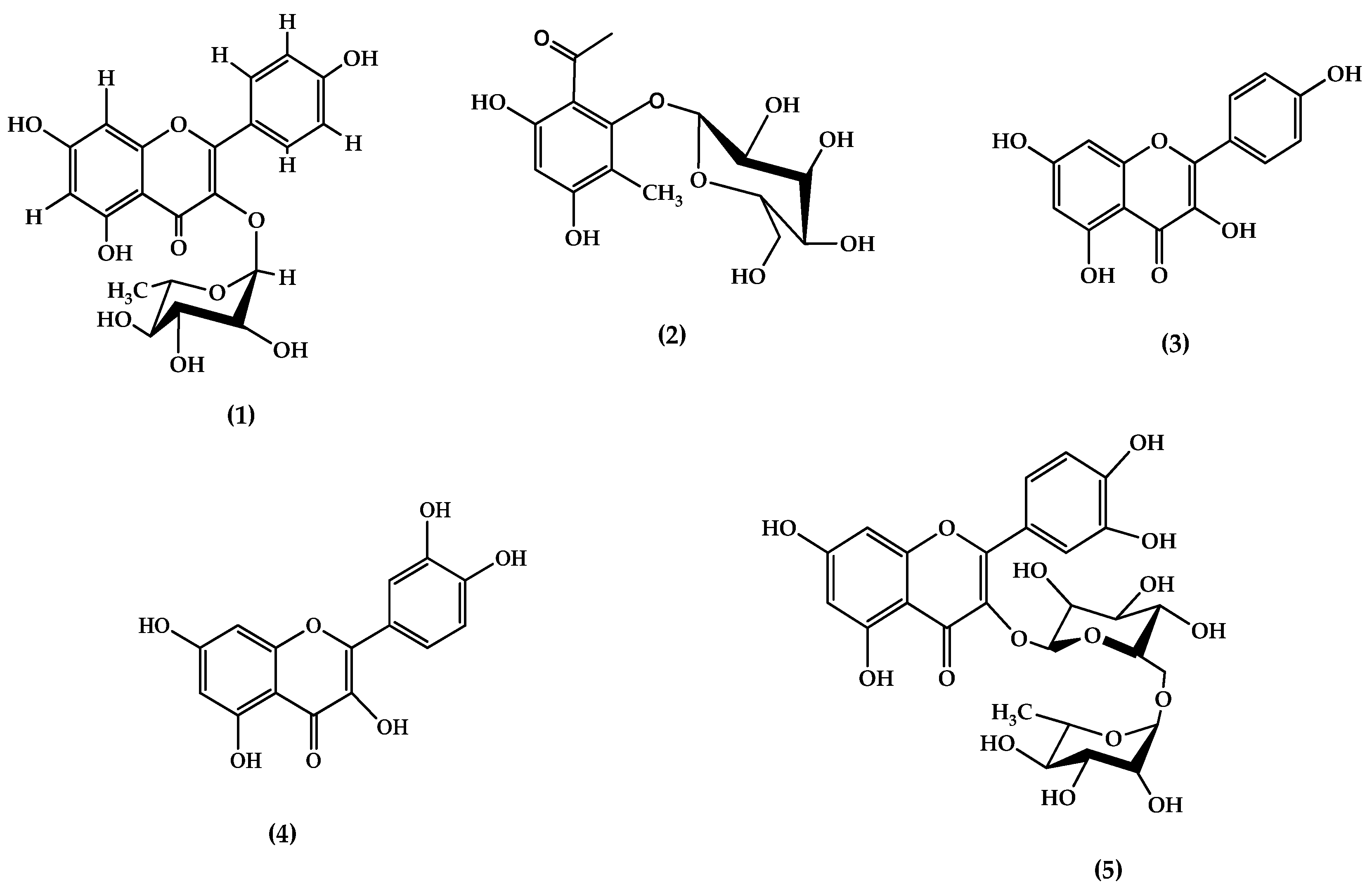
The methanolic extract of GLE leaves was subjected to column chromatography followed by further purification on a Sephadex LH-20 column using a DCM/MeOH (7:3) solvent system to afford five compounds (15; Figure 2). The detailed spectroscopic data for each compound are described in the following.

3.1.1. Compound 1

Compound 1 was isolated as a yellow powder. Its UV-Vis spectrum displayed maximum absorption bands at λmax 254 nm and 335 nm (Figure S1), which suggested a flavonol skeleton [38,39]. This is because flavonoids are known to show two strong absorption bands in the ranges of 220–296 nm and 300–600 nm [40], which is attributed to strong absorption of UV-Vis light by the π–π* type molecular orbital electronic transitions of the two aromatic conjugated π–electron ring systems [38,39]. The MS/MS (ESI-TOF) spectrum of compound 1 showed [M-H], i.e., [C21H20O10–H] at m/z 431.2, consistent with previous authors’ work [41]. Upon collision-induced dissociation, the ion lost a sugar moiety to form a base peak at m/z 485.1, i.e., [C15H10O6–H] [42]. This further fragmented to give peaks at m/z 241.2, 179.1, 151.1, and 109.2 (Figures S2 and S3). These fragment ions are characteristic of flavonol compounds, arising from the cleavage of the flavonoid’s core structure. By comparison with the published literature [41,43,44], compound 1 was tentatively identified as kaempferol-3-O-rhamnopyranoside.
These spectral data were further compared with NMR results, which largely confirmed that the resonances corresponded to aromatic and glycosidic protons and carbons (Table 1). The 13C NMR showed 18 carbon signals (Figure S4), of which 17 carbon signals were in the range of δC 74.0–176.8 ppm and δC 17.8 ppm. The latter is characteristic of a methyl group in the rhamnose sugar moiety [45] while the signal at δC 176.8 ppm attests to the presence of a carbonyl carbon (C4) in the C-ring [46]. The 1H NMR showed four doublet signals within the aromatic region at δH 6.17 (2.0 Hz, 1H), 6.20 (2.0 Hz, 1H), 6.93 (8.7 Hz, 2H), and 7.71 (8.7 Hz, 2H) ppm (Figure S5), where two proton pairs were equivalent and signals at δH 6.17 and 6.20 ppm (d, 2.0 Hz) were meta coupled protons H6 and H8, respectively [47]. The 1H NMR of the sugar region showed an anomeric proton signal at δH 5.85 ppm (d, 1.3 Hz, H1″) (Figure S6), with J = 1.3 Hz indicating an α-linkage of the sugar moiety to the aglycone [48]. The proton signal at δH 1.38 ppm (d, 6.2 Hz) belonged to the H6″ of the sugar moiety. The DQF-COSY (Figure S7) gave cross peaks whose chemical assignments were consistent with the α-rhamnopyranosyl moiety literature [30,48].
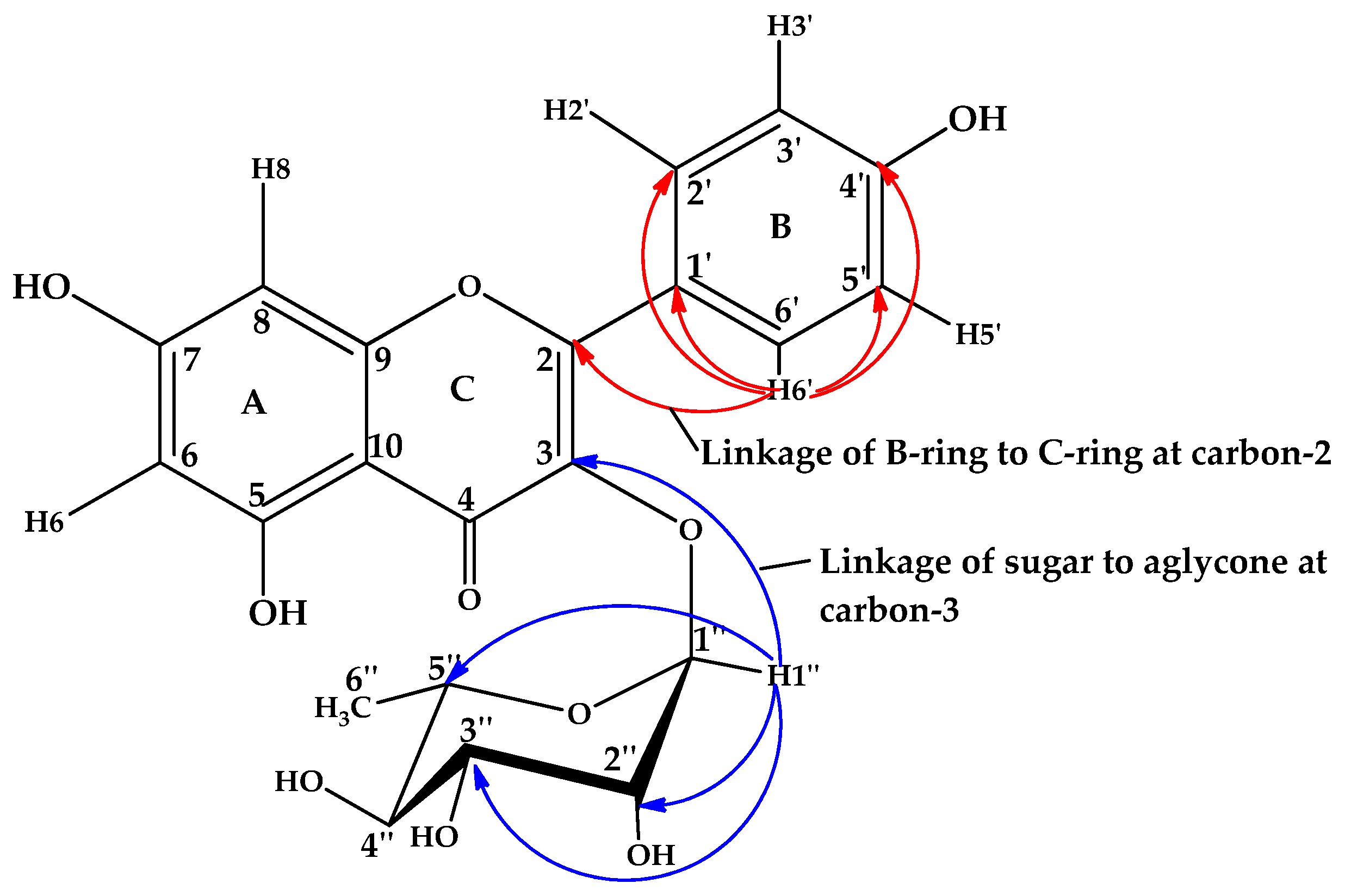
The HSQC correlation (Figure S8) between aromatic protons at δH 7.71 and 6.93 ppm with their respective aromatic carbons at δC 128.1 and 112.5 ppm indicated two ortho-coupled doublet signals, revealing the presence of an AA′BB′ system of ring B of the aglycone assignable to C2′, 6′/H2′, 6′ and C3′, 5′/H3′, 5′, respectively [30,48]. The HMBC spectrum (Figure S9) confirmed the linkage of the rhamnopyranosyl group to the carbon C3 of the aglycone at δ 5.85/136.7 ppm (C3/H1″), while a cross peak at δ 7.71/128.1 ppm (C2/H2′, 6′) showed coupling between carbon C2 of the C–ring and protons H2′ and H6′ of the B-ring, confirming that the B–ring is joined to the C–ring at carbon C2 [49] (Figure 3). Upon comparison with the literature [30,47,48,50], the observed spectral data confirmed that compound 1 is kaempferol-3-O-α-rhamnopyranoside, which is also known as afzelin (Figure 2).
Although kaempferol-3-O-α-rhamnopyranoside is hereby identified for the first time in the Gouania genus, it has been previously isolated from the extracts of leaves and twigs of Lindera neesiana [51], Cercis chinensis flowers [52], Raphanus raphanistrum [41] and Sedum caespitosum aerial parts [53], and Cornus macrophylla and Stenocarpus sinuatus leaves [50,54].

3.1.2. Compound 2

Obtained as a brownish amorphous solid, compound 2 had a UV-Vis spectrum with maximum characteristic absorbance wavelengths of 270 nm and 364 nm (Figure S10). These wavelengths are typical of phenolic compounds [38,39], and corresponds to π–π* and a combination of π–π* and n–π* transitions in the aromatic system and excitations in attached chromophores, respectively [38]. The MS/MS (ESI-TOF) spectrum of compound 2 (Figure S11) showed [M + H]+, C19H21O9+ at m/z 345.1, which fragmented with a sugar loss (C6H11O6, M-179) [42] to form a radical cation, [C9H9O3 + H]+ at m/z 166.2. This was followed by loss of a methyoxy radical (CH3O˙) to form a fragment ion C8H7O2+ at m/z 135.4; then the loss of formaldehyde (CH2O) to give the benzylideneoxonium at m/z 105.1 (C7H5O+) [42], a characteristic fragment of the aromatic ketones (Figure S12). Comparing the spectral data with the literature [38,55,56,57,58], compound 2 was tentatively assigned to be 4,6-dihydroxy-3-methylacetophenone-2-O-β-D-glucopyranoside.
The 1D 1H NMR showed one proton signal at δ 6.37 (3H, singlet), which belonged to the aromatic region [55], indicating that the aromatic ring has one proton (Table 2). The part of the 1D 1H NMR spectrum between δH 3.0 and 5.5 ppm represented the sugar protons [55]. The signal at δH 5.03 (d, J = 7.8 Hz) represented the anomeric proton, with a coupling constant of J = 7.8 Hz, indicating a β-linkage of the sugar moiety [57]. The sugar protons at δH 5.03 (J = 7.8 Hz), δH 4.40 (dd, J = 7.8, 8.2 Hz), δH 3.54 (dd, J = 9.0, 9.1 Hz), δH 3.20 (dd, J = 9.1, 9.7 Hz), δH 3.35 (ddd, J = 2.3, 6.0, 9.7 Hz), δH 3.48 (dd, J = 2.2, 12.1 Hz), and δH 3.17 (dd, J = 5.7, 12.5 Hz) were in agreement with a glucopyranosyl unit [57,58]. The 13C NMR data of compound 2 revealed the presence of a phloroglucinol-type aromatic ring signal at δC 159.61, 147.85, 139.40, 97.57, 92.71, and 61.07 ppm [59], and the acetyl group of acetophenone at δC 208.57 and 29.34 ppm [58]. These spectral data confirmed that compound 2 is 4,6-dihydroxy-3-methylacetophenone-2-O-β-D-glucopyranoside.
This acetophenone glycoside was previously isolated from the flower buds of Syzygium aromaticum [58]. Such acetophenones and their derivatives, including triterpenic acid (2,4-dihydroxy-6-methoxyl-3-methyl-acetophenone) and its 2-β-glucoside [60], 2,4-dihydroxy-6-methoxyl-methylene-3-methyl-acetophenone and 2-dihydroxy-6-methoxyl-methylene-3-methyl-acetophenone-4-O-beta-D-glucopyranoside [61], and 2,4-dihydroxy-6-methoxy-acetophenone 4-O-β-D-glucopyranoside [62] were previously reported in the roots of Euphorbia ebracteolata and aerial parts of Artemisia annua.

3.1.3. Compound 3

Compound 3 was isolated as a yellow crystalline solid which was sparingly soluble in water but dissolved in ethanol. Its UV-Vis spectrum showed maximum absorbances at wavelengths of 256 and 365 nm (Figure S13A), suggesting a conjugated π system chromophore of a flavonoid compound [39,63]. The colored nature of the compound suggested that it possesses a flavonoid nucleus, since most flavonoids are colored [64]. The observed intense absorption bands in the range 250–280 nm and 340–400 nm indicated the presence of a 5, 7, C-4′-trioxygenated flavone [38]. Most flavones and flavonols are known to exhibit two major absorption bands (i) 320–385 nm, which represent the B-ring absorption, and (ii) 250–285 nm, corresponding to the A-ring absorption [65,66]. The rapid decomposition of compound 3 in alkaline medium (sodium ethoxide; Figure S13B) showed that the C-3 and C-4′ contained free hydroxyl groups. The rapid decomposition was evidenced by the shift of the absorbance band to a low intense peak at 315 nm. Flavonols, whose C-3 and C-4′ hydroxyl groups are protected by either methylation or glycoxidation, are stable in alkaline media. This stability is not influenced by the number or substitution of hydroxyl groups at other common positions of flavonols. The presence of a hydroxyl group at C-7 was confirmed by reacting compound 3 with a sodium acetate/ethanol solution, which showed a shift in frequency in the benzenoid region (250–280 nm) due to the ionization of the C7 hydroxyl group that affects the aromaticity in the A- and C-rings of the flavonoid (Figure S13C) [67]. It was concluded from the UV-Vis spectral data that compound 3 is a flavonol with free hydroxyl groups at C-3, C-4′, and C-7.
The UV-Vis data was further supported by LC–MS/MS. The mass spectrum of compound 3 (Figure S14) had [M + H], corresponding to C15H10O6 (calculated molecular weight: 286.1 g/mol). This also formed its base peak, suggesting that it has stable resonance structures, which increased its stability. In addition, the spectrum suggested that the flavonol in compound 3 was less branched, or lacked the sugar moieties, which would otherwise rapidly fragment to give a base peak with lower mass than the molecular ion [42]. Upon collision-induced dissociation of the [M-H], i.e., m/z 285.1, compound 3 fragmented to give m/z 241.2 (C14H9O4) due to the loss of a CO2 molecule (M-44), followed by the loss of a carbon monoxide molecule to give a fragment peak at m/z 212.0. Further fragmentation gave peaks at m/z 179.1, m/z 151.1, and m/z 109.2. This detailed structural fragmentation (Figure S15) was compared with published spectroscopic data [44,68,69]. The compound was tentatively identified as kaempferol, a well-known flavonol (Figure 1). To further confirm this, the HPLC profile of the methanolic extract was run against standard kaempferol. There was a peak matching the HPLC profile of the extract and that of the standard at retention times of 5.358 and 5.368 min, respectively (Figure S16). Based on the matching peaks of the extract and the standard and their corresponding retention times as previously compared in the literature [69,70], it was concluded that kaempferol is compound 3 isolated from the methanol extract.
Although free kaempferol (compound 3) and its derivative (compound 1) are hereby reported in GLE leaves for the first time, it was previously isolated from the crude extract of Gouania longipetala leaf extract [71]. Several kaempferol derivatives have been reported in the Gouania species. For example, kaempferol-3-O-α-L-rhamnoside in Gouania obtusifolia leaves [34], kaempferol-3-O-(6-O-E-coumaroyl)-β-D-galactopyranosyl-(1→2)-α-L-rhamnopyranoside, kaempferol-3-O-(6-O-E-feruloyl)-β-D-galactopyranosyl-(1→2)-α-L-rhamnopyranoside, kaempferol-3-O-α-L-rhamnopyranosyl-(1→6)-β-D-galactopyranoside, kaempferol-3-O-α-L-rhamnopyranosyl-(1→6)-β-D-glucopyranoside, kaempferol-3-O-α-L-rhamnopyranoside, kaempferol-3-O-β-D-xylopyranosyl-(1→2)-α-L-rhamnopyranoside, kaempferol-3-O-β-D-galactopyranosyl-(1→2)-α-L-rhamnopyranoside in aerial parts of Gouania longipetala [30], and kaempferol-3-O-(6-O-E-caffeoyl)-β-D-galactopyranosyl-(1→2)-α-L-rhamnopyranoside in aerial parts of Gouania leptostachya [36]. Kaempferol is a flavonol aglycone biophenolic compound that has also been isolated in several plants, including Albizia chinensis [49], Brassica rapa, Psidium guajava, and other fruits and vegetables such as onions, broccoli, tea, grapes, tomatoes, and strawberries [72,73].

3.1.4. Compound 4

Compound 4 was obtained as a yellow solid. Its UV-Vis spectrum showed maximum wavelengths of 250 nm and 375 nm (Figure S17), which suggested a typical flavonol skeleton [67,74]. It exhibited properties that were comparable to compound 3. For example, it showed a rapid decomposition with a characteristic shift of its absorbance band to a low intense peak at 315 nm when it was treated with alkaline sodium ethoxide. This clearly confirmed that like compound 3, compound 4 should be a flavonol possessing a free hydroxyl group and a carbon-3 of the C-ring, and a free hydroxyl group at C-4′ of the B-ring [49,67]. The presence of a hydroxyl group at C-7, as carried out for compound 3, was also confirmed, since compound 4 upon reacting with sodium acetate/ethanol solution had a noticeable shift in frequency in the benzenoid region (250–280 nm).
The mass spectrum of compound 4 (Figure S18) showed [M + H], with a molecular formula of C15H10O7 (calculated molecular weight: 302.1 g/mol) and a base peak at m/z 151.1. Upon collision-induced dissociation of [M-H], i.e., m/z 301.1, compound 4 fragmented to give m/z 271.2 ([C14H8O6-H]) due to the loss of hydrogen and carbon monoxide (M-29), followed by the loss of a carbon monoxide molecule (M-29-28) to give the fragment peak observed at m/z 243.9 (C13H8O5-H). Further fragmentation gave peaks at m/z 112.8, 151.1, 179.1, 228.0, and 256.0. The overall fragmentation pattern (Figure S19) was consistent with previously published spectroscopic data [44,68,69,70]. This led to the identification of compound 4 as quercetin. To further confirm this, the HPLC profile of the methanolic extract of GLE leaves was run against standard quercetin. A peak in the HPLC profile of the extract that matched that of the standard at retention times of 4.408 min and 4.328 min, respectively, was identified (Figure S20). It was then concluded that compound 4 is quercetin.
Quercetin is one of the most important bioflavonoids reported in various species in kingdom Plantae [75]. Its derivatives such as quercetin 3-O-α-L-rhamnoside [34], quercetin-3-O-β-D-galactopyranosyl-(1→2)-α-L-rhamnopyranoside, quercetin-3-O-α-L-rhamnopyranosyl-(1→6)-β-D-galactopyranoside [30], quercetin-3-O-β-D-xylopyranosyl-(1→2)-α-L-rhamnopyranoside, and quercetin-3-O-6-E-p-coumaroyl-β-D-glucopyranosyl-(1→2)-α-L-rhamnopyranoside [69] were previously isolated from Gouania obtusifolia leaves, Gouania longipetala aerial parts, and Gouania leptostachya leaves, respectively. However, this is the first time the free form of quercetin is being reported in the Gouania genus. Quercetin has been isolated from several plant species, including Albizia chinensis [49] and Prunus serrulata [76] leaves.

3.1.5. Compound 5

Compound 5 was isolated as a light yellow solid, and had characteristically strong UV-Vis absorption at wavelengths of 256 nm and 379 nm (Figure S21), which strongly indicated that it possesses a flavonol skeleton [67,74]. To gain further insights, the compound was treated with alkaline sodium ethoxide, but it did not show any bathochromic shifts in the absorbance band. This indicated that the compound is a flavonol without a free hydroxyl group on carbon-3 of the C-ring, since the free hydroxyl group at C-4′ of the B-ring can react only if a free hydroxyl group was present at carbon-3 of the C-ring [67]. Interestingly, the reaction of compound 5 with sodium acetate/ethanol solution caused a shift in the wavelength in the benzenoid region (250–280 nm), which indicated the presence of a free hydroxyl group at C-7 of the A-ring. This data suggested that compound 5 is glycosylated at C-3 and has a free hydroxyl at C-7 [67].
The mass spectrum of compound 5 (Figure S22) showed [M + H], corresponding to the molecular formula C27H30O16 (calculated molar mass = 610.2 g/mol). The spectrum indicated the abundance of m/z 463.2 (attributed to the loss of C6H12O4), m/z 446.0 (from the loss of C6H12O5), and the base peak at m/z 301.1, supposedly due to the loss of two sugar units (C12H22O9). It was clear that the base peak was formed after the loss of the two sugar units, converting it to quercetin (compound 4) at m/z 301.1. This further fragmented to form other fragment peaks similar to those of quercetin (Figure S19). This suggested that quercetin (compound 4) forms the flavonoid skeleton of compound 5. Comparison of its fragmentation pattern (Figure S23) with published literature records [68,77] led to its identification as rutin, a bioflavonoid sometimes called quercetin-3-O-rutinoside, rutoside, or sophorin, among other name variants. This flavonoid is one of the most widely recognized phenolic compounds found in various plant species [78], but this is the first time that it is being reported in the Gouania genus.
Taken together, the results of chromatographic isolation and characterization of the methanolic extract of GLE leaves showed that it is rich in polyphenols. Members of the Gouania genus are known to be rich in terpenoids and phenolic compounds, with up to five phenolic compounds, twenty flavonoids, and thirty-four terpenoids reported to date [28,29,30,31,32,33,34,35,36]. It is worth noting that this is the first time that the kaempferol derivative (compound 1), the acetophenone (compound 2), as well as free forms of kaempferol (compound 3), quercetin (compound 4), and rutin (compound 5) are being reported in GLE and the Gouania genus. Kaempferol, quercetin, and rutin are known for their intriguing bioactivities such as antioxidant, antidiabetic, cytoprotective, vasoprotective, anticancer, anti-asthma, anti-inflammatory, and antimicrobial properties [79,80], which could validate the use of GLE leaves for treating ailments in traditional medicine. The possible synergistic effect of these multispectral pharmacologically active compounds as well administration of GLE herbal preparations for up to one week could explain why this species may be effective in traditional treatment of diseases. This resonates well with the report from southwestern Uganda where this species is considered by local communities to be effective against all diseases [81]. While some of the compounds (35) reported in this study have been previously isolated from other plant species, their isolation from GLE points to the distinct ecological roles or medicinal properties specific to this species for survival in the environment in which it grows. As an example, the acetophenone derivative with a glucopyranoside ring identified in GLE (compound 2) could be biosynthesized to fend off pests [82], which also sheds light on the perspective of using this plant against babesiosis [18].

3.2. Antibacterial Potential of Isolated Polyphenols

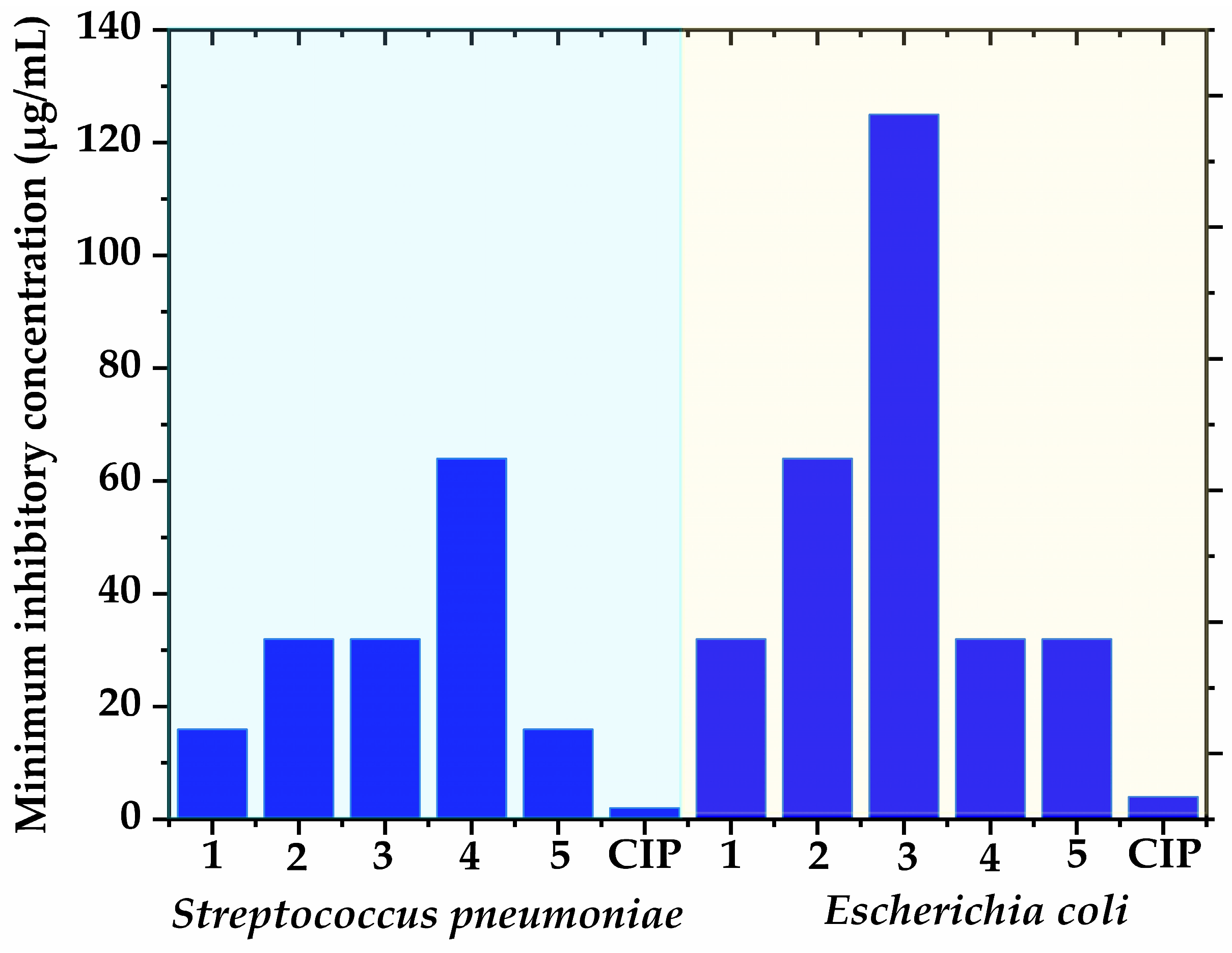
The compounds isolated from the methanolic leaf extract of GLE (15) exhibited antibacterial activity against S. pneumoniae and E. coli with MIC between 16 µg/mL and 125 µg/mL (Figure 4). The isolated compounds had higher bioactivity, as all had lower MIC compared to GLE leaf extracts reported by Gumisiriza et al. [7]. This lends credence to the traditional use of GLE for treating various infections in Africa. Most of the isolated compounds in this study have been previously reported to have antibacterial activity. For example, afzelin inhibited the growth of Pseudomonas aeruginosa with a MIC of 31 μg/mL [50]. The acetophenone (compound 2) has no reported antimicrobial activity, but 4-hydroxy3(isopentent-2-yl) acetophenone isolated from Helichrvsum italicum was previously reported to have antibacterial activity against Bacillus species and Staphylococcus epidermidis, and E. coli with MIC values of >100 µg/mL and 25 µg/mL, respectively [83]. Kaempferol isolated from Dodonaea viscosa leaves had MIC ranging from 16 to 63 µg/mL against E. coli, Enterococcus faecalis, Staphylococcus aureus, and Pseudomonas aeruginosa [84]. Quercetin is a ubiquitous flavonoid with antibacterial activity against several microorganisms, including Actinobacillus actinomycetemocomitans, Lactobacillus acidophilus, Streptococcus mutans, Streptococcus sobrinus, Streptococcus sanguis, and Prevotella intermedia (MIC = 1000–8000 µg/mL) [85]. Rutin is a glycoside combining the flavonol quercetin and the disaccharide rutinose. It equally possesses antibacterial and antibiofilm properties against multidrug-resistant Pseudomonas aeruginosa at higher concentrations (MIC = 512 µg/mL) [86].
The MIC obtained in this study varied between the microorganisms, and this is likely due to their genetic differences as well as the structure and physicochemical properties of the compounds such as their solubility, molecular size, stability, and lipophilicity [87]. For example, the biological effects of rutin in vivo are known to be limited by its low bioavailability [80]. Considering structure–bioactivity relationships, a previous study cited that quercitrin (afzelin with OH at C3) elicited no antibacterial activity, while kaempferol (afzelin without a rhamnose group) tended to exhibit inferior antibacterial activity to afzelin. Their observations led to the postulation that the hydroxyl group at C3 of the C-ring of the flavone skeleton and rhamnose group may be involved in the antibacterial effect of this compound [50]. Similarly, quercetin’s antibacterial activity has been attributed to its solubility and interaction with bacterial cell membranes, a property largely influenced by the presence of its hydroxyl groups [88].
The antimicrobial activity of flavonoids, phenolic and flavonoid glycosides is attributed to the interaction of the compounds with the bacterial membrane proteins or lipids leading to bacterial membrane stabilization and disruption, inhibition of membrane-bound enzymes, modulation of immune response, and lipid raft disruption. For example, quercetin inhibits the supercoiling activity of gyrase B of E. coli and induces DNA cleavage [89]. Kaempferol is also able to directly inhibit DNA helicases such as SAPriA in Staphylococcus aureus [90]. Kaempferol-3-O-α-rhamnopyranoside, kaempferol, quercetin, and rutin inhibit quorum sensing which interferes with bacterial communication systems, reducing virulence factors and biofilm formation [91]. For example, rutin had antibiofilm potential against Klebsiella pneumoniae strains isolated from hospitalized patients [92]. On the other hand, membrane permeabilization and rupture is another known mechanism of the antibacterial activity of kaempferol and quercetin, which is mediated through increasing bacterial membrane permeability, interfering with bacterial energy metabolism, reducing pathogenicity, which induces cytoplasmic leakage [93].

3.3. Free Radical Scavenging Results

All the compounds showed radical scavenging activities with IC50 from 18.6 ± 1.30 µg/mL to 28.1 ± 0.09 µg/mL (Table 3). The antioxidant potential of the compounds based on the IC50 values could be arranged as quercetin (compound 4) > kaempferol (compound 3) > kaempferol-3-O-α-rhamnopyranoside (compound 1) > 4,6-dihydroxy-3-methylacetophenone-2-O-β-D-glucopyranoside (compound 2) > rutin (compound 5). The obtained IC50 were, however, higher than that of ascorbic acid (14.6 ± 0.15 µg/mL) but the IC50 of kaempferol (compound 3) and quercetin (compound 4) were not significantly different from that of the positive control (ascorbic acid) as per Dunnett’s T3 multiple comparisons test after Brown–Forsythe and Welch ANOVA (p > 0.05). These results indicate that kaempferol and quercetin from the methanolic extract of GLE leaves exhibit potent antioxidant activity similar to ascorbic acid, highlighting their potential as natural antioxidant agents.
The isolated compounds (15) are known antioxidants. For example, Adhikari-Devkota et al. [51] reported that kaempferol-3-O-α-rhamnopyranoside characterized from Lindera neesiana aerial parts had an IC50 of 201 μg/mL in the DPPH assay, which is higher than in the present study. Tatsimo et al. [94] reported that kaempferol-3-O-α-rhamnopyranoside elicited antioxidant activity with an IC50 value of 6.44 μg/mL, which is better than what is observed in the present study. Kaempferol-3-O-α-L-rhamnoside, a kaempferol glycoside isolated from Pithecellobium dulce leaves, also exhibited strong antioxidant activity with an IC50 value of 14.6 μg/mL [95]. The compound 4,6-dihydroxy-3-methylacetophenone-2-O-β-D-glucopyranoside (compound 2) has not been studied for its antioxidant activity before. However, kaempferol (compound 3) previously exhibited superior antioxidant potential (IC50 = 4.506 μg/mL) to butylated hydroxytoluene (IC50 = 10.33 μg/mL) [96]. The antioxidant activity of kaempferol optimally isolated from Ocimum basilicum leaves (IC50 = 0.68 μg/mL) was recently reported to be higher than that of ascorbic acid (IC50 = 0.79 μg/mL) [97]. Further, quercetin and rutin (isolated in this study) are among the most studied antioxidants [98].

4. Conclusions

Five known bioactive polyphenols (compounds 15) were identified from GLE leaves for the first time. This is the first time that the kaempferol derivative, the acetophenone as well as free forms of quercetin, kaempferol, and rutin are being reported in the Gouania genus. The compounds exhibited antibacterial and antioxidant activities, supporting their claimed use in traditional medicine. The comparable antioxidant activities of kaempferol and quercetin to that of ascorbic acid suggests that this species could be a source of natural antioxidant agents that could be exploited. In addition, the possible synergistic effects of these multispectral pharmacologically active compounds could explain why GLE herbal preparations are effective against a wide range of diseases. Although some of the compounds reported in this study have been previously isolated from other plant species, their isolation from GLE points to the distinct ecological roles and/or medicinal properties specific to this species for survival in the environment in which it grows. Further research should aim to identify the compounds associated with other bioactivities that traditional healers have pointed to; this could lead to the isolation of more bioactive molecules. In vivo studies using animal models to evaluate the efficacy and safety of the isolated compounds from this species could provide stronger evidence of their bioactivity, dosage, safety, and toxicity, which would complement the observed in vitro antibacterial and antioxidant activities.

Supplementary Materials

The following supporting information can be downloaded at: https://www.mdpi.com/article/10.3390/microbiolres15040140/s1, Figure S1. UV-Vis spectrum of compound 1; Figure S2. Mass spectrum of compound 1; Figure S3. Fragmentation pattern of compound 1; Figure S4. Expanded 13C NMR spectrum of compound 1; Figure S5. 1H NMR spectrum (aromatic region) of compound 1; Figure S6. 1H NMR spectrum (sugar region) of compound 1; Figure S7. COSY spectrum of compound 1; Figure S8. The 1H-13C HSQC spectrum of compound 1; Figure S9. The 1H-13C HMBC spectrum of compound 1; Figure S10. UV-Vis spectrum of compound 2; Figure S11. Mass spectrum of compound 2; Figure S12. Fragmentation pattern of compound 2; Figure S13. UV-vis spectrum of compound 3 in (A) methanol, (B) sodium ethoxide, and (C) sodium acetate/methanol solution; Figure S14. Mass spectrum of compound 3; Figure S15. Fragmentation pattern of compound 3; Figure S16. HPLC profile peak matching of (a) kaempferol standard with (b) methanolic extract of GLE; Figure S17. UV-Vis spectrum of compound 4; Figure S18. Mass spectrum of compound 4; Figure S19. Fragmentation pattern of compound 4; Figure S20. HPLC profile peak matching of (a) standard quercetin with (b) methanolic extract of GLE; Figure S21. UV-Vis spectrum of compound 5; Figure S22. Mass spectrum of compound 5; Figure S23. Fragmentation pattern of compound 5.

Author Contributions

Conceptualization, H.G. and E.A.O.; methodology, H.G., E.A.O., T.O., J.B.L. and D.C.S.; software, H.G. and T.O.; validation, E.A.O., L.M., R.A., T.O., J.B.L. and D.C.S.; formal analysis, H.G.; investigation, H.G.; resources, E.A.O., L.M., R.A., T.O., J.B.L. and D.C.S.; data curation, H.G. and T.O.; writing—original draft preparation, H.G. and T.O.; writing—review and editing, E.A.O., L.M., R.A., T.O., J.B.L. and D.C.S.; visualization, H.G. and T.O.; project administration, H.G.; funding acquisition, H.G., E.A.O., L.M., R.A., T.O., J.B.L. and D.C.S. All authors have read and agreed to the published version of the manuscript.

Funding

This research was funded by Organization for the Prohibition of Chemical Weapons (OPCW) and Mbarara University of Science and Technology, Uganda under grant numbers L/ICA/ICB-122/22 and Pf/Conf-HG01/22, respectively.

Institutional Review Board Statement

Mbarara University of Science and Technology Research Ethics Committee (protocol no. 19/08-17) and Uganda National Council for Science and Technology (No. NS34ES) approved the study.

Informed Consent Statement

Not applicable.

Data Availability Statement

Data will be available upon reasonable request from the corresponding authors.

Conflicts of Interest

The authors declare no conflicts of interest.

References

  1. GBD 2019 Antimicrobial Resistance Collaborators. Global mortality associated with 33 bacterial pathogens in 2019: A systematic analysis for the Global Burden of Disease Study 2019. Lancet 2023, 400, 2221–2248. [Google Scholar]
  2. Pashkov, E.A.; Pak, A.V.; Pashkov, E.P.; Bykov, A.S.; Budanova, E.V.; Poddubikov, A.V.; Svitich, O.A.; Zverev, V.V. The prospects for the use of drugs based on the phenomenon of RNA interference against HIV infection. Probl. Virol. 2022, 67, 278–289. [Google Scholar] [CrossRef] [PubMed]
  3. Tabuti, J.R.S.; Obakiro, S.B.; Nabatanzi, A.; Anywar, G.; Nambejja, C.; Mutyaba, M.R.; Omara, T.; Waako, P. Medicinal plants used for treatment of malaria by indigenous communities of Tororo District, Eastern Uganda. Trop. Med. Health 2023, 51, 34. [Google Scholar] [CrossRef] [PubMed]
  4. Ssenku, J.E.; Okurut, S.A.; Namuli, A.; Kudamba, A.; Tugume, P.; Matovu, P.; Wasige, G.; Kafeero, H.M.; Walusansa, A. Medicinal plant use, conservation, and the associated traditional knowledge in rural communities in Eastern Uganda. Trop. Med. Health 2022, 50, 39. [Google Scholar] [CrossRef] [PubMed]
  5. Omara, T. Antimalarial Plants Used across Kenyan Communities. Evid.-Based Complement. Altern. Med. 2020, 2020, 4538602. [Google Scholar] [CrossRef]
  6. Okello, D.; Kang, Y. Exploring antimalarial herbal plants across communities in Uganda based on electronic data. Evid.-Based Complement. Altern. Med. 2019, 2019, 3057180. [Google Scholar] [CrossRef]
  7. Gumisiriza, H.; Birungi, G.; Lejju, J.; Olet, E.; Kembabazi, O.; Duncan, C.; Sesaazi, C.D. Ethnobotany and Antimicrobial Activity of Gouania longispicata Engl. J. Complement. Med. Res. 2020, 11, 86–94. [Google Scholar] [CrossRef]
  8. Giday, M.; Asfaw, Z.; Woldu, Z. Ethnomedicinal study of plants used by Sheko ethnic group of Ethiopia. J. Ethnopharmacol. 2010, 132, 75–85. [Google Scholar] [CrossRef]
  9. Irakiza, R.; Vedaste, M.; Elias, B.; Nyirambangutse, B.; Serge, N.; Marc, N. Assessment of traditional ecological knowledge and beliefs in the utilisationof important plant species: The case of Buhanga sacred forest, Rwanda. Koedoe 2016, 58, a1348. [Google Scholar] [CrossRef]
  10. Mollel, N.P.; Otieno, J.N.; Sitoni, D.K. Medicinal plants traded in Arusha city, Tanzania. J. Med. Plants Stud. 2022, 10, 175–182. [Google Scholar]
  11. Hamill, F.; Apio, S.; Mubiru, N.; Mosang, M.; Bukenya-Ziraba, R.; Maganyi, O.; Soejarto, D. Traditional herbal drugs of southern Uganda, I. J. Ethnopharmacol. 2000, 70, 281–300. [Google Scholar] [CrossRef] [PubMed]
  12. Kudamba, A.; Kasolo, J.N.; Bbosa, G.S.; Lugaajju, A.; Wabinga, H.; Niyonzima, N.; Ocan, M.; Damani, A.M.; Kafeero, H.M.; Ssenku, J.E.; et al. Medicinal plants used in the management of cancers by residents in the Elgon Sub-Region, Uganda. BMC Complement. Med. Ther. 2023, 23, 450. [Google Scholar] [CrossRef] [PubMed]
  13. Gumisiriza, H.; Sesaazi, C.D.; Olet, E.A.; Kembabazi, O.; Birungi, G. Medicinal plants used to treat “African” diseases by the local communities of Bwambara sub-county in Rukungiri District, Western Uganda. J. Ethnopharmacol. 2021, 268, 113578. [Google Scholar] [CrossRef] [PubMed]
  14. Tabuti, J.R.; Kukunda, C.B.; Kaweesi, D.; Kasilo, O.M. Herbal medicine use in the districts of Nakapiripirit, Pallisa, Kanungu, and Mukono in Uganda. J. Ethnobiol. Ethnomed. 2012, 8, 35. [Google Scholar] [CrossRef]
  15. Olsthoorn, F.M.O. Determinants of Spatial Patterns of Human Activity in Bwindi Impenetrable National Park. Master’s Thesis, Imperial College London, London, UK, 2017. [Google Scholar]
  16. Kyoshabire, M.; Katuura, E.; Cunningham, A.B.; Hoeft, R. Medicinal plants and herbalist preferences around Bwindi Impenetrable National Park. J. Med. Plants Res. 2017, 11, 161–170. [Google Scholar]
  17. Tanno, T. Plant utilization of the Mbuti pygmies with Special Reference to their material culture and use of wild vegetable foods. Afr. Study Monogr. 1981, 1, 1–53. [Google Scholar]
  18. Chifundera, K. Livestock diseases and the traditional medicine in the bushi area, Kivu province, Democratic Republic of Congo. Afr. Stud. Monogr. 1998, 19, 13–33. [Google Scholar]
  19. Ganas, J.; Ortmann, S.; Robbins, M.M. Food preferences of wild mountain gorillas. Am. J. Primatol. 2008, 70, 927–938. [Google Scholar] [CrossRef] [PubMed]
  20. Rothman, J.M.; Dierenfeld, E.S.; Molina, D.O.; Shaw, A.V.; Hintz, H.F.; Pell, A.N. Nutritional chemistry of foods eaten by gorillas in Bwindi Impenetrable National Park, Uganda. Am. J. Primatol. 2006, 68, 675–691. [Google Scholar] [CrossRef]
  21. Yamagiwa, J.; Basabose, A.K.; Kaleme, K.; Yumoto, T. Diet of Grauer’s Gorillas in the Montane Forest of Kahuzi, Democratic Republic of Congo. Int. J. Primatol. 2005, 26, 1345–1373. [Google Scholar] [CrossRef]
  22. Krief, S.; Hladik, C.M.; Haxaire, C. Ethnomedicinal and bioactive properties of plants ingested by wild chimpanzees in Uganda. J. Ethnopharmacol. 2005, 101, 1–15. [Google Scholar] [CrossRef] [PubMed]
  23. Freymann, E.; Carvalho, S.; Garbe, L.A.; Dwi Ghazhelia, D.; Hobaiter, C.; Huffman, M.A.; Muhumuza, G.; Schulz, L.; Sempebwa, D.; Wald, F.; et al. Pharmacological and behavioral investigation of putative self-medicative plants in Budongo chimpanzee diets. PLoS ONE 2024, 19, e0305219. [Google Scholar] [CrossRef] [PubMed]
  24. Demgne, O.M.F.; Tchinda, C.F.; Mbaveng, A.T.; Beng, V.P.; Kuete, V. Antibacterial and antibiotic-potentiating activities of nine Cameroonian medicinal plants against multidrug-resistant bacteria expressing active efflux pumps. Investig. Med. Chem. Pharmacol. 2022, 5, 58. [Google Scholar] [CrossRef]
  25. Migabo, H.; Izere, C.; Habyarimana, T.; Josiane, U.K.N.; Nsabayezu, E.; Niyonzima, F.N. Phytochemistry analysis and anti-bacterial properties of Rwandese medicinal plants. NeuroQuantology 2022, 20, 965–978. [Google Scholar]
  26. Gumisiriza, H.; Birungi, G.; Omara, T.; Lejju, J.B.; Sesaazi, C.D. Polyphenolic content, antioxidant activity and acute toxicity of Gouania longispicata Engl. leaves. LIANBS, 2025; in press. [Google Scholar]
  27. Ezeja, M.I.; Anaga, A.O.; Asuzu, I.U. Antidiabetic, antilipidemic, and antioxidant activities of Gouania longipetala methanol leaf extract in alloxan-induced diabetic rats. Pharm. Biol. 2015, 53, 605–614. [Google Scholar] [CrossRef] [PubMed]
  28. Nair, S.P.; Rao, J.M. Gouanic acid from the leaves of Gouania microcarpa. Phytochemistry 1993, 33, 711–712. [Google Scholar] [CrossRef]
  29. Giacomelli, S.R.; Maldaner, G.; Stücker, C.; Marasciulo, C.; Schmidt, J.; Wessjohann, L.; Dalcol, I.I.; Morel, A.F. Triterpenoids from Gouania ulmifolia. Planta Med. 2007, 73, 499–501. [Google Scholar] [CrossRef]
  30. Gossan, M.A.; Yao-kouassi, A.; Coffy, A.A.; Harakat, D.; Voutquenne-Nazabadioko, L. New acylated flavonol glycosides from the aerial parts of Gouania longipetala. Phytochem. Lett. 2015, 11, 306–310. [Google Scholar] [CrossRef]
  31. Gossan, D.P.; Alabdul Magid, A.; Yao-Kouassi, P.A.; Ahibo, C.A.; Josse, J.; Gangloff, S.C.; Morjani, H.; Voutquenne-Nazabadioko, L. Triterpene glycosides from the aerial parts of Gouania longipetala. Phytochemistry 2017, 134, 71–77. [Google Scholar] [CrossRef]
  32. Kennelly, E.J.; Lewis, W.H.; Winter, R.E.; Johnson, S.; Elvin-Lewis, M.; Gossling, J. Triterpenoid saponins from Gouania lupuloides. J. Nat. Prod. 1993, 56, 402–410. [Google Scholar] [CrossRef] [PubMed]
  33. Désiré, S.; Ernestine, N.; Bruno, T.B.; Lazare, S.S.; Ulrich, D.D.; Lateef, M.; Schneider, B.; Ali, M.S.; Barthélemy, N. A new dammarane type triterpene glucoside from the aerial parts of Gouania longipetala (Rhamnaceae). Nat. Prod. Res. 2021, 35, 3192–3203. [Google Scholar] [CrossRef] [PubMed]
  34. Li, X.; Ohtsuki, T.; Shindo, S.; Sato, M.; Koyano, T.; Preeprame, S.; Kowithayakorn, T.; Ishibashi, M. Mangiferin identified in a screening study guided by neuraminidase inhibitory activity. Planta Med. 2007, 73, 1195–1196. [Google Scholar] [CrossRef] [PubMed]
  35. Hang, N.T.; Bich Thu, N.T.; Le Ba, V.; Van On, T.; Khoi, N.M.; Do, T.H. Characterisation of four new triterpenoid saponins with nitric oxide inhibitory activity from aerial parts of Gouania leptostachya. Nat. Prod. Res. 2022, 36, 5999–6005. [Google Scholar] [CrossRef] [PubMed]
  36. Hang, N.T.; Thu, N.T.B.; Vinh, L.B.; Phong, N.V.; On, T.V.; Lee, K.Y. A New Ceanothane-Type Triterpenoid Saponin Isolated from Gouania leptostachya DC. var. tonkinensis Pit. and Its Underlying Anti-Inflammatory Effects. J. Microbiol. Biotechnol. 2023, 33, 941–948. [Google Scholar] [CrossRef]
  37. Omara, T.; Kiprop, A.K.; Kosgei, V.J. Intraspecific variation of phytochemicals, antioxidant, and antibacterial activities of different solvent extracts of Albizia coriaria leaves from some agroecological zones of Uganda. Evid.-Based Complement. Altern. Med. 2021, 2021, 2335454. [Google Scholar] [CrossRef]
  38. Mabasa, X.E.; Mathomu, L.M.; Madala, N.E.; Musie, E.M.; Sigidi, M.T. Molecular Spectroscopic (FTIR and UV-Vis) and Hyphenated Chromatographic (UHPLC-qTOF-MS) Analysis and In Vitro Bioactivities of the Momordica balsamina Leaf Extract. Biochem. Res. Int. 2021, 2021, 2854217. [Google Scholar] [CrossRef]
  39. Guemari, F.; Laouini, S.E.; Rebiai, A.; Bouafia, A.; Meneceur, S.; Tliba, A.; Majrashi, K.A.; Alshareef, S.A.; Menaa, F.; Barhoum, A. UV-Visible Spectroscopic Technique-Data Mining Tool as a Reliable, Fast, and Cost-Effective Method for the Prediction of Total Polyphenol Contents: Validation in a Bunch of Medicinal Plant Extracts. Appl. Sci. 2022, 12, 9430. [Google Scholar] [CrossRef]
  40. Sisa, M.; Bonnet, S.L.; Ferreira, D.; Van der Westhuizen, J.H. Photochemistry of Flavonoids. Molecules 2010, 15, 5196–5245. [Google Scholar] [CrossRef] [PubMed]
  41. Ibrahim, L.F.; Elkhateeb, A.; Marzouk, M.M.; Hussein, S.R.; Abdel-Hameed, E.S.; Kassem, M.E.S. Flavonoid investigation, LC–ESI-MS profile and cytotoxic activity of Raphanus raphanistrum L. (Brassicaceae). J. Chem. Pharm. Res. 2016, 8, 786–793. [Google Scholar]
  42. Nicolescu, T. Interpretation of Mass Spectra. In Mass Spectrometry; IntechOpen: London, UK, 2017; pp. 23–78. [Google Scholar]
  43. Fabre, N.; Rustan, I.; de Hoffmann, E.; Quetin-Leclercq, J. Determination of flavone, flavonol, and flavanone aglycones by negative ion liquid chromatography electrospray ion trap mass spectrometry. J. Am. Soc. Mass. Spectrom. 2001, 12, 707–715. [Google Scholar] [CrossRef] [PubMed]
  44. Chen, Y.; Yu, H.; Wu, H.; Pan, Y.; Wang, K.; Jin, Y.; Zhang, C. Characterization and Quantification by LC-MS/MS of the Chemical Components of the Heating Products of the Flavonoids Extract in Pollen Typhae for Transformation Rule Exploration. Molecules 2015, 20, 18352–18366. [Google Scholar] [CrossRef] [PubMed]
  45. Mair, C.E.; Grienke, U.; Wilhelm, A.; Urban, E.; Zehl, M.; Schmidtke, M.; Rollinger, J.M. Anti-Influenza Triterpene Saponins from the Bark of Burkea africana. J. Nat. Prod. 2018, 81, 515–523. [Google Scholar] [CrossRef] [PubMed]
  46. Malee, T. Chemical Constituents from the Leaves and Stems of Citrus aurantifolia Swingle. Master’s Thesis, Prince of Songkla University, Hat Yai, Thailand, 2011. [Google Scholar]
  47. Hussain, J.; Ali, L.; Khan, A.L.; Rehman, N.U.; Jabeen, F.; Kim, J.-S.; Al-Harrasi, A. Isolation and Bioactivities of the Flavonoids Morin and Morin-3-O-β-D-glucopyranoside from Acridocarpus orientalis—A Wild Arabian Medicinal Plant. Molecules 2014, 19, 17763–17772. [Google Scholar] [CrossRef]
  48. Ek, S.; Kartimo, H.; Mattila, S.; Tolonen, A. Characterization of phenolic compounds from lingonberry (Vaccinium vitis-idaea). J. Agric. Food Chem. 2006, 54, 9834–9842. [Google Scholar] [CrossRef]
  49. Ghaly, N.S.; Melek, F.R.; Abdelwahed, N.A.M. Flavonoids from Albizia chinensis of Egypt. Rev. Latinoam. Química 2010, 38, 153–158. [Google Scholar]
  50. Lee, S.Y.; So, Y.-J.; Shin, M.S.; Cho, J.Y.; Lee, J. Antibacterial Effects of Afzelin Isolated from Cornus macrophylla on Pseudomonas aeruginosa, A Leading Cause of Illness in Immunocompromised Individuals. Molecules 2014, 19, 3173–3180. [Google Scholar] [CrossRef]
  51. Adhikari-Devkota, A.; Dirar, A.I.; Kurizaki, A.; Tsushiro, K.; Devkota, H.P. Extraction and Isolation of Kaempferol Glycosides from the Leaves and Twigs of Lindera neesiana. Separations 2019, 6, 10. [Google Scholar] [CrossRef]
  52. Zhang, J.; Zhou, L.; Cui, L.; Liu, Z.; Wei, J.; Kang, W. Antioxidant and α-glucosidase inhibitiory activity of Cercis chinensis flowers. Food Sci. Hum. Wellness 2020, 9, 313–319. [Google Scholar] [CrossRef]
  53. Söhretoğlu, D.; Sabuncuoğlu, S. Secondary metabolites, cytotoxic response by neutral red retention and protective effect against H2O2 induced cytotoxicity of Sedum caespitosum. Nat. Prod. Commun. 2012, 7, 39–40. [Google Scholar] [CrossRef]
  54. Younis, M.M.; Ayoub, I.M.; George, M.Y.; Mostafa, N.M.; Eldahshan, O.A. In vivo hepatoprotective and nephroprotective effects of Stenocarpus sinuatus leaf extract against ifosfamide-induced toxicity in rats. Arch. Pharm. 2024, 357, e2300438. [Google Scholar] [CrossRef] [PubMed]
  55. Brown, G.D.; Bauer, J.; Osborn, H.M.I.; Kuemmerle, R. A Solution NMR Approach To Determine the Chemical Structures of Carbohydrates Using the Hydroxyl Groups as Starting Points. ACS Omega 2018, 3, 17957–17975. [Google Scholar] [CrossRef] [PubMed]
  56. Ayräs, P.; Lötjönen, S.; Widén, C.J. NMR spectroscopy of naturally occurring phloroglucinol derivatives. Planta Med. 1981, 42, 187–194. [Google Scholar] [CrossRef] [PubMed]
  57. Andersen, Ø.M.; Fossen, T. Characterization of anthocyanins by NMR. In Current Protocals in Analytical Chemistry; Wiley: Hoboken, NJ, USA, 2003; pp. F.1.4.1–F.1.4.23. [Google Scholar]
  58. Ryu, B.; Kim, H.M.; Woo, J.H.; Choi, J.H.; Jang, D.S. A new acetophenone glycoside from the flower buds of Syzygium aromaticum (cloves). Fitoterapia 2016, 115, 46–51. [Google Scholar] [CrossRef] [PubMed]
  59. Liang, C.; Staerk, D.; Kongstad, K.T. Potential of Myrtus communis Linn. as a bifunctional food: Dual high-resolution PTP1B and α-glucosidase inhibition profiling combined with HPLC-HRMS and NMR for identification of antidiabetic triterpenoids and phloroglucinol derivatives. J. Funct. Foods 2020, 64, 103623. [Google Scholar] [CrossRef]
  60. Zhang, H.; Ding, Y.; Chen, G.; Dong, Y.; Zhu, Y. Studies on Active Constituents of Root of Euphorbia ebracteolata Hayata Euphorbiaceae. J. Integr. Plant Biol. 1987, 29, 4. [Google Scholar]
  61. Zhang, N.; Cai, H.; Cai, B.; Yang, H.; Li, J.; Yang, G. Two new cytotoxic acetophenone derivatives from Euphorbia ebracteolata Hayata. Fitoterapia 2010, 81, 385–388. [Google Scholar] [CrossRef]
  62. Singh, A.K.; Pathak, V.; Agrawal, P.K. Annphenone, a phenolic acetophenone from Artemisia annua. Phytochemistry 1997, 44, 555–557. [Google Scholar] [CrossRef]
  63. Petroviciu, I.; Albu, F.; Medvedovici, A. LC/MS and LC/MS/MS based protocol for identification of dyes in historic textiles. Microchem. J. 2010, 95, 247–254. [Google Scholar] [CrossRef]
  64. Tsimogiannis, D.; Oreopoulou, V. Classification of Phenolic Compounds in Plants. In Polyphenols in Plants, 2nd ed.; Isolation, Purification and Extract Preparation; Academic Press: Cambridge, MA, USA, 2019; pp. 263–284. [Google Scholar]
  65. Kumar, S.; Pandey, A.K. Chemistry and biological activities of flavonoids: An overview. Sci. World J. 2013, 2013, 162750. [Google Scholar] [CrossRef]
  66. Jomová, K.; Hudecova, L.; Lauro, P.; Simunkova, M.; Alwasel, S.H.; Alhazza, I.M.; Valko, M. A Switch between Antioxidant and Prooxidant Properties of the Phenolic Compounds Myricetin, Morin, 3′,4′-Dihydroxyflavone, Taxifolin and 4-Hydroxy-Coumarin in the Presence of Copper(II) Ions: A Spectroscopic, Absorption Titration and DNA Damage Study. Molecules 2019, 24, 4335. [Google Scholar] [CrossRef] [PubMed]
  67. Scott, A.I. Interpretation of the Ultraviolet Spectra of Natural Products. In International Series of Monographs on Organic Chemistry; Elsevier: Amsterdam, The Netherlands, 1964; Volume 690, pp. 313–362. [Google Scholar]
  68. Satheeshkumar, N.; Shantikumar, S.; Komali, M. Identification and Quantification of Aldose Reductase Inhibitory Flavonoids in Herbal Formulation and Extract of Gymnema sylvestre Using HPLC-PDA and LC-MS/MS. Chromatogr. Res. Int. 2014, 2014, 518175. [Google Scholar] [CrossRef]
  69. Thuy, N.T.K.; Trang, D.T.; Thi, M.; Trang, N.; Bay, N.K.; Tai, B.H. Flavonol glycosides and dammarane saponin from Gouania leptostachya. Vietnam J. Chem. 2019, 57, 277–280. [Google Scholar] [CrossRef]
  70. Kaurinovic, B.; Vastag, D. Flavonoids and Phenolic Acids as Potential Natural Antioxidants. In Antioxidants; Shalaby, E., Ed.; IntechOpen: London, UK, 2019; pp. 1–20. [Google Scholar]
  71. Nwachukwu, K.C.; Aloh, G.S.; Egbachukwu, S.I.; Ijioma, S.N.; Alli, L.A.; Ugwuanyi, K.C.; Onwuchekwa, B.U.; Okoh, M.P. Phytochemical composition, GC-MS analysis and toxicological profiling of Gouania longipetala leaf extract in rats. Afr. J. Biotechnol. 2023, 22, 192–201. [Google Scholar]
  72. Omar, S.H. Discovery and Development of Neuroprotective Agents from Natural Products. In Natural Product Drug Discovery; Elsevier: Amsterdam, The Netherlands, 2018; pp. 103–148. [Google Scholar]
  73. Periferakis, A.; Periferakis, K.; Badarau, I.A.; Petran, E.M.; Popa, D.C.; Caruntu, A.; Costache, R.S.; Scheau, C.; Caruntu, C.; Costache, D.O. Kaempferol: Antimicrobial Properties, Sources, Clinical, and Traditional Applications. Int. J. Mol. Sci. 2022, 23, 15054. [Google Scholar] [CrossRef] [PubMed]
  74. Sandosh, T.A.; Peter, M.P.J.; Raj, J.Y. Phytochemical Analysis of Stylosanthes fruticosa using UV-VIS, FTIR and GC-MS. Res. J. Chem. Sci. 2013, 3, 14–23. [Google Scholar]
  75. Anand, D.A.V.; Arulmoli, R.; Parasuraman, S. Overviews of Biological Importance of Quercetin: A Bioactive Flavonoid. Pharmacogn. Rev. 2016, 10, 84–89. [Google Scholar]
  76. Şengönül, H.; Demircan, O. Isolation and characterization of quercetin from Prunus Serrulata leaf extract in the synthesis of 708 Fe3O4 nanoparticles. Inorg. Chem. Commun. 2024, 159, 111688. [Google Scholar] [CrossRef]
  77. March, R.; Brodbelt, J. Analysis of flavonoids: Tandem mass spectrometry, computational methods, and NMR. J. Mass. Spectrom. 2008, 43, 1581–1617. [Google Scholar] [CrossRef]
  78. Sun, W.; Shahrajabian, M.H. Therapeutic Potential of Phenolic Compounds in Medicinal Plants-Natural Health Products for Human Health. Molecules 2023, 28, 1845. [Google Scholar] [CrossRef]
  79. Jan, R.; Khan, M.; Asaf, S.; Lubna; Asif, S.; Kim, K.-M. Bioactivity and Therapeutic Potential of Kaempferol and Quercetin: New Insights for Plant and Human Health. Plants 2022, 11, 2623. [Google Scholar] [CrossRef] [PubMed]
  80. Gullón, B.; Lú-Chau, T.A.; Moreira, M.T.; Lema, J.M.; Eibes, G. Rutin: A review on extraction, identification and purification methods, biological activities and approaches to enhance its bioavailability. Trends Food Sci. Technol. 2017, 67, 220–235. [Google Scholar]
  81. Gumisiriza, H.; Birungi, G.; Olet, E.A.; Sesaazi, C.D. Medicinal plant species used by local communities around Queen Elizabeth National Park, Maramagambo Central Forest Reserve and Ihimbo Central Forest Reserve, South western Uganda. J. Ethnopharmacol. 2019, 239, 111926. [Google Scholar] [CrossRef]
  82. Ahmadpourmir, H.; Attar, H.; Asili, J.; Soheili, V.; Taghizadeh, S.F.; Shakeri, A. Natural-derived acetophenones: Chemistry and pharmacological activities. Nat. Prod. Bioprospect. 2024, 14, 28. [Google Scholar] [CrossRef] [PubMed]
  83. Tomás-Barberán, F.; Iniesta-Sanmartín, E.; Tomás-Lorente, F.; Rumbero, A. Antimicrobial phenolic compounds from three Spanish Helichrysum species. Phytochemistry 1990, 29, 1093–1095. [Google Scholar] [CrossRef]
  84. Teffo, L.S.; Aderogba, M.A.; Eloff, J.N. Antibacterial and antioxidant activities of four kaempferol methyl ethers isolated from Dodonaea viscosa Jacq. var. angustifolia leaf extracts. S. Afr. J. Bot. 2010, 76, 25–29. [Google Scholar] [CrossRef]
  85. Liu, Y.; Li, L.; Feng, J.; Lou, B.; Zhou, X.; Wu, H. Antibacterial activity of quercetin on oral infectious pathogens. Afr. J. Microbiol. Res. 2011, 5, 5358–5361. [Google Scholar]
  86. Almuhanna, Y.; Alshalani, A.; AlSudais, H.; Alanazi, F.; Alissa, M.; Asad, M.; Joseph, B. Antibacterial, Antibiofilm, and Wound Healing Activities of Rutin and Quercetin and Their Interaction with Gentamicin on Excision Wounds in Diabetic Mice. Biology 2024, 13, 676. [Google Scholar] [CrossRef]
  87. Bubonja-Šonje, M.; Knežević, S.; Abram, M. Challenges to antimicrobial susceptibility testing of plant-derived polyphenolic compounds. Arh. Hig. Rada Toksikol. 2020, 71, 300–311. [Google Scholar] [CrossRef]
  88. Osonga, F.J.; Akgul, A.; Miller, R.M.; Eshun, G.B.; Yazgan, I.; Akgul, A.; Sadik, O.A. Antimicrobial Activity of a New Class of Phosphorylated and Modified Flavonoids. ACS Omega 2019, 4, 12865–12871. [Google Scholar] [CrossRef]
  89. Plaper, A.; Golob, M.; Hafner, I.; Oblak, M.; Solmajer, T.; Jerala, R. Characterization of quercetin binding site on DNA gyrase. Biochem. Biophys. Res. Commun. 2003, 306, 530–536. [Google Scholar] [PubMed]
  90. Huang, Y.H.; Huang, C.C.; Chen, C.C.; Yang, K.J.; Huang, C.Y. Inhibition of Staphylococcus aureus PriA Helicase by Flavonol Kaempferol. Protein J. 2015, 34, 169–172. [Google Scholar] [CrossRef] [PubMed]
  91. Zeng, Y.; Nikitkova, A.; Abdelsalam, H.; Li, J.; Xiao, J. Activity of quercetin and kaemferol against Streptococcus mutans biofilm. Arch. Oral Biol. 2019, 98, 9–16. [Google Scholar] [CrossRef]
  92. Wang, Z.; Ding, Z.; Li, Z.; Ding, Y.; Jiang, F.; Liu, J. Antioxidant and antibacterial study of 10 flavonoids revealed rutin as a potential antibiofilm agent in Klebsiella pneumoniae strains isolated from hospitalized patients. Microb. Pathog. 2021, 159, 105121. [Google Scholar] [CrossRef]
  93. Czerkas, K.; Olchowik-Grabarek, E.; Łomanowska, M.; Abdulladjanova, N.; Sękowski, S. Antibacterial Activity of Plant Polyphenols Belonging to the Tannins against Streptococcus mutans—Potential against Dental Caries. Molecules 2024, 29, 879. [Google Scholar] [CrossRef]
  94. Tatsimo, S.J.; Tamokou, J.D.D.; Havyarimana, L.; Csupor, D.; Forgo, P.; Hohmann, J.; Kuiate, J.R.; Tane, P. Antimicrobial and antioxidant activity of kaempferol rhamnoside derivatives from Bryophyllum pinnatum. BMC Res. Notes 2012, 5, 158. [Google Scholar] [CrossRef]
  95. Akter, M.; Parvin, M.S.; Hasan, M.M.; Rahman, M.A.A.; Islam, M.E. Anti-tumor and antioxidant activity of kaempferol-3-O-alpha-L-rhamnoside (Afzelin) isolated from Pithecellobium dulce leaves. BMC Complement. Med. Ther. 2022, 22, 169. [Google Scholar] [CrossRef] [PubMed]
  96. Tian, C.; Liu, X.; Chang, Y.; Wang, R.; Lv, T.; Cui, C.; Liu, M. Investigation of the anti-inflammatory and antioxidant activities of luteolin, kaempferol, apigenin and quercetin. S. Afr. J. Bot. 2021, 137, 257–264. [Google Scholar] [CrossRef]
  97. Altemimi, A.B.; Mohammed, M.J.; Yi-Chen, L.; Watson, D.G.; Lakhssassi, N.; Cacciola, F.; Ibrahim, S.A. Optimization of Ultrasonicated Kaempferol Extraction from Ocimum basilicum Using a Box–Behnken Design and Its Densitometric Validation. Foods 2020, 9, 1379. [Google Scholar] [CrossRef]
  98. Qi, W.; Qi, W.; Xiong, D.; Long, M. Quercetin: Its Antioxidant Mechanism, Antibacterial Properties and Potential Application in Prevention and Control of Toxipathy. Molecules 2022, 27, 6545. [Google Scholar] [CrossRef]
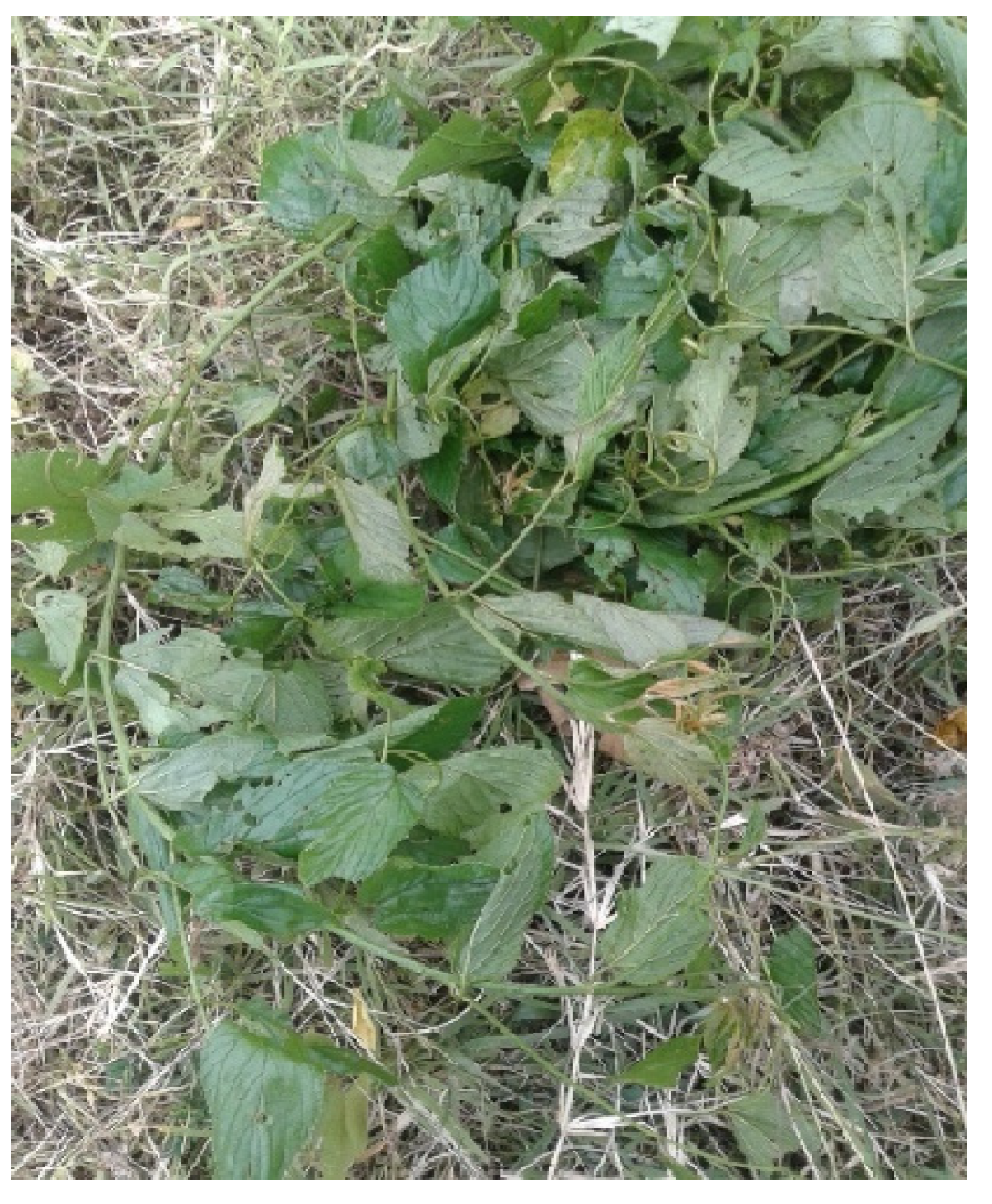
Figure 1. Gouania longispicata shoot sampled from Rukungiri District, Western Uganda. Photo taken by Hannington Gumisiriza.
Microbiolres 15 00140 g001
Figure 2. Structures of compounds characterized in methanolic extract of GLE leaves.
Microbiolres 15 00140 g002
Figure 3. Visualized HMBC correlations in compound 1 isolated from methanolic extract of GLE leaves. The linkages between the sugar to the aglycone and the B-ring to the C-ring are shown.
Microbiolres 15 00140 g003
Figure 4. Minimum inhibitory concentration of isolated compounds from methanolic extract of GLE against S. pneumoniae and E. coli. Values are means of replicates (n = 3). The numbers 15 refer to the isolated compounds: kaempferol-3-O-α-rhamnopyranoside (compound 1), 4,6-dihydroxy-3-methylacetophenone-2-O-β-D-glucopyranoside (compound 2), kaempferol (compound 3), quercetin (compound 4), and rutin (compound 5), respectively, while CIP = Ciprofloxacin.
Microbiolres 15 00140 g004
Table 1. 13CNMR data of compound 1 isolated from methanolic extract of GLE leaves.
PositionδH (ppm), Multiplicity, J (Hz) 1δC (ppm)
Aglycone
2 158.4
3 136.7
4 176.8
5 162.4
66.17, d, 2.097.8
7 164.8
86.20, d, 2.094.9
9 158.4
10 104.8
1′ 121.0
2′7.71, d, 8.7128.1
3′6.93, d, 8.7112.5
4′ 160.1
5′6.93, d, 8.7112.5
6′7.71, d, 8.7128.1
3-O-α- rhamnopyranosyl
1″5.85, d, 1.3101.1
2″4.39, dd, 3.5, 1.282.9
3″4.06, dd, 9.3, 3.571.8
4″3.75, t, 9.276.8
5″3.80, m74.0
6″1.38, d, 6.217.8
1 Multiplicity is represented as follows: singlet (s); doublet (d); doublet of a doublet (dd); triplet (t); multiplet (m).
Table 2. 13CNMR spectral data for compound 2 isolated from methanolic extract of GLE leaves.
Carbon δH (ppm), Multiplicity, J (Hz) 1δC
1-61.07
2-139.40
3-97.57
4-147.85
56.42, s92.71
6-159.61
β-D-GlucoseO-glycosylation
1′5.03, d (7.8)72.12
2′4.40, dd (7.8, 8.2)73.39
3′3.54, dd (9.0, 9.1)76.55
4′3.20, dd (9.1, 9.7)69.82
5′3.35, ddd (2.3, 6.0, 9.7)70.14
6′3.48, dd (2.2, 12.1)
3.17, dd (5.7, 12.5)
65.61
COCH3 208.57
COCH32.56, s29.34
-CH32.14, s9.75
1 Multiplicity is represented as follows: singlet (s); doublet (d); doublet of a doublet (dd); doublet of a doublet of a doublet (ddd).
Table 3. Half maximal inhibitory concentration (IC50) of isolated polyphenols from methanolic extract of GLE leaves.
CompoundHalf Maximal Inhibitory Concentration (µg/mL) 1
Kaempferol-3-O-α-rhamnopyranoside (compound 1)20.0 ± 0.45 a *
4,6-dihydroxy-3-methylacetophenone-2-O-β-D-glucopyranoside
(compound 2)
26.3 ± 0.12 b *
Kaempferol (compound 3)19.8 ± 1.03 a,c,f
Quercetin (compound 4)18.6 ± 1.30 a,c,d,f
Rutin (compound 5)28.1 ± 0.09 e *
Ascorbic acid (positive control)14.6 ± 0.15 c,f
1 Values are mean ± standard deviation of triplicates. Means carrying different alphabetical letters are significantly different as per Dunnett’s multiple comparisons test (p < 0.05). * Significantly different from that of ascorbic acid at p < 0.01.
Disclaimer/Publisher’s Note: The statements, opinions and data contained in all publications are solely those of the individual author(s) and contributor(s) and not of MDPI and/or the editor(s). MDPI and/or the editor(s) disclaim responsibility for any injury to people or property resulting from any ideas, methods, instructions or products referred to in the content.