Article

Antimicrobial and Antioxidant Activities of 18β-Glycyrrhetinic Acid Biotransformed by Aspergillus niger

by
Shaymaa Wagdy El-Far
1,
Mahmoud A. Al-Saman
2,
Fatma I. Abou-Elazm
3,
Rania Ibrahim Shebl
4 and
Asmaa Abdella
2,*
1
Division of Pharmaceutical Microbiology, Department of Pharmaceutics and Industrial Pharmacy, College of Pharmacy, Taif University, P.O. Box 11099, Taif 21944, Saudi Arabia
2
Department of Industrial Biotechnology, Genetic Engineering and Biotechnology Research Institute, University of Sadat City, Sadat City 32897, Egypt
3
Department of Microbiology and Immunology, Faculty of Pharmacy, Misr University for Science and Technology, Giza 32361, Egypt
4
Department of Microbiology and Immunology, Faculty of Pharmacy, Ahram Candian University, Giza 12451, Egypt
*
Author to whom correspondence should be addressed.
Microbiol. Res. 2024, 15(4), 1993-2006; https://doi.org/10.3390/microbiolres15040133
Submission received: 21 August 2024 / Revised: 22 September 2024 / Accepted: 26 September 2024 / Published: 29 September 2024

Abstract

:
The search for novel plant-based antioxidant and antibacterial medication has garnered a lot of attention lately. Glycyrrhiza glabra, known as licorice, is one of the most important medicinal plants. The primary component of Glycyrrhiza glabra is glycyrrhizin, which is biotransformed into 18α- and 18β-glycyrrhetinic acid for a variety of medicinal purposes. The goal of this study was to improve the bioavailability of glycyrrhizin by its biotransformation into glycyrrhetinic acid by Aspergillus niger. The biotransformation process was optimized using response surface methodology. A two-level Plackett–Burman design was employed to identify the factors that had a significant impact on the process of biotransformation. The three main variables were pH, glycerrhizin concentration, and incubation time. These three medium components were further optimized using a 3-level Box–Behnken design, and their optimum levels were pH of 8, an incubation period of 6 days, and a glycyrrhizin concentration of 1%. Using these optimum conditions, the maximum level obtained was 159% greater than in the screening experiment. Regarding the antimicrobial activity of glycyrrhizin extract, Bacillus subtilis emerged as the most sensitive organism with the lowest MIC (60 µg/mL) and the highest zone of inhibition (17 mm). The most resistant organism was Pseudomonas aeruginosa, which had the highest MIC (400 µg/mL) and the smallest zone of inhibition (10 mm). In the case of glycyrrhetinic acid, Bacillus subtilis was the most sensitive organism with the highest zone of inhibition (32 mm) and the lowest MIC (20 µg/mL). Pseudomonas aeruginosa was the most resistant organism, with the lowest zone of inhibition (18 mm), and the highest MIC (140 µg/mL). The antioxidant activity of glycyrrhizin extract increased from 12.81% at a concentration of 63 µg/100 µL to 41.41% at a concentration of 1000 µg/100 µL, while that of glycyrrhetinic acid extract increased from 35.5% at a concentration of 63 µg/100 µL to 76.85% at a concentration of 1000 µg/100 µL. The present study concluded that biotransformation of glycyrrhizin into glycyrrhetinic acid increased its bioavailability and antioxidant and antimicrobial activities. Glycyrrhizin and glycyrrhetinic acid might be used as a natural antimicrobial and antioxidant in pharmaceutical industries

1. Introduction

The study of plant-based natural medicines has attracted a lot of attention lately in an effort to develop novel antioxidant and antimicrobial agents [1]. The licorice root, Glycyrrhiza glabra, belongs to the Fabaceae family and is commonly used as a flavoring and sweetener in food. [2]. According to clinical and experimental research, there are additional beneficial pharmacological properties of licorice root, including anti-inflammatory, antiviral, antimicrobial, antioxidant, anticancer, immune-modulating, hepatoprotective, and cardioprotective effects [3].
Numerous compounds, including triterpene saponins, flavonoids, isoflavonoids, and chalcone, have been isolated from licorice [4]. The major constituent of licorice is a triterpenoid saponin glycoside, glycyrrhizin (glycyrrhizinic acid), which is 50 times sweeter than sucrose and safe for use in diabetes [5]. Glycyrrhizin (glycyrrhizinic acid) is a triterpenoid saponin that is the glucosiduronide derivative of 3β-Hydroxy-11-oxo-18β,20β-olean-12-en-29-oic acid) [6]. The aglycon of glycyrrhizinic acid is glycyrrhetic acid which exists as two isomers, namely the trans form (α-glycyrrhetic acid) and the cis form (β glycyrrhetic acid (Figure 1) [7]. Glycyrrhizic acid’s bioactive properties are related to its metabolite, 18β-glycyrrhetinic acid, which is produced through enzymatic hydrolysis [8]. 18β-glycyrrhetinic acid is used for its anti-tumor, anti-virus, antibacterial, and antioxidant properties in processed food [9].
Studies reveal that 18β-glycyrrhetinic acid possesses potent antimicrobial characteristics. Methicillin-resistant Staphylococcus aureus (MRSA) and other antibiotic-resistant bacteria can be effectively combated by 18β-glycyrrhetinic acid, which inhibits the expression of virulence genes and the bacteria’s ability to survive [10]. 18β-glycyrrhetinic acid has been shown to have significant antioxidant activity. Melekoglu et al. [11] found that rats treated with 18β-glycyrrhetinic acid had significantly higher levels of antioxidant defense system parameters, such as MDA, GSH, SOD, and CAT.
18β-glycyrrhetinic acid is 20 times more biologically active than glycyrrhizic acid [12]. 18β-glycyrrhetinic acid can be produced through chemical or biological hydrolysis. Chemical hydrolysis has the disadvantages of poor selectivity and the requirement of harsh conditions, thus causing environmental pollution [13]. Biotransformation is the preferred method for preparing 18β-glycyrrhetinic acid due to its high yield, specificity, and environmental compatibility, outperforming conventional chemical approaches [14].
Screening for biocatalysts, including microorganisms and enzymes with high catalytic specificity and biotransformation efficacy, is crucial for the industrial production of 18β-glycyrrhetinic acid [15]. Numerous microorganisms can produce β-D-Glucuronidase (EC 3.2.1.31), which converts glycyrrhizic acid to 18β-glycyrrhetinic acid [14]. Response surface methodology (RSM) when compared to univariate strategies, allows for an optimization of biotransformation that results in fewer experimental units and more precise results. [16].
The study aims to optimize the biotransformation of glycyrrhizin to 18β-glycyrrhetinic acid by Aspergillus niger using response surface methodology (RSM). The study aims also to evaluate the antioxidant and antimicrobial properties of biotransformed 18β-glycyrrhetinic acid.

2. Materials and Methods

2.1. Chemicals

Glycyrrhizin (GL) (glycyrrhizic acid ammonium salt) ≥ 95.0% (Biochemika, St. Louis, MO, USA) and 18-ß-Glycyrrhetinic acid (GA), 97.0% (Sigma-Aldrich, St. Louis, MO, USA) were used in this study.

2.2. Source of Plant

Licorice roots were obtained from the Department of Medicinal and Aromatic Plants Research, Horticulture Research Institute (HRI), Agriculture Research Center, Ministry of Agriculture, Giza, Egypt. They were washed, dried at 35 °C, ground to a fine powder, and stored at −18 °C.

2.3. Microorganisms

Aspergillus niger NRRL 3122 (Agricultural Research Service Culture Collection, Peoria, IL, USA) was grown on potato-dextrose-agar (PDA) plates at 4 °C. The bacterial strains used in this study to test the antimicrobial activity of glycyrrhizin and glycyrrhetinic acid were Escherichia coli ATCC 25922, Pseudomonas aeruginosa ATCC 27853, Staphylococcus aureus ATCC 25923, Salmonella typhimurium ATCC 23852, and Bacillus subtilis ATCC 21332 cultured in trypticase soy agar (TSA) and trypticase soy broth (TSB) media (Difco, Sparks, MD, USA).

2.4. Crude Glycyrrhizin Extraction

The ground licorice root was extracted with hot water at 60 °C for 3 h. The aqueous extract was then treated with concentrated sulfuric acid to acidify it to pH 1.8. An insoluble brown precipitate (crude glycyrrhizin) was formed and separated by centrifugation at 6000 rpm/15 min. Glycyrrhizin was dissolved in distilled water and alkalinized to a pH of approximately 5 to 6.5.

2.5. Preparation of the Fermentation Culture for the Biotransformation of Glycyrrhizin to Glycyrrhetinic Acid by Aspergilus niger

A spore suspension of Aspergillus niger at a concentration of 2 × 10⁷ spores was prepared by rubbing spores from 7-day PDA plates with 10 mL of distilled water. A total of 0.1 mL of spore suspension was inoculated in 100 mL of sterile fermentation medium consisting of the following (g/L): glucose, 10; yeast extract, 3; corn steep liquor, 8; crude glycyrrhizin, 6; NaNO3, 3; MgSO4·7H2O, 0.5; K2HPO4, 0.1, and the pH was adjusted to 6.0. The culture was incubated at 120 rpm for 7 days at 30 °C. Following fermentation, the culture was centrifuged at 6000 rpm for 15 min (Centurion Scientific LTD Model 1020 series, Chichester, UK).

2.6. Multifactorial Experiments for Optimizing the Biotransformation of Glycyrrhizin to Glycyrrhetinic Acid

2.6.1. Screening of Factors Affecting the Biotransformation of Glycyrrhizin to Glycyrrhetinic Acid Using a Plackett–Burman Design

The Plackett–Burman experimental design [17], which is a fractional factorial design, was employed to demonstrate the relative significance of different environmental factors on the production of glycyrrhetinic acid in liquid cultures. This model was used to identify and assess the key variables that affect the response; it does not explain how the factors interact. When it is difficult to determine which factors have the greatest potential to affect the dependent variable, this design is very helpful for screening a large number of independent variables.
Seven independent variables (Table 1) were screened in twelve runs organized according to the Plackett–Burman design matrix (Table 2). The studied factors were initial pH, temperature, concentration of glycyrrhizin (GL), incubation time, glucose concentration, yeast extract concentration, and corn steep liquor (CSL) concentration.
A high level (+) and low level (−) were tested for every variable. Each trial was performed twice, and the final data were calculated using the mean of the duplicates. The test answers (dependent variable) were the averages of the GA production. The main effect of each variable was computed as the difference between the average of measurements taken at the high value (+) and the low value (−). The Plackett-Burman design was applied using the subsequent first-order model:
Y = βo + Σ βiXi
Y (Glycyrrhetinic acid concentration (mg/mL), βo (the model intercept), βi (the linear coefficient for each factor), and Xi (the concentration of each factor).
The impact of the corresponding factor on the production of glycyrrhetinic acid was indicated by the magnitude of the coefficient βi and its positive or negative value. A 95% confidence level (p < 0.05) was used to ascertain the significance of each factor’s impact on the production of glycyrrhetinic acid.

2.6.2. Optimization of Significant Variables Using a Box–Behnken Design

Using a Box–Behnken design, the response surface methodology (RSM) was employed to further optimize the variables that were found to have a significant impact on the production of glycyrrhetinic acid from the Plackett–Burman experiments [18]. The design, which included 15 experiments with 3 factors, 3 levels, and 3 center points, was used to fit a second-order response surface. After fitting the data to a second-order polynomial equation, SAS JMP version 18 (SAS Institute, Cary, NC, USA) was used to determine the ideal concentration of each factor for the production of glycyrrhetinic acid.
The glycyrrhetinic acid production was fitted with the following quadratic equation using multiple regression and the least squares method:
Y = βo + Σ βi Xi + Σ βij XiXj+ Σ βiiXi 2.
where Y is the response (concentration of glycyrrhetinic acid), βo is the model intercept, βi is the model’s linear coefficient, βii is the quadratic coefficient, βij is the interaction coefficient, and Xi and Xj are the levels of the factors in coded values.

2.7. Extraction of of Glycyrrhizin (GL) and Glycyrrhetinic Acid (GA)

The fermentation culture was centrifuged at 6000 rpm for 15 min and GA was extracted from the supernatant using a mixture of chloroform and water (1:1, v/v). The chloroform layer containing GA was collected, evaporated to 1 mL, and finally analyzed by high performance liquid chromatography (HPLC) to identify and quantify the GA [19].

2.8. Determination of Glycyrrhizin (GL) and Glycyrrhetinic Acid (GA)

Glycyrrhetinic acid was quantified using HPLC, which included a quaternary G1311A chromatographic pump, a variable wavelength G1314A detector, and a Zorbax 300SB C18 column (4.5 mm × 250 mm). GA’s mobile phase was acetonitrile–water–acetic acid (80:20:1, v/v), flowing at a rate of 1.5 mL/min, while the GL’s mobile phase was (40:60:1, v/v), flowing at a rate of 1.0 mL/min. The column was kept at room temperature, and the chromatograms were monitored at a wavelength of 254 nm. The sample injection volume was 20 µL. The concentrations of the GA samples were calculated using their peak areas [20].

2.9. Antimicrobial Activity of the Biotransformed Glycyrrhetinic Acid

2.9.1. Paper Disc Diffusion Assay

Tryptone soy agar (TSA) was evenly covered with 100 µL of bacterial cell suspension. A 25 µL extract prepared at a concentration of 10 mg/mL was impregnated onto filter paper discs (Whatman No. 41) with a diameter of 6 mm. For twenty-four hours, plates were incubated at 37 °C. After measuring the zone of inhibition (ZOI) precisely, the means of three replicates were determined. Discs of streptomycin (10 μg/mL, Sigma, St. Louis, MO, USA) were used as positive controls [21].

2.9.2. Determination of Minimal Inhibitory Concentration

The bacterial strains under investigation were subjected to tests using glycyrrhizin and glycyrrhetinic acid extract to ascertain their respective minimal inhibitory concentrations (MIC) through a quantitative assay [22]. ρ-iodonitro-tetrazolium violet (INT) was employed in the broth microdilution method as a bacterial growth indicator. After being spread out on TSA plates, 50 µL from each well containing serial concentrations of glycyrrhizin and glycyrrhetinic acid extract was incubated for 24 h at 37 °C.

2.10. Antioxidant Activity of the Biotransformed Glycyrrhetinic Acid

A total of 100 µL of each sample concentration (6.5–1000 µg/100 µL) was mixed with 500 µL of methanolic solution of 0.1 Mm DPPH radicals and the mixture was vortexed for 10 s at room temperature. Against a blank of methanol without DPPH, the decrease in absorption at 515 nm was measured after 30, 60, and 90 min of mixing using a spectrophotometer (Shimadzu, Kyoto, Japan). The % of inhibition was calculated from the following equation [23]:
% inhibition = [(absorbance of control − absorbance of test sample)/absorbance of control] × 100

2.11. Statistical Analysis

An analysis of variance (ANOVA) was performed on all the data. Each item was examined in three samples, and the mean and standard deviation values were reported. To ascertain whether mean differences between variables were statistically significant (p ≤ 0.05), Duncan’s multiple range tests were employed. SPSS 16 was used to perform all of the analyses.

3. Results and Discussion

3.1. Multifactorial Designs for Optimizing the Biotransformation of Glycyrrhizin into Glycyrrhetinic Acid

3.1.1. Screening of Factors Affecting Biotransformation of Glycyrrhizin to Glycyrrhetinic Acid Using a Plackett–Burman Design

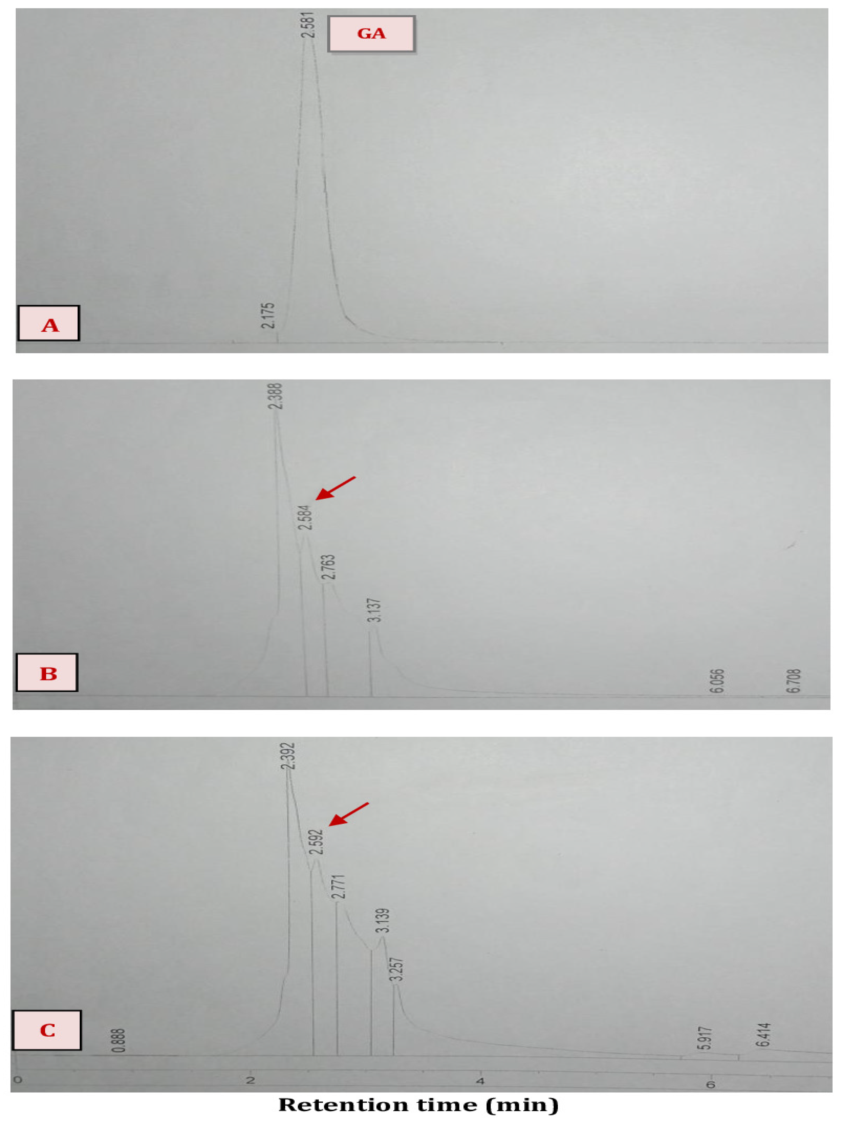
Quantitative determination of 18α-GA acid was carried out by using a HPLC (Figure 2, Table 2) depicts the wide variation in glycyrrhetinic acid concentration, from 9.3–80.3 mg/100 mL in the Plackett–Burman design, highlighting the importance of optimizing biotransformation conditions. The main effects of the examined factors on biotransformation of glycyrrhizin into glycyrrhetinic acid are identified and presented in Table 3. The correlation between the glycyrrhetinic acid concentration and the seven factors is described by the following linear correlation model:
Glycyrrhetinic acid concentration = 35.32 + 10.63 (pH-6) + −2.95 (temperature—27.5/2.5) + 
8.69 (GL%—0.75/0.25) + 13.084 (incubation time—5) + 0.477 (glucose%—2) + −3.71 (yeast
extract %—0.3/0.1) + −2.93 (CSL%—0.75/0.25).                   
Table 2. Randomized Plackett–Burman experimental design for variables affecting biotransformation of glycyrrhizin into glycyrrhetinic acid using a Plackett–Burman design.
pHTemperatureGlycyrrhizin Conc. (g/100 mL)Incubation Time (days)Glucose Conc. (g/100 mL)Yeast Extract Conc.
(g/100 mL)
CSL Conc. (g/100 mL)Glycyrrhetinic Acid (mg/g)
−−+−−+−5251410.40.528.98
−+−+++−5300.5630.40.523.1
−+++−−−5301610.20.548.31
+−−+−++7250.5610.4150.17
+−−−+−−7250.5430.20.531.77
−+−−+−+5300.5430.219.3
−−−+−−+5250.5610.2128.34
−−+−+++5251430.4110.11
+++−−−+7301410.2136.21
+++++++7301630.4160.21
+−+++−−7251630.20.580.3
++−−−+−7300.5410.40.517.0.5
Table 3 demonstrates the significant effects of factors with p-values < 0.05 on the biotransformation of glycyrrhizin into glycyrrhetinic acid. These factors were selected for further optimization using the Box –Behnken design.
Significant factors were found to be pH, incubation time, and glycyrrhizin concentration, with corresponding p-values of 0.0086 (t ratio = 4.81), 0.0041 (t ratio = 5.92), and 0.0171 (t ratio = 3.93), respectively. The results indicated that at the higher concentration level tested, each of these factors had a significant impact on raising the concentration of glycyrrhetinic acid. This shows that the higher concentrations utilized in the experiment rather than the lower concentrations would be the optimal concentrations of these compounds for glycyrrhetinic acid concentration. Similar results were reported by Quan et al. [24], who found that the biotransformation of glycyrrhizin to glycyrrhetinic acid was significantly influenced by both the cultivation time and the concentration of glycyrrhizin.

3.1.2. Optimization of Significant Variables Using a Box–Behnken Design

The significant factors (pH, GL%, and glucose%) were tested at three different levels to find the optimal level for the production of glycyrrhetinic acid in order to determine the optimal response region.
The factors’ design matrix and the measured glycyrrhetinic acid concentration for each experimental run are shown in Table 4 in both coded and natural units. The factors’ design matrix and the measured glycyrrhetinic acid concentration for each experimental run are shown in Table 4 in both coded and natural units. The highest concentration of glycyrrhetinic acid (128.25 mg/g) was recorded during trial number 8, which had a pH of 8, an incubation period of 6 days, and a GL% of 1%. Trial No. 1, which had a pH of 9, an incubation period of 10 days, and a GL% of 2%, showed the lowest glycyrrhetinic acid concentration of 66.39 mg/g. The maximum yield of glycyrrhetinic acid obtained after optimization was higher than that reported in the previous literature (13.3, 25.2, and 48 mg/gL, respectively) [8,25,26].
A second-order polynomial equation was fitted to the experimental glycyrrhetinic acid concentration in order to determine the ideal conditions for the biotransformation of glycyrrhizin into glycyrrhetinic acid.
Glycyrrhetinic acid concentration (mg/g) = 175.393 + −46.005 (incubation time) + 48.148        
(pH) + −31.268 (GL%) + 2.42(pH) (incubation time) + 1.55 (GL%) (incubation time) +        
0.115 (GL%) (pH) + 0.755 (incubation time) (incubation time) + −4.45 (pH) (pH) + 3.67 (GL%) (GL%)
The Student’s t-distribution, associated p-values, and parameter estimates for the variables that were determined to have the greatest impacts on the concentration of glycyrrhetinic acid are shown in Table 5. It was found that incubation time (p = 0.0327) was a significant factor.

3.1.3. Localization of the Optimum Condition

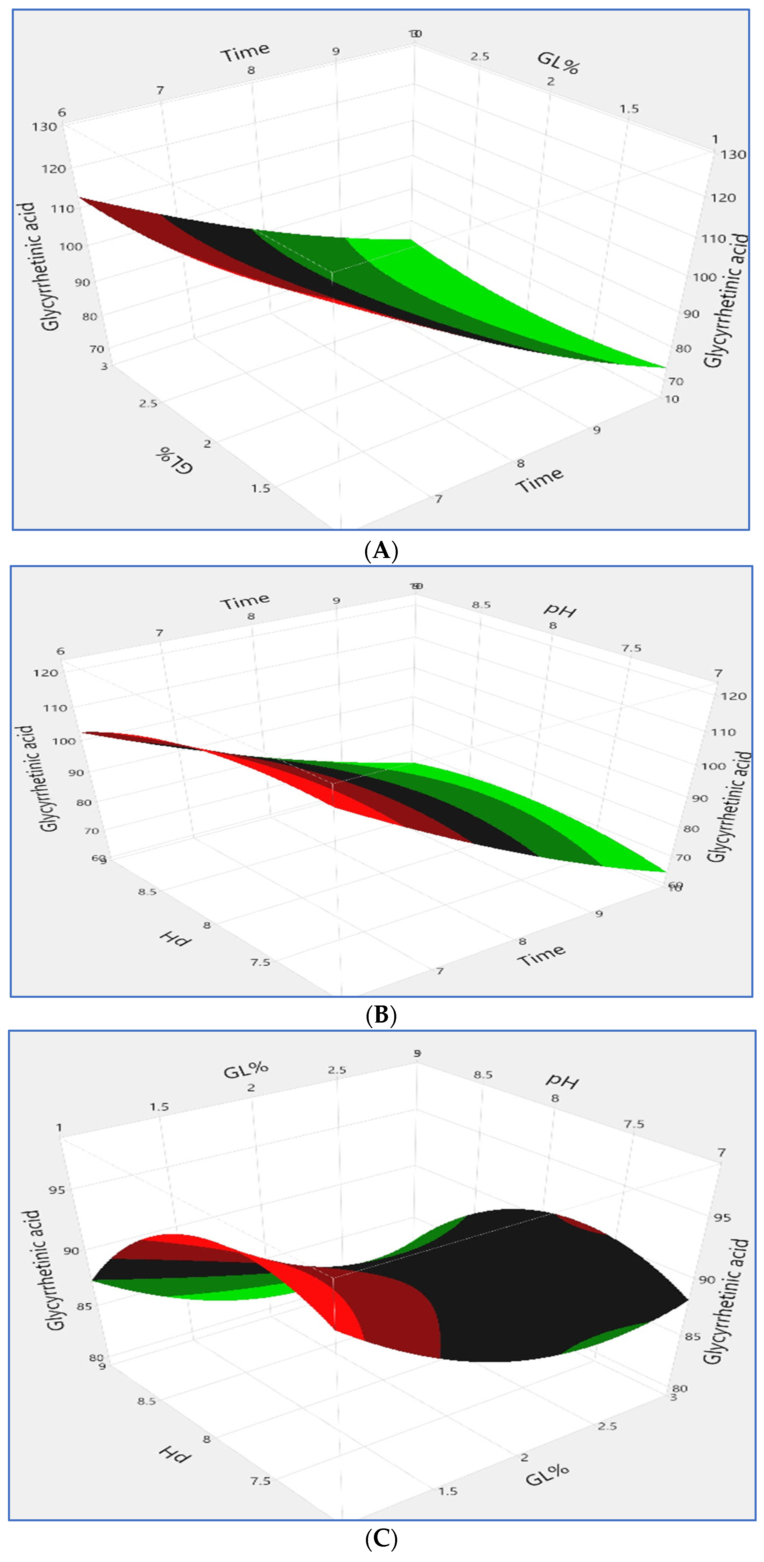
The three-dimensional responses for glycyrrhetinic acid concentration are shown in Figure 3 These responses were obtained by choosing two factors and maintaining the third factor’s value at its central value. A response surface plo3A. A response surface plot of time vs. GL% with pH maintained at zero (pH = 8) is shown in Figure 3B. The pH vs. GL% with the time set to zero (8 h) is shown in Figure 3C. The plots revealed that the highest concentration of glycyrrhetinic acid was achieved at pH 8, an incubation period of 6 days, and a 1% GL%.

3.1.4. Model Validation

The optimal condition that resulted from the optimization experiment was compared to the model’s prediction and experimentally validated (Table 4). The polynomial model’s predicted value of 125.59 mg/g for the mean glycyrrhetinic acid concentration was comparable to the experimentally determined value of 128.25 mg/g. To determine the goodness of fit, the determination coefficient (R2) was computed. R2 = 0.97 in this instance for the conditions under investigation shows that 97% of the variation in the total data can be explained by the response model [27]. After optimization, the amount of glycyrrhetinic acid was 159% higher than it was in the screening experiment.

3.2. Antimicrobial Activity of Glycyrrhizin and the Biotransformed Glycyrrhetinic Acid

The extracts of crude glycyrrhizin and the biotransformed glycyrrhetinic acid were investigated for their antimicrobial activity against different strains of bacteria (Bacillus subtilis, Escherichia coli, Pseudomonas aeruginosa, Salmonella typhimurium, and Staphylococcus aureus). Antimicrobial activity was determined qualitatively and quantitatively based on the diameters of inhibition zones and MIC values (Figure S1).
Table 6 provides a summary of the findings which showed that both crude glycyrrhizin and glycyrrhetinic acid were active against Gram-negative strains (E. coli, P. aeruginosa and S. typhimurium) and Gram-positive strains (S. aureus, and Bacillus subtilis). It can also be concluded that glycyrrhetinic acid extract was more effective than glycyrrhizin extract with all strains. Also, it was clear that Gram-positive bacteria were more sensitive than Gram- negative bacteria with all strains.
The bacteria with the highest resistance was Pseudomonas aeruginosa. With glycyrrhizin extract, it displayed the lowest inhibition zone (10 ± 0.4 mm) and the highest MIC (400 µg/mL); with bio-transformed glycyrrhetinic acid extract, on the other hand, it displayed the lowest inhibition zone (18 ± 1.3 mm) and the highest MIC (140 µg/mL). Bacillus subtilis was the most sensitive, scoring the highest inhibition zone (17 ± 1.1mm) and the lowest MIC (60 µg/mL) when using glycyrrhizin extract; however, when using the biotransformed glycyrrhetinic acid extract, it had the highest inhibition zone (32 ± 1.7mm) and the highest MIC (20 µg/mL).
Glycyrrhizic acid acts on the permeability of biofilm formation, the cell membrane, and efflux pump activity to inhibit bacterial growth [28]. Furthermore, glycyrrhizic acid modifies bacterial characteristics, such as viability and efflux pump activity, to reduce multidrug resistance [29].
Because of its higher bioavailability, biotransformed glycyrrhetic acid is evidently much more antibacterial than glycyrrhizin [30]. According to Kim et al. [31], 18β-glycyrrhetinic acid inhibited protein, RNA, and DNA synthesis. 18βGA exhibited bactericidal effectiveness by reducing the expression of saeR, mecA, and sbi, the three main virulence genes of MRSA [32]. According to Oyama et al. [33], glycyrrhetinic acid’s strong antibacterial activity results from the blockage of several metabolic pathways for amino acids and carbohydrates. Significant alterations in intracellular polar metabolite profiles were observed upon exposure to 18β-glycyrrhetinic acid. These included elevated levels of succinate and citrate, as well as notable decreases in multiple amino acids, including branched chain amino acids [34]. Methicillin-resistant Staphylococcus aureus (MRSA) was shown to exhibit inhibition of multiple pathways involved in amino acid and carbohydrate metabolism when exposed to 18β-glycyrrhetinic acid [35].
The greater sensitivity of Gram-positive bacteria, which may result from the relative impermeability of Gram-negative bacteria’s outer membranes, can account for the high activity against Gram-positive bacteria [36]. 18β-glycyrrhetinic acid appears to be a useful disruptor of biofilms. Treating planktonic MRSA cultures with 18β-glycyrrhetinic acid significantly reduces micro-aggregation, and this could be due to interference with the aggregation of cells [35]. Triterpenes’ antibacterial action, which includes the reduction and inhibition of biofilm formation, has only been demonstrated to impact Gram-positive bacteria [34].

3.3. Antioxidant Activity of Glycyrrhizin and the Biotransformed Glycyrrhetinic Acid

Table 7 shows that both glycyrrhizin and biotransformed glycyrrhetinic acid scavenged the DPPH radical in a dose-dependent manner over time.
The concentration of 1000 µg/100 µL (41.41%) yielded the highest scavenging percentage in the case of crude glycyrrhizin extract, followed by 500 µg/100 µL (34%), and 250 µg/100 µL (25.51%), respectively (p < 0.05). The highest scavenging percentage (76.85%) was observed in the case of biotransformed glycyrrhetinic acid at a concentration of 1000 µg/100 µL. This was followed by 500 µg/100 µL (67.79%) and 250 µg/100 µL (54.25%), respectively (p < 0.05).
Glycyrrhizin scavenges 1,1-diphenyl-2-picrylhydrazyl (DPPH) radicals instead of hydroxyl or superoxide anion radicals [37]. By capturing a solvated electron, glycyrrhizin can prevent molecular oxygen from capturing it [38]. Glycyrrhizic acid can also activate the nuclear factor Nrf2 through redox regulation by Keap1, and this can affect the levels of ROS within cells through additional mechanisms [39].
Glycyrrhetinic acid increases the expression of the antioxidant enzyme HO-1, which activates the intracellular antioxidant system [40]. Treatment with glycyrrhetinic acid was shown to decrease the activities of the matrix metalloproteinases MMP-1 and MMP-3 as well as some cytokines, such as IL-6, TNF-a, and IL-10, while increasing the activities of important antioxidant enzymes, such as SOD and GSH-Px [41]. Li et al. [42] stated that glycyrrhizic acid administration reduces lipid peroxidation and increases antioxidant status.

4. Conclusions

Based on the above results, our study demonstrates into the potential biotransformation of glycyrrhizin into glycyrrhetinic acid by Aspergillus niger using a response surface methodology. It also offers novel insights into the improvement of bioavailability of the biotransformed glycyrrhetinic acid. It also studied the potent antioxidant and antimicrobial activities of glycyrrhizin and glycyrrhetinic acid extracts. These effects can be extended to more applications of Glycyrrhiza glabra. The mechanism of the antimicrobial and antioxidant activities of Glycyrrhiza glabra needs further investigation.

Supplementary Materials

The following supporting information can be downloaded at: https://www.mdpi.com/article/10.3390/microbiolres15040133/s1, Figure S1: Disc diffusion test for the growth inhibition of E. coli.

Author Contributions

S.W.E.-F., methodology, funding; M.A.A.-S., conceptualization, methodology, tables, figures; F.I.A.-E., formal analysis, funding; R.I.S., software, writing; A.A., methodology, writing, and editing. All authors have read and agreed to the published version of the manuscript.

Funding

This research received no external funding.

Institutional Review Board Statement

Not applicable.

Informed Consent Statement

Not applicable.

Data Availability Statement

Available when required.

Conflicts of Interest

The authors declare no conflicts of interest.

References

  1. Abdallah, E.M.; Alhatlani, B.Y.; Menezes, R.d.P.; Martins, C.H.G. Back to Nature: Medicinal Plants as Promising Sources for Antibacterial Drugs in the Post-Antibiotic Era. Plants 2023, 12, 3077. [Google Scholar] [CrossRef]
  2. Wahab, S.; Annadurai, S.; Abullais, S.S.; Das, G.; Ahmad, W.; Ahmad, M.F.; Kandasamy, G.; Vasudevan, R.; Ali, M.S.; Amir, M. Glycyrrhiza glabra (Licorice): A Comprehensive Review on Its Phytochemistry, Biological Activities, Clinical Evidence and Toxicology. Plants 2021, 10, 2751. [Google Scholar] [CrossRef]
  3. AlDehlawi, H.; Jazzar, A. The Power of Licorice (Radix glycyrrhizae) to Improve Oral Health: A Comprehensive Review of Its Pharmacological Properties and Clinical Implications. Healthcare 2023, 11, 2887. [Google Scholar] [CrossRef]
  4. Tuli, H.S.; Garg, V.K.; Mehta, J.K.; Kaur, G.; Mohapatra, R.K.; Dhama, K.; Sak, K.; Kumar, A.; Varol, M.; Aggarwal, D.; et al. Licorice (Glycyrrhiza glabra L.)-Derived Phytochemicals Target Multiple Signaling Pathways to Confer Oncopreventive and Oncotherapeutic Effects. OncoTargets Ther. 2022, 15, 1419–1448. [Google Scholar] [CrossRef] [PubMed]
  5. Pastorino, G.; Cornara, L.; Soares, S.; Rodrigues, F.; Oliveira, M.B.P.P. Liquorice (Glycyrrhiza glabra): A phytochemical and pharmacological review. Phytother. Res. 2018, 32, 2323–2339. [Google Scholar] [CrossRef]
  6. Shinu, P.; Gupta, G.L.; Sharma, M.; Khan, S.; Goyal, M.; Nair, A.B.; Kumar, M.; Soliman, W.E.; Rahman, A.; Attimarad, M.; et al. Pharmacological Features of 18β-Glycyrrhetinic Acid: A Pentacyclic Triterpenoid of Therapeutic Potential. Plants 2023, 12, 1086. [Google Scholar] [CrossRef]
  7. Sabbioni, C.; Mandrioli, R.; Ferranti, A.; Bugamelli, F.; Saracino, M.A.; Forti, G.C.; Fanali, S.; Raggi, M.A. Separation and analysis of glycyrrhizin, 18beta-glycyrrhetic acid and 18alpha-glycyrrhetic acid in liquorice roots by means of capillary zone electrophoresis. J. Chromatogr. A 2005, 15, 65–71. [Google Scholar] [CrossRef]
  8. Chamoli, A.; Hasan, M.; Ahmad, M.; Rashid, M.; Ali, B.; Panda, B.P. Microbial Screening for Biotransformation of Glycyrrhizin into 18α-Glycyrrhetinic Acid and 18β-Glycyrrhetinic Acid and their HPLC Analysis. Acta Sci. Microbiol. 2019, 2, 62–66. [Google Scholar] [CrossRef]
  9. Feng, S.; Chun Li, C.; Xu, X.; Wang, X. Screening strains for directed biosynthesis of β-D-mono-glucuronide-glycyrrhizin and kinetics of enzyme production. J. Mol. Catal. B Enzym. 2022, 43, 63–67. [Google Scholar] [CrossRef]
  10. Chen, L.; Gong, J.; Yong, X.; Li, Y.; Wang, S. A review of typical biological activities of glycyrrhetinic acid and its derivatives. RSC Adv. 2024, 14, 6557–6597. [Google Scholar] [CrossRef]
  11. Melekoglu, R.; Cifitci, O.; Eraslan, S.; Alan, S.; Basak, N. The Protective Effects of Glycyrrhetinic Acid and Chrysin against Ischemia-Reperfusion Injury in Rat Ovaries. BioMed. Res. Int. 2018, 2018, 5421308. [Google Scholar]
  12. Lu, D.; Hui, L.; Yan, D.; Ping-Kai, O. Biocatalytic properties of a novel crude glycyrrhizin hydrolase from the liver of the domestic duck. J. Mol. Catal. B Enzym. 2006, 43, 148–152. [Google Scholar] [CrossRef] [PubMed]
  13. Wang, R.; Lin, C.; Liu, J.; Yu, F.; Gao, X.P. Pressured microwave-assisted hydrolysis of crude glycyrrhizic acid for preparation of glycyrrhetinic acid. Chin. J. Chem. Eng. 2012, 20, 152–157. [Google Scholar] [CrossRef]
  14. Xu, Y.; Feng, X.; Jia, J.; Chen, X.; Jiang, T.; Rasool, A.; Lv, B.; Qu, L.; Li, C. A novel βglucuronidase from Talaromyces pinophilus Li-93 precisely hydrolyzes glycyrrhizin into glycyrrhetinic acid 3-O-mono-β-D-glucuronide. Appl. Environ. Microbiol. 2018, 84, e00755-18. [Google Scholar] [CrossRef]
  15. Zhang, Q.; Gao, B.; Xiao, Y.; Yang, H.; Wang, Y.; Du, L.; Zhu, D. Purification and characterization of a novel β-glucuronidase precisely converts glycyrrhizin to glycyrrhetinic acid 3-O-mono-β-D-glucuronide from plant endophytic Chaetomium globosum DX-THS3. Int. J. Biol. Macromol. 2020, 159, 782–792. [Google Scholar] [CrossRef]
  16. Chellapandi, P.; Jani, H.M. Production of endoglucanase by the native strains of Streptomyces isolates in submerged fermentation. Braz. J. Microbiol. 2008, 39, 122–127. [Google Scholar] [CrossRef] [PubMed]
  17. Plackett, R.L.; Burman, J.P. The design of optimum multifactorial experiments. Biometrika 1946, 33, 305–325. [Google Scholar] [CrossRef]
  18. Box, G.E.P.; Behnken, D.W. Some new three level designs for the study of quantitative variables. Technometrics 1960, 2, 455–475. [Google Scholar] [CrossRef]
  19. Siracusa, L.; Saija, A.; Cristani, M.; Climino, F.; Arrigo, M.D.; Trombetta, D.; Rao, F.; Ruberto, G. Phytocomplexes from liquorice (Glycyrrhiza glabra L.) leaves–chemical characterization and evaluation of their antioxidant, anti-genotoxic and anti-inflammatory activity. Fitoterapia 2011, 82, 546–555. [Google Scholar] [CrossRef]
  20. Nocca, G.; Callà, C.; Santini, S.A.; Amalfitano, A.; Marigo, L.; Rossetti, D.V.; Spagnuolo, G.; Cordaro, M. Quantitative determination of 18-β-glycyrrhetinic Acid in HepG2 cell line by High Performance Liquid Chromatography method. Int. J. Anal. Chem. 2018, 2018, 5673186. [Google Scholar] [CrossRef]
  21. Balouiri, M.; Sadiki, M.; Ibnsouda, S.K. Methods for in vitro evaluating antimicrobial activity: A review. J. Pharm. Anal. 2015, 6, 71–79. [Google Scholar] [CrossRef] [PubMed]
  22. Kowalska-Krochmal, B.; Dudek-Wicher, R. The Minimum Inhibitory Concentration of Antibiotics: Methods, Interpretation, Clinical Relevance. Pathogens 2021, 10, 165. [Google Scholar] [CrossRef] [PubMed]
  23. Ramadan, M.F.; Kroh, L.W.; Moersel, J.T. Radical scavenging activity of black cumin (Nigella sativa L.), coriander (Coriandrum sativum L.) and niger (Guizotia abyssinica Cass.) crude seed oils and oil fractions. J. Agric. Food Chem. 2003, 51, 6961–6969. [Google Scholar] [CrossRef] [PubMed]
  24. Quan, Y.; Wang, L.; Liu, Y.; Cong, J.; Xie, S.; Wu, X. Optimization of Fermentation Medium for Glycyrrhizin Biotransformation to Monoglucuronyl-glycyrrhetinic Acid by Plackett-Burman and Box-Behnken Design. Korean Chem. Eng. Res. 2015, 53, 321–326. [Google Scholar] [CrossRef]
  25. Ahmad, M.; Jalaluddin, M.; Panda, B.P. Bioconversion of glycyrrhizin of Glycyrrhiza glabra root extract to 18β-glycyrrhetinic acid. Drug Devel Ther. 2016, 7, 683–688. [Google Scholar]
  26. Ahmad, M.; Jalaluddin, M.; Pand, B.P. Enrichment of biologically active 18-β glycyrrhetinic acid in Glycyrrhiza glabra root by solid state fermentation. Ann Microbiol. 2014, 64, 683–688. [Google Scholar] [CrossRef]
  27. Mahdavi, V.; Monajem, A. Statistical Optimization for Oxidation of Ethyl Benzene over Co- Mn/SBA-15 catalyst by Box-Behnken Design. Korean J. Chem. Eng. 2013, 33, 2178–2185. [Google Scholar] [CrossRef]
  28. Chakotiya, A.S.; Tanwar, A.; Narula, A.; Sharma, R.K. Alternative to antibiotics against Pseudomonas aeruginosa: Effects of Glycyrrhiza glabra on membrane permeability and inhibition of efflux activity and biofilm formation in Pseudomonas aeruginosa and its in vitro time-kill activity. Microb. Pathog. 2016, 98, 98–105. [Google Scholar] [CrossRef]
  29. Hazlett, L.D.; Ekanayaka, S.A.; McClellan, S.A.; Francis, R. Glycyrrhizin Use for Multi-Drug Resistant Pseudomonas aeruginosa: In Vitro and In Vivo Studies. Investig. Opthalmology Vis. Sci. 2019, 60, 2978–2989. [Google Scholar] [CrossRef]
  30. Kowalska, A.; Kalinowska-Lis, U. 18b-Glycyrrhetinic acid: Its core biological properties and dermatological applications. Int. J. Cosmet. Sci. 2019, 41, 325–331. [Google Scholar] [CrossRef]
  31. Kim, H.K.; Park, Y.; Kim, H.N.; Choi, B.H.; Jeong, H.G.; Lee, D.G.; Hahm, K.S. Antimicrobial mechanism of β-glycyrrhetinic acid isolated from licorice, Glycyrrhiza glabra Hyung Keun. Biotechnol. Lett. 2002, 24, 1899–1902. [Google Scholar] [CrossRef]
  32. Long, D.R.; Mead, J.; Hendricks, J.M.; Hardy, M.E.; Voyich, J.M. 18β-Glycyrrhetinic acid inhibits methicillin-resistant Staphylococcus aureus survival and attenuates virulence gene expression. Antimicrob. Agents Chemother. 2013, 57, 241–247. [Google Scholar] [CrossRef] [PubMed]
  33. Oyama, K.; Kawada-Matsuo, M.; Oogai, Y.; Hayashi, T.; Nakamura, N.; Komatsuzawa, H. Antibacterial Effects of Glycyrrhetinic Acid and Its Derivatives on Staphylococcus aureus. PLoS ONE 2016, 11, e0165831. [Google Scholar] [CrossRef] [PubMed]
  34. Verstraeten, S.; Catteau, L.; Boukricha, L.; Quetin-Leclercq, J.; Mingeot-Leclercq, M.-P. Effect of Ursolic and Oleanolic Acids on Lipid Membranes: Studies on MRSA and Models of Membranes. Antibiotics 2021, 10, 1381. [Google Scholar] [CrossRef]
  35. Weaver, A.J., Jr.; Borgogna, T.R.; O’Shea-Stone, G.; Peters, T.R.; Copié, V.; Voyich, J.; Teintze, M. 18β-Glycyrrhetinic Acid Induces Metabolic Changes and Reduces Staphylococcus aureus Bacterial Cell-to-Cell Interactions. Antibiotics 2022, 11, 781. [Google Scholar] [CrossRef] [PubMed]
  36. Schelz, Z.; Molnar, J.; Hohmann, J. Antimicrobial and antiplasmid activities of essential oils. Fitoterapia 2006, 77, 279–285. [Google Scholar] [CrossRef] [PubMed]
  37. Imai, P.; Takagi, Y.; Iwazaki, A.; Nakanishi, N. Radical scavenging ability of glycyrrhizin. Free Radic. Antioxid. 2013, 3, 40–42. [Google Scholar] [CrossRef]
  38. Ageeva, A.A.; Kruppa, A.I.; Magin, I.M.; Babenko, S.V.; Leshina, T.V.; Polyakov, N.E. New Aspects of the Antioxidant Activity of Glycyrrhizin Revealed by the CIDNP Technique. Antioxidants 2022, 11, 1591. [Google Scholar] [CrossRef]
  39. Polyakova, N.E.; Leshinaa, T.V. Physicochemical Approaches to the Study of the Antioxidant Activity of Glycyrrhizin. Russ. J. Phys. Chem. A 2023, 97, 828–835. [Google Scholar] [CrossRef]
  40. Lefaki, M.; Papaevgeniou, N.; Tur, J.A.; Vorgias, C.E.; Sykiotis, G.P.; Chondrogianni, N. The dietary triterpenoid 18α-Glycyrrhetinic acid protects from MMC-induced genotoxicity through the ERK/Nrf2 pathway. Redox Biol. 2020, 28, 101317. [Google Scholar] [CrossRef]
  41. Kong, S.Z.; Chen, H.M.; Yu, X.T.; Zhang, X.; Feng, X.X.; Kang, X.H.; Li, W.-J.; Huang, N.; Luo, H.; Su, Z.-R. The protective effect of 18b-Glycyrrhetinic acid against UV irradiation induced photoaging in mice. Exp. Gerontol. 2015, 61, 147–155. [Google Scholar] [CrossRef] [PubMed]
  42. Li, X.L.; Zhou, A.G.; Zhang, L.; Chen, W.J. Antioxidant Status and Immune Activity of Glycyrrhizin in Allergic Rhinitis Mice. Int. J. Mol. Sci. 2011, 12, 905–916. [Google Scholar] [CrossRef] [PubMed]
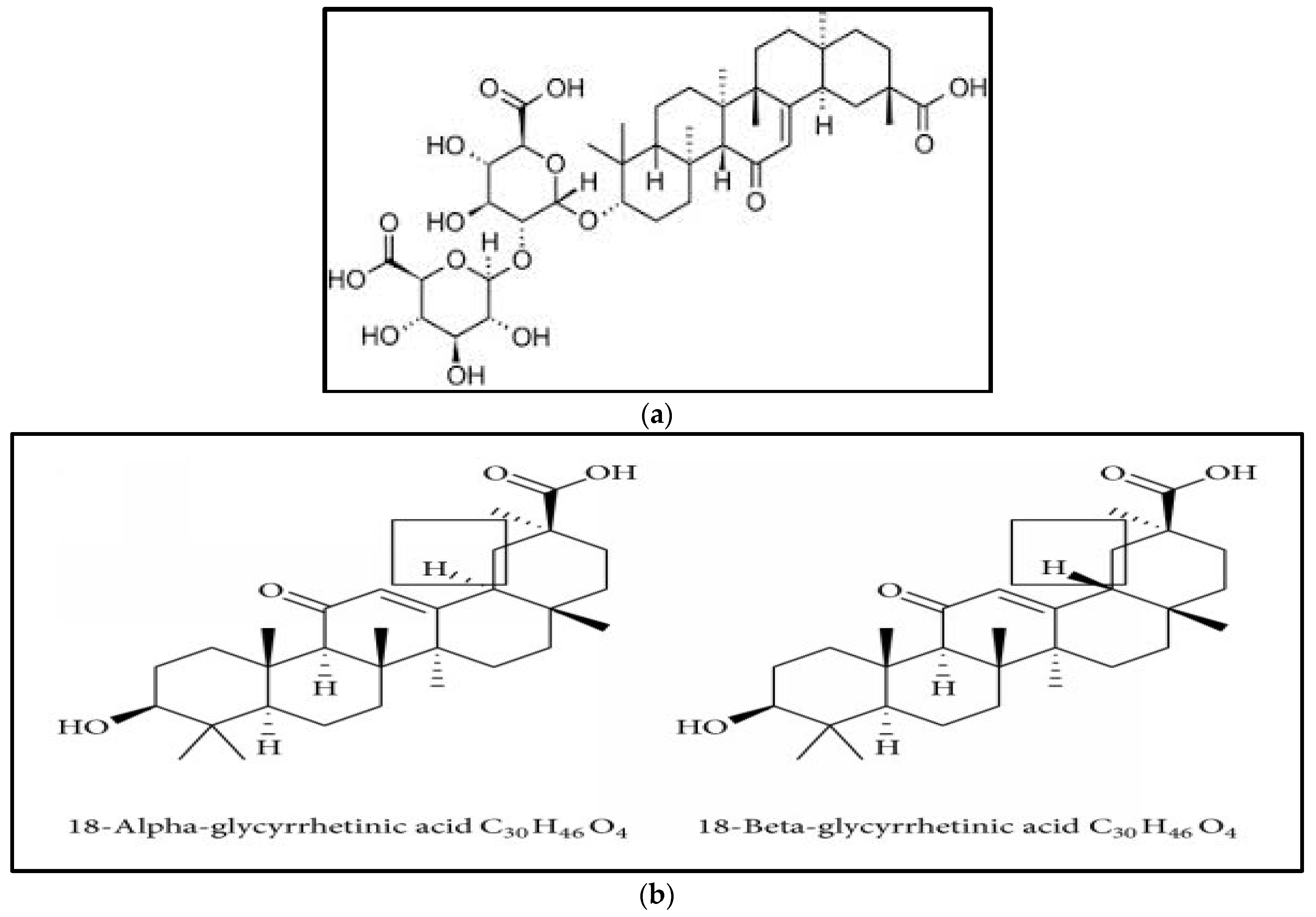
Figure 1. Chemical structures of (a) glycyrrhizin and (b) 18α-glycyrrhetinic acid and 18β-glycyrrhetinic acid.
Microbiolres 15 00133 g001
Figure 2. HPLC profiles of biotransformation of glycyrrhizin (GL) after incubation with Aspergillus niger NRRL 3122. (A) Chromatogram of standard 18β-glycyrrhetinic acid (B) One of the chromatograms of the biotransformation of glycyrrhizin using the Plackett–Burman design, chromatography conditions: column, Zorbax 21 300SB C18 column (4.5 mm × 250 mm); mobile phase, acetonitrile–water—acetic acid (80:20:1, v/v/v) at a flow rate of 1.5 mL/min; UV detector; detective wavelength, 254 nm; injection volume, 10 μL. (C) One of the chromatograms of the biotransformation of glycyrrhizin using the Box–Behnken design, Chromatography conditions: column, Zorbax 21 300SB C18 column (4.5 mm × 250 mm); mobile phase, acetonitrile–water–acetic acid (80:20:1, v/v/v) at a flow rate of 1.5 mL/min; UV detector; detective wavelength, 254 nm; injection volume, 10 μL.
Microbiolres 15 00133 g002
Figure 3. Response surface curves from Box–Behnken experiments for time vs. GL% (A), time vs. pH (B), and pH vs. GL% (C) for the biotransformation of glycyrrhizin into glycyrrhetinic acid by Aspergillus niger.
Microbiolres 15 00133 g003
Table 1. Variables and their levels employed in a Plackett–Burman design affecting the biotransformation of glycyrrhizin into glycyrrhetinic acid by Aspergillus niger.
FactorLow Level (−1)High Level (+1)
pH57
Temperature (°C)2530
Glycyrrhizin (g/100 mL)0.51
Incubation time (day)46
Glucose (g/100 mL)13
Yeast extract (g/100 mL)0.20.4
CSL (g/100 mL)0.51
Table 3. Analysis of the effects of seven variables on biotransformation of glycyrrhizin into glycyrrhetinic acid using a Plackett–Burman design.
Parameter Estimates
TermEstimateStd Errort RatioProb > |t|Uncoded Estimate
Intercept35.3208332.21148615.97<0.0001 *−68.46
pH (5, 7)10.6308332.2114864.810.0086 *10.630833
Temperature (25, 30)−2.95752.211486−1.340.2521−1.183
GL% (0.5, 1)8.69916672.2114863.930.0171 *34.796667
Incubation time (4, 6)13.0841672.2114865.920.0041 *13.084167
Glucose% (1, 3)0.47752.2114860.220.83960.4775
Yeast extract% (0.2, 0.4)−3.71752.211486−1.680.1681−37.175
CSL% (0.5, 1)−2.9308332.211486−1.330.2557−11.72333
Significant differences * p < 0.05.
Table 4. Box–Behnken factorial experimental design and responses of glycyrrhetinic acid concentration as affected by incubation time, pH, and glycyrrhizin concentrations.
PatternIncubation TimepHGL%Glycyrrhetinic Acid mg/g
Actual ValuePredicted Value
++0109266.3966.67
−0+683111.41112.91
00088283.289.66
0++89385.1982.25
0−−87192.8195.74
0−+87390.2288.99
+0+108370.6773.32
−0−681128.25125.59
0+−89187.2388.45
00088297.2389.66
+−0107265.4464
+0−108175.1173.6
−−0672119.76119.47
00088288.5689.66
−+0692101.34102.77
Table 5. Analysis of the effects of the most significant variables (incubation time, pH, and glycyrrhizin concentrations) on glycyrrhetinic acid concentration using a Box–Benken design.
Parameter Estimates
TermEstimateStd Errort RatioProb > |t|
Intercept175.39333211.24060.830.4442
Incubation time−46.0052115.70847−2.930.0327 *
pH48.14791746.182081.040.3449
Glycyrrhizin conc.−31.2683326.46856−1.180.2906
Incubation time × pH2.421251.3380021.810.1301
Incubation time × glycyrrhizin conc.1.551.3380021.160.2990
pH × Glycyrrhizin conc.0.1152.6760040.040.9674
Incubation time × incubation time0.75552080.6963181.090.3274
pH × pH−4.4529172.785274−1.600.1708
Glycyrrhizin conc. × glycyrrhizin conc.3.67458332.7852741.320.2443
Significant differences * p < 0.05.
Table 6. The antibacterial activity of crude glycyrrhizin and biotransformed glycyrrhetinic acid against strains of bacterial pathogens, as indicated by the minimal inhibitory concentration (MIC, μg/mL) and zone of inhibition diameter (ZOI, mm).
Bacterial Strain Crude GlycyrrhizinBioconverted Glycyrrhetinic Acid
ZOI (mm) * MIC (µg/mL) **ZOI (mm) MIC (µg/mL)
Bacillus subtilis17 ± 1.1 a6032 ± 1.7 a20
Escherichia coli12 ± 0.7 b20022 ± 1.2 c80
Pseudomonas aeruginosa10 ± 0.4 c40018 ± 1.3 d140
Salmonella typhimurium12 ± 0.8 b22021 ± 1.1 c100
Staphylococcus aureus15 ± 0.9 a10029 ± 1.4 b40
* Zone of inhibition (ZOI) diameters included the diameter of the paper disc (6 mm) ± standard deviations. ** MIC: minimal inhibitory concentration (the lowest concentration that prevents growth). In the same column, different superscript letters denote significant differences at p < 0.05.
Table 7. Radical scavenging activity of crude glycyrrhizin and biotransformed glycyrrhetinic acid at different concentrations and incubation times.
Radical Scavenging Activity *
Crude GlycyrrhizinBiotransformed Glycyrrhetinic Acid
Incubation Time Incubation Time
Conc. **30 min60 min90 minConc.
Mean ± SE
30 min60 min90 minConc.
Mean ± SE
100045.92 ± 0.0541.77 ± 0.0535.73 ± 0.0641.14 ± 4.1880.58 ± 0.0677.64 ± 0.0572.33 ± 0.0776.85 ± 3.41
50036.71 ± 0.0534.67 ± 0.0530.61 ± 0.0534.00 ± 2.5371.84 ± 0.0668.93 ± 0.0562.59 ± 0.0567.79 ± 3.86
25029.59 ± 0.0425.51 ± 0.0521.44 ± 0.0525.51 ± 3.3358.74 ± 0.0555.02 ± 0.0449.00 ± 0.0554.25 ± 4.01
12522.49 ± 0.0520.43 ± 0.0616.37 ± 0.0419.76 ± 2.5049.51 ± 0.0546.09 ± 0.0441.03 ± 0.0545.54 ± 3.48
6315.97 ± 0.0512.81 ± 0.0409.66 ± 0.0412.81 ± 2.5839.32 ± 0.0436.86 ± 0.0330.33 ± 0.0435.50 ± 3.80
Incubation time
mean ± SE
30.14 ± 10.5027.04 ± 10.2322.76 ± 9.42 60.00 ± 14.8556.91 ± 14.8151.06 ± 14.97
* Radical scavenging activity given as % inhibition. Triplicate samples were analyzed, and the main values and the SD were given. ** Concentration μg/100 μL. % inhibition for standard ascorbic acid was 100%.
Disclaimer/Publisher’s Note: The statements, opinions and data contained in all publications are solely those of the individual author(s) and contributor(s) and not of MDPI and/or the editor(s). MDPI and/or the editor(s) disclaim responsibility for any injury to people or property resulting from any ideas, methods, instructions or products referred to in the content.