Article

Modulation of the Toll-like Receptor 3-Mediated Intestinal Immune Response by Water Kefir

1
Laboratory of Immunobiotechnology, Reference Centre for Lactobacilli (CERELA-CONICET), San Miguel de Tucumán 4000, Argentina
2
Food and Feed Immunology Group, Laboratory of Animal Food Function, Graduate School of Agricultural Science, Tohoku University, Sendai 980-8572, Japan
3
Livestock Immunology Unit, International Education and Research Centre for Food and Agricultural Immunology (CFAI), Graduate School of Agricultural Science, Tohoku University, Sendai 980-8572, Japan
4
Department of Food, Agriculture and Environment, Miyagi University, Sendai 980-8572, Japan
*
Authors to whom correspondence should be addressed.
These authors contributed equally to this work.
JSPS fellow.
Microbiol. Res. 2024, 15(3), 1239-1250; https://doi.org/10.3390/microbiolres15030083
Submission received: 1 June 2024 / Revised: 6 July 2024 / Accepted: 17 July 2024 / Published: 20 July 2024

Abstract

:
Kefir has been associated with beneficial effects on its host’s health. The previous works examining the impact of kefir on the immune system focused on milk kefir or the exopolysaccharides and bacterial strains derived from it, while water kefir has not been evaluated. Furthermore, studies have focused on kefir’s ability to modulate immune system hemostasis and exert anti-inflammatory effects, while its specific action on antiviral immunity has not been investigated. Thus, the aim of this work was to examine the potential immunomodulatory effects of water kefir on the intestinal innate antiviral immunity mediated by Toll-like receptor-3 (TLR3). Adult BALB/c mice fed water kefir ad libitum, diluted 1:5, 1:10, or 1:20 in the drinking water, for 6 consecutive days. On day 7, the treated groups and the untreated control mice received an intraperitoneal injection of the TLR3 agonist poly(I:C). Two days after the TLR3 activation, the intestinal damage and the innate immune response were studied. The intraperitoneal administration of poly(I:C) induced inflammatory-mediated intestinal tissue damage, characterized by the upregulation of interferons (IFNs), pro-inflammatory mediators (TNF-α, IL-15, IL-6), and factors involved in epithelial destruction (RAE-1 and NKG2D). The histological analysis of small intestinal samples showed that mice receiving water kefir 1:5 exhibited reduced edema and a lower inflammatory cell infiltration. Kefir-treated mice had significantly lower levels of serum LDH, AST, and ALT as well as intestinal TNF-α, IL-15, IL-6, RAE-1, and NKG2D. This group also showed higher concentrations of intestinal IFN-β, IFN-γ, and IL-10. The treatment with 1:10 of water kefir reduced intestinal damage and modulated cytokines but its effect was significantly lower than the 1:5 treatment, while the water kefir 1:20 did not modify the parameters evaluated compared to control mice. The results indicate that water kefir exerts its immunomodulatory effects in a dose-dependent manner. The in vivo studies allow us to speculate that water kefir can induce two beneficial effects on the intestinal TLR3-mediated immune response: the enhancement of antiviral defenses and the protection against the inflammatory-mediated tissue damage. These protective effects of water kefir require further exploration to understand how water kefir, or its specific molecules/strains, can influence the immune response and to determine the extent of its protection against a real viral challenge.

1. Introduction

The pattern recognition receptors (PRRs) expressed in the intestinal mucosa, such as Toll-like receptors (TLRs), can recognize different microbial antigens inducing the activation of innate immunity that is essential for the protection of the host [1]. Among PRRs, TLR3 specifically recognizes genomic double-stranded RNA (dsRNA) from viruses or dsRNA synthesized during viral replication. Studies using purified genomic dsRNA, the synthetic dsRNA analog polyinosinic:polycytidylic acid [poly(I:C)] and TLR3 knockout mice have highlighted the crucial role of this receptor in intestinal antiviral immunity [2,3]. TLR3 activation in the gut occurs in response to viral challenges, enabling cells to detect viruses and develop resistance. Gut TLR3 has been shown to exert both beneficial and detrimental effects. On the one hand, the induction of interferons (IFNs), cytokines, and chemokines production by TLR3 activation enhances antiviral mechanisms and coordinates immune responses that help to eliminate the virus and infected cells [1]. On the other hand, it was shown that both poly(I:C) and purified rotavirus dsRNA can cause severe mucosal damage in the gut via TLR3-dependent pathways, which involve the injury of the intestinal epithelium through the interaction of the retinoic acid early inducible-1 (RAE-1), expressed on intestinal epithelial cells (IECs), with the NKG2D receptor, expressed on intestinal intraepithelial lymphocytes (IELs) [2,3]. Then, an efficient modulation of TLR3-mediated immunity is important to protect the host against virus without damaging the intestinal mucosa.
Probiotics have seen a significant increase in use in the food industry and consumers over the past few decades [4,5]. This is due to the wide range of beneficial properties they have, including their capacity to increase the resistance to infections. In fact, several research works described the ability of certain probiotic strains to modulate host defenses and improve the resistance to viral infections (reviewed in [6,7]). In this sense, we have reported that the immunomodulatory probiotic strains Lacticaseibacillus rhamnosus CRL1505, Lactiplantibacillus plantarum CRL1506, and Lactobacillus delbrueckii TUA4408L modulate the intestinal innate antiviral immune response increasing protection against gastrointestinal viruses [8,9,10,11]. We showed that these strains are capable of modulating the innate immune response triggered by the activation of TLR3 in the gut, enhancing the production of IFNs (IFN-α, IFN-β, and IFN-γ) and antiviral factors, and at the same time beneficially regulating the balance of proinflammatory (TNF-α, IL-1β, and IL-6), regulatory cytokines (IL-10 and IL-27) and intestinal RAE-1/NKG2D expressions. The influence of the probiotic strains on the response to TLR3 signaling activation is translated into a more efficient elimination of viruses by IECs as well as in the protection of the intestinal tissue against the inflammatory damage mediated by IELs [8,10,11].
Kefir is a microbial symbiont mixture that produces jelly-like grains as it grows. It contains both lactic acid bacteria (LAB) (Lactobacillus, Lactococcus, Leuconostoc, Acetobacter, and Streptococcus spp.) and yeasts (Kluyveromyces, Torula, Candida, and Saccharomyces spp.), which are surrounded by a polysaccharide matrix called kefiran [12,13]. Kefir can be classified into two main types: dairy kefir, made from milk, and non-dairy kefir, which is prepared with water and sucrose, honey, or sugar cane. Kefir is fermented using either commercial lyophilized starter cultures, traditional kefir grains, or the remaining product after kefir grains are removed [12,13]. Comparative studies of milk kefir and water kefir described differences in microbiological, dry matter, protein, ash, and mineral contents, as well as differences in the intrinsic structures of the grains as revealed by scanning electron microscopy [14]. In addition, the microbial and chemical composition of water kefir appears to be dependent on the geographical origin of the grains and the fermentation substrate and conditions [12]. Traditionally, water kefir is produced on a small scale, and it is not common to use defined starter cultures in its preparation. However, as water kefir increases in popularity as a healthy beverage, it is important to gain insight into its composition and define its health-beneficial properties.
The regular consumption of kefir is linked to several health benefits such as improved lactose digestion and tolerance, antibacterial, hypocholesterolemic, and anti-hypertensive effects, as well as antioxidant, anti-carcinogenic, and immunomodulatory activities [15,16,17]. Kefir, specific LAB strains isolated from kefir, and kefiran have been reported to possess immunomodulatory effects. It was shown that the feeding of mice with milk kefir increased levels of IL-4, IL-6, and IL-10 in the lamina propria of the small intestine, as well as the concentrations of IgA [18]. Similarly, feeding milk kefir to rats with metabolic syndromes reduced the expression of IL-1β and increased IL-10 in adipose tissue [19]. The oral administration of the exopolysaccharide produced by Lactobacillus kefiranofaciens, a strain isolated from kefir, increased the numbers of IgA+, IL-6+, and IL-10+ cells in the small and large intestine lamina propria [20]. In line with this report, it was shown that kefiran diminished the expression of IL-1β and TNF-α, as well as nuclear factor kappa B (NF-kB) activation in monocytes and was able to reduce and enhance the levels of IL-1β and IL-10, respectively, in dendritic cells (DCs) [21]. The administration of Lentilactobacillus kefiri CIDCA 8348 to mice increased IgA in feces, reduced expression of the proinflammatory mediators IFN-γ, GM-CSF, and IL-1β in Peyer’s patches, and increased intestinal IL-10 [22]. More recently, the effect of kefir milk and the strain Lacticaseibacillus paracasei Z2, isolated from kefir grains, on the immune response was comparatively evaluated in mice [23]. The work demonstrated that both treatments were able to significantly increase intestinal mucin gene (muc-1 and muc-2) expressions, as well as intestinal IgA production, being the isolated strain more efficient than kefir to induce the immunomodulatory effects.
These previous works examining the impact of kefir on the immune system focused on milk kefir or the exopolysaccharides and LAB strains derived from it, while water kefir has not been evaluated. Furthermore, studies have focused on kefir’s ability to modulate immune system hemostasis and exert anti-inflammatory effects, while its specific action on antiviral immunity has not been investigated. Thus, the aim of this work was to examine the potential immunomodulatory effects of water kefir on the intestinal innate antiviral immunity. For this purpose, mice were fed water kefir in different dilutions and then challenged with an intraperitoneal injection of the TLR3 agonist poly(I:C). The intestinal innate antiviral immune response and the gut damage were evaluated.

2. Materials and Methods

2.1. Water Kefir Elaboration

Water kefir was produced by adding kefir grains and mascabo sugar in sterile water 5% w/v, with the addition of raisins. The fermentation was performed at room temperature for 24 h in plastic bottles covered with canvas scrap adjusted with a rubber band. The end of fermentation was established by measuring pH. After 24 h, acidification dropped two pH points compared to the initial value (pH = 6.5–6.0). Then, the grains were recovered by sieving, conserved in a plastic recipient and stored at 4 °C for the next fermentations. The filtered liquid was collected in glass bottles for further dilutions and determinations. Once the bottles were collected, they were closed hermetically and stored in the refrigerator at 4 °C until use.

2.2. Animals, Feeding Procedures, and Administration of Poly(I:C)

Male 5-week-old BALB/c mice were obtained from the closed colony kept at the animal facilities of the Reference Center for Lactobacilli (CERELA-CONICET, Tucuman, Argentina). Animals were housed in plastic cages in a controlled atmosphere (22 ± 2 °C temperature, 55 ± 2% humidity) with a 12 h light/dark cycle. All experiments were carried out in compliance with the Guide for Care and Use of Laboratory Animals and approved by the Ethical Committee of Animal Care at CERELA, Argentina (protocol number BIOT-CRL/14).
Mice were allocated randomly to groups and each group consisted of 5 animals. Mice were fed water kefir ad libitum, diluted 1:5, 1:10, or 1:20 in the drinking water, for 6 consecutive days. Mice without water kefir treatment were used as controls. The treated groups and the untreated control mice were fed a conventional balanced diet ad libitum. On day 7, mice were injected intraperitoneally with 100 μL of PBS containing 30 μg of poly(I:C) (high molecular weight polyinosine-polycytidylic acid, InvivoGene, San Diego, CA, USA) according to our previous publications [8,10]. This route and dose of poly(I:C) administration induce TLR3-mediated intestinal inflammation and damage [8,10]. Two days after the activation of TLR3 by poly(I:C) administration, mice were anaesthetized to obtain samples of blood and small intestine. Blood samples were obtained from the cardiac puncture. Intestinal tissue and fluid were obtained as described before [8]. Briefly, the small intestine was flushed with 5 mL of PBS and the fluid was centrifuged (10,000× g, 4 °C 10 min) to separate particulate material. The supernatant was kept frozen at −80 °C until use.

2.3. Histological Studies of Small Intestine Injury

Small intestines were removed and processed for paraffin inclusion following the Sainte-Marie technique as previously described [8]. Serial paraffin sections (4 µm) were stained with hematoxylin-eosin followed by light microscopy examination. The analysis was performed blindly in 10–12 fields of vision for each sample. Photomicrographs were captured and the images were then cropped and corrected for brightness and contrast, but otherwise were not manipulated. The presence and extent of the edema, inflammatory infiltration, and mucosal erosion were evaluated according to previous publications [8].

2.4. Serum Enzymes Activities

Blood samples obtained by cardiac puncture were collected in tubes without the use of anticoagulants to obtain serum. Lactate dehydrogenase (LDH), alanine transaminase (ALT), and aspartate transaminase (AST) activities were determined in the serum to evaluate general and hepatic toxicity, according to our previous publications [8], by measuring the formation of the reduced form of nicotinamide adenine dinucleotide (NAD) using the Wiener reagents and procedures (Wiener Lab, Buenos Aires, Argentina). LDH, ALT, and AST activities were expressed as units per liter of serum.

2.5. Intestinal and Serum Cytokines

Cytokine concentrations in serum and intestinal fluid samples were measured with commercially available enzyme-linked immunosorbent assay (ELISA) technique kits following the manufacturer’s recommendations (R&D Systems, Minnneapolis, MN, USA). IFN-β (Mouse IFN-beta ELISA Kit, sensitivity: 15.5 pg/mL), IFN-γ (Mouse IFN-gamma Quantikine ELISA Kit, sensitivity: 2 pg/mL), IL-6 (Mouse IL-6 Quantikine ELISA Kit, sensitivity: 1.8 pg/mL), IL-10 (Mouse IL-10 Quantikine ELISA Kit, sensitivity: 5.2 pg/mL), IL-15 (Mouse IL-15 DuoSet ELISA kit, sensitivity: 0.125 ng/mL), and TNF-α (Mouse TNF-alpha ELISA Kit—Quantikine, sensitivity: 7.21 pg/mL) were used.

2.6. Quantitative Expression Analysis by Real-Time PCR

Two-step real-time quantitative PCR was performed to characterize the expression of TLR3, RIG-I, RAE-1, and NKG2D in intestinal samples following the methodology previously described [8]. Briefly, total RNA was isolated from small intestinal tissue samples using TRIzol reagent (Invitrogen, Carlsbad, CA, USA) and cDNA was synthesized with a Quantitect reverse transcription (RT) kit (Qiagen, Tokyo, Japan) according to the manufacturer’s recommendations. Real-time quantitative PCR was carried out with a 7300 real-time PCR system (Applied Biosystems, Warrington, UK) and the Platinum SYBR green qPCR SuperMix (Invitrogen). The primers and the PCR cycling conditions were described previously [8]. The expression of β-actin was used to normalize cDNA levels for differences in total cDNA levels in the samples. Results are shown as relative index related to the expression of factors in basal conditions set as 1.

2.7. Statistical Analysis

The experiments were conducted in 5 mice per group (n = 5 for each assessed parameter). The data were presented as mean ± standard deviation (SD). After verification of the normal distribution of data, a two-way ANOVA was used. Tukey’s test (for pairwise comparisons of the means) was used to test for differences between the groups, with significance set at p < 0.05 or p < 0.01.

3. Results

3.1. Water Kefir Reduces Poly(I:C)-Induced Damage

The potential protective effect of water kefir on TLR3-induced damage was first evaluated by measuring the percentage of body weight loss and the biochemical markers LDH, ALT, and AST in serum samples. Poly(I:C) challenge induced a 10 ± 2% of body weight loss in the control group while animals fed water kefir showed values of 3 ± 1, 4 ± 1, and 7 ± 2% for 1:5, 1:10, and 1:20 dilutions, respectively. Healthy mice without inflammatory challenges had values of LDH, ALT, and AST of approximately 400, 30, and 40 UI/L, respectively. Water kefir administration did not modify the levels of serum LDH, ALT, and AST in basal conditions. The intraperitoneal administration of poly(I:C) significantly increased the levels of serum LDH, ALT, and AST in the four experimental groups compared to basal levels (Figure 1).
However, mice receiving water kefir has significantly lower serum AST than poly(I:C) controls. Of note, the activities of serum LDH and ALT were lower than controls only in mice treated with water kefir diluted in 1:5 and 1:10 (Figure 1). Serum LDH was also lower in water kefir 1:20 than controls. For all the serum enzymes, the lowest values of activities were found in mice treated with water kefir 1:5 while the highest were found in the water kefir 1:20 group, suggesting a dose-dependent effect.
Histopathological analysis of small intestinal tissue samples was also performed to corroborate the protective effect of water kefir against TLR3-induced damage (Figure 2). Control mice showed intestinal histological alterations characterized mainly by edema, inflammatory infiltration, and mild mucosal erosion. Water kefir treatment significantly reduced intestinal damage (Figure 2). The effect was more notorious in mice receiving water kefir diluted in 1:5, which showed significantly lower edema and inflammatory infiltration. In contrast, the water kefir 1:20 group had slight improvements compared to the control group.

3.2. Water Kefir Differentially Modulates Cytokine Response to Poly(I:C)

We and others reported previously that the intraperitoneal administration of poly(I:C) induces changes in intestinal and serum cytokines characterized by alterations in the levels of type I IFNs, inflammatory cytokines, and chemokines, as well as in regulatory cytokines [2,3,8,10,24]. Among the cytokines differentially regulated by poly(I:C) are IFN-β, IFN-γ, TNF-α, IL-15, IL-6, and IL-10. Thus, we next assessed the effect of water kefir administration on the levels of these six cytokines in intestinal fluid samples before and after the activation of TLR3. The study of intestinal cytokines in basal conditions before the challenge with poly(I:C) showed that water kefir did not modify the levels of TNF-α, IL-15, IL-6, or IL-10 for all the dilutions evaluated. It was observed slight but significant increases in the levels of IFN-β and IFN-γ when mice treated with water kefir 1:5 and the control group were compared: 91.4 ± 2.3 (control) vs. 145.6 ± 3.4 pg/mL (water kefir 1:5) for IFN-β, and 47.5 ± 2.1 (control) vs. 71.8 ± 4.5 pg/mL (water kefir 1:5) for IFN-γ.
Poly(I:C) administration significantly increased the levels of the proinflammatory factors TNF-α, IL-15, and IL-6 as well as IFN-β and IFN-γ in the intestine of all the experimental groups (Figure 3). However, the levels of IFN-β and IFN-γ were higher while concentration of the proinflammatory cytokines were lower in mice treated with water kefir 1:5 than poly(I:C) controls. In addition, animals receiving water kefir 1:5 had significantly higher levels of intestinal IL-10 than controls (Figure 3). The administration of water kefir 1:10 increased intestinal IFN-β and IL-10 and reduced TNF-α, IL-15, and IL-6 although the concentrations of these factors did not reach the levels found in water kefir 1:5. Of note, water kefir 1:20 only reduced the levels of intestinal TNF-α (Figure 3).
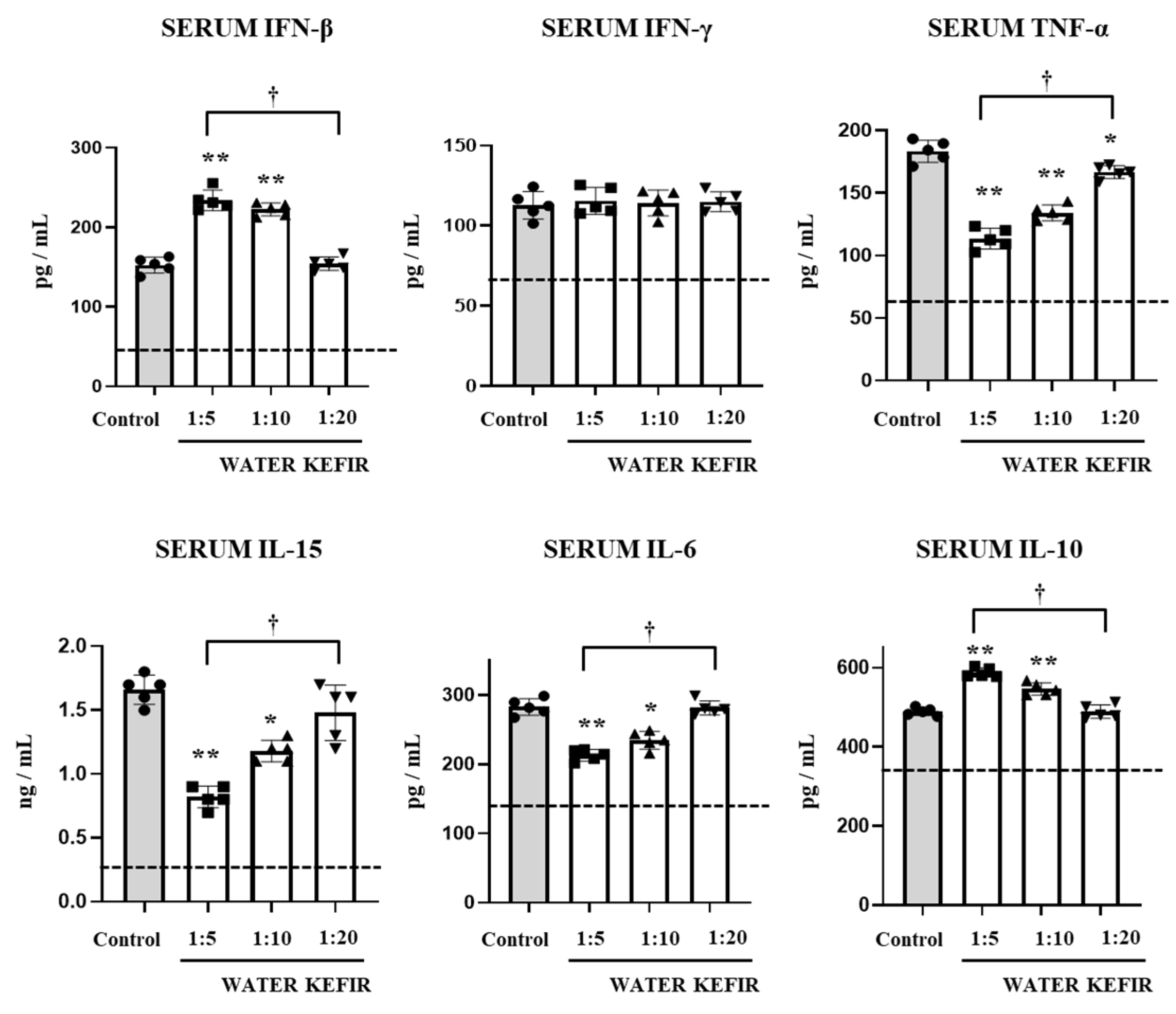
We also evaluated the levels of the same six immunological factors in serum samples. Before the challenge with poly(I:C), none of the doses of water kefir studied induced changes in the levels of the cytokines evaluated. Enhanced concentrations of serum IFN-β, IFN-γ, TNF-α, IL-15, IL-6, and IL-10 were found in all the experimental groups after the challenge with poly(I:C) in comparison with basal conditions (Figure 4). Like the results described for intestinal cytokines, in mice treated with water kefir 1:5 the levels of TNF-α, IL-15, and IL-6 were lower and the concentrations of IL-10 and IFN-β were higher than control mice (Figure 4). Of note, water kefir 1:5 did not increase serum IFN-γ levels as it did in the intestine. The administration of water kefir 1:10 increased serum IFN-β and IL-10 and reduced TNF-α, IL-15, and IL-6 although the concentrations of these factors did not reach the levels found in water kefir 1:5. On the other hand, water kefir 1:20 only reduced the levels of serum TNF-α (Figure 4).
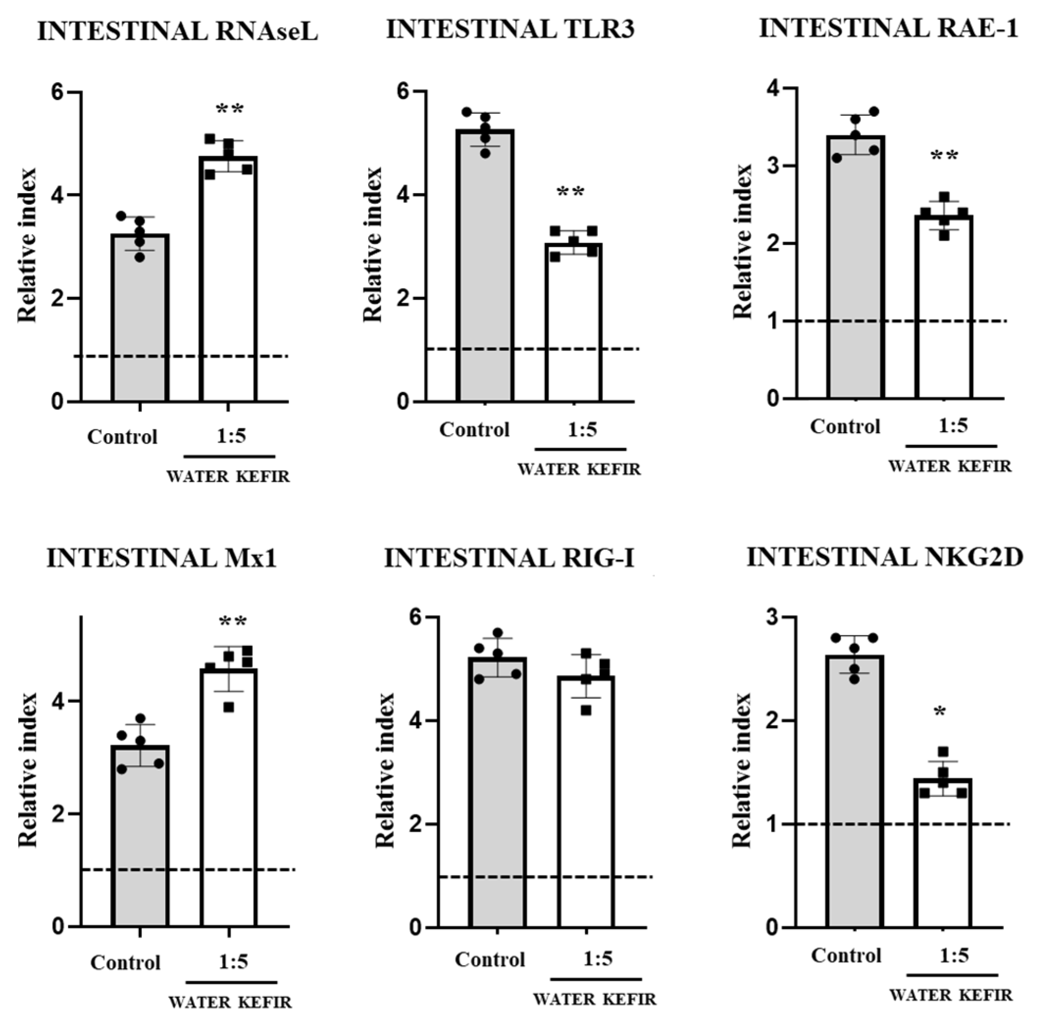
Finally, we evaluated the expression of the antiviral factors RNAseL, Mx1, the PRRs TLR3, and RIG-I, as well as the factors RAE-1 and NKG2D that are involved in the interaction of IECs and IELs, in the intestine of mice. For these experiments, we selected water kefir 1:5. As shown in Figure 5, the challenge with poly(I:C) increased the expression of the six factors evaluated compared to basal levels in both experimental groups. However, the levels of RNAseL and Mx1 were higher in water kefir-treated mice than in controls. In addition, expression levels of TLR3, RAE-1, and NKG2D were significantly lower in mice treated with water kefir 1:5 than controls (Figure 5). No differences were found between the two groups when RIG-I was analyzed.

4. Discussion

The immunomodulatory effects of milk kefir, specific LAB strains isolated from kefir, and kefiran have been reported previously [19,20,21,22,23]. Those studies demonstrated that milk kefir has the potential to regulate intestinal immune hemostasis and modulate inflammatory responses. In contrast, the immunomodulatory effects of water kefir were not investigated. The potential beneficial effect of water kefir should be studied considering that the immunoregulatory activities of milk kefir cannot be automatically extrapolated to water kefir because of the chemical and microbiological differences between the two foods [14]. In addition, the influence of kefir on mucosal antiviral immune response was not investigated before. The present study demonstrated for the first time the immunomodulatory effects of water kefir on the TLR3-mediated intestinal antiviral innate immune response in a murine model.
Using IEC and DC in vitro cultures as well as a murine model in which TLR3-mediated intestinal immune response is triggered by the intraperitoneal administration of poly(I:C), we demonstrated that L. delbrueckii TUA4408L, L. rhamnosus CRL1505, and L. plantarum CRL1506 differentially modulated the intestinal cytokine profile, conducting to an enhanced production of antiviral factors as well as to a protection against tissue damage [8,10,11]. The detailed immunological studies carried out in our previous works allowed us to select parameters for a rapid and accurate evaluation of the immunomodulatory potential of foods and LAB strains, including the determination of serum enzymes, the concentration of intestinal and blood IFN-β, IFN-γ, TNF-α, IL-15, IL-6, and IL-10, and the expression of RNAseL, Mx1, RAE-1, NKG2D, RIG-I, and TLR3. Using the mice model of TLR3-mediated intestinal damage and the selected biomarkers, we demonstrated here that water kefir administration can improve antiviral innate immunity and reduce inflammatory damage in a dose dependent manner.
(a) Modulation of the intestinal innate antiviral immune response by water kefir. The production of IFN-β and IFN-γ in response to viral infections in the intestinal mucosa trigger the expression of several antiviral genes that improve the protective response of epithelial cells by restricting the replication of the pathogens [25,26]. IFNs also help to coordinate the cellular antiviral response by activating macrophages and T cells that eliminate virus-infected cells [25,26]. Considering that water kefir, particularly the 1:5 dilution, was able to enhance the production of IFN-β and IFN-γ in response to TLR3 activation, it is tempting to speculate that it could play a protective role in response to an intestinal virus infection. In fact, the treatment of mice with water kefir 1:5 significantly augmented the expression of RNAseL and Mx1, antiviral factors involved in the protection of the intestinal mucosa against viruses such as rotavirus and porcine epidemic diarrhea virus [27]. The 2′-5′-oligoadenylate synthetase (OAS)–RNAseL pathway is activated by dsRNA viral molecules inducing the enzyme degrading activities of RNAseL, restricting viral infection [28]. Furthermore, the products obtained by the RNAseL degradation activities amplify innate immune mechanisms through their recognition of the PRRs, RIG-I, and MDA5. On the other hand, Mx1, also called MxA, inhibits a wide range of RNA and DNA viruses replicating in the cytoplasm [29]. In support of the hypothesis that water kefir could be effective in reducing viral replication in the intestinal epithelium, our previous in vitro studies using porcine IECs showed that the immunomodulatory bifidobacteria strains differentially regulated gene expression after poly(I:C) stimulation inducing enhancements of IFNs, Mx1, OAS1, and RNAseL [30]. The changes induced in IECs by bifidobacteria significantly reduced rotavirus replication.
Previous transcriptomic studies performed in IECs with immunomodulatory lactobacilli and bifidobacteria strains showed that the mechanism by which antiviral defenses are upregulated would be mediated by a differential modulation of the negative regulator of the TLR signaling A20 [8,10,11]. It was shown that the challenge of HT29 cells, which are human colon epithelial cells, with rotavirus increased the expression of several IFN inducible genes, including OAS1, Mx1, IL-18, and IITP3 [31]. Of note, the co-stimulation of HT29 cells with poly(I:C) and probiotic strains significantly reduced A20 expression levels and augmented IFNs and antiviral factors [32]. In line with these findings, we observed that immunomodulatory bacteria can reduce A20 expression in IECs leading to an improved activation of the interferon regulatory factor 3 (IRF3) and NF-κB signaling pathways, which increase the expression of not only IFNs, Mx1, and RNAseL, but also several other antiviral factors, including OASL, Mx2, OAS2, RNAse4, IFIT1, and IFIT3 [10]. Then, further in vitro studies in IEC cultures are necessary to determine if water kefir has a similar mechanism of action to improve antiviral defenses. In addition, studying the effect of water kefir on the response to a real viral intestinal infection, such as that caused by rotavirus, is an important point for future research.
(b) Modulation of the intestinal TLR3-mediated inflammatory damage by water kefir. The balance of pro- and anti-inflammatory cytokines in the mucosal tissues during the course of viral infections has to be tightly regulated to allow an efficient clearance of the pathogen with minimal tissue damage [33,34]. As described in previous studies [2,3] and here, the intraperitoneal administration of poly(I:C) induced inflammatory-mediated intestinal tissue damage in mice, characterized by increased inflammatory cell infiltration, enhancement of TNF-α and IL-6, and reduction in the immunoregulatory cytokine IL-10. In addition, enhanced levels of IL-15, RAE-1, and NKG2D are found in the gut after poly(I:C) challenge, which are important mediators of TLR3-induced small intestinal injury. IL-15 is produced by IECs after the abnormal TLR3 activation, and this cytokine participates in epithelial damage through the recruitment of IELs that eliminate IECs and produce mucosal erosion [2,3]. IL-15 stimulates the cytotoxic activity of CD3+NK1.1+ IELs and makes them more potent killers of virus-infected IECs [35]. RAE-1, which is a high-affinity ligand for NKG2D, is minimally detected on the healthy intestinal tissues of mice [36]. However, the TLR3 activation increases intestinal RAE-1 expression, particularly in IECs, allowing their destruction by NKG2D-expressing IELs [2,3]. In fact, the obstruction of the RAE-1-NKG2D interaction significantly reduces the cytotoxic effect of IELs on IECs and diminishes intestinal injury in mice stimulated with purified rotaviral dsRNA [2,3]. Of note, mice receiving water kefir 1:5 exhibited significantly reduced intestinal damage after poly(I:C) exposure. Water kefir-treated mice had attenuated levels of the proinflammatory cytokines TNF-α, IL-15, and IL-6, and expression of RAE-1 and NKG2D while the levels of IL-10, which is a key anti-inflammatory cytokine in the gut [33,34], were higher than in the controls. Thus, the changes in the balance of intestinal TNF-α/IL-10 as well as the IL-15/RAE-1/NKG2D interactions would be involved in the protection of water kefir against intestinal damage. How water kefir can induce this differential balance of inflammatory and anti-inflammatory factors in the context of the TLR3-mediated innate response is a topic that must be investigated to provide the scientific basis to allow water kefir to be proposed as a modulator of the intestinal damage induced by viral inflammation.
Another important point for future research is to determine which factors present in water kefir are responsible for the immunomodulatory effect observed in the context of TLR3-mediated intestinal immunity, considering that different metabolites including kefiran, as well as different types of bacteria and yeasts, can be found in this food. It is also necessary to investigate whether it is a synergistic effect of all the factors. A detailed study of the concentrations of the metabolic products as well as the microbiological composition could help to establish not only which components would have the biological effect but also to elucidate why a dose-dependent effect was observed. Furthermore, it would be important to establish if this specific composition is related to the immunomodulatory effect and if it is possible to modulate antiviral intestinal immunity with water kefir obtained in other regions and in different ways. Metagenomic analysis and studies of chemical composition of water kefir produced in different conditions or in the same conditions at different moments coupled with in vivo studies such as the one carried out in this work, could provide some clues in this regard.
In conclusion, although kefir has been extensively studied for its beneficial effects on health, no specific scientific research has been conducted so far on its impact in antiviral immunity. Our in vivo studies allowed us to speculate that water kefir can induce two beneficial effects on the intestinal TLR3-mediated immune response: the enhancement of antiviral defenses and the protection against inflammatory-mediated tissue damage. These protective effects of water kefir require further exploration to understand how water kefir, or its specific molecules/strains, can influence the immune response, and to determine the extent of its protection against a real viral challenge. Comparative experiments of milk kefir and water kefir are also necessary to determine whether the ability to modulate TLR3-mediated antiviral immunity is an effect achieved only by the latter or not. This work represents a first step to propose water kefir as a dietary intervention for managing and preventing gastrointestinal disorders associated with viral infections.

Author Contributions

Conceptualization, H.K. and J.V.; methodology, S.D.M., R.O.M., M.E., L.A., Y.I., F.N., K.N. and Y.S.; formal analysis, R.O.M. and S.D.M.; investigation, L.A.; resources, H.K. and J.V.; writing—original draft preparation, S.D.M. and J.V.; writing—review and editing, H.K.; visualization, J.V.; supervision, J.V.; project administration, H.K. and J.V.; funding acquisition, H.K. and J.V. All authors have read and agreed to the published version of the manuscript.

Funding

This study was supported by the ANPCyT–FONCyT grants PICT-2016-0410 and PICT-2021-I-A-00705 to J.V. This study was also supported by a Grant-in-Aid for Scientific Research (A) (23H00354, H.K.), a Challenging Research (Exploratory, 23K18072, H.K.) and Grant-in-Aid for JSPS fellows (24KJ0434, Y.I.) from the Japan Society for the Promotion of Science (JSPS); by the Research Program on Development of Innovative Technology grants (JPJ007097) from the Project of the Bio-oriented Technology Research Advancement Institution (BRAIN), by the Japan Racing Association to H.K. and by AMED Grant Number JP21zf0127001.

Institutional Review Board Statement

The study was conducted in accordance with the Declaration of Helsinki and approved by the CERELA-CONICET Animal Care and Ethics Committee under the BIOT-CRL/14 protocol, April 2014.

Informed Consent Statement

Not applicable.

Data Availability Statement

The data presented in this study are available throughout the article.

Conflicts of Interest

The authors declare no conflicts of interest.

References

  1. Luan, X.; Wang, L.; Song, G.; Zhou, W. Innate immune responses to RNA: Sensing and signaling. Front. Immunol. 2024, 15, 1287940. [Google Scholar] [CrossRef] [PubMed]
  2. Zhou, R.; Wei, H.; Sun, R.; Tian, Z. Recognition of double-stranded RNA by TLR3 induces severe small intestinal injury in mice. J. Immunol. 2007, 178, 4548–4556. [Google Scholar] [CrossRef] [PubMed]
  3. Zhou, R.; Wei, H.; Sun, R.; Zhang, J.; Tian, Z. NKG2D recognition mediates Toll-like receptor 3 signaling-induced breakdown of epithelial homeostasis in the small intestines of mice. Proc. Natl. Acad. Sci. USA 2007, 104, 7512–7515. [Google Scholar] [CrossRef] [PubMed]
  4. FAO/WHO Expert Consultation on Evaluation of Health and Nutritional Properties of Probiotics in Food Including Powder Milk with Live Lactic Acid Bacteria. 2001. Available online: https://openknowledge.fao.org/server/api/core/bitstreams/382476b3-4d54-4175-803f-2f26f3526256/content (accessed on 4 April 2024).
  5. Das, T.K.; Pradhan, S.; Chakrabarti, S.; Mondal, K.C.; Ghosh, K. Current status of probiotic and related health benefits. Appl. Food Res. 2022, 2, 100185. [Google Scholar] [CrossRef]
  6. Villena, J.; Li, C.; Vizoso-Pinto, M.G.; Sacur, J.; Ren, L.; Kitazawa, H. Lactiplantibacillus plantarum as a Potential Adjuvant and Delivery System for the Development of SARS-CoV-2 Oral Vaccines. Microorganisms 2021, 9, 683. [Google Scholar] [CrossRef] [PubMed]
  7. Harper, A.; Vijayakumar, V.; Ouwehand, A.C.; Ter Haar, J.; Obis, D.; Espadaler, J.; Binda, S.; Desiraju, S.; Day, R. Viral Infections, the Microbiome, and Probiotics. Front. Cell Infect. Microbiol. 2021, 10, 596166. [Google Scholar] [CrossRef] [PubMed]
  8. Tada, A.; Zelaya, H.; Clua, P.; Salva, S.; Alvarez, S.; Kitazawa, H.; Villena, J. Immunobiotic Lactobacillus strains reduce small intestinal injury induced by intraepithelial lymphocytes after Toll-like receptor 3 activation. Inflamm. Res. 2016, 65, 771–783. [Google Scholar] [CrossRef] [PubMed]
  9. Kanmani, P.; Albarracin, L.; Kobayashi, H.; Hebert, E.M.; Saavedra, L.; Komatsu, R.; Gatica, B.; Miyazaki, A.; Ikeda-Ohtsubo, W.; Suda, Y.; et al. Genomic Characterization of Lactobacillus delbrueckii TUA4408L and Evaluation of the Antiviral Activities of its Extracellular Polysaccharides in Porcine Intestinal Epithelial Cells. Front. Immunol. 2018, 9, 2178. [Google Scholar] [CrossRef] [PubMed]
  10. Albarracin, L.; Garcia-Castillo, V.; Masumizu, Y.; Indo, Y.; Islam, M.A.; Suda, Y.; Garcia-Cancino, A.; Aso, H.; Takahashi, H.; Kitazawa, H.; et al. Efficient Selection of New Immunobiotic Strains With Antiviral Effects in Local and Distal Mucosal Sites by Using Porcine Intestinal Epitheliocytes. Front. Immunol. 2020, 11, 543. [Google Scholar] [CrossRef]
  11. Mizuno, H.; Arce, L.; Tomotsune, K.; Albarracin, L.; Funabashi, R.; Vera, D.; Islam, M.A.; Vizoso-Pinto, M.G.; Takahashi, H.; Sasaki, Y.; et al. Lipoteichoic Acid Is Involved in the Ability of the Immunobiotic Strain Lactobacillus plantarum CRL1506 to Modulate the Intestinal Antiviral Innate Immunity Triggered by TLR3 Activation. Front. Immunol. 2020, 11, 571. [Google Scholar] [CrossRef]
  12. Lynch, K.M.; Wilkinson, S.; Daenen, L.; Arendt, E.K. An update on water kefir: Microbiology, composition and production. Int. J. Food Microbiol. 2021, 345, 109128. [Google Scholar] [CrossRef]
  13. Azizi, N.F.; Kumar, M.R.; Yeap, S.K.; Abdullah, J.O.; Khalid, M.; Omar, A.R.; Alitheen, N.B. Kefir and its biological activities. Foods 2021, 10, 1210. [Google Scholar] [CrossRef]
  14. Gökırmaklı, Ç.; Güzel-Seydim, Z.B. Water kefir grains vs. milk kefir grains: Physical, microbial and chemical comparison. J. Appl. Microbiol. 2022, 132, 4349–4358. [Google Scholar] [CrossRef] [PubMed]
  15. Sharifi, M.; Moridnia, A.; Mortazavi, D.; Salehi, M.; Bagheri, M.; Sheikhi, A. Kefir: A powerful probiotics with anticancer properties. Med. Oncol. 2017, 34, 183. [Google Scholar] [CrossRef] [PubMed]
  16. Peluzio, M.D.C.G.; Dias, M.D.M.E.; Martinez, J.A.; Milagro, F.I. Kefir and intestinal microbiota modulation: Implications in human health. Front. Nutr. 2021, 8, 638740. [Google Scholar] [CrossRef] [PubMed]
  17. Egea, M.B.; Santos, D.C.D.; Oliveira Filho, J.G.; Ores, J.D.C.; Takeuchi, K.P.; Lemes, A.C. A review of nondairy kefir products: Their characteristics and potential human health benefits. Crit. Rev. Food Sci. Nutr. 2022, 62, 1536–1552. [Google Scholar] [CrossRef]
  18. Vinderola, C.G.; Duarte, J.; Thangavel, D.; Perdigón, G.; Farnworth, E.; Matar, C. Immunomodulating capacity of kefir. J. Dairy Res. 2005, 72, 195–202. [Google Scholar] [CrossRef]
  19. Rosa, D.D.; Grześkowiak, Ł.M.; Ferreira, C.L.; Fonseca, A.C.; Reis, S.A.; Dias, M.M.; Siqueira, N.P.; Silva, L.L.; Neves, C.A.; Oliveira, L.L.; et al. Kefir reduces insulin resistance and inflammatory cytokine expression in an animal model of metabolic syndrome. Food Funct. 2016, 7, 3390–3401. [Google Scholar] [CrossRef]
  20. Vinderola, G.; Perdigón, G.; Duarte, J.; Farnworth, E.; Matar, C. Effects of the oral administration of the exopolysaccharide produced by Lactobacillus kefiranofaciens on the gut mucosal immunity. Cytokine 2006, 36, 254–260. [Google Scholar] [CrossRef]
  21. Bahari, A.; Shahabi-Ghahfarrokhi, I.; Koolivand, D. Kefiran ameliorates malfunctions in primary and functional immune cells caused by lipopolysaccharides. Int. J. Biol. Macromol. 2020, 165, 619–624. [Google Scholar] [CrossRef]
  22. Carasi, P.; Racedo, S.M.; Jacquot, C.; Romanin, D.E.; Serradell, M.A.; Urdaci, M.C. Impact of kefir derived Lactobacillus kefiri on the mucosal immune response and gut microbiota. J. Immunol. Res. 2015, 2015, 361604. [Google Scholar] [CrossRef] [PubMed]
  23. Karaffová, V.; Mudroňová, D.; Mad’ar, M.; Hrčková, G.; Faixová, D.; Gancarčíková, S.; Ševčíková, Z.; Nemcová, R. Differences in Immune Response and Biochemical Parameters of Mice Fed by Kefir Milk and Lacticaseibacillus paracasei Isolated from the Kefir Grains. Microorganisms 2021, 9, 831. [Google Scholar] [CrossRef]
  24. Lee, G.A.; Chang, Y.W.; Lin, W.L.; Yang, Y.C.S.; Chen, W.J.; Huang, F.H.; Liu, Y.R. Modulatory Effects of Heat-Inactivated Streptococcus Thermophilus Strain 7 on the Inflammatory Response: A Study on an Animal Model with TLR3-Induced Intestinal Injury. Microorganisms 2023, 11, 278. [Google Scholar] [CrossRef]
  25. Zhang, E.; Fang, M.; Jones, C.; Minze, L.J.; Xing, J.; Zhang, Z. Mechanisms involved in controlling RNA virus-induced intestinal inflammation. Cell Mol. Life Sci. 2022, 79, 313. [Google Scholar] [CrossRef]
  26. Wright, A.P.; Nice, T.J. Role of type-I and type-III interferons in gastrointestinal homeostasis and pathogenesis. Curr. Opin. Immunol. 2024, 86, 102412. [Google Scholar] [CrossRef]
  27. Sánchez-Tacuba, L.; Rojas, M.; Arias, C.F.; López, S. Rotavirus controls activation of the 2′-5′-oligoadenylate synthetase/RNase L pathway using at least two distinct mechanisms. J. Virol. 2015, 89, 12145–12153. [Google Scholar] [CrossRef] [PubMed]
  28. Dai, J.; Yi, G.; Philip, A.A.; Patton, J.T. Rotavirus NSP1 Subverts the Antiviral Oligoadenylate Synthetase-RNase L Pathway by Inducing RNase L Degradation. mBio 2022, 13, e0299522. [Google Scholar] [CrossRef]
  29. McKellar, J.; Arnaud-Arnould, M.; Chaloin, L.; Tauziet, M.; Arpin-André, C.; Pourcelot, O.; Blaise, M.; Moncorgé, O.; Goujon, C. An evolutionarily conserved N-terminal leucine is essential for MX1 GTPase antiviral activity against different families of RNA viruses. J. Biol. Chem. 2023, 299, 102747. [Google Scholar] [CrossRef]
  30. Ishizuka, T.; Kanmani, P.; Kobayashi, H.; Miyazaki, A.; Soma, J.; Suda, Y.; Aso, H.; Nochi, T.; Iwabuchi, N.; Xiao, J.Z.; et al. Immunobiotic bifidobacteria strains modulate rotavirus immune response in porcine intestinal epitheliocytes via pattern recognition receptor signaling. PLoS ONE 2016, 11, e0152416. [Google Scholar] [CrossRef]
  31. Bagchi, P.; Nandi, S.; Chattopadhyay, S.; Bhowmick, R.; Halder, U.C.; Nayak, M.K.; Kobayashi, N.; Chawla-Sarkar, M. Identification of common human host genes involved in pathogenesis of different rotavirus strains: An attempt to recognize probable antiviral targets. Virus Res. 2012, 169, 144–153. [Google Scholar] [CrossRef]
  32. Macpherson, C.; Audy, J.; Mathieu, O.; Tompkins, T.A. Multistrain probiotic mod ulation of intestinal epithelial cells’ immune response to a double-stranded RNA ligand, poly(I-C). Appl. Environ. Microbiol. 2014, 80, 1692–1700. [Google Scholar] [CrossRef] [PubMed]
  33. Kidess, E.; Kleerebezem, M.; Brugman, S. Colonizing Microbes, IL-10 and IL-22: Keeping the Peace at the Mucosal Surface. Front. Microbiol. 2021, 12, 729053. [Google Scholar] [CrossRef] [PubMed]
  34. Nguyen, H.D.; Aljamaei, H.M.; Stadnyk, A.W. The Production and Function of Endogenous Interleukin-10 in Intestinal Epithelial Cells and Gut Homeostasis. Cell Mol. Gastroenterol. Hepatol. 2021, 12, 1343–1352. [Google Scholar] [CrossRef]
  35. Ebert, E.C. Interleukin 15 is a potent stimulant of intraepithelial lymphocytes. Gastroenterology 1998, 115, 1439–1445. [Google Scholar] [CrossRef]
  36. Cerwenka, A.; Bakker, A.B.; McClanahan, T.; Wagner, J.; Wu, J.; Phillips, J.H.; Lanier, L.L. Retinoic acid early inducible genes define a ligand family for the activating NKG2D receptor in mice. Immunity 2000, 12, 721–727. [Google Scholar] [CrossRef] [PubMed]
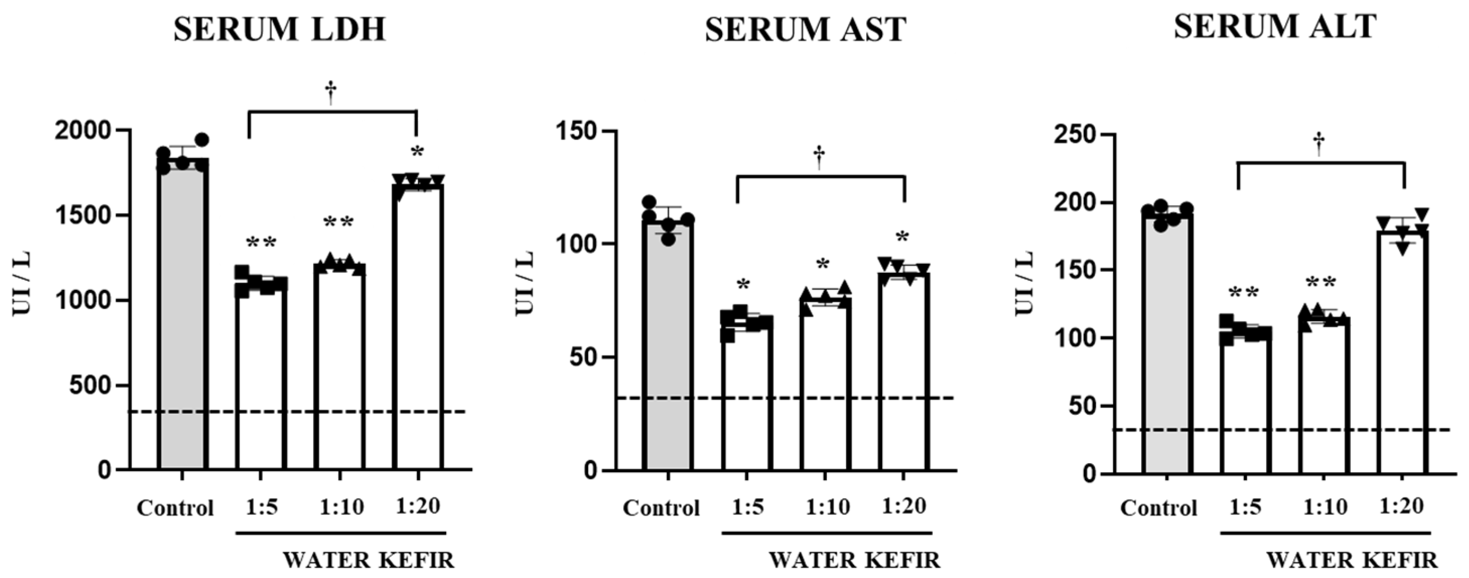
Figure 1. Effect of water kefir on serum biochemical markers. Mice were fed water kefir in different dilutions (1:5, 1:10 and 1:20) for 6 days and challenged on day 7 with an intraperitoneal injection of the viral pathogen-associated molecular pattern poly(I:C). Mice without water kefir treatment and challenged with poly(I:C) were used as control. The activities of serum enzymes alanine aminotransferase (ALT), aspartate aminotransferase (AST), and lactate dehydrogenase (LDH) were determined 2 days after TLR3 activation. The dotted lines indicate the parameter values under basal conditions. The results are expressed as mean ± SD. Significant differences were shown compared to the poly(I:C)-treated control group at p < 0.05 (*) or p < 0.01 (**). Significant differences were shown between the indicated groups at p < 0.05 (†).
Microbiolres 15 00083 g001
Figure 2. Effect of water kefir on intestinal damage. Mice were fed water kefir in different dilutions (1:5, 1:10 and 1:20) for 6 days and challenged on day 7 with an intraperitoneal injection of the viral pathogen-associated molecular pattern poly(I:C). Mice without water kefir treatment and challenged with poly(I:C) were used as control. The histopathological analysis of small intestine was performed 2 days after TLR3 activation. Hematoxylin-eosin stain of histological slices of small intestine, micrographs at 10× and 40× are shown. E: edema, I: inflammatory infiltration.
Microbiolres 15 00083 g002
Figure 3. Effect of water kefir on intestinal cytokines. Mice were fed water kefir in different dilutions (1:5, 1:10, and 1:20) for 6 days and challenged on day 7 with an intraperitoneal injection of the viral pathogen-associated molecular pattern poly(I:C). Mice without water kefir treatment and challenged with poly(I:C) were used as control. The concentration of intestinal IFN-β, IFN-γ, TNF-α, IL-15, IL-6, and IL-10 were determined 2 days after TLR3 activation. The dotted lines indicate the parameter values under basal conditions. The results are expressed as mean ± SD. Significant differences were shown compared to the poly(I:C)-treated control group at p < 0.05 (*) or p < 0.01 (**). Significant differences were shown between the indicated groups at p < 0.05 (†).
Microbiolres 15 00083 g003
Figure 4. Effect of water kefir on serum cytokines. Mice were fed water kefir in different dilutions (1:5, 1:10, and 1:20) for 6 days and challenged on day 7 with an intraperitoneal injection of the viral pathogen-associated molecular pattern poly(I:C). Mice without water kefir treatment and challenged with poly(I:C) were used as control. The concentration of serum IFN-β, IFN-γ, TNF-α, IL-15, IL-6, and IL-10 were determined 2 days after TLR3 activation. The dotted lines indicate the parameter values under basal conditions. The results are expressed as mean ± SD. Significant differences were shown compared to the poly(I:C)-treated control group at p < 0.05 (*) or p < 0.01 (**). Significant differences were shown between the indicated groups at p < 0.05 (†).
Microbiolres 15 00083 g004
Figure 5. Effect of water kefir on intestinal immune factors expression. Mice were fed water kefir in a dilution of 1:5 for 6 days and challenged on day 7 with an intraperitoneal injection of the viral pathogen-associated molecular pattern poly(I:C). Mice without water kefir treatment and challenged with poly(I:C) were used as control. The expressions of RNAseL, Mx1, TLR3, RIG-I, RAE-1, and NKG2D were determined 2 days after TLR3 activation. The dotted lines indicate the parameter values under basal conditions. The results are expressed as mean ± SD. Significant differences were shown compared to the poly(I:C)-treated control group at p < 0.05 (*) or p < 0.01 (**).
Microbiolres 15 00083 g005
Disclaimer/Publisher’s Note: The statements, opinions and data contained in all publications are solely those of the individual author(s) and contributor(s) and not of MDPI and/or the editor(s). MDPI and/or the editor(s) disclaim responsibility for any injury to people or property resulting from any ideas, methods, instructions or products referred to in the content.