Article

Biodegradation of Free Cyanide by a New Isolated Alkaliphilic Bacillus licheniformis Strain

by
Daniel Uribe-Ramírez
,
Eliseo Cristiani-Urbina
* and
Liliana Morales-Barrera
*
Departamento de Ingeniería Bioquímica, Escuela Nacional de Ciencias Biológicas, Instituto Politécnico Nacional, Av. Wilfrido Massieu s/n, Unidad Profesional Adolfo López Mateos, Delegación Gustavo A. Madero, Ciudad de México 07738, Mexico
*
Authors to whom correspondence should be addressed.
Microbiol. Res. 2024, 15(1), 33-49; https://doi.org/10.3390/microbiolres15010003
Submission received: 22 November 2023 / Revised: 14 December 2023 / Accepted: 18 December 2023 / Published: 21 December 2023

Abstract

:
Microbial treatment of free-cyanide-polluted wastewater is a cost-effective, efficient, and eco-friendly method. Free-cyanide-degrading microbial cultures were isolated from different sources using batch-enrichment culture techniques, with acetate as the carbon source. Five microbial cultures were able to tolerate and grow at 1500 mg/L free cyanide, which was used as the only nitrogen source under strongly alkaline conditions (pH = 11). Among them, one bacterial strain (B11) was selected for further study because of its high free-cyanide-biodegradation efficiency. Bacterial strain B11 was molecularly identified as Bacillus licheniformis CDBB B11. Free cyanide inhibited the growth rate of B. licheniformis CDBB B11 at initial cyanide concentrations >75 mg/L. Despite this, the bacterial strain demonstrated 100% cyanide-biodegradation efficiency at initial cyanide concentrations ranging from 25 to 75 mg/L, which decreased to 32% as the initial cyanide concentration increased from 75 to 1500 mg/L. Free-cyanide biodegradation corresponds to bacterial growth and ammonia accumulation in the culture medium. The alkaliphilic B. licheniformis CDBB B11 strain is a robust candidate for the detoxification of free-cyanide-laden wastewater because it tolerates and efficiently degrades free cyanide at concentrations of up to 1500 mg/L.

1. Introduction

Cyanide is produced in high concentrations by anthropogenic activities such as coke plants, petroleum refining, dye manufacturing, and the chemical, mining, and metal finishing industries [1,2,3,4,5,6]. Cyanide concentrations in the effluents of such industries are typically between 0.01 and 10 mg/L; however, some effluents from the electroplating industries have been reported to have cyanide levels greater than 100,000 mg/L [1].
Free cyanide (cyanide ion (CN) and hydrocyanic acid (HCN)), simple cyanide compounds that dissociate easily (e.g., KCN and NaCN), metal cyanide complexes, and organic cyanide complexes can all exist in wastewater [1,3,7,8,9,10].
Cyanides have multiple toxic effects on living organisms, the most important of which is their ability to inhibit cellular respiration by binding to iron at the catalytic center of mitochondrial cytochrome c oxidase, which prevents the transport of electrons from cytochrome a3 to oxygen. Consequently, aerobic oxidative metabolism and phosphorylation are compromised, resulting in hypoxia. Thus, cells can no longer produce ATP aerobically to meet their energy requirements or perform their cellular functions [11,12,13].
Cyanide contamination seriously threatens the environment, the health of humans and other living beings, and the sustainable development of people and society. As a result, it is essential to treat cyanide-containing wastewater to remove it. Various cyanide-detoxification strategies have been developed, including physical, chemical, and/or biological treatment methods. However, biological methods have economic and environmental advantages over conventional physical and chemical methods because they are less expensive, more efficient, and do not add or create new persistent toxic chemicals, as seen in other treatment methods; therefore, biological methods are environmentally friendly [10,14,15,16].
Although cyanide is toxic to microorganisms at low concentrations, it has been reported that several bacteria, filamentous fungi, yeasts, and algae degrade it through hydrolytic, oxidative, reductive, and/or substitution/transfer pathways, allowing microbial cells to use cyanide as a carbon and/or nitrogen source for growth [1,3,8,17].
Most research on cyanide biodegradation has been conducted under acidic or neutral pH conditions. However, at these pH conditions, almost all cyanide (≥99%) is found as the highly toxic and volatile HCN (pKa = 9.3 at 20 °C) that evaporates easily, causing environmental and health risks to humans and other living beings [3,18,19,20]. Furthermore, under these pH conditions, the removal of cyanide from aqueous solutions is due to volatilization (a physical mechanism) and the action of microorganisms (a biological mechanism) [3]. Consequently, there is great interest in the biological treatment of cyanide under alkaline conditions, preferably at pH > 10, at which most of the free cyanide in solution is in the form of the cyanide anion (CN) (≥99%) and cyanide loss through volatilization is limited [3,19,21]. However, the microbial biodegradation of cyanide in highly alkaline solutions has been a great challenge in the application of bioremediation techniques because the rate and extent of cyanide biodegradation both decrease significantly at pH values higher than 10 [14].
Likewise, most microorganisms capable of assimilating cyanide can only use it as a nitrogen source, but not as a carbon source; therefore, in most cyanide-biodegradation studies, an external source of carbon, often a reducing sugar such as glucose, has been provided for cell growth [3,19,20,21]. However, cyanide can react with the aldehyde and ketone groups of reducing sugars (Kiliani–Fischer reaction) to form cyanohydrins, thus masking the microbial biodegradation of cyanide [19,22]. In this regard, cyanide removal from aqueous solutions under aerobic and anaerobic conditions in the presence of reducing sugars is independent of microbial metabolism [21]. Therefore, in cyanide-biodegradation studies, the use of aldehyde- and ketone-containing compounds should be avoided as carbon sources for the growth of microorganisms [19,23]. It is, therefore, critical to isolate microorganisms capable of degrading cyanide in strongly alkaline aqueous solutions (pH ≥ 10) and to use simple carbon sources devoid of aldehyde or ketone groups to ensure that cyanide removal is due to biological activity rather than abiotic factors. Despite its importance, information on this topic is scarce.
As a result, we focused our efforts on isolating microbial strains capable of tolerating and biodegrading high free-cyanide concentrations under highly alkaline (pH = 11.0) conditions using acetate as the carbon and energy source. Subsequently, the microbial strain with the highest cyanide-biodegradation efficiency was selected and identified at the molecular level. Finally, with the selected microbial strain, kinetic studies of cell growth, cyanide biodegradation, and ammonia accumulation in culture media were performed at various initial concentrations of cyanide.

2. Materials and Methods

2.1. Isolation Sources of Microorganisms

We used soil samples exposed to industrial wastewater, agrochemicals, hydrocarbons, heavy metals, gold and silver mining industry tailings, and activated sludge (Table 1) as sources of microorganisms capable of degrading cyanide.

2.2. Microbial Enrichment Cultures

Enrichment cultures were prepared in mineral medium with the following composition per liter of 100 mM phosphate buffer, pH 11.0: MgSO4·7H2O, 0.018 mg; CoCl2, 0.013 mg; CaCl2, 0.004 mg; ZnSO4, 0.004 mg; Na2Mo4·2H2O, 0.002 mg; MnSO4·H2O, 0.01 mg; NiSO4·6H2O, 0.01 mg; CuSO4·7H2O, 0.01 mg; and FeCl3·6H2O, 0.005 mg [24]. 1 g/L sodium acetate was used as a carbon and energy source and 20 mg/L free cyanide (as sodium cyanide) as the only nitrogen source. All reagents were obtained from JT Baker (Avantor Performance Materials, Inc., Xalostoc, Estado de México, Mexico) and Sigma-Aldrich (Sigma-Aldrich, Co., St. Louis, MO, USA). Because cyanide has a boiling point of 25.7 °C, the culture media containing cyanide were sterilized by microfiltration using Whatman® GF/F glass microfiber filters with a pore size of 0.7 μm (Cytiva™, Amersham, UK). The pH of the culture media was tested on a regular basis throughout the investigation, and when necessary, it was corrected to 11.0 by adding 0.1 M NaOH.
For enrichment cultures, isolation-source samples (0.5 g) were added to 250 mL Erlenmeyer flasks containing 50 mL of sterile culture medium with an initial cyanide anion concentration of 20 mg/L. The flasks were incubated at room temperature (approximately 20 ± 2 °C) at 120 rpm for 5 days. Aliquots of 5 mL of culture samples were used to successively inoculate flasks containing fresh culture medium with an initial free-cyanide concentration of 20 mg/L. The samples were incubated under the aforementioned conditions until the degradation of cyanide by the microbial cultures became evident upon observing cell growth. This procedure was repeated, but the initial cyanide concentration was gradually increased to 1500 mg/L (50, 100, 200, 500, 1000, and 1500 mg/L).
A preliminary study of cyanide degradation was conducted using the microbial cultures obtained after the enrichment period to select the microbial culture that exhibited the highest cyanide-degradation efficiency. These studies were performed in Erlenmeyer flasks containing mineral medium supplemented with 1 g/L of sodium acetate, 1500 mg/L of free cyanide, and a 0.1 g/L initial biomass concentration of the microbial culture. Simultaneously, a biomass-free control was used to detect potential cyanide losses due to volatilization and/or adsorption onto the glass. The flasks were incubated for 210 h at 20 ± 2 °C and 120 rpm. The residual cyanide concentration in each of the tested microbial cultures was evaluated at the end of the incubation period, and the cyanide-degradation efficiency was assessed. The microbial culture with the highest cyanide-degradation efficiency was selected for further investigation.

2.3. Microbial Isolation and Morphological Characterization

The selected microbial culture was 10-fold serially diluted up to 10−6 and 0.2 mL aliquots were dispersed in triplicate on nutrient agar plates. After incubating the plates at 30 °C for 48 h, single bacterial colonies were collected from the plates and the streak plate method was used to ensure purity.
Morphological observations were recorded on the surface appearance, shape, color, edge, elevation, opacity, and texture or consistency of colonies of pure cultures of the isolated bacteria grown on nutrient agar at 30 °C for 48 h. Microscopic parameters examined included Gram staining and cell shape [25,26].

2.4. Molecular Identification of the Selected Bacterial Isolate

Molecular identification of the selected bacterial isolate was conducted by the National Collection of Microbial Strains and Cell Cultures of the Center for Research and Advanced Studies of the National Polytechnic Institute (CINVESTAV-IPN) based on the sequence of a 16S ribosomal RNA fragment.
Briefly, the genomic DNA of the selected bacterial strain was extracted using the ZR Fungal/Bacterial DNA MiniPrep kit (Zymo Research, Irvine, CA, USA). From the total DNA obtained, the 16S rRNA gene was amplified with the universal oligonucleotides 27F (5′ AGAGTTTGATCMTGGCTCAG 3′) and 1492R (5′ TACGGYTACCTTGTTACGACTT 3′) according to the method described by Lane [27]. The integrity of the expected fragment (1500 nucleotides) was confirmed using 1% (w/v) agarose gel electrophoresis at 100 V for 1 h. The 16S rRNA gene was sequenced using an ABI Prism 310 Genetic Analyzer PE (Applied Biosystems, San Francisco, CA, USA) to obtain two sequences for each oligonucleotide. The sequences were edited manually using Chromas 2.4 (2012) software (Technelysium Pty. Ltd., Brisbane, Australia) to form a consensus sequence. To detect commonalities between biological sequences, consensus sequences were compared using the nucleotide BLAST program of the National Center for Biotechnology Information (NCBI, Bethesda, MD, USA). The consensus sequence was aligned with the BLAST search results using the Clustal W algorithm included within MEGA software (version 6.0; Mega Software Technologies, Delhi, India) [28] to create a dendrogram using the neighbor-joining method to discover commonalities between sequences of defined species types.

2.5. Effect of Carbon Source (Acetate) Concentration on Free-Cyanide Biodegradation by the Selected Bacterial Strain

To investigate the influence of the initial acetate concentration on free-cyanide degradation by the selected bacterial strain, four initial acetate concentrations (1, 2, 4, and 6 g/L) were tested, and their effects on the degradation of two initial free-cyanide concentrations (400 and 1500 mg/L) were determined. The tests were performed in 500-mL Erlenmeyer flasks containing 100 mL of culture medium, with an initial biomass concentration of 0.1 g/L. The flasks were incubated for 120 h at 20 ± 2 °C and 120 rpm. Bacterial cultures were sampled periodically, and their biomass and residual cyanide concentrations were determined.

2.6. Kinetic Study of Cell Growth, Cyanide Biodegradation, and Ammonia Accumulation

The performance assessment of a biological cyanide-degradation process requires a kinetic evaluation of the microorganisms’ proliferation and capacity to degrade various levels of cyanide as a substrate [6]. Therefore, kinetic studies of cell growth and cyanide biodegradation by selected bacterial strain were performed at various initial cyanide concentrations. Because ammonia (NH3) is a metabolic product of the biodegradation of cyanide through hydrolytic, reductive, oxidative, and substitution/transfer pathways [3,29], the presence and concentration of ammonia in the cultures of selected bacterial strain were evaluated in this study.
Kinetic studies were performed in 1000-mL Erlenmeyer flasks containing 200 mL of mineral medium supplemented with sodium acetate (1 g/L) and free cyanide at initial concentrations ranging from 0 to 1500 mg/L (0, 25, 50, 75, 100, 200, 400, 600, 800, 1000, and 1500 mg/L). The flasks were inoculated with a small volume of concentrated cell suspension of the selected bacterial strain, resulting in an initial biomass concentration of 0.1 g/L for all bacterial cultures. To detect probable cyanide losses due to volatilization and/or adsorption on the glass, a biomass-free control was run concurrently for each initial cyanide concentration assayed. The flasks were shaken in an incubator shaker for 10 days at 120 rpm and 20 ± 2 °C. Bacterial culture samples were collected every 24 h, and biomass, residual cyanide, and ammonia concentrations were determined.

2.7. Evaluation of Kinetic Parameters of Cell Growth, Cyanide Biodegradation, and Ammonia Accumulation

The maximum specific growth rate and overall biomass volumetric productivity were calculated using Equations (1) and (2), respectively:
μ m a x = l n x 2 l n ( x 1 ) t 2 t 1
r x = x f x 0 t f t 0
where μmax is the maximum specific growth rate (1/h); rx is the overall volumetric productivity of biomass (g/L·h); x1 and x2 are the biomass concentrations at the beginning and end of the exponential growth phase, respectively (g/L); t1 and t2 are the incubation times at the beginning and end of the exponential growth phase, respectively (h); x0 and xf are the biomass concentrations at the beginning and end of incubation period, respectively (g/L); and t0 = 0 h and tf are the initial and final incubation times (h), respectively.
The cyanide-biodegradation performance of the selected bacterial culture was assessed using Equations (3) and (4), which calculate the efficiency and volumetric rate of cyanide biodegradation, respectively:
E = S 0 S f S 0 × 100
r S = S f S 0 t f t 0  
where E is the overall cyanide-biodegradation percentage (%); rS is the overall volumetric biodegradation rate of free cyanide (mg/L·h); and S0 and Sf are the cyanide concentrations at the beginning and end of incubation period (g/L), respectively.
The overall volumetric rate of ammonia accumulation in the culture medium was calculated as follows:
r p = p f p 0 t f t 0
where rp is the overall volumetric rate of ammonia accumulation (mg/L·h), p0 is the ammonia concentration at the initial time of incubation (g/L) and pf is the ammonia concentration at the final incubation time (g/L).

2.8. Statistical Analysis

All experiments were performed at least three times to statistically evaluate the data obtained with two-way ANOVA using Tukey’s multiple comparison method and GraphPad Prism version 8.0.4 program (GraphPad Software, Boston, MA, USA).

2.9. Analytical Methods

The biomass concentration was determined using the dry weight of the cell mass. The culture samples were filtered through pre-weighed Whatman GF/F glass microfiber filters (Cytiva™, Amersham, UK) with a pore size of 0.7 μm, which were rinsed twice with sterile distilled water and subsequently dried to constant weight at 60 °C. The biomass concentration was calculated using the weight differential of the microfiber filter before and after filtration and drying.
The filtrates were used to measure the pH using a potentiometer and to quantify the concentrations of residual cyanide and ammonia using Hach Water Analysis Handbook techniques 8027 and 8038 [30].

3. Results and Discussion

3.1. Screening and Selection of Cyanide-Degrading Microbial Cultures

It is crucial to have acclimatized and adapted microbial cultures to achieve strong cyanide-degrading efficiency and low mortality values in microbial cells. The current study involved the enrichment of microbial cultures capable of degrading cyanide under alkaline conditions.
Microbial cultures capable of growing under strongly alkaline conditions (pH 11) using 20 mg/L free cyanide as the sole nitrogen source were obtained from all 13 isolation sources used in the present study (Table 2). These microbial cultures were named by prefixing the letter B, followed by a number corresponding to the isolation-source number (for example, B1 corresponds to the name of the microbial culture that was isolated from isolation source 1, soil contaminated with wastewater from the pulp and paper industry).
However, as the initial free-cyanide concentration increased from 20 to 1500 mg/L during the enrichment period of alkaliphilic cyanide-degrading microorganisms, the number of microbial cultures that could tolerate and grow on cyanide decreased from 13 to 5 (Table 2). The five final microbial cultures were labeled B1, B4, B8, B11, and B12 (Table 2).
The ability of these five microbial cultures (B1, B4, B8, B11, and B12) to biodegrade free cyanide from aqueous solutions was investigated using an initial free-cyanide concentration of 1500 mg/L (Table 3). To detect possible abiotic cyanide losses, a biomass-free control (abiotic control) was also tested.
Table 3 shows that, despite the fact that the experiments were performed at pH 11, where most of the cyanide in the aqueous solution is in the form of cyanide anions and the loss of cyanide through volatilization is limited, 28.7 mg/L of cyanide was not recovered from the biomass-free control, representing a cyanide loss of approximately 1.91%. This loss of cyanide can be attributed to the presence of a small amount of hydrocyanic acid in solution [31]. Few biological cyanide-removal studies have used biomass-free controls to quantify abiotic cyanide losses; however, Tiong et al. [32] and Vallenas-Arévalo et al. [33] showed 6-11% and 43% abiotic cyanide losses at pH 10, respectively.
All microbial cultures were able to remove more free cyanide (173–504.5 mg/L; 11.5–33.6%) than was lost abiotically (28.7 mg/L; 1.91%) (Table 3). Similarly, it was found that the microbial culture B11 degraded the most cyanide (504.5 mg/L; 33.6%), followed by the B1 culture (348.6 mg/L; 23.24%), with a significant difference between both cultures (p < 0.05). These microbial cultures removed more cyanide than those reported in other studies. Mekuto et al. [34] reported that Pseudomonas aeruginosa removed 200 mg/L of cyanide at a pH of 10, and Karamba et al. [24] reported that Serratia marcescens removed 98 mg/L of cyanide at pH 7, using glucose as a carbon and energy source.
Cyanide is highly toxic to microorganisms, and most are unable to survive at high cyanide concentrations [8]. This investigation yielded several microbial cultures capable of tolerating and degrading a high cyanide concentration (1500 mg/L); therefore, these cultures have the potential to be exploited in the bioremediation of cyanide-laden wastewater. Furthermore, the results showed that microbial culture B11 had the greatest capacity for cyanide degradation under the strongly alkaline conditions tested in this study; therefore, this microbial culture was selected for subsequent studies.

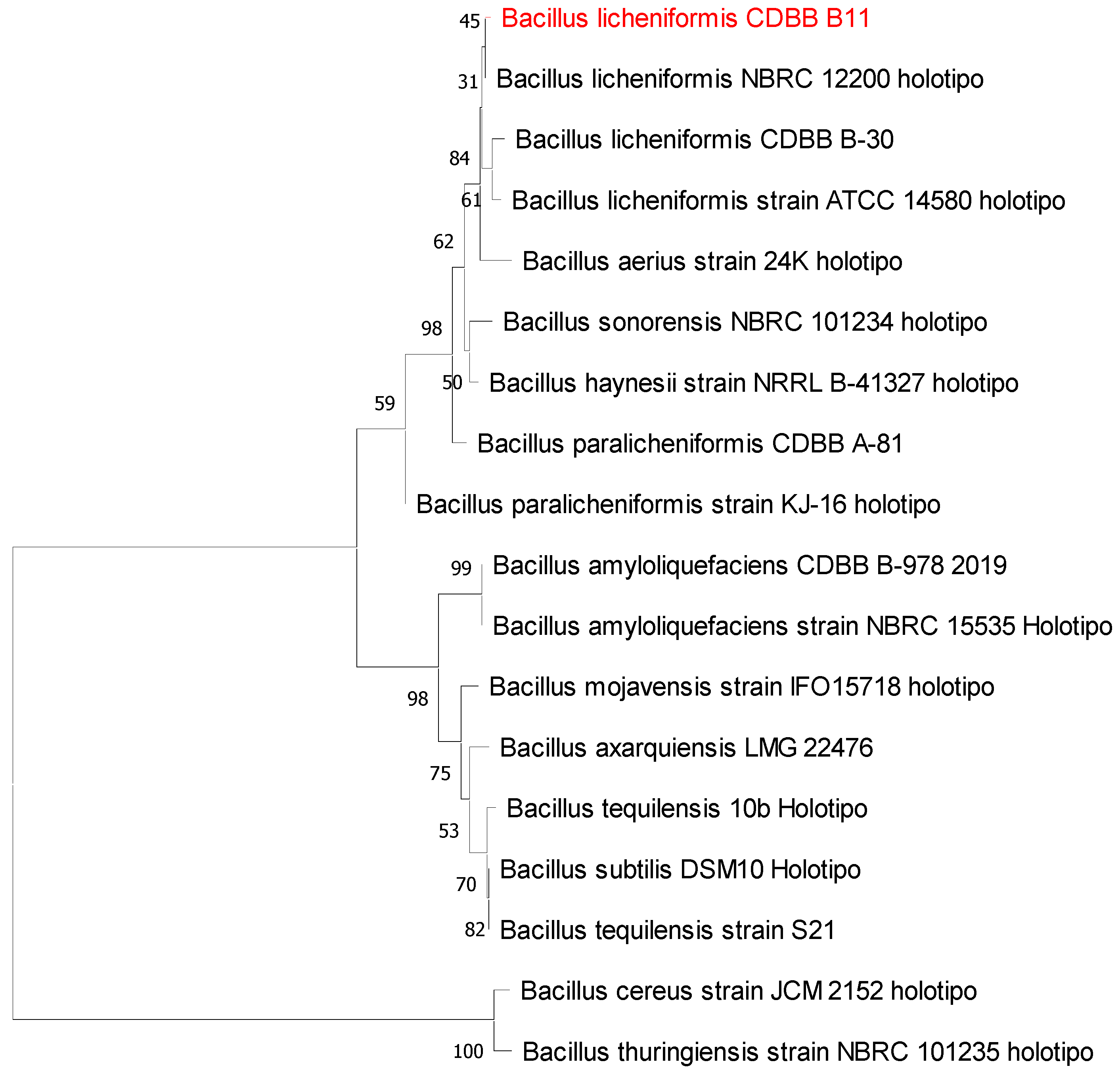
3.2. Isolation, Morphological Characterization, and Molecular Identification of Selected Cyanide-Degrading Bacterium

Microscopic observations showed that microbial culture B11 consisted only of bacteria with identical microscopic morphology. Similarly, only bacterial colonies with identical morphologies were recovered after serial 10-fold dilutions and streak plating on nutrient agar medium. These results suggest that a pure bacterial culture was isolated under a selective pressure of 1500 mg/L free cyanide. The purity of the bacterium was confirmed by careful examination of bacterial colonies and their microscopic morphologies.
The colony morphology and cell microscopic characteristics of the B11 isolate grown on nutrient agar for 48 h at 30 °C are shown in Figure 1 and Table 4.
These characteristics are consistent with those reported for the genus Bacillus [35,36].
Bacterial isolate B11 was molecularly identified as Bacillus licheniformis (NCBI Sequence Viewer, accession number: MG977020.1) by the 16S rRNA gene region (1465 nt), showing a sequence coverage of 100% and a sequence similarity of 100%, and it will be further referred to as Bacillus licheniformis CDBB B11. Figure 2 shows the dendrogram obtained for the strain CDBB B11.
Bacillus licheniformis is a bacterium commonly found in soil. It forms part of the subtilis group along with Bacillus subtilis and Bacillus pumilus. It can grow in alkaline conditions and at high temperatures (optimal growth temperature = 50 °C but can survive at much higher temperatures) [37].
Previous studies on the cyanotrophic potential of some Bacillus species have been performed using reducing sugar as a carbon and energy source and/or culture media at neutral pH and/or at high temperatures (>40 °C) [38,39,40]. However, all of these culture conditions lead to the volatilization or spontaneous transformation of cyanide [3,8,19,20,21].
Similarly, some Bacillus species have been reported to be capable of degrading cyanide in pure culture [14,33,41] or in a consortium [42,43] in culture media containing glycerol or whey as carbon and energy sources, or in Luria Bertani medium (15 g/L peptone + 5 g/L yeast extract + 10 g/L NaCl) at pH 9.5–10. Using acetate as a carbon and energy source, the Bacillus licheniformis CDBB B11 strain isolated in this study was able to tolerate and degrade high concentrations of free cyanide (1500 mg/L) under conditions that were at least ten times more alkaline (pH 11) than previously reported with other Bacillus strains.

3.3. Effect of Acetate Concentration on Cyanide Biodegradation by B. licheniformis CDBB B11

The concentration of carbon and energy sources for microbial growth can have a significant influence on cyanide biodegradation [7]. The effect of acetate concentration on cell growth and cyanide biodegradation by B. licheniformis CDBB B11 was investigated using four different concentrations of acetate (1, 2, 4, and 6 g/L) and two different initial cyanide concentrations (400 and 1500 mg/L). The maximum increase in cell mass and cyanide-biodegradation efficiency are shown in Table 5.
At the two initial cyanide concentrations assayed, the increase in acetate concentration caused an increase in biomass production (maximum increase of cell mass) and cyanide-biodegradation efficiency. These results are consistent with those of Guadalima and Monteros [7], who found that increasing sucrose concentrations improved the efficiency of cyanide degradation by a microbial consortium at pH 10.5.
However, in the current study, the greatest difference in cyanide-degradation efficiency was reached between acetate concentrations of 6 and 1 g/L and was 12 and 9% for the initial cyanide concentrations of 400 and 1500 mg/L, respectively. These differences in cyanide-degradation efficiency are minor and do not justify the use of culture media containing more than 1 g/L acetate. Additionally, in the biological treatment of cyanide-laden wastewater, the addition of carbon and energy sources should be minimized because it would impair the economics of the process. Therefore, subsequent experiments were performed using 1 g/L of acetate.

3.4. Kinetic Study of Cell Growth, Cyanide Biodegradation, and Ammonia Accumulation by Bacillus licheniformis CDBB B11 at Different Initial Free-Cyanide Concentrations

It is crucial to evaluate the kinetic performance of a microbial culture for cyanide biodegradation at various initial cyanide concentrations because low cyanide concentrations can limit cell growth and cyanide biodegradation due to a lack of a nitrogen source. As the cyanide concentration increases, so do its toxic and inhibitory effects, limiting microbial cell growth and cyanide biodegradation [44,45].
Batch cultures of Bacillus licheniformis CDBB B11 were grown in culture media containing 1 g/L acetate at a pH of 11.0, and the initial cyanide concentration was varied from 0 to 1500 mg/L to evaluate the kinetic performance of the bacterium at different initial cyanide concentrations.
Figure 3 shows the effects of varying initial cyanide concentrations on the kinetic profile of cell growth, maximum specific growth rate, and overall biomass volumetric productivity of B. licheniformis CDBB B11.
When no cyanide was added to the culture medium (initial cyanide concentration = 0 mg/L), the biomass concentration decreased over time (Figure 3A). Cell lysis occurred because the bacterium lacked a nitrogen source (cyanide) for growth and reproduction. At higher initial cyanide concentrations, the cell concentration increased as the cyanide concentration increased to 800 mg/L, which could be attributed to cyanide providing the amount of nitrogen required for bacterial growth. At initial cyanide concentrations greater than 800 mg/L, the cell concentrations were similar (p > 0.05; Figure 3A). The formation of spores was observed in all cyanide-containing cultures after 24 h of incubation.
The specific growth rate of B. licheniformis CDBB B11 increased as the initial free-cyanide concentration increased from 0 to 75 mg/L (Figure 3B). However, at higher initial concentrations, cyanide inhibited the growth of the bacterium and caused its specific growth rate to decrease linearly with respect to the initial cyanide concentration. The intercept of this straight line along the x-axis provides an inhibition parameter (Ki) of 2581 mg cyanide/L, which represents the maximum initial cyanide concentration above which B. licheniformis CDBB B11 does not grow in batch cultures; that is, Ki is the initial cyanide concentration at which the specific growth rate of the bacterium is zero. An experiment was conducted at an initial cyanide concentration of 2581 mg/L, and after 240 h of incubation, no growth was detected, confirming the Ki value.
The overall biomass volumetric productivity increased as the initial cyanide concentration increased from 25 to 800 mg/L. At higher initial concentrations, biomass productivity remained practically constant, with a value of approximately 1.9 mg/L·h (Figure 3C).
Figure 4 shows the effect of the initial cyanide concentration on the time-dependent variation of cyanide biodegradation and cyanide-biodegradation efficiency, as well as on the overall volumetric rate of cyanide biodegradation by B. licheniformis CDBB B11. The data in Figure 4 are the results after deducting the abiotic losses of cyanide (≤1.91%),and represent the data for free cyanide degraded by the biological activity of the bacterial strain.
The amount of cyanide degraded by B. licheniformis CDBB B11 depended on the incubation time and initial cyanide concentration (Figure 4A). In general, cyanide biodegradation by the bacterial strain increased gradually with incubation time, reaching a maximum value that remained constant thereafter.
Similarly, increasing the initial cyanide concentration from 25 to 1000 mg/L increased the amount of free cyanide biodegraded by B. licheniformis CDBB B11 from 25 to 477.65 mg/L. However, no significant changes were found in the amount of free cyanide biodegraded when initial cyanide concentrations of 1000 and 1500 mg/L were used at incubation times equal to or greater than 144 h (p > 0.05).
The growth and cyanide-biodegradation patterns of Bacillus licheniformis CDBB B11 (Figure 3A and Figure 4A) clearly indicate that biomass concentration and cyanide biodegradation are directly related because the cultures with the highest cell concentrations also biodegraded the most cyanide.
The cyanide-biodegradation efficiency of Bacillus licheniformis CDBB B11 increased as the incubation time increased until it reached a maximum value, which remained constant thereafter (Figure 4B). Similarly, Bacillus licheniformis CDBB B11 was able to biodegrade all free cyanide present in the culture media when the initial cyanide concentration was less than 100 mg/L, and the free-cyanide-biodegradation efficiency was therefore 100%. However, the free-cyanide-biodegradation efficiency decreased from 100% to 32% as the initial free-cyanide concentration increased from 75 to 1500 mg/L. These results are consistent with those of Akinpelu et al. [46], who found that the free-cyanide-removal efficiency of Fusarium oxysporum decreased from 77% to 58% when the initial free-cyanide concentration increased from 100 to 200 mg/L. Similarly, the free-cyanide-removal efficiency of Trametes versicolor decreased from approximately 80% to 30% as the initial cyanide concentration increased from 25 to 400 mg/L [47]. Aerococcus viridans T1 removed 86.7% of free cyanide at an initial concentration of 150 mg/L and 84.1% of free cyanide at an initial concentration of 200 mg/L [5]. Furthermore, a bacterial consortium isolated from gold-mine-tailing sediments demonstrated a cyanide-removal efficiency greater than 90% in the initial cyanide concentration range of 5–120 mg/L [48].
The alkaliphilic Bacillus licheniformis strain isolated in this study tolerated high concentrations of free cyanide (1500 mg/L), and its cyanide-biodegradation capacity was similar to or higher than that of other alkaliphilic Bacillus strains. Bacillus sp. CN-22 [41], Bacillus subtilis [14], and a Bacillus consortium [42] removed 96.69%, 87%, and 60% of free cyanide from culture media with initial cyanide concentrations of 200, 500, and 400 mg/L at pH values of 10.3, 10, and 9.5, respectively. Furthermore, Vallenas-Arévalo et al. [33] reported a cyanide-removal efficiency of 99% at an initial concentration of 500 mg/L and pH 10 using a B. licheniformis strain isolated from gold-mine tailings; however, approximately 43% of this cyanide-removal efficiency was due to abiotic factors, and only 56% was removed by the bacterial strain.
The overall volumetric biodegradation rate of cyanide increased from 0.0986 to 1.99 mg/L·h as the initial cyanide concentration increased from 25 to 1000 mg/L and remained practically constant at an initial cyanide concentration of 1500 mg/L. This pattern might be attributed to the fact that higher cell densities were attained at higher starting cyanide concentrations, which led to greater biodegradation of cyanide from the culture medium.
Cyanide-degrading microorganisms can use cyanide as a nitrogen source, converting it into less toxic or non-toxic products, and ammonia (NH3) is one of the main products of cyanide bacterial biodegradation [3,5,48,49,50]. Therefore, in the present study, ammonia production was investigated with the aim of confirming cyanide biodegradation by B. licheniformis CDBB B11, because this anion was the only source of nitrogen available in the culture media.
Ammonia accumulation (Figure 5A) was detected in all cyanide-containing culture media from the beginning to the end of growth (Figure 3A) and cyanide-biodegradation kinetics were observed (Figure 4A). These results indicated that these three parameters, cell growth, cyanide biodegradation, and ammonia accumulation, are closely related.
As the initial free-cyanide concentration increased from 25 to 1000 mg/L, so the ammonia concentration in the culture media also increased from 4 to 90 mg/L. The latter ammonia concentration was similar to that attained at an initial cyanide concentration of 1500 mg/L (Figure 5A). Other studies have also found that the ammonia concentration increases with an increase in the initial cyanide concentration [51,52].
Although the amount of cyanide biodegraded by the bacterial strain (Figure 4A) and ammonia accumulation in the culture media (Figure 5A) increased with increasing initial cyanide concentrations, the cyanide-biodegradation efficiency decreased (Figure 4B). This may be attributed to the toxic effects of cyanide and the fact that ammonia inhibits or suppresses the synthesis of enzymes responsible for cyanide degradation, making the biodegradation process slower and inefficient [46,52,53,54].
With respect to the initial cyanide concentration, both the overall volumetric rate of ammonia accumulation (Figure 5B) and overall volumetric rate of cyanide biodegradation (Figure 4B) increased with increasing initial cyanide concentration from 25 to 1000 mg/L and subsequently remained constant at 1500 mg/L. These results support the hypothesis that cyanide biodegradation and ammonia accumulation are closely related.

4. Conclusions

The present study focused on isolating microbial cultures that could tolerate and grow in high free-cyanide concentrations using this toxic compound as the only supply of nitrogen and acetate as the carbon and energy source under strongly alkaline conditions (pH = 11). Bacterial strain B11, molecularly identified as Bacillus licheniformis CDBB B11, was selected for further research based on its high cyanide-biodegradation efficiency. Kinetic characterization of B. licheniformis CDBB B11 cell growth and free-cyanide biodegradation showed that this bacterium demonstrated remarkable tolerance and effective free-cyanide biodegradation at initial concentrations of up to 1500 mg/L. Furthermore, B. licheniformis CDBB B11 produced ammonia as a by-product of cyanide biodegradation. Bacterial growth and the breakdown of cyanide occurred simultaneously with ammonia accumulation in the culture media. The alkaliphilic B. licheniformis CDBB B11 strain examined in this study has the potential to bioremediate and detoxify free-cyanide-laden wastewaters and other waters. This strain clearly has the potential for high cyanide-biodegradative activity.

Author Contributions

Conceptualization, E.C.-U. and L.M.-B.; methodology, D.U.-R., L.M.-B. and E.C.-U.; software, D.U.-R.; formal analysis, D.U.-R., L.M.-B. and E.C.-U.; investigation, D.U.-R.; resources, L.M.-B. and E.C.-U.; data curation, D.U.-R.; writing—original draft preparation, D.U.-R. and E.C.-U.; writing—review and editing, D.U.-R., L.M.-B. and E.C.-U.; visualization, D.U.-R. and L.M.-B.; supervision, E.C.-U.; project administration, E.C.-U.; funding acquisition, L.M.-B. and E.C.-U. All authors have read and agreed to the published version of the manuscript.

Funding

This research was funded by the Instituto Politécnico Nacional, Secretaría de Investigación y Posgrado under Grant Numbers 20231441 and 20230270.

Institutional Review Board Statement

Not applicable.

Informed Consent Statement

Not applicable.

Data Availability Statement

All relevant data are within the paper.

Acknowledgments

The authors are grateful for the technical support provided by the Colección Nacional de Cepas Microbianas y Cultivos Celulares (“National Collection of Microbial Strains and Cell Cultures”) of the Centro de Investigación y de Estudios Avanzados, IPN (CINVESTAV, IPN). The CONAHCYT (Consejo Nacional de Humanidades, Ciencias y Tecnologías) awarded a graduate scholarship to the co-author D.U.-R. E.C.-U. and L.M.-B. hold grants from EDI-IPN, COFAA-IPN, and SNI-CONAHCYT.

Conflicts of Interest

The authors declare no conflicts of interest.

References

  1. Dwivedi, N.; Balomajumder, C.; Mondal, P. Applications of microorganisms in biodegradation of cyanide from wastewater. In Advances in Microbial Biotechnology. Current Trends and Future Prospects; Kumar, P., Patra, J.J., Chandra, P., Eds.; Apple Academic Press, Inc.: Oakville, ON, Canada, 2019. [Google Scholar]
  2. Zuhra, K.; Szabo, C. The two faces of cyanide: An environmental toxin and a potential novel mammalian gasotransmitter. FEBS J. 2022, 289, 2481–2515. [Google Scholar] [CrossRef] [PubMed]
  3. Alvillo-Rivera, A.; Garrido-Hoyos, S.; Buitrón, G.; Thangarasu-Sarasvathi, P.; Rosano-Ortega, G. Biological treatment for the degradation of cyanide: A review. J. Mater. Res. Technol. 2021, 12, 1418–1433. [Google Scholar] [CrossRef]
  4. Alvillo-Rivera, A.; Garrido-Hoyos, S.; Buitrón, G. Cyanide treatment of mining tailings using suspended biomass and moving bed biomass reactors. Environ. Sci. Pollut. Res. 2022, 29, 37458–37470. [Google Scholar] [CrossRef] [PubMed]
  5. Jiang, W.; Lu, Y.; Feng, Z.; Yu, H.; Ma, P.; Zhu, J.; Wang, Y.; Sun, J. Biodegradation of cyanide by a new isolated Aerococcus viridans and optimization of degradation conditions by response surface methodology. Sustainability 2022, 14, 15560. [Google Scholar] [CrossRef]
  6. Manogaran, M.; Yakasai, H.M.; Karamba, K.I. Kinetics of cyanide degradation by Azotobacter vinelandii. J. Biochem. Microbiol. Biotechnol. 2019, 7, 38–42. [Google Scholar] [CrossRef]
  7. Guadalima, M.P.G.; Monteros, D.A.N. Evaluation of the rotational speed and carbon source on the biological removal of free cyanide present on gold mine wastewater, using a rotating biological contactor. J. Water Process Eng. 2018, 23, 84–90. [Google Scholar] [CrossRef]
  8. Cosmos, A.; Erdenekhuyag, B.-O.; Yao, G.; Li, H.; Zhao, J.; Laijun, W.; Luy, X. Principles and methods of bio-detoxification of cyanide contaminants. J. Mater. Cycles Waste Manag. 2020, 22, 939–954. [Google Scholar] [CrossRef]
  9. Luque-Almagro, V.M.; Cabello, P.; Sáez, L.P.; Olaya-Abril, A.; Moreno-Vivián, C.; Roldán, M.D. Exploring anaerobic environments for cyanide and cyano-derivatives microbial degradation. Appl. Microbiol. Biotechnol. 2018, 102, 1067–1074. [Google Scholar] [CrossRef]
  10. Razanamahandry, L.C.; Onwordi, C.T.; Saban, W.; Bashir, A.K.H.; Mekuto, L.; Malenga, E.; Manikandan, E.; Fosso-Kankeu, E.; Maaza, M.; Ntwampe, S.K.O. Performance of various cyanide degrading bacteria on the biodegradation of free cyanide in water. J. Hazard. Mater. 2019, 380, 120900. [Google Scholar] [CrossRef]
  11. Bhattacharya, R.; Flora, S.J.S. Chapter 19—Cyanide toxicity and its treatment. In Handbook of Toxicology of Chemical Warfare Agents; Gupta, R.C., Ed.; Academic Press: San Diego, CA, USA, 2009; pp. 255–270. [Google Scholar] [CrossRef]
  12. Terada, A.; Komatsu, D.; Ogawa, T.; Flamandita, D.; Sahlan, M.; Nishimura, M.; Yohda, M. Isolation of cyanide-degrading bacteria and molecular characterization of its cyanide-degrading nitrilase. Biotechnol. Appl. Biochem. 2022, 69, 183–189. [Google Scholar] [CrossRef]
  13. Maduh, E.U.; Turek, J.J.; Borowitz, J.L.; Rebar, A.; Isom, G.E. Cyanide-induced neurotoxicity: Calcium mediation of morphological changes in neuronal cells. Toxicol. Appl. Pharmacol. 1990, 103, 214–221. [Google Scholar] [CrossRef] [PubMed]
  14. Rosario, C.G.A.; Vallenas-Arévalo, A.T.; Arévalo, S.J.; Espinosa, D.C.R.; Tenório, J.A.S. Biodegradation of cyanide using a Bacillus subtilis isolated from artisanal gold mining tailings. Braz. J. Chem. Eng. 2023, 40, 129–136. [Google Scholar] [CrossRef]
  15. Sharma, M.; Akhter, Y.; Chatterjee, S. A review on remediation of cyanide containing industrial wastes using biological systems with special reference to enzymatic degradation. World J. Microbiol. Biotechnol. 2019, 35, 70. [Google Scholar] [CrossRef] [PubMed]
  16. Shin, D.; Park, J.; Park, H.; Lee, J.-C.; Kim, M.-S.; Lee, J. Key microbes and metabolic potentials contributing to cyanide biodegradation in stirred-tank bioreactors treating gold mining effluent. Miner. Process. Extr. Metall. Rev. 2020, 41, 85–95. [Google Scholar] [CrossRef]
  17. Sáez, L.P.; Cabello, P.; Ibañez, M.I.; Luque-Almagro, V.M.; Roldán, M.D.; Moreno-Vivián, C. Cyanate assimilation by the alkaliphilic cyanide-degrading bacterium Pseudomonas pseudoalcaligenes CECT5344: Mutational analysis of the cyn gene cluster. Int. J. Mol. Sci. 2019, 20, 3008. [Google Scholar] [CrossRef] [PubMed]
  18. Cabello, P.; Luque-Almagro, V.M.; Olaya-Abril, A.; Sáez, L.P.; Moreno-Vivián, C.; Roldán, M.D. Assimilation of cyanide and cyano-derivatives by Pseudomonas pseudoalcaligenes CECT5344: From omic approaches to biotechnological applications. FEMS Microbiol. Lett. 2018, 365, fny032. [Google Scholar] [CrossRef] [PubMed]
  19. Luque-Almagro, V.M.; Huertas, M.-J.; Martínez-Luque, M.; Moreno-Vivián, C.; Roldán, M.D.; García-Gil, J.; Castillo, F.; Blasco, R. Bacterial degradation of cyanide and its metal complexes under alkaline conditions. Appl. Environ. Microbiol. 2005, 71, 940–947. [Google Scholar] [CrossRef] [PubMed]
  20. Patil, Y.B.; Paknikar, K.M. Biodetoxification of silver-cyanide from electroplating industry wastewater. Lett. Appl. Microbiol. 2000, 30, 33–37. [Google Scholar] [CrossRef]
  21. Hope, K.M.; Knowles, C.J. The anaerobic utilization of cyanide in the presence of sugars by microbial cultures can involve an abiotic process. FEMS Microbiol. Lett. 1991, 80, 217–220. [Google Scholar] [CrossRef]
  22. Hartley, T.F.; Philcox, J.C.; Willoughby, J. Two methods for monitoring the photodecomposition of sodium nitroprusside in aqueous and glucose solutions. J. Pharm. Sci. 1985, 74, 668–671. [Google Scholar] [CrossRef]
  23. Moradkhani, M.; Yaghmaei, S.; Nejad, Z.G. Biodegradation of cyanide under alkaline conditions by a strain of Pseudomonas putida isolated from gold mine soil and optimization of process variables through response surface methodology. Period. Polytech. Chem. Eng. 2018, 62, 265–273. [Google Scholar] [CrossRef]
  24. Karamba, K.I.; Shukor, M.Y.; Syed, M.A.; Zulkharnain, A.; Yasid, N.A.; Khalid, A.; Khalil, K.A.; Ahmad, S.A. Isolation, screening and characterisation of cyanide-degrading Serratia marcescens strain aq07. J. Chem. Pharm. Sci. 2015, 8, 401–406. [Google Scholar]
  25. Ibrahim, S.N.M.M.; Amalia, S.E.; Agatha, B.; Lestari, M.Y.; Saputra, A.D.; Ikmala, N.L.F.; Ryanto, F.H.A.R.; Dewi, P.A.; Anam, M.K.; Nafidiastri, F.A.; et al. Screening and identifying of cellulolytic bacteria from Alas Purwo National Park. AIP Conf. Proc. 2018, 2002, 020064. [Google Scholar] [CrossRef]
  26. Ni’matuzahroh; Fitriani, N.; Nuswantara, E.N.; Affandi, M.; Prasongsuk, S.; Kurniawan, S.B. Isolation and characterization of Schmutzdecke in slow sand filter for treating domestic wastewater. J. Ecol. Eng. 2022, 23, 76–88. [Google Scholar] [CrossRef]
  27. Lane, D.J. 16S/23S rRNA sequencing. In Nucleic Acid Techniques in Bacterial Systematics; Stackebrandt, E., Goodfellow, M., Eds.; John Wiley & Sons: New York, NY, USA, 1991; pp. 115–147. [Google Scholar]
  28. Tamura, K.; Stecher, G.; Peterson, D.; Filipski, A.; Kumar, S. MEGA6: Molecular evolutionary genetics analysis version 6.0. Mol. Biol. Evol. 2013, 30, 2725–2729. [Google Scholar] [CrossRef] [PubMed]
  29. Malmir, N.; Fard, N.A.; Aminzadeh, S.; Moghaddassi-Jahromi, Z.; Mekuto, L. An overview of emerging cyanide bioremediation methods. Processes 2022, 10, 1724. [Google Scholar] [CrossRef]
  30. Hach Company. Hach Water Analysis Handbook, 5th ed.; Hach Company: Loveland, CO, USA, 2008. [Google Scholar]
  31. Pérez-Domínguez, J.C.; Higuera-Cobos, Ó.F. Comportamiento electroquímico del cianuro. Ing. Desarro. 2008, 24, 63–76. [Google Scholar]
  32. Tiong, B.; Bahari, Z.M.; Lee, N.; Jaafar, J.; Ibrahim, Z.; Shahir, S. Cyanide degradation by Pseudomonas pseudoalcaligenes strain W2 isolated from mining effluent. Sains Malays. 2015, 44, 233–238. [Google Scholar] [CrossRef]
  33. Vallenas-Arévalo, A.T.; Rosario, C.G.A.; Espinosa, D.C.R.; Tenório, J.A.S. Bacterial degradation of free cyanide in alkaline medium using Bacillus licheniformis strain. In Energy Technology 2018; Sun, Z., Wang, C., Guillen, D.P., Neelameggham, N.R., Zhang, L., Howarter, J.A., Wang, T., Olivetti, E., Zhang, M., Verhulst, D., et al., Eds.; Springer International Publishing: Cham, Switzerland, 2018; pp. 367–373. [Google Scholar] [CrossRef]
  34. Mekuto, L.; Ntwampe, S.K.O.; Kena, M.; Golela, M.T.; Amodu, O.S. Free cyanide and thiocyanate biodegradation by Pseudomonas aeruginosa STK 03 capable of heterotrophic nitrification under alkaline conditions. 3 Biotech 2016, 6, 6. [Google Scholar] [CrossRef]
  35. Logan, N.A.; Vos, P.D. Bacillus. In Bergey’s Manual of Systematics of Archaea and Bacteria, 1st ed.; Whitman, W.B., Rainey, F., Kämpfer, P., Trujillo, M., Chun, J., DeVos, P., Hedlund, B., Dedysh, S., Eds.; John Wiley & Sons, Inc., in Association with Bergey’s Manual Trust: Hoboken, NJ, USA, 2015; pp. 1–163. [Google Scholar] [CrossRef]
  36. Slepecky, R.A.; Hemphill, H.E. The genus Bacillus−Nonmedical. In The Prokaryotes. A Handbook on the Biology of Bacteria: Bacteria: Firmicutes, Cyanobacteria, 3rd ed.; Dworkin, M., Falkow, S., Rosenberg, E., Schleifer, K.-H., Stackebrandt, E., Eds.; Springer: New York, NY, USA, 2006; Volume 4, pp. 530–562. [Google Scholar] [CrossRef]
  37. Salkinoja-Salonen, S.; Vuorio, R.; Andersson, M.A.; Kämpfer, P.; Andersson, M.C.; Honkanen-Buzalski, T.; Scoging, A.C. Toxigenic strains of Bacillus licheniformis related to food poisoning. Appl. Environ. Microbiol. 1999, 65, 4637–4645. [Google Scholar] [CrossRef]
  38. Arutchelvan, V.; Elangovan, R.; Venkatesh, K.R.; Nagarajan, S. Biodegradation of cyanide using Bacillus megaterium. J. Ind. Pollut. Control 2005, 21, 247–254. [Google Scholar]
  39. Nwokoro, O.; Dibua, M.E.U. Degradation of soil cyanide by single and mixed cultures of Pseudomonas stutzeri and Bacillus subtilis. Arch. Ind. Hyg. Toxicol. 2014, 65, 113–119. [Google Scholar] [CrossRef] [PubMed]
  40. Safa, Z.J.; Aminzadeh, S.; Zamani, M.; Motallebi, M. Significant increase in cyanide degradation by Bacillus sp. M01 PTCC 1908 with response surface methodology optimization. AMB Express 2017, 7, 200. [Google Scholar] [CrossRef] [PubMed]
  41. Wu, C.-F.; Xu, X.-M.; Zhu, Q.; Deng, M.-C.; Feng, L.; Peng, J.; Yuan, J.-P.; Wang, J.-H. An effective method for the detoxification of cyanide-rich wastewater by Bacillus sp. CN-22. Appl. Microbiol. Biotechnol. 2014, 98, 3801–3807. [Google Scholar] [CrossRef] [PubMed]
  42. Mekuto, L.; Jackson, V.A.; Ntwampe, S.K.O. Biodegradation of free cyanide using Bacillus sp. consortium dominated by Bacillus safensis, lichenformis and tequilensis strains: A bioprocess supported solely with whey. J. Bioremediat. Biodegrad. 2013, 18, 2–7. [Google Scholar] [CrossRef]
  43. Mekuto, L.; Ntwampe, S.K.O.; Jackson, V.A. Biodegradation of free cyanide and subsequent utilisation of biodegradation by-products by Bacillus consortia: Optimisation using response surface methodology. Environ. Sci. Pollut. Res. 2015, 22, 10434–10443. [Google Scholar] [CrossRef] [PubMed]
  44. Karamba, K.I.; Ahmad, S.A.; Zulkharnain, A.; Yasid, N.A.; Ibrahim, S.; Shukor, M.Y. Batch growth kinetic studies of locally isolated cyanide-degrading Serratia marcescens strain AQ07. 3 Biotech 2018, 8, 11. [Google Scholar] [CrossRef] [PubMed]
  45. Singh, N.; Balomajumder, C. Batch growth kinetic studies for elimination of phenol and cyanide using mixed microbial culture. J. Water Process Eng. 2016, 11, 130–137. [Google Scholar] [CrossRef]
  46. Akinpelu, E.A.; Ntwampe, S.K.O.; Mpongwana, N.; Nchu, F.; Ojumu, T.V. Biodegradation kinetics of free cyanide in Fusarium oxysporum-beta vulgaris waste-metal (As, Cu, Fe, Pb, Zn) cultures under alkaline conditions. BioResources 2016, 11, 2470–2482. [Google Scholar] [CrossRef]
  47. Cabuk, A.; Unal, A.T.; Kolankaya, N. Biodegradation of cyanide by a white rot fungus, Trametes versicolor. Biotechnol. Lett. 2006, 28, 1313–1317. [Google Scholar] [CrossRef]
  48. Alvarado-López, M.J.; Garrido-Hoyos, S.E.; Raynal-Gutiérrez, M.E.; El-Kassis, E.G.; Luque-Almagro, V.M.; Rosano-Ortega, G. Cyanide biodegradation by a native bacterial consortium and its potential for goldmine tailing biotreatment. Water 2023, 15, 1595. [Google Scholar] [CrossRef]
  49. Maiga, Y.; Young, S.; Orner, K.D.; Mihelcic, J.R.; Harwood, V.J.; Ouattara, A.S. Isolation and assessment of cyanide biodegradation potential of indigenous bacteria from contaminated soil. J. Environ. Prot. 2022, 13, 716–731. [Google Scholar] [CrossRef]
  50. Mirizadeh, S.; Yaghmaei, S.; Nejad, Z.G. Biodegradation of cyanide by a new isolated strain under alkaline conditions and optimization by response surface methodology (RSM). J. Environ. Health Sci. Eng. 2014, 12, 85. [Google Scholar] [CrossRef] [PubMed]
  51. Sirianuntapiboon, S.; Chuamkaew, C. Packed cage rotating biological contactor system for treatment of cyanide wastewater. Bioresour. Technol. 2007, 98, 266–272. [Google Scholar] [CrossRef]
  52. Supromin, N.; Potivichayanon, S.; Toensakes, R. Degradation of metal cyanide from real electroplating wastewater by mixed culture of SUTS 1 and SUTS 2. In Proceedings of the 3rd International Conference on Biological, Chemical and Environmental Sciences (BCES-2015), Kuala Lumpur, Malaysia, 21–22 September 2015. [Google Scholar] [CrossRef]
  53. Kao, C.M.; Lin, C.C.; Liu, J.K.; Chen, Y.L.; Wu, L.T.; Chen, S.C. Biodegradation of the metal-cyano complex tetracyanonickelate (II) by Klebsiella oxytoca. Enzym. Microb. Technol. 2004, 35, 405–410. [Google Scholar] [CrossRef]
  54. Suh, Y.-J.; Park, J.M.; Yang, J.-W. Biodegradation of cyanide compounds by Pseudomonas fluorescens immobilized on zeolite. Enzym. Microb. Technol. 1994, 16, 529–533. [Google Scholar] [CrossRef]
Figure 1. Colonial (A) and microscopic (B) morphologies of the bacterial isolate B11.
Microbiolres 15 00003 g001
Figure 2. Phylogenetic relationships between the 16S rRNA gene sequence of the bacterial strain isolated in this work (CDBB B11—in red) and those of members related to Bacillus licheniformis.
Microbiolres 15 00003 g002
Figure 3. Effect of initial free-cyanide concentration on the kinetic profile of cell growth (A), specific growth rate (B), and biomass productivity (C) of B. licheniformis CDBB B11.
Microbiolres 15 00003 g003
Figure 4. Effect of initial free-cyanide concentration on the kinetic profile of cyanide biodegraded (A), cyanide-biodegradation efficiency (B), and volumetric rate of cyanide biodegradation (C).
Microbiolres 15 00003 g004
Figure 5. Effect of initial free-cyanide concentration on the kinetic profile of ammonia accumulation (A) and volumetric rate of ammonia accumulation (B).
Microbiolres 15 00003 g005
Table 1. Isolation sources of microorganisms.
Isolation Source Geographical Coordinates
1Soil contaminated with wastewater from pulp and paper industries
(San Rafael, Mexico State, Mexico)
19°12′34.3″ N
98°45′18.9″ W
2Agrochemical-contaminated soil [1]
(San Rafael, Mexico State, Mexico)
19°11′40.3″ N
98°45′23.3″ W
3Industrial wastewater-contaminated soil [1]
(Ixtapaluca, Mexico State, Mexico)
19°17′20.3″ N
98°52′58.4″ W
4Industrial wastewater-contaminated soil [2]
(Chalco, Mexico State, Mexico)
19°16′09.9″ N
98°51′33.2″ W
5Agrochemical-contaminated soil [3]
(Chalco, Mexico State, Mexico)
19°14′09.9″ N
98°53′12.6″ W
6Surface-water source (La Compañia River) that receives domestic and industrial effluents
(Chalco, Mexico State, Mexico)
19°16′28.7″ N
98°51′51.6″ W
7Heavy metal and hydrocarbon-contaminated soil from a manganese mine
(Tianguistengo, Hidalgo, Mexico)
20°45′00″ N
98°40′00″ W
8Heavy metal and hydrocarbon-contaminated soil from an ex-refinery
(Azcapotzalco, Mexico City, Mexico)
19°29′28.73″ N
99°11′25.04″ W
9Hydrocarbon-contaminated soil
(Tamalín, Veracruz, Mexico)
21°20′21.08″ N
97°48′44.13″ W
10Heavy metal and hydrocarbon-contaminated swamp [1]
(Minatitlán, Veracruz, Mexico)
17°59′03.4″ N
94°29′00.2″ W
11Heavy metal and hydrocarbon-contaminated swamp [2]
(Minatitlán, Veracruz, Mexico)
17°59′03.4″ N
94°29′00.2″ W
12Tailings from the gold and silver mining industry
(Ocampo, Chihuahua, Mexico)
28°11′34.8″ N
108°24′14.7″ W
13Activated sludge
(Ixtapaluca, Mexico State, Mexico)
19°17′34.3″ N
98°53′41.0″ W
Table 2. Cell growth of enrichment cultures at different initial free-cyanide concentrations.
Enrichment CultureInitial Cyanide Concentration (mg/L)
205010020050010001500
B1+++++++
B2+
B3++
B4+++++++
B5+
B6+++
B7+++
B8+++++++
B9+
B10+
B11+++++++
B12+++++++
B13+++
(+)—growth; (−)—not growth.
Table 3. Cyanide removal by enriched cultures at an initial free-cyanide concentration of 1500 mg/L.
Enrichment CultureCyanide Removal
(mg/L)(%)
B1348.6 ± 14.5923.24 ± 0.97
B4200.7 ± 13.7813.38 ± 0.92
B8173 ± 11.8111.53 ± 0.79
B11504.5 ± 19.8133.63 ± 1.41
B12205.5 ± 12.4013.7 ± 0.83
Biomass-free control28.7 ± 5.551.91 ± 0.1
Table 4. Macroscopic and microscopic characteristics of the bacterial isolate B11.
Colony MorphologyCell MorphologySpore-Forming Bacteria
Shape:IrregularShape:Short rod-shapedPositive
Color:BeigeGram:Positive
Edge:Entire
Surface appearance:Rough and matte
Elevation:Plane convex
Opacity:Opaque
Texture:Dry
Table 5. Effect of acetate concentration on cyanide biodegradation by B. licheniformis CDBB B11.
Acetate
Concentration (g/L)
Maximum Increase of Cell Mass
(g/L)
Cyanide Biodegradation Efficiency (%)
400 mg
of Cyanide/L
1500 mg
of Cyanide/L
400 mg
of Cyanide/L
1500 mg
of Cyanide/L
10.345 ± 0.0350.55 ± 0.02875 ± 0.8144 ± 1.08
20.655 ± 0.0350.99 ± 0.01479 ± 0.0547 ± 0.14
40.71 ± 0.0141.38 ± 0.05685 ± 0.2850 ± 0.61
60.785 ± 0.0631.41 ± 0.00787 ± 0.4353 ± 0.44
Disclaimer/Publisher’s Note: The statements, opinions and data contained in all publications are solely those of the individual author(s) and contributor(s) and not of MDPI and/or the editor(s). MDPI and/or the editor(s) disclaim responsibility for any injury to people or property resulting from any ideas, methods, instructions or products referred to in the content.

