Article

Poly(3-Hydroxybutyrate) Biosynthesis from [U-13C6]D-Glucose by Ralstonia eutropha NCIMB 11599 and Recombinant Escherichia coli

by
Ramamoorthi M. Sivashankari
,
Yuki Miyahara
and
Takeharu Tsuge
*
Department of Materials Science and Engineering, Tokyo Institute of Technology, 4259 Nagatsuta, Midori-ku, Yokohama 226-8502, Japan
*
Author to whom correspondence should be addressed.
Microbiol. Res. 2023, 14(4), 1894-1906; https://doi.org/10.3390/microbiolres14040129
Submission received: 8 October 2023 / Revised: 9 November 2023 / Accepted: 10 November 2023 / Published: 12 November 2023

Abstract

:
The use of stable isotope-labeled polymers in in situ biodegradation tests provides detailed information on the degradation process. As isotope-labeled raw chemicals are generally expensive, it is desirable to prepare polymer samples with high production yields and high isotope-labeling ratios. The biodegradable plastic poly[(R)-3-hydroxybutyrate)] (P(3HB)) is produced by microorganisms. In this study, to produce carbon 13 (13C)-labeled P(3HB) from [U-13C6]D-glucose (13C-glucose), the culture conditions needed for high production yields and high 13C-labeling ratios were investigated using Ralstonia eutropha NCIMB 11599 and recombinant Escherichia coli JM109. We found that over 10 g/L of P(3HB) could be obtained when these microorganisms were cultured in Luria-Bertani (LB3) medium containing 3 g/L NaCl and 40 g/L 13C-glucose, while 1.4–4.7 g/L of P(3HB) was obtained when a mineral salt (MS) medium containing 20 g/L 13C-glucose was used. The 13C-labeling ratio of P(3HB) was determined by 1H nuclear magnetic resonance and gas chromatography-mass spectrometry (GC-MS), and both analytical methods yielded nearly identical results. High 13C-labeling ratios (97.6 atom% by GC-MS) were observed in the MS medium, whereas low 13C-labeling ratios (88.8–94.4 atom% by GC-MS) were observed in the LB3 medium. Isotope effects were observed for the P(3HB) content in cells cultured in the LB3 medium and the polydispersity of P(3HB).

1. Introduction

Polyhydroxyalkanoates (PHAs) are aliphatic polyesters that are stored in microbial cells in preparation for starvation. Recently, PHAs have attracted considerable interest because of their potential applications as biodegradable plastics [1,2,3]. Among PHAs, poly[(R)-3-hydroxybutyrate] (P(3HB)) is the most common PHA synthesized by microorganisms, and 3HB-based copolymers are used as practical biodegradable plastics. P(3HB) and 3HB-based copolymers are biodegradable in a wide range of environments, such as compost, soil, river water, and marine water [4].
During PHA biodegradation, PHA depolymerases secreted by microorganisms first attach to the polyester surface and then hydrolyze the ester bonds, after which the resulting oligomers are taken up by cells, metabolized, and mineralized into carbon dioxide and water [4]. P(3HB) and 3HB-based copolymers show excellent biodegradability in the environment [4,5], as elucidated by weight loss and biochemical oxygen demand (BOD) measurements of the sample polymers [4]. However, these methods have certain limitations. Weight-loss measurements cannot determine whether the polymer has been mineralized to carbon dioxide in situ, and the BOD method cannot be used for in situ measurements in actual degradation environments. Polymer samples composed of stable isotopes have been proposed to overcome these limitations [6,7,8,9,10]. For example, carbon-13 (13C) is a stable carbon isotope that occurs naturally at a rate of 1.1%. Therefore, when monitoring in situ degradation, a substantial increase in the abundance of 13C in the surrounding environment is indicative of mineralization.
In addition, stable isotope probing can be used to identify the types of microorganisms that have assimilated polymer degradation products [11,12,13]. In this method, the DNAs of environmental microorganisms enriched with 13C is extracted by centrifugation based on density differences, and the type of microorganism is determined based on the 16S ribosomal RNA sequence. Thus, polymers enriched with stable isotopes can be used to identify assimilating bacteria in field studies. During PHA degradation, a biofilm forms on the surface of PHA, and a large number of bacteria that do not retain PHA depolymerase can be detected by examining the bacterial flora [4]. The relationship between bacteria that directly degrade PHA and those that assimilate degradation products has not yet been clearly elucidated.
13C-labeled PHAs are useful substrates for in situ biodegradation tests that provide insights into the degradation process. Because 13C-enriched raw chemicals are usually expensive, efficient PHA synthesis from raw chemicals aids in conducting experiments. In this study, we investigated a method for the efficient synthesis of 13C-labeled P(3HB) from [U-13C6]D-glucose. The production yield and 13C-labeling ratio of P(3HB) were compared using the glucose-utilizing mutant strain Ralstonia eutropha NCIMB 11599 and recombinant Escherichia coli JM109 harboring pGEM-phaCABRe as production hosts.

2. Materials and Methods

2.1. Bacterial Strain and Plasmid

The wild-type R. eutropha (Cupriavidus necator) H16 strain cannot utilize glucose; therefore, the mutant strain R. eutropha NCIMB 11599, that can utilize glucose, was used as a host for P(3HB) production [14]. In contrast, E. coli JM109 was employed as the recombinant host for P(3HB) production. The plasmid pGEM-phaCABRe [15] carrying the PHA biosynthetic genes (phaCABRe) from R. eutropha H16 was introduced into E. coli JM109. The phaC gene encodes PHA synthase (PhaC). The phaA and phaB genes encode 3-ketothiolase (PhaA) and NADPH-dependent acetoacetyl-CoA reductase (PhaB), respectively, which provide (R)-3HB-CoA as the substrate for PhaC.

2.2. Culture Conditions

For the preculture of R. eutropha NCIMB 11599 and E. coli JM109, Luria-Bertani (LB) medium (10 g tryptone, 5 g yeast extract, and 10 g NaCl per liter of water) was used [16]. For the main culture, 3 g/L NaCl-containing LB medium (hereafter referred to as LB3 medium) containing 40 g/L glucose was used as a complete medium, while mineral salt (MS) medium (9 g/L Na2HPO4·12H2O, 1.5 g/L KH2PO4, 0.5 g/L NH4Cl, and 1.4 g/L MgSO4·7H2O) containing 20 g/L glucose and 1 mL/L of trace metal solution was used as a minimal medium. The trace metal solution consisted of the following (per liter of 0.1 M HCl): 0.22 g of CoCl2·6H2O, 9.7 g of FeCl3, 7.8 g of CaCl2, 0.12 g of NiCl2·6H2O, 0.11 g of CrCl3·6H2O, and 0.16 g of CuSO4·5H2O. As an exception, in experiments investigating the effect of NaCl concentration on P(3HB) synthesis, the NaCl concentration in the LB medium was varied from 0 to 10 g/L. To maintain the plasmid during culture, 50 mg/L ampicillin was added to the E. coli culture. The main culture was initiated by adding 1% (v/v) of the inoculum.
As for the main culture, the cells were incubated in a 5 mL culture medium in a 16.5 × 165 mm test tube at 30 ℃ with reciprocal shaking at 160 rpm for 72 h. 13C-labeled P(3HB) was biosynthesized using [U-13C6]D-glucose (Cambridge Isotope Laboratories, Inc., Andover, MA, USA; 99% 13C enrichment; hereafter referred to as 13C-glucose). Following incubation, the cells were harvested by centrifugation and lyophilized for approximately three days. The cell supernatant was used to measure the glucose concentration in the culture medium using a glucose assay kit (Fujifilm Wako Pure Chemical Corporation, Tokyo, Japan).

2.3. Quantitative Analysis of P(3HB)

The P(3HB) concentration and content were determined by gas chromatography (GC) using a Shimadzu GC-2014s (Shimadzu, Kyoto, Japan) equipped with a flame ionization detector (FID). The lyophilized cells were methanolyzed to convert P(3HB) into 3HB-methyl ester constituents in the presence of 15% (v/v) sulfuric acid and 85% (v/v) methanol for GC-FID analysis. The methanolysis reaction was carried out at 100 °C for 140 min. The methanolyzed samples were allowed to cool to room temperature, and 1 mL of deionized water was added to separate the polar and non-polar components. The non-polar fraction containing 3HB-methyl ester was filtered, and an equal volume of chloroform solution containing 0.1% (w/v) methyl-n-octanoate as an internal standard was added to prepare the final sample for GC-FID analysis. The samples were injected through the GC capillary column InertCap 1 (30 m × 0.25 mm, GL Science, Tokyo, Japan). The column temperature was initially set at 90 °C for 2 min, increased to 110 °C at a rate of 5 °C/min, and then increased to 280 °C at a rate of 20 °C/min. The signal peak area was calculated to determine the P(3HB) concentration and P(3HB) content of the dried cells.

2.4. Polymer Extraction

Polymer samples used for nuclear magnetic resonance (NMR) and gel permeation chromatography (GPC) were extracted from lyophilized cells. The lyophilized cells were dissolved in chloroform (1 mg PHA: 2 mL chloroform) at room temperature for three days, after which the cell residue was filtered, and the resulting polymer solution was purified by precipitation in cold methanol. The polymers were then collected using filter paper and dried in a fume hood for 2–3 days.

2.5. Gel Permeation Chromatography

The molecular weight of the polymers synthesized in this study was determined by GPC using a Shimadzu Nexera GPC system with an RI-504 refractive index detector (Shodex, Tokyo, Japan) equipped with two KF-406 LHQ joint-columns (at 40 °C, Shodex). Chloroform was used as the mobile phase at a flow rate of 0.3 mL/min. The polymer sample concentration and injection volume were set at 1 mg/mL and 10 μL, respectively. Polystyrene standards with low polydispersity were analyzed as reference standards to construct a calibration curve.

2.6. Nuclear Magnetic Resonance

The 13C-labeling ratio of the biosynthesized P(3HB) was analyzed by NMR using an Avance III 400 A spectrometer (Bruker BioSpin, Rheinstetten, Germany). A 400 MHz 1H NMR spectroscopic analysis was performed on approximately 10–15 mg of the purified polymer samples dissolved in 1 mL of deuterated chloroform. The 13C-labeling ratio of the methine and methyl carbons in P(3HB) was determined by calculating the ratio of the peak areas originating from 1H-12C and 1H-13C in the NMR spectrum.

2.7. Gas Chromatography-Mass Spectrometry

Trimethylsilylation of the 3HB methyl ester prepared for GC-FID analysis was performed to determine the 13C-labeling ratio of P(3HB) using gas chromatography-mass spectrometry (GC-MS) [17,18]. The 200 μL of the 3HB methyl ester dissolved in chloroform, 300 μL dimethylformamide, and 100 μL of bis(trimethylsilyl)trifluoroacetamide (BSTFA) as the silylating agent were added to a screw cap glass tube, and the mixture was then heated at 70 °C for 30 min, with intermittent stirring. After the completion of the reaction, the mixture was cooled to room temperature. Subsequently, 1 mL of ultrapure water and 1 mL of hexane were added, and the mixture was vigorously stirred. The upper layer, consisting of the hexane phase, was collected as a sample after allowing it to stand. This sample was then subjected to GC-MS analysis.
Trimethylsilylated 3HB methyl ester was quantified using a Shimadzu GCMC-QC2010 equipped with InertCap 1 (GL Science). The separated sample was ionized using electron impact ionization. The SCAN mode was used for qualitative analysis and the selected ion monitoring (SIM) mode was used for quantitative analysis. The SCAN mode is a qualitative analysis method based on the mass fragment in each peak of the detected total ion chromatogram and is excellent for qualitative analysis because the mass fragment is unique to each compound. The SIM mode selects arbitrary ions and measures changes in the number of ions over time. The 13C-labeling ratio of P(3HB) was calculated from SIM mode analysis using the following formula:
%13C-labeling ratio = 100 × (MS13-GlcMSGlc)/Nc
where MS13-Glc and MSGlc are the average molecular masses of trimethylsilylated 3HB methyl ester synthesized from 13C-glucose and natural glucose, respectively. Nc is the number of carbon atoms present in the 3HB repeating unit, and an Nc of 4 was used. In this study, MSGlu was determined as 175.16 by GC-MS analysis, in the presence of natural isotopes such as carbon, silicon, and oxygen.

3. Results

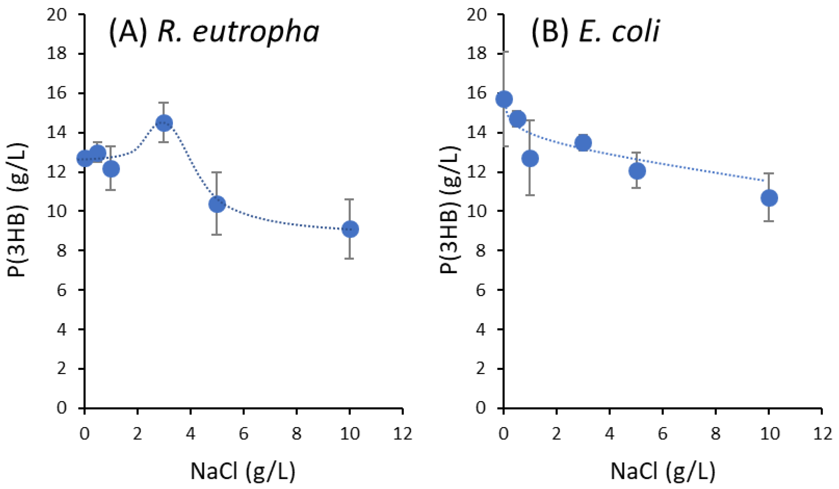
3.1. Effect of NaCl Concentration on P(3HB) Synthesis from Natural Glucose

LB medium generally contains 10 g/L NaCl [16], and is frequently used for culturing E. coli; however, it can also be used for culturing R. eutropha. In contrast, R. eutropha is usually cultured in a nutrient-rich medium that does not contain NaCl [19,20]. First, we investigated the NaCl preferences of these bacteria for P(3HB) synthesis to verify whether the same medium composition can be used for the culture tests in this study. Using R. eutropha NCIMB 11599 [14] and recombinant E. coli JM109 harboring pGEM-phaCABRe [15], the effect of NaCl concentration in the LB medium on P(3HB) synthesis was examined. To this end, the NaCl concentration in the LB medium was varied from 0 to 10 g/L with supplementation of 40 g/L natural glucose.
P(3HB) synthesis by R. eutropha NCIMB 11599 at different NaCl concentrations is shown in Figure 1A. The highest P(3HB) concentration of 14.5 g/L was obtained with 3 g/L NaCl in LB medium. Figure 1B shows the synthesis of P(3HB) using recombinant E. coli JM109 at different NaCl concentrations. In the culture test, the highest P(3HB) concentration of 15.7 g/L was obtained without NaCl addition, whereas the P(3HB) concentration decreased to 10.7 g/L with the addition of 10 g/L NaCl, which differed from the trend shown by R. eutropha. However, even at an NaCl concentration of 3 g/L, the recombinant E. coli showed a relatively high P(3HB) concentration of 13.5 g/L. Based on these observations, we cultured both strains in LB medium supplemented with 3 g/L NaCl. This medium is hereafter referred to as LB3 medium.

3.2. P(3HB) Synthesis from 13C-Glucose by R. eutropha NCIMB 11599

R. eutropha NCIMB 11599 was cultured in LB3, and MS media supplemented with 20 and 40 g/L 13C-glucose, respectively. The results were compared with those of cultures containing natural glucose. The culture results are presented in Table 1. When LB3 medium supplemented with 40 g/L 13C-glucose was used, cell growth was nearly identical to the one observed when using natural glucose; however, the P(3HB) content and concentration decreased. This implied that 13C-glucose slightly reduced the activity of P(3HB) synthesis in LB3 medium due to isotope effects. In contrast, almost all 13C-glucose was consumed during the cultivation, resulting in a P(3HB) yield of 0.26 g-P(3HB)/g-glucose, which was also lower than that from natural glucose (0.38 g-polymer/g-glucose).
In the MS medium, similar cell growth, P(3HB) content, and P(3HB) concentration were observed between cultures using 13C-glucose and natural glucose (Table 1). The P(3HB) yields from 13C-glucose and natural glucose were 0.28 and 0.29 g-P(3HB)/g-glucose, respectively. No difference was observed between 13C-glucose and natural glucose when the MS medium was used.

3.3. P(3HB) Synthesis from 13C-Glucose by Recombinant E. coli JM109

Recombinant E. coli JM109 harboring the plasmid pGEM-phaCABRe was cultured in LB3 and MS media supplemented with 13C-glucose or natural glucose. The culture results are shown in Table 1. When LB3 medium supplemented with 13C-glucose was used, slightly higher cell growth was observed than with natural glucose, although the P(3HB) content and P(3HB) concentration decreased. This indicates that the isotope effect of 13C-glucose slightly inhibited P(3HB) synthesis in the LB3 medium, as was similarly observed for R. eutropha NCIMB 11599. Almost all 13C-glucose was consumed during the cultivation, and the P(3HB) yield from 13C-glucose was 0.26 g-polymer/g-glucose, which was also a lower value than that for natural glucose (0.35 g-polymer/g-glucose).
In the MS medium, slightly higher cell growth, P(3HB) content, and P(3HB) concentration were observed when the cells were cultured on 13C-glucose than on natural glucose (Table 1). This trend was different from the one obtained using the LB3 medium. In contrast, 13C-glucose consumption during the cultivation was only 13.1 g/L, and the P(3HB) yield from 13C-glucose was as low as 0.11 g-polymer/g-glucose.

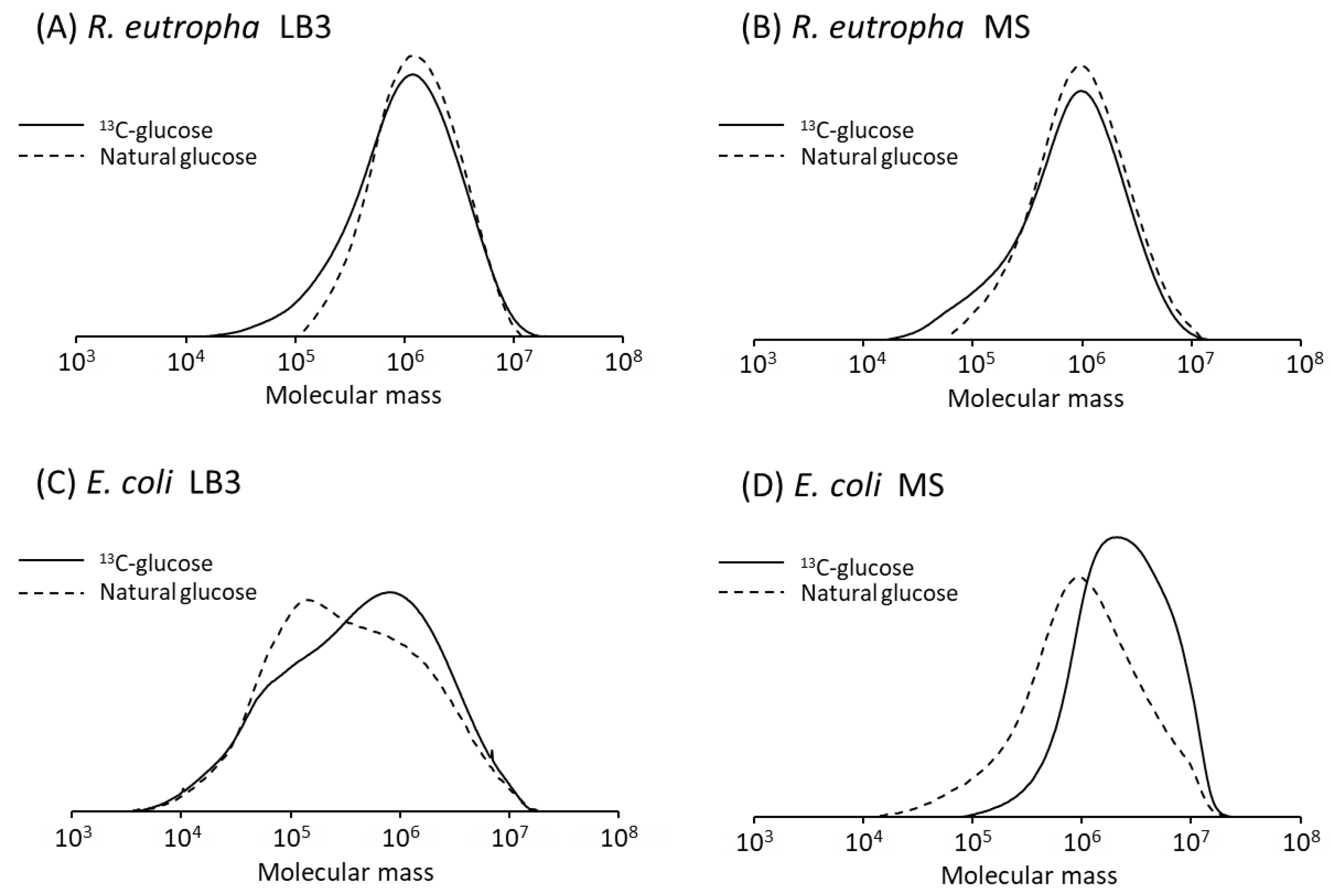
3.4. Molecular Weight of P(3HB)

The molecular weights of P(3HB) synthesized using 13C-glucose and natural glucose were determined using GPC. The results are presented in Table 2 and Figure 2. P(3HB) synthesized by R. eutropha NCIMB 11599 showed weight-average molecular weight (Mw) of (1.77–1.87) × 106 for LB3 medium and (1.36–1.52) × 106 for MS medium. Thus, no significant effect of 13C-glucose on the molecular weight of P(3HB) was observed in R. eutropha NCIMB 11599. However, the polydispersity (Mw/Mn, Mn is the number-average molecular weight) of P(3HB) synthesized from 13C-glucose, whose values are 3.37–3.41, being larger than those from natural glucose (2.08–2.45).
In contrast, the Mw and Mw/Mn of P(3HB) synthesized by recombinant E. coli JM109 in LB3 medium were (1.04–1.27) × 106 and 8.67–9.87, respectively. The high polydispersity suggests that these polymer samples have a wide distribution of molecular weights (Figure 2C) owing to abnormal polymerization. Using 13C-glucose further increased the polydispersity of P(3HB). According to a previous study [15], when recombinant E. coli JM109 was cultured in LB medium supplemented with natural glucose, the Mw exceeded 3 × 10⁶ with a narrow polydispersity of 1.8. In a previous study, shake flasks were used as the culture vessels [15], whereas test tubes were used in the present study. The difference in the molecular weights of P(3HB) may be due to the aeration capacity of each culture vessel.
In the MS medium, the Mw and Mw/Mn of P(3HB) synthesized from 13C-glucose by recombinant E. coli JM109 were 3.60 × 10⁶ and 2.30, respectively. Recombinant E. coli is known to synthesize ultra-high molecular weight (UHMW)-P(3HB) with Mw > 3 × 10⁶ [15,21], and this study also demonstrated that UHMW-P(3HB) can be synthesized using the MS medium supplemented with 13C-glucose. However, P(3HB) synthesized from natural glucose did not reach Mw of 3 × 10⁶, most likely because of an anaerobic metabolism due to insufficient aeration of the test tube culture. The endogenous ethanol generated during anaerobic metabolism in E. coli greatly influences the molecular weight of PHA [21].
As described above, isotope effects were commonly observed in the polydispersity of P(3HB). 13C-glucose tended to increase the polydispersity of P(3HB), except in one of the culture conditions.

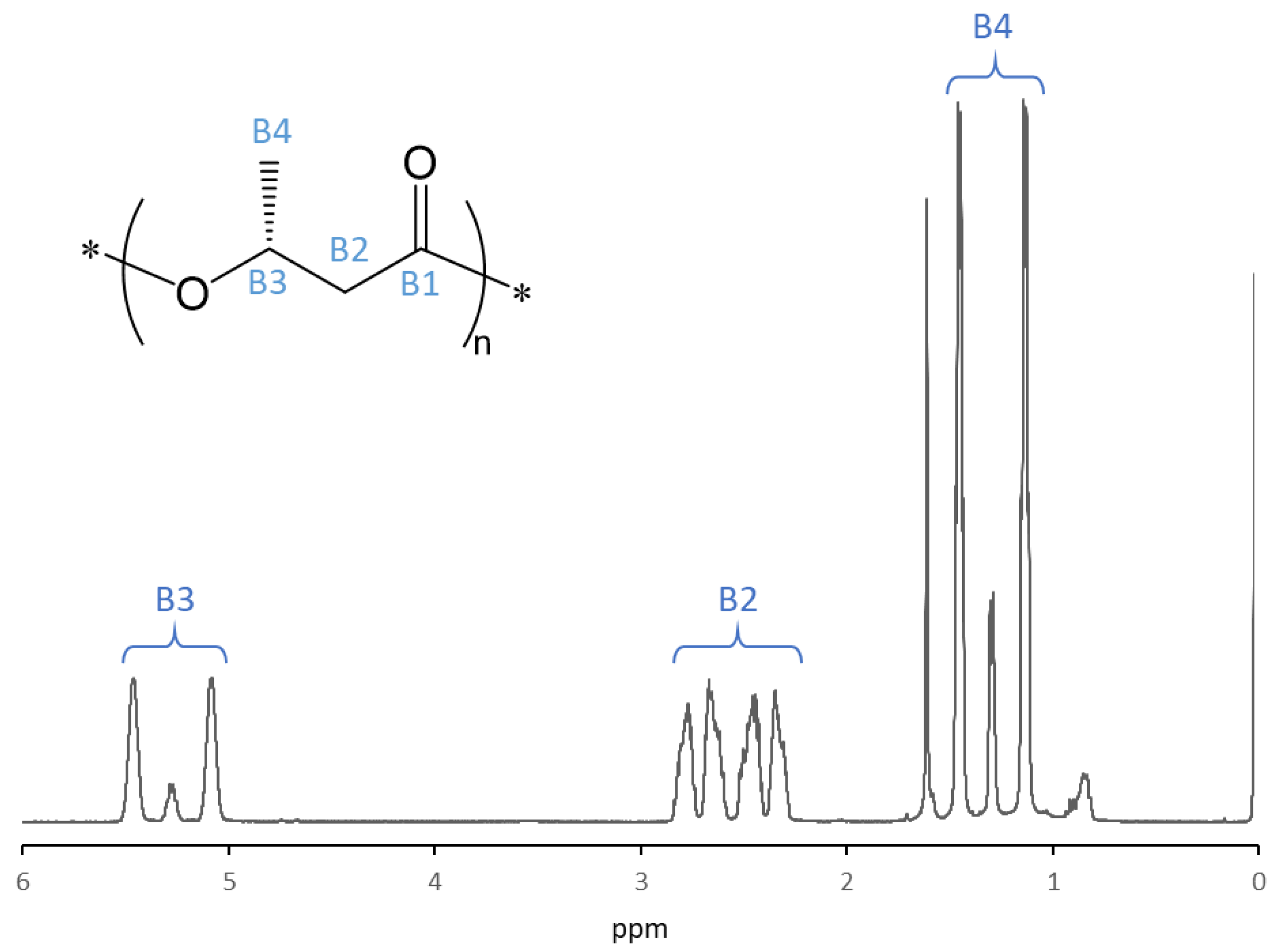
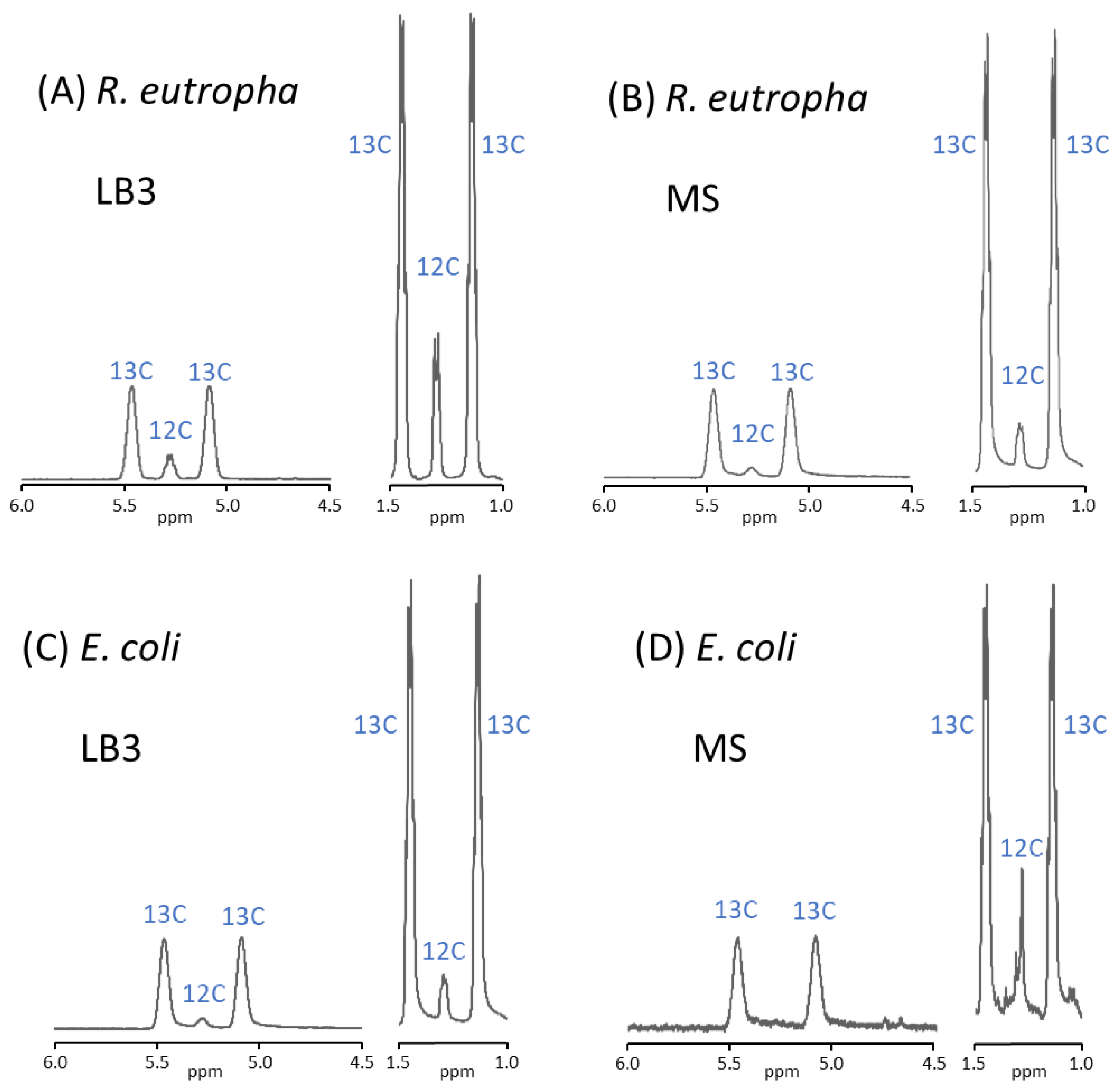
3.5. 1H NMR Analysis of P(3HB)

1H nuclear magnetic resonance (NMR) analysis was performed on P(3HB) synthesized from 13C-glucose. The spin-spin coupling of 1H atoms to an adjoining magnetically active 13C atom (1H-13C) in 1H NMR results in a different chemical shift from that of 1H-12C. Furthermore, 1H resonance showed excellent quantification. Thus, the 13C-labeling ratios of the methine carbon (>CH−) and methyl carbon (−CH3) of the 3HB unit were calculated based on the 1H NMR spectra.
Figure 3 shows 1H NMR spectrum of P(3HB) synthesized from 13C-glucose by R. eutropha NCIMB 11599 in the LB3 medium. Figure 4 shows enlarged spectra of P(3HB) biosynthesized from 13C-glucose under all conditions. Table 2 summarizes the 13C-labeling ratios of the methine (B3) and methyl (B4) carbons in the 3HB repeating unit, as shown in Figure 3.
For the methylene (B2) carbon, the 1H-13C and 1H-12C signals overlapped; thus, they were not used to calculate the 13C-labeling ratio. Additionally, the 13C-labeling ratio of the carbonyl (B1) carbon could not be calculated from the 1H NMR analysis because it lacked protons. The average 13C-labeling ratios of the B3 and B4 carbons are listed in Table 2. The average 13C-labeling ratio of P(3HB) synthesized by R. eutropha NCIMB 11599 was 88.0 atom% in LB3 medium and 97.6 atom% in MS medium.
In contrast, the 13C-labeling ratio of P(3HB) synthesized by recombinant E. coli JM109 was 94.3 atom% in the LB3 medium and 95.1 atom% in the MS medium. P(3HB) synthesized by E. coli JM109 in MS medium showed quite different 13C-labeling ratios depending on the carbon position; the 13C-labeling ratios of B3 and B4 were 100 and 90.1 atom%, respectively (Figure 4D). Although it is unclear why this difference occurred in this sample, it may have been influenced by the low sample concentration used for the measurement. The signal may be buried in the background noise.
Among the four conditions tested, the highest and second-highest 13C-labeling ratios were observed when R. eutropha NCIMB 11599 and recombinant E. coli JM109 were cultured in MS medium. In contrast, the lowest 13C-labeling ratio (88.0 atom% was observed when R. eutropha NCIMB 11599 was cultured in the LB3 medium. However, the 13C labeling ratio determined by 1H NMR had the disadvantage that it was limited to specific carbons.

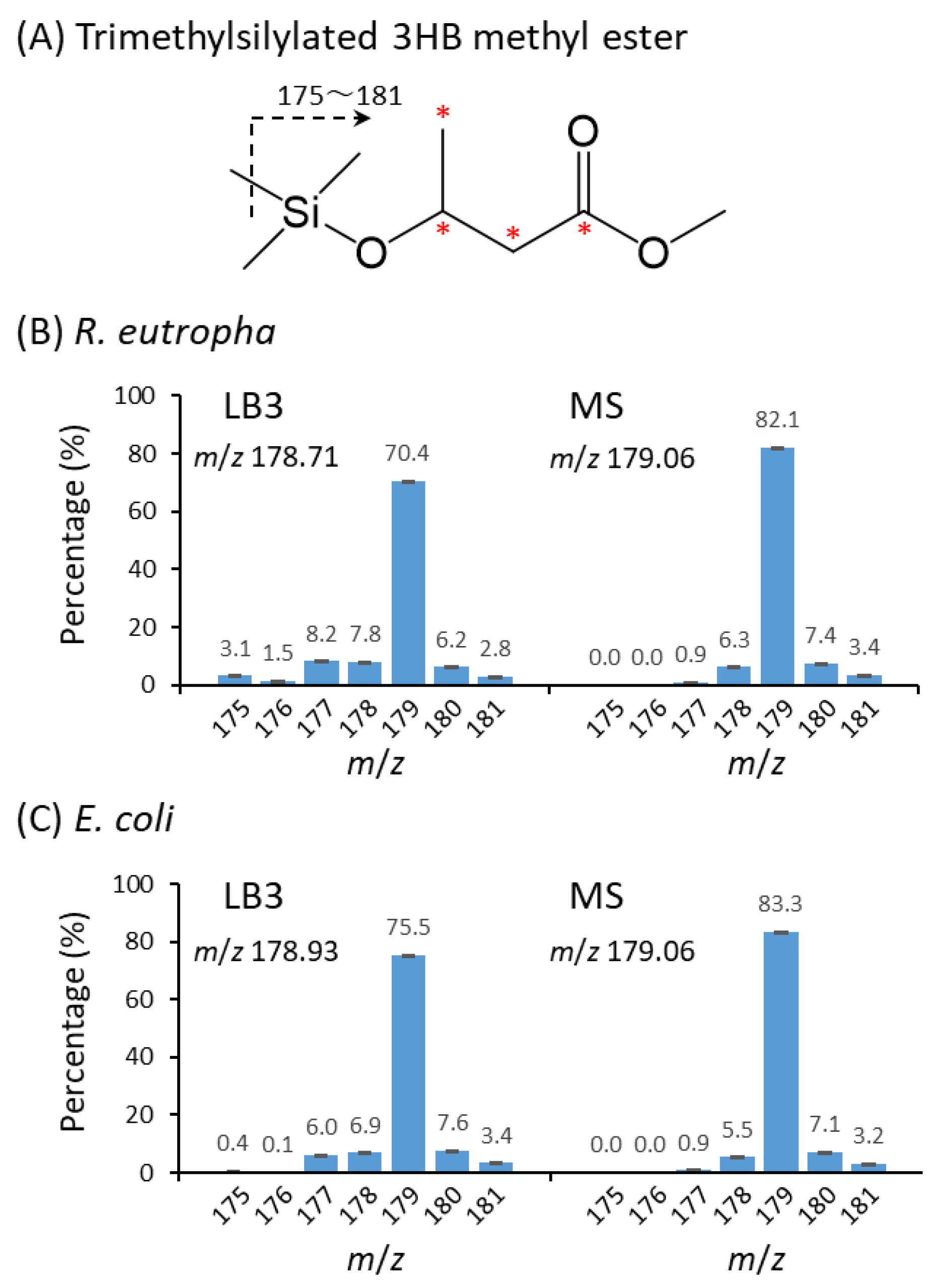
3.6. GC-MS Analysis of P(3HB)

The 13C-labeling ratio of P(3HB) was determined using gas chromatography-mass spectrometry (GC-MS). To analyze the P(3HB) samples using GC-MS, the samples were methanolyzed and converted into 3HB-methyl esters. However, as 3HB-methyl ester yielded a low intensity of the parent ion in the GC-MS analysis, it was unsuitable for determining the 13C-labeling ratio. Therefore, to increase the intensity of the parent ion, 3HB methyl ester was further silylated and subjected to GC-MS analysis [17,18]. Silylated 3HB methyl ester yielded a signal at m/z 175 when all four carbons in the 3HB unit were not labeled with 13C, whereas it yielded a signal at m/z 179 when all four carbons in the 3HB unit were labeled with 13C. In reality, the detected mass was distributed in the range of m/z 175–181 because there were trace amounts of natural isotopes in the silylated 3HB methyl ester, namely, the silicon used for silylation, the carbons that constitute tetramethylsilane and methyl ester, and the oxygens that constitute the ester bond and carbonyl group.
Figure 5 shows the molecular mass distributions of the silylated 3HB methyl esters analyzed by GC-MS. The 13C-labeling ratios of P(3HB) calculated using GC-MS analysis are listed in Table 2. The 13C-labeling ratios obtained by GC-MS analysis were in the range of 88.8 to 97.6 atom%, which were in good agreement with those obtained by 1H NMR analysis, except for recombinant E. coli JM109 cultured in MS medium. This indicated that the 13C-labeling ratios determined by GC-MS were reliable, and that this method was more suitable than the 1H NMR because the overall 13C-labeling ratio could be determined.

4. Discussion

Previous studies have reported on culturing of R. eutropha using 13C-labeled substrates; most have focused on the analysis of metabolic routes in bacterial cells [22,23,24,25,26,27,28]. For example, fatty acids labeled with 13C on specific carbon(s) were used for studying the metabolic pathway of PHA via the β-oxidation pathway [22,23]. Additionally, using 13C-labeled amino acids, such as leucine and valine, PHA monomer-supplying pathways from amino acids have been studied [24]. To the best of our knowledge, there have been no reports on the biosynthesis of PHA highly labeled with 13C for use in biodegradation tests. This is the first study to investigate an efficient 13C-labeling method for P(3HB) biosynthesis.
Additionally, the findings of this study reveal the isotope effects, which are the differences between 13C-labeled and non-labeled carbon sources, for P(3HB) biosynthesis. The most significant difference was observed in the P(3HB) content of cells cultured in the LB3 medium. However, the reason the isotope effect was observed only under these conditions has not yet been elucidated. Another isotope effect was observed in the molecular weight distribution of P(3HB) synthesized under all of the conditions tested. As the molecular weight and polydispersity of P(3HB) are easily changed by the intracellular conditions present [21], 13C-glucose may lead to a slightly different metabolic state from that of natural glucose.
In summary, when a large amount of 13C-labeled P(3HB) is required, a P(3HB) yield of over 10 g/L can be obtained by culturing recombinant E. coli JM109 in LB3 medium supplemented with 40 g/L 13C-glucose. The obtained P(3HB) showed 94.4 atom% 13C-labeling ratio. Meanwhile, 97.6 atom% of 13C-labeling ratio in P(3HB) was obtained at 4.7 g/L by culturing R. eutropha NCIMB 11599 in the MS medium supplemented with 20 g/L 13C-glucose. Because the 13C enrichment of the 13C-glucose used in this study was 99 atom% according to the supplier’s information, the observed 13C-labeling ratio was very close to the upper limit. In a previous study, autotrophic production of stable isotope-labeled arginine in R. eutropha H16 was studied, and isotope enrichments of 98.8%–99.4% for arginine were achieved [28], which were slightly higher than the labeling ratios observed in this study. To prepare 13C-labeled UHMW-P(3HB) (Mw > 3.0 × 10⁶), recombinant E. coli JM109 was cultured in MS medium supplemented with 13C-glucose. Unfortunately, this biosynthesis method did not result in high P(3HB) concentration and P(3HB) yield (1.4 g/L, 0.11 g-polymer/g-glucose, respectively).
The lowest 13C-labeling ratio (88.8 atom% by GC-MS) was observed when R. eutropha NCIMB 11599 was cultured in the LB3 medium supplemented with 13C-glucose. The main reason for this was that R. eutropha NCIMB 11599 metabolized the amino acids contained in the LB3 medium to produce 3HB monomers [29,30]. In addition, a slight decrease in the 13C-labeling ratio (94.4 atom% by GC-MS) was observed in recombinant E. coli JM109 grown on LB3 medium, suggesting that a small amount of amino acid was metabolized to the 3HB monomer. The abundant amino acids in LB medium, such as alanine and leucine [31], may be degraded to acetyl-CoA in cells to form 3HB monomers via the dimerization of acetyl-CoA by the action of 3-ketothiolase (PhaA) and NADPH-dependent acetoacetyl-CoA reductase (PhaB). Alternatively, (S)-3HB-CoA, an intermediate metabolite of lysine degradation [32], may isomerize to (R)-3HB-CoA and then polymerize to P(3HB). The differences in the 13C-labeling ratio between the MS and LB3 media (8.8 atom% and 3.2 atom% for R. eutropha and E. coli, respectively) indicated the amount of carbon derived from the LB3 medium components other than the glucose in P(3HB). This study is the first to show how much carbon is converted into P(3HB) from the components of the medium other than the main carbon source.
To compare the P(3HB) synthesis under the same culture conditions, LB3 and MS media were used for both R. eutropha and E. coli. Generally, E. coli is cultured in LB medium containing 10 g/L NaCl as the complete medium. R. eutropha is usually cultured in the nutrient-rich medium containing polypeptone and meat extract, but not NaCl [19,20]. When R. eutropha was cultured in the LB medium containing 10 g/L NaCl, the P(3HB) concentration was relatively low. However, in the present study, we found that the LB medium containing 3 g/L NaCl maximized P(3HB) synthesis in R. eutropha NCIMB 11599 (Figure 1). Based on these results, we performed culture experiments using 3 g/L NaCl-containing LB medium. In contrast, in E. coli, P(3HB) synthesis tended to decrease slightly as the NaCl concentration increased. Previously, PHA synthesis and secretory production of hydroxyalkanoate oligoesters using recombinant E. coli were investigated [33,34,35,36]. Increasing the NaCl concentration in the culture medium resulted in an increased intracellular PHA concentration and HA oligoester secretion when PHA synthase from Pseudomonas sp. 61-3 was used [33]. In contrast, increasing the NaCl concentration negatively affected P(3HB) synthesis and 3HB oligoester secretion when PHA synthase from Bacillus cereus YB-4 was used [34]. The osmotic pressure caused by NaCl can influence PHA synthase via the host reaction. Thus, the behavior of PHA synthases in response to the host reaction may differ depending on the PHA synthase used.
R. eutropha is a hydrogen-oxidizing chemoautotrophic bacterium [37,38,39,40,41,42]. R. eutropha NCIMB 11599 can grow in a mixture of hydrogen, oxygen, and carbon dioxide [42]. Meanwhile, it has been suggested that R. eutropha can fix carbon dioxide even under heterotrophic culture conditions [43]. Furthermore, carbon dioxide fixation appears to be promoted by the presence of glycerol, a substrate for R. eutropha that is hardly assimilated under heterotrophic culture conditions [44,45,46,47]. In this study, a high P(3HB) yield of 0.28 g-polymer/g-glucose was observed when R. eutropha NCIMB 11599 was cultured in MS medium supplemented with 20 g/L 13C-glucose. The P(3HB) yield was approximately three-fold higher than that of the recombinant E. coli. Moreover, the 13C-labeling ratio of P(3HB) remained high at approximately 98 atom%. Under these culture conditions, no amino acids in the culture medium could be converted to the 3HB monomer unit; thus, another mechanism may be involved in the enhancement of the P(3HB) yield from glucose. One possibility is that the carbon dioxide generated by the glycolysis of glucose and the tricarboxylic acid cycle may be re-fixed via the Calvin–Benson–Bassham cycle, as suggested in a previous study [43]. As the basis for this possibility, the present ratio of m/z 178 (6.3 ± 0.1%, Figure 5B), in which one out of four carbons in 3HB repeating unit is 12C (carbon that may come from the atmosphere), for the sample from R. eutropha cultured in MS medium was slightly higher than that of E. coli (5.5 ± 0.1%). Although this difference is statistically significant, it is unclear whether it is a difference reflecting metabolic backgrounds, and further studies are needed to confirm this hypothesis.

5. Conclusions

In this study, we investigated the culture conditions for the synthesis of 13C-labeled P(3HB) from 13C-glucose using R. eutropha NCIMB 11599 and recombinant E. coli JM109 as hosts. Culturing recombinant E. coli in LB3 medium yielded a large amount of P(3HB) (10.4 g/L, 94.4 atom% by GC-MS), while culturing R. eutropha in MS medium yielded highly 13C-labeled P(3HB) (4.7 g/L, 97.6 atom% by GC-MS). In contrast, culturing E. coli in MS medium yielded 13C-labeled UHMW-P(3HB). In addition to the technical aspects of 13C-labeled P(3HB) production, the findings of this study provide important insights into the metabolic differences between R. eutropha and E. coli. R. eutropha showed a greater capacity to convert medium components other than glucose to 3HB than E. coli. Furthermore, isotope effects were observed on the P(3HB) content in the cells when cultured in LB3 medium and the molecular weight distribution of P(3HB) synthesized under all of the conditions tested.

Author Contributions

Conceptualization, R.M.S. and T.T.; methodology, R.M.S., Y.M. and T.T.; software, R.M.S. and T.T.; validation, R.M.S., Y.M. and T.T.; formal analysis, R.M.S., Y.M. and T.T.; investigation, R.M.S. and Y.M.; resources, T.T.; data curation, Y.M. and T.T.; writing—original draft preparation, R.M.S.; writing—review and editing, T.T.; visualization, R.M.S. and T.T.; supervision, T.T.; project administration, T.T.; funding acquisition, T.T. All authors have read and agreed to the published version of the manuscript.

Funding

This study is based on the results obtained from project JPNP18016 commissioned by the New Energy and Industrial Technology Development Organization (NEDO).

Institutional Review Board Statement

Not applicable.

Informed Consent Statement

Not applicable.

Data Availability Statement

All data can be found in the article.

Acknowledgments

The authors gratefully acknowledge the Materials Analysis Division, Open Facility Center, Tokyo Institute of Technology, for supporting NMR and TG measurements.

Conflicts of Interest

The authors declare no conflict of interest.

References

  1. Sudesh, K.; Abe, H.; Doi, Y. Synthesis, structure and properties of polyhydroxyalkanoates: Biological polyesters. Prog. Polym. Sci. 2000, 25, 1503–1555. [Google Scholar] [CrossRef]
  2. Chen, G.Q.; Hajnal, I.; Wu, H.; Lv, L.; Ye, J. Engineering biosynthesis mechanisms for diversifying polyhydroxyalkanoates. Trends Biotechnol. 2015, 33, 565–574. [Google Scholar] [CrossRef] [PubMed]
  3. Mukherjee, A.; Koller, M. Microbial polyHydroxyAlkanoate (PHA) biopolymers—Intrinsically natural. Bioengineering 2023, 10, 855. [Google Scholar] [CrossRef] [PubMed]
  4. Suzuki, M.; Tachibana, Y.; Kasuya, K.I. Biodegradability of poly(3-hydroxyalkanoate) and poly(ε-caprolactone) via biological carbon cycles in marine environments. Polym. J. 2021, 53, 47–66. [Google Scholar] [CrossRef]
  5. Narancic, T.; Verstichel, V.; Chaganti, S.R.; Morales-Gamez, L.; Kenny, S.T.; De Wilde, B.; Padamati, R.B.; O’Connor, K.E. Biodegradable plastic blends create new possibilities for end-of-life management of plastics but they are not a panacea for plastic pollution. Environ. Sci. Technol. 2018, 52, 10441–10452. [Google Scholar] [CrossRef]
  6. Goudriaan, M.; Morales, V.H.; van der Meer, M.T.J.; Mets, A.; Ndhlovu, R.T.; van Heerwaarden, J.; Simon, S.; Heuer, V.B.; Hinrichs, K.U.; Niemann, H. A stable isotope assay with 13C-labeled polyethylene to investigate plastic mineralization mediated by Rhodococcus ruber. Mar. Pollut. Bull. 2023, 186, 114369. [Google Scholar] [CrossRef]
  7. Zumstein, M.T.; Schintlmeister, A.; Nelson, T.F.; Baumgartner, R.; Woebken, D.; Wagner, M.; Kohler, H.E.; McNeill, K.; Sander, M. Biodegradation of synthetic polymers in soils: Tracking carbon into CO2 and microbial biomass. Sci. Adv. 2018, 4, eaas9024. [Google Scholar] [CrossRef] [PubMed]
  8. Saito, T.; Shiraki, M.; Tatsumichi, M. Study on development of the evaluation method of biodegradable plastics by detecting 13CO2. Kobunshi Ronbunshu 1993, 50, 781–783. [Google Scholar] [CrossRef]
  9. Yang, Y.; Yang, J.; Wu, W.M.; Zhao, J.; Song, Y.; Gao, L.; Yang, R.; Jiang, L. Biodegradation and mineralization of polystyrene by plastic-eating mealworms: Part 1. Chemical and physical characterization and isotopic tests. Environ. Sci. Technol. 2015, 49, 12080–12086. [Google Scholar] [CrossRef]
  10. Vaksmaa, A.; Polerecky, L.; Dombrowski, N.; Kienhuis, M.V.; Posthuma, I.; Gerritse, J.; Boekhout, T.; Niemann, H. Polyethylene degradation and assimilation by the marine yeast Rhodotorula mucilaginosa. ISME Commun. 2023, 3, 68. [Google Scholar] [CrossRef]
  11. Radajewski, S.; Ineson, P.; Parekh, N.R.; Murrell, J.C. Stable-isotope probing as a tool in microbial ecology. Nature 2000, 403, 646–649. [Google Scholar] [CrossRef] [PubMed]
  12. Dumont, M.G.; Murrell, J.C. Stable isotope probing—Linking microbial identity to function. Nat. Rev. Microbiol. 2005, 3, 499–504. [Google Scholar] [CrossRef]
  13. Neufeld, J.D.; Dumont, M.G.; Vohra, J.; Murrell, J.C. Methodological considerations for the use of stable isotope probing in microbial ecology. Microb. Ecol. 2007, 53, 435–442. [Google Scholar] [CrossRef]
  14. Orita, I.; Iwazawa, R.; Nakamura, S.; Fukui, T. Identification of mutation points in Cupriavidus necator NCIMB 11599 and genetic reconstitution of glucose-utilization ability in wild strain H16 for polyhydroxyalkanoate production. J. Biosci. Bioeng. 2012, 113, 63–69. [Google Scholar] [CrossRef] [PubMed]
  15. Agus, J.; Kahar, P.; Abe, H.; Doi, Y.; Tsuge, T. Molecular weight characterization of poly[(R)-3-hydroxybutyrate] synthesized by genetically engineered strains of Escherichia coli. Polym. Degrad. Stabil. 2006, 91, 1138–1146. [Google Scholar] [CrossRef]
  16. Sambrook, J.; Fritsch, E.F.; Maniatis, T. Molecular Cloning: A Laboratory Manual, 2nd ed.; Cold Spring Harbor Laboratory Press: Cold Spring Harbor, NY, USA, 1989. [Google Scholar]
  17. Lee, E.Y.; Choi, C.Y. Gas chromatography—Mass spectrometric analysis and its application to a screening procedure for novel bacterial polyhydroxyalkanoic acids containing long chain saturated and unsaturated monomers. J. Ferment. Bioeng. 1995, 80, 408–414. [Google Scholar] [CrossRef]
  18. Ashby, R.D.; Solaiman, D.K.Y.; Foglia, T.A. The synthesis of short-and medium-chain-length poly(hydroxyalkanoate) mixtures from glucose- or alkanoic acid-grown Pseudomonas oleovorans. J. Ind. Microbiol. Biotechnol. 2002, 28, 147–153. [Google Scholar] [CrossRef]
  19. Doi, Y.; Kawaguchi, Y.; Nakamura, Y.; Kunioka, M. Nuclear magnetic resonance studies of poly(3-hydroxybutyrate) and polyphosphate metabolism in Alcaligenes eutrophus. Appl. Environ. Microbiol. 1989, 55, 2932–2938. [Google Scholar] [CrossRef]
  20. Saegusa, H.; Shiraki, M.; Kanai, C.; Saito, T. Cloning of an intracellular poly[D(−)-3-hydroxybutyrate] depolymerase gene from Ralstonia eutropha H16 and characterization of the gene product. J. Bacteriol. 2001, 183, 94–100. [Google Scholar] [CrossRef]
  21. Tsuge, T. Fundamental factors determining the molecular weight of polyhydroxyalkanoate during biosynthesis. Polym. J. 2016, 48, 1051–1057. [Google Scholar] [CrossRef]
  22. Huijberts, G.N.; de Rijk, T.C.; de Waard, P.; Eggink, G. 13C nuclear magnetic resonance studies of Pseudomonas putida fatty acid metabolic routes involved in poly(3-hydroxyalkanoate) synthesis. J. Bacteriol. 1994, 176, 1661–1666. [Google Scholar] [CrossRef]
  23. Fukui, T.; Kato, M.; Matsusaki, H.; Iwata, T.; Doi, Y. Morphological and 13C-nuclear magnetic resonance studies for polyhydroxyalkanoate biosynthesis in Pseudomonas sp. 61-3. FEMS. Microbiol. Lett. 1998, 164, 219–225. [Google Scholar] [CrossRef]
  24. Saika, A.; Ushimaru, K.; Mizuno, S.; Tsuge, T. Genome-based analysis and gene dosage studies provide new insight into 3-hydroxy-4-methylvalerate biosynthesis in Ralstonia eutropha. J. Bacteriol. 2015, 197, 1350–1359. [Google Scholar] [CrossRef] [PubMed]
  25. Shah, D.T.; Berger, P.A.; Tran, M.; Asrar, J.; Madden, L.A.; Anderson, A.J. Synthesis of poly(3-hydroxybutyrate) by Ralstonia eutropha in the presence of 13C-labeled ethylene glycol. Macromolecules 2000, 33, 6624–6626. [Google Scholar] [CrossRef]
  26. Liu, C.; Nangle, S.N.; Colón, B.C.; Silver, P.A.; Nocera, D.G. 13C-Labeling the carbon-fixation pathway of a highly efficient artificial photosynthetic system. Faraday Discuss. 2017, 198, 529–537. [Google Scholar] [CrossRef] [PubMed]
  27. Ludevese-Pascual, G.; Ahmed, F.; De Troch, M.; Amar, E.; Laranja, J.L.; Bode, S.; Boeckx, P.; Bossier, P.; De Schryver, P. Determination of poly-β-hydroxybutyrate assimilation by postlarval whiteleg shrimp, Litopenaeus vannamei using stable 13C isotope tracing. J. World Aquac. Soc. 2021, 52, 184–194. [Google Scholar] [CrossRef]
  28. Lütte, S.; Pohlmann, A.; Zaychikov, E.; Schwartz, E.; Becher, J.R.; Heumann, H.; Friedrich, B. Autotrophic production of stable-isotope-labeled arginine in Ralstonia eutropha strain H16. Appl. Environ. Microbiol. 2012, 78, 7884–7890. [Google Scholar] [CrossRef]
  29. Fraser-Smith, E.C.; Austin, M.A.; Reed, L.L. Utilization of amino acids as a source of nitrogen by Hydrogenomonas eutropha. J. Bacteriol. 1969, 97, 457–459. [Google Scholar] [CrossRef] [PubMed]
  30. Iskandaryan, M.; Blbulyan, S.; Sahakyan, M.; Vassilian, A.; Trchounian, K.; Poladyan, A. L-amino acids affect the hydrogenase activity and growth of Ralstonia eutropha H16. AMB Express 2023, 13, 33. [Google Scholar] [CrossRef]
  31. Sezonov, G.; Joseleau-Petit, D.; d’Ari, R. Escherichia coli physiology in Luria-Bertani broth. J. Bacteriol. 2007, 189, 8746–8749. [Google Scholar] [CrossRef] [PubMed]
  32. Kanehisa, M.; Goto, S.; Kawashima, S.; Nakaya, A. The KEGG databases at GenomeNet. Nucleic Acids Res. 2002, 30, 42–46. [Google Scholar] [CrossRef]
  33. Mizuno, S.; Sakurai, T.; Nabasama, M.; Kawakami, K.; Hiroe, A.; Taguchi, S.; Tsuge, T. The influence of medium composition on the microbial secretory production of hydroxyalkanoate oligomers. J. Gen. Appl. Microbiol. 2021, 67, 134–141. [Google Scholar] [CrossRef] [PubMed]
  34. Sakurai, T.; Mizuno, S.; Miyahara, Y.; Hiroe, A.; Taguchi, S.; Tsuge, T. Optimization of culture conditions for secretory production of 3-hydroxybutyrate oligomers using recombinant Escherichia coli. Front. Bioeng. Biotechnol. 2022, 10, 829134. [Google Scholar] [CrossRef] [PubMed]
  35. Utsunomia, C.; Matsumoto, K.I.; Taguchi, S. Microbial secretion of D-lactate-based oligomers. ACS Sustain. Chem. Eng. 2017, 5, 2360–2367. [Google Scholar] [CrossRef]
  36. Nduko, J.M.; Taguchi, S. Microbial production of biodegradable lactate-based polymers and oligomeric building blocks from renewable and waste resources. Front. Bioeng. Biotechnol. 2021, 8, 618077. [Google Scholar] [CrossRef] [PubMed]
  37. Pohlmann, A.; Fricke, W.F.; Reinecke, F.; Kusian, B.; Liesegang, H.; Cramm, R.; Eitinger, T.; Ewering, C.; Pötter, M.; Schwartz, E.; et al. Genome sequence of the bioplastic-producing “Knallgas” bacterium Ralstonia eutropha H16. Nat. Biotechnol. 2006, 24, 1257–1262. [Google Scholar] [CrossRef]
  38. Panich, J.; Fong, B.; Singer, S.W. Metabolic engineering of Cupriavidus necator H16 for sustainable biofuels from CO2. Trends Biotechnol. 2021, 39, 412–424. [Google Scholar] [CrossRef]
  39. Heinrich, D.; Raberg, M.; Steinbüchel, A. Studies on the aerobic utilization of synthesis gas (syngas) by wild type and recombinant strains of Ralstonia eutropha H16. Microb. Biotechnol. 2018, 11, 647–656. [Google Scholar] [CrossRef]
  40. Brigham, C. Perspectives for the biotechnological production of biofuels from CO2 and H2 using Ralstonia eutropha and other ‘Knallgas’ bacteria. Appl. Microbiol. Biotechnol. 2019, 103, 2113–2120. [Google Scholar] [CrossRef]
  41. Garcia-Gonzalez, L.; Mozumder, M.S.I.; Dubreuil, M.; Volcke, E.I.; De Wever, H. Sustainable autotrophic production of polyhydroxybutyrate (PHB) from CO2 using a two-stage cultivation system. Catal. Today 2015, 257, 237–245. [Google Scholar] [CrossRef]
  42. Miyahara, Y.; Wang, C.T.; Ishii-Hyakutake, M.; Tsuge, T. Continuous supply of non-combustible gas mixture for safe autotrophic culture to produce polyhydroxyalkanoate by hydrogen-oxidizing bacteria. Bioengineering 2022, 9, 586. [Google Scholar] [CrossRef]
  43. Shimizu, R.; Dempo, Y.; Nakayama, Y.; Nakamura, S.; Bamba, T.; Fukusaki, E.; Fukui, T. New insight into the role of the Calvin cycle: Reutilization of CO2 emitted through sugar degradation. Sci. Rep. 2015, 5, 11617. [Google Scholar] [CrossRef]
  44. Thorbecke, R.; Yamamoto, M.; Miyahara, Y.; Oota, M.; Mizuno, S.; Tsuge, T. The gene dosage effect of carbonic anhydrase on the biosynthesis of poly(3-hydroxybutyrate) under autotrophic and mixotrophic culture conditions. Polym. J. 2021, 53, 209–213. [Google Scholar] [CrossRef]
  45. Schwartz, E.; Voigt, B.; Zühlke, D.; Pohlmann, A.; Lenz, O.; Albrecht, D.; Schwarze, A.; Kohlmann, Y.; Krause, C.; Hecker, M.; et al. A proteomic view of the facultatively chemolithoautotrophic lifestyle of Ralstonia eutropha H16. Proteomics 2009, 9, 5132–5142. [Google Scholar] [CrossRef] [PubMed]
  46. Strittmatter, C.S.; Eggers, J.; Biesgen, V.; Pauels, I.; Becker, F.; Steinbüchel, A. The reliance of glycerol utilization by Cupriavidus necator on CO2 fixation and improved glycerol catabolism. Appl. Microbiol. Biotechnol. 2022, 106, 2541–2555. [Google Scholar] [CrossRef] [PubMed]
  47. Friedrich, C.G.; Friedrich, B.; Bowien, B. Formation of enzymes of autotrophic metabolism during heterotrophic growth of Alcaligenes eutrophus. Microbiology 1981, 122, 69–78. [Google Scholar] [CrossRef]
Figure 1. Effect of NaCl concentration in LB medium on P(3HB) production by (A) Ralstonia eutropha NCIMB 11599 and (B) recombinant Escherichia coli JM109 harboring pGEM-phaCABRe from natural glucose. Cells were cultured in 5 mL media in the test tube at 30 °C for 72 h. Results are the averages of values from three individual experiments (n = 3).
Microbiolres 14 00129 g001
Figure 2. Molecular mass of P(3HB) biosynthesized by (A,B) Ralstonia eutropha NCIMB 11599 and (C,D) recombinant Escherichia coli JM109. The culture medium used was (A,C) LB3 or (B,D) MS supplemented with 13C-glucose (solid line) or natural glucose (dotted line).
Microbiolres 14 00129 g002
Figure 3. The 400 Mz 1H NMR spectrum of P(3HB) biosynthesized by R. eutropha NCIMB 11599 in LB3 medium supplementing 40 g/L 13C-glucose.
Microbiolres 14 00129 g003
Figure 4. The enlarged 1H NMR spectra of 1.0–1.5 ppm (methyl proton, B4) and 4.5–6.0 ppm (methine proton, B3). 12C and 13C represent 1H-12C resonance and 1H-13C resonance, respectively. The positions of B3 and B4 are shown in Figure 3.
Microbiolres 14 00129 g004
Figure 5. GC-MS analysis of P(3HB) after methanolysis and trimethylsilylated treatment. (A) The chemical structure of trimethylsilylated 3HB methyl ester. The m/z of the parent ion is in the range of 175–181, depending on the involved isotopes. The red asterisks (*) denote the 13C-labeling positions. (B) The m/z distribution of 3HB-derived parent ions biosynthesized by Ralstonia eutropha NCIMB 11599 (n = 3). (C) The m/z distribution of 3HB-derived parent ions biosynthesized by recombinant Escherichia coli JM109 (n = 3). The average molecular masses of trimethylsilylated 3HB methyl ester are also shown in (B,C).
Microbiolres 14 00129 g005
Table 1. Biosynthesis of P(3HB) from glucose using Ralstonia eutropha NCIMB 11599 and Escherichia coli JM109 harboring pGEM-phaCABRe.
StrainCulture
Medium
Type of
Glucose
Initial
Glucose
(g/L)
Dry Cell wt.
(g/L)
P(3HB)
Content
(wt%)
P(3HB)
(g/L)
Glucose
Consumption
(g/L)
P(3HB) Yield
(g-polymer/
g-glc)
R. eutrophaLB3Natural4016.3 ± 0.882 ± 612.6 ± 3.538.6 ± 0.30.38 ± 0.03
LB3[U-13C6]4016.2 ± 0.664 ± 110.0 ± 0.338.2 ± 0.00.26 ± 0.01
MSNatural205.4 ± 1.784 ± 24.5 ± 0.115.8 ± 0.10.29 ± 0.00
MS[U-13C6]205.7 ± 0.182 ± 04.7 ± 0.116.7 ± 0.10.28 ± 0.00
E. coliLB3Natural4015.7 ± 0.283 ± 413.5 ± 0.438.2 ± 0.20.35 ± 0.01
LB3[U-13C6]4016.5 ± 0.163 ± 410.4 ± 0.739.9 ± 0.10.26 ± 0.01
MSNatural202.2 ± 0.053 ± 11.2 ± 0.012.9 ± 0.10.09 ± 0.00
MS[U-13C6]202.6 ± 0.157 ± 61.4 ± 0.113.1 ± 0.30.11 ± 0.01
Cells were cultured in 5 mL media in a test tube at 30 °C for 72 h. LB3 medium denotes LB medium containing 3 g/L NaCl. MS medium denotes mineral salt medium. Results are the averages of values from three individual experiments (n = 3).
Table 2. Molecular weight and 13C-labeling ratio of P(3HB).
StrainCulture
Medium
Type of
Glucose
Molecular Weight a13C-Labeling Ratio (atom%)
Mw
(×106)
Mw/Mnby 1H-NMR bby GC-MS c
B3B4Average
R. eutrophaLB3Natural1.872.08----
LB3[U-13C6]1.773.4188.887.288.088.8 ± 1.4
MSNatural1.522.45----
MS[U-13C6]1.363.3798.197.197.697.6 ± 0.0
E. coliLB3Natural1.048.67----
LB3[U-13C6]1.279.8793.894.794.394.4 ± 0.3
MSNatural2.063.31----
MS[U-13C6]3.602.3010090.195.197.6 ± 0.1
LB3 medium denotes LB medium containing 3 g/L NaCl. MS medium denotes a mineral salt medium. a Determined by GPC. b B3 and B4 denote the methine and methyl protons of P(3HB), respectively (Figure 3). c Results are the averages of values from three individual experiments (n = 3). -, not determined.
Disclaimer/Publisher’s Note: The statements, opinions and data contained in all publications are solely those of the individual author(s) and contributor(s) and not of MDPI and/or the editor(s). MDPI and/or the editor(s) disclaim responsibility for any injury to people or property resulting from any ideas, methods, instructions or products referred to in the content.

