Communication

The Molecular Epidemiology of Enterovirus in a Birth Cohort in Nepal

1
Center for International Health, University of Bergen, N-5020 Bergen, Norway
2
Walter Reed/AFRIMS Research Unit Nepal, Kathmandu 44600, Nepal
3
Department of Research, Innlandet Hospital Trust, N-2381 Lillehammer, Norway
4
Department of Virology, Norwegian Institute of Public Health, N-0213 Oslo, Norway
5
Institute of Clinical Medicine, University of Oslo, N-0318 Oslo, Norway
6
Department of Microbiology, Oslo University Hospital, N-0424 Oslo, Norway
*
Author to whom correspondence should be addressed.
Microbiol. Res. 2023, 14(3), 909-917; https://doi.org/10.3390/microbiolres14030063
Submission received: 30 May 2023 / Revised: 29 June 2023 / Accepted: 3 July 2023 / Published: 20 July 2023

Abstract

:
Acute gastroenteritis (AGE) has a major impact on morbidity and mortality worldwide. The viral aetiology of diarrhoeal diseases may remain unknown due to limited diagnostic facilities. Non-polio enteroviruses (NPEVs) are the third most frequent pathogen detected in stool specimens from AGE cases, yet their potential role in AGE is uncertain. In Nepal, limited data are available on NPEVs, due to both the lack of an adequate surveillance program and the availability of tests. The global polio eradication initiative effort of the WHO has eradicated the incidence of poliomyelitis and acute flaccid paralysis (AFP) from many parts of the world, including Nepal. However, cases of AFP associated with NPEVs have been reported in different countries, including the neighbouring India. This study aims to investigate the diarrhoeal stool samples from a birth cohort until the age of 36 months for NPEVs and the genotype diversity of NPEV in community children with diarrhoea. A total of 280 longitudinal diarrhoeal stool samples that were negative for other enteric pathogens were tested using RT-PCRs. NPEVs was detected in 97 stool specimens (34.6%) and were significantly more frequent in infants up to one year of age. This study identified 17 various NPEV types, with the dominating species being Enterovirus B (EV-B). Ten different types of echoviruses were recorded in this study, with the two rare NPEVs B74 and A120. Based on prevalence, seasonality, and diversity, further studies are warranted to investigate the role of enterovirus in diarrhoeal disease.

1. Introduction

Non-polio enteroviruses (NPEVs) are single-stranded RNA viruses belonging to four species of enterovirus (A to D) in the family Picornavirididae. These viruses are responsible for a range of manifestations in humans, covering asymptomatic, mild, and severe and life-threatening illness [1,2]. Some NPEVs have been associated with acute flaccid paralysis (AFP), with a clinical presentation similar to poliomyelitis caused by poliovirus. Moreover, some strains of NPEVs have been reported to cause infections such as hand, foot, and mouth disease, meningitis, encephalitis, paralysis, and myocarditis. NPEVs are transmitted through the faecal–oral route and are common in infants, mostly presenting as an asymptomatic infection [3].
The global effort led by the World Health Organization has reduced the global incidence of poliomyelitis. With this successful eradication program, wild poliovirus, which is a cause of AFP, has been eliminated from most parts of the world, including Nepal. However, cases of AFP associated with NPEVs have, in later years, been reported in different countries, including India [4]. In 2014, a large outbreak associated with enterovirus D68 (EV-D68) with severe respiratory illness and AFP was reported [5]. Even though EV infections are asymptomatic, they have shown associations with many diseases, for example, aseptic meningitis is caused by echovirus 30, EV-A71 [6], EV-D70, and CV-A24 cause acute haemorrhagic conjunctivitis, and several coxsackievirus types, A6 (CV-A6) and CV-A10, typically cause hand, foot, and mouth disease (HFMD) [7].
Studies on NPEVs have not confirmed any association of EV infection with diarrheal diseases. However, EV infection has been reported and documented in a number of studies investigating acute diarrhoea. Echovirus 11, CV-A6, CV-B2, PV3, CV-B4, E18, and CV-A2 were detected in the stool of children with acute diarrhoea in the Hebei province of China [8,9]. Echovirus type 6 was reported in an outbreak of gastroenteritis among children in Japan [10]. Similarly, E11 and Coxsackie B6 have been reported in acute gastroenteritis in different countries such as Malaysia [11], India [12], and Brazil [13].
There is no NPEVs surveillance program in Nepal. The epidemiological studies on EV infection and information on the genotypic diversity of EVs are limited. Therefore, this study aimed to determine the prevalence of NPEVs in stool samples that were negative for other common diarrheal etiologies in children below 3 years of age and perform a molecular characterization of the EVs circulating in children with diarrhea in Nepal. This study will provide scientific information for assessing the requirement of establishing an NPEVs surveillance program for generating robust data for public health planning.

2. Materials and Methods

2.1. Study Population and Data Collection

The Malnutrition due to Enteric Diseases (MAL-ED) study conducted in Nepal was part of a multi-centre birth cohort study carried out in 8 different countries. The MAL-ED study in Nepal enrolled 240 newborn babies from the Bhaktapur community. The newborns within 17 days of birth were enrolled over the period of two years, then followed up with different measurements until three years of age. The cohort represents a peri urban community in Nepal, one of south Asia’s least developed countries. Detailed information about the site was described in a previously published paper [14]. The MAL-ED study consists of eight birth cohorts worldwide followed throughout the first years of life (originally 0 to 36 months of age). Active surveillance for infectious diseases, general child health information, and basic dietary intake was undertaken by visiting each home twice per week. Upon enrolment, each child’s date of birth, sex, and birth weight (if available) were recorded, information about the initiation of breastfeeding was noted, and the child’s length, weight, and head circumference were measured. Stool samples were collected every month from birth until 24 months, then quarterly, as well as during each episode of diarrhoea (defined as ≥3 loose stools in a 24 h period and separated by ≥2 diarrhoea-free days).

2.2. Study Type and Sample Size

Our study is a retrospective study that includes the diarrheal stool samples collected between 2010–2015 from 131 children (0–36 months age) enrolled in the birth cohort of the MAL-ED study. In total, 280 stool samples that tested negative for bacteria (Salmonella, Shigella, Campylobacter, Vibrio, Aeromonas, Plesiomonas, and E. coli), parasites (Giardia, E. Histolytica, Cryptosporidium, and Cyclospora), and enteric viruses (Astrovirus, Rotavirus, and Adenovirus) were included in the study. The details of the testing procedure for the different pathogens included in the MAL-ED project were described in previously published articles [14,15].

2.3. Ethic Approval

The study was approved by the ethics committee of the Nepal Health Research Council (NHRC) as a national IRB of Nepal (Reg. 222/2015) and the Norwegian regional committee for medical and health research ethics (REK-sør-øst B 42335). Written informed consent was obtained from the guardians of every child participating.

2.4. Nucleic Acid Extraction

A 10% suspension of faecal samples was prepared in 0.85% NaCl and centrifuged at 4000× g for 20 min. One hundred and forty microliters of the supernatant were used as a starting material for the viral nucleic acid extraction, which was performed manually by using the QiaAmp Viral RNA Mini Kit (QIAGEN, Hilden, Germany) according to the manufacturers’ instructions. The RNA extracted material was used as a template for the real-time RT-PCRs.

2.5. Real Time RT PCR of EV

Real-time RT-PCRs were performed on the stool samples using primers that targeted the highly conserved region suitable for the NPEV types (A to D) in the Picornavirididae family. The primers applied for the screening of enterovirus were published by Rotbart et al., (1990) [16,17,18,19] using genus-specific primers targeted to the 5′ non-translated region (NTR), generating 120 base pairs amplicon from the 5′ NTR of entero- and rhinoviruses. The forward primer was located in the 450–470 genomic region in the 5′ NTR region, as were several other primers described previously for the detection of enteroviruses [18,20,21,22,23]. The genome was amplified using the One Step RT-PCR Kit (QIAGEN, Hilden, Germany) on the Rotor Gene cycler system (Corbett/QIAGEN, Hilden, Germany), applying 30 min at 50 °C and 15 min at 95 °C, followed by 45 cycles of 15 s at 94 °C and 60 s at 63 °C.

2.6. Enterovirus Typing

The samples positive for enterovirus detected using a real-time PCR (Ct value < 30) were further evaluated using Sanger sequencing to determine the serotype. A portion of the VP1 region (capsid gene) was used for molecular typing, as described in the Nix et al. protocol [24]. The resulting DNA templates were sequenced on an ABI Prism 3500 Genetic Analyzer using the Big Dye Terminator Cycle Sequencing Kit v.1.1 (Applied Biosystems, Foster City, CA, USA) with primers AN89 and AN88. The sequences were analysed and aligned using Sequencer version 5.4.6 (GeneCodes) against the reference strains. The NCBI basic local alignment search tool (BLAST) was used to identify the serotypes. The sequences for the stool specimens described here have been deposited in the GenBank sequence database (accession numbers OQ348761 to OQ348783).

2.7. Statistical Analysis

The descriptive variables were recorded as means and standard deviations. A chi-square test of independence was performed to examine the relation between the age categories and seasonality. p values of < 0.05 were considered to be statistically significant.

3. Results

In 131 children, 71 males and 60 females, NPEVs were detected in 49.6% (65/131) of them. The median age of the children included was 7.45 months. The minimum and maximum breastfeeding lengths among the enrolled children were recorded as 10 days and 6.6 months, respectively. The longitudinal number of stool samples collected in this study varied from one to a maximum of six diarrheal stool samples per individual child. In total, 280 diarrheal stool samples were analysed from 131 children. Out of the 280 stool samples collected, EV was detected in 34.6% of them (97/280). The associations of EV with gender, different age groups, and season are shown in Table 1. Infants up to one year of age showed a higher frequency of NPEV detection compared to older age groups (Figure 1).
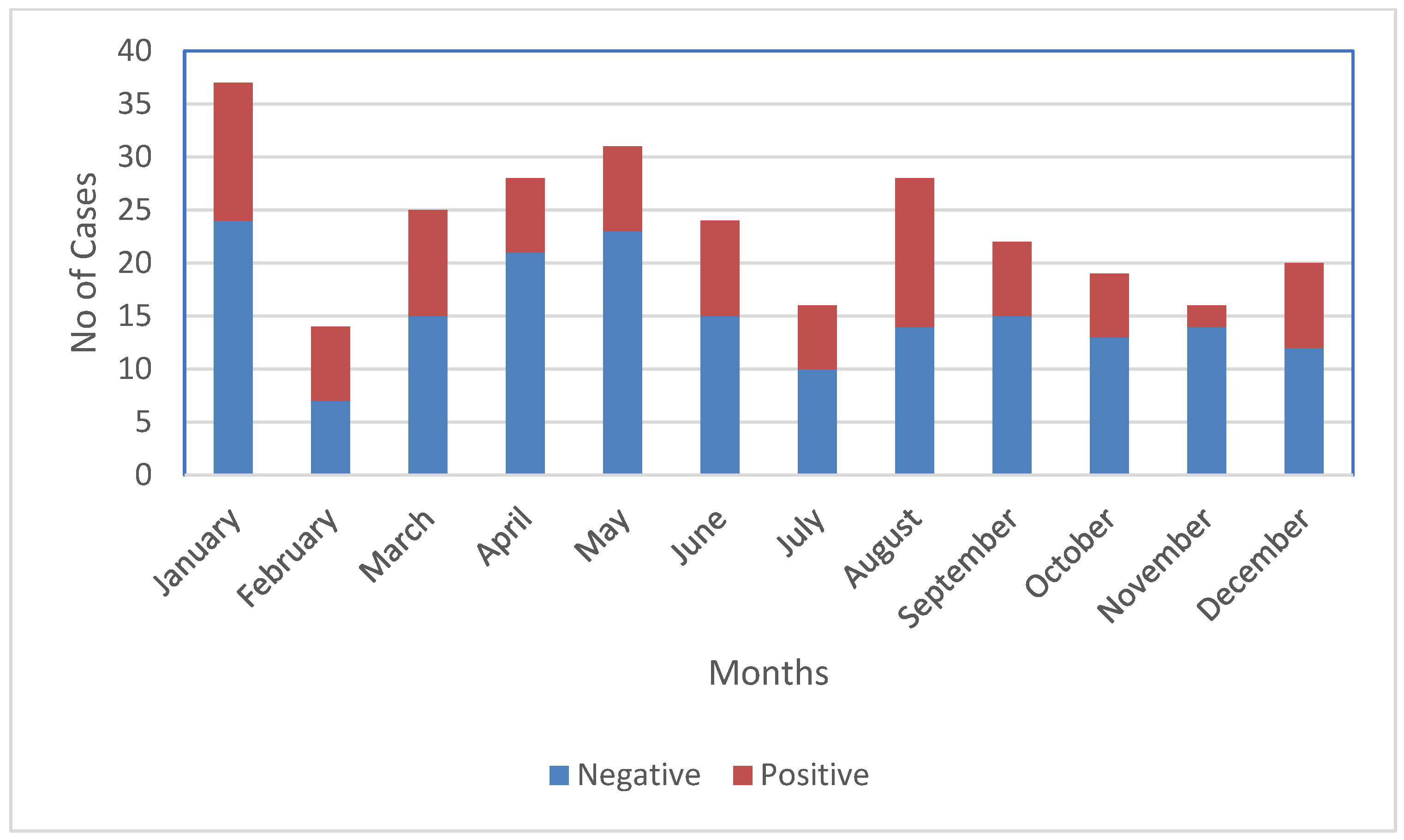
NPEVs were found all year round, with the highest number being detected in the months of January and August (Figure 2); overall, most cases occurred in the summer and winter (Table 1). However, there was no statistically significant association between NPEV detection and season (p > 0.05). Multiple episodes of NPEV infections were detected in 19 children (2 episodes in 10 children, 3 episodes in 6 children, and 4 episodes in 3 children.
Two species of Enteroviruses, Enterovirus A and B (EV-A and EV-B) with 17 different genotypes were identified (Table 2). The majority of the genotyped viruses, 78.3%, belonged to the EV-B species. Echoviruses were the most detected types and were found in 60.9% of the sequenced stool samples, followed by Coxsackievirus B and A (CV-B and CV-A). Ten different Echoviruses were detected in the stool samples. The uncommon serotypes EV-A120 and EV-B74 were also identified in the stool samples from the children.

4. Discussion

This is the first study investigating NPEV infections in stool samples from a birth cohort of infants and young children in Nepal. In our study, 34.6% of the diarrhoea stool samples from children that were negative for other enteric pathogens tested in the MAL-ED study were positive for EVs. The prevalence of EVs in children with gastroenteritis has been reported in different countries, ranging from 24 to 40% [9]. In an Indian study, 18–21% of NPEV-associated infections were reported in children with diarrhoea, raising the question of their significance in diarrhoea [25,26]. The high prevalence of NPEVs shows the value of monitoring NPEVs in children, even after the eradication of polio, to understand the epidemiology for planning appropriate public health measures.
NPEV infections occur globally, exhibiting a peak in summer and autumn in temperate climates [3]. We observed NPEVs in the samples collected almost year-round and the incidence peaked in the months of January and August. This is in contrast to the findings of Shobha et al. [27] and Bubba et al. [28], who reported a high prevalence of NPEVs in the autumn and winter.
Most NPEV infections accompanied by clinical symptoms result in unspecific febrile illness, although more severe disease can occur with the replication of the virus in various target organs, including the central nervous system. The primary replication of NPEVs takes place in either the respiratory or gastrointestinal tract, resulting in the shedding of the virus in stool; still, enteroviruses do not seem to cause gastrointestinal diseases [29]. This could be reason for the NPEVs detected in healthy children being asymptomatic and not being regarded as diarrheal pathogens [30,31]. Nevertheless, the significant association of enterovirus infection with gastroenteritis disease and persistent diarrhoea has been described in many studies [25,32,33].
EV-A is known to be the main pathogen responsible for hand, foot, and mouth disease (HFMD) [34] and there have been reports of EV-A invading the nervous system, with the majority of these occurring in Asia [35]. EV-A71, CV-A16, and CV-A6 are common types associated with HFMD outbreaks. The emergence of CV-A4 and CV-A2 has been reported in China [36,37], Korea [38], and Thailand [39], which has been associated with HFMD or herpangina outbreaks, even though these cases were less severe than ones with EV-A71 [40]. In our study, we identified CV-A4 and CV-A2 serotypes in children less than 5 years of age with symptoms of diarrhoea, which had not been reported previously. In addition to the more common NPEV types, we found the recently characterized EV-A120 in one of the children with diarrhoea. There have been no reported cases of EV-A120 in Nepal to date. The first case of EV-A120 was reported in Madagascar in 2011, which was isolated from the faeces of a healthy 3-year-old child [3]. Later, a few countries such as Papua New Guinea, Nigeria, France, Pakistan, and Tajikistan isolated the EV-A120 strain. In Papua New Guinea and Nigeria, the EV-A120 strains were isolated from patients with AFP and in France, Pakistan, and other parts of Africa, the EV-A120 strains were isolated from wastewater [41]. The phylogenetic characteristics of EV-A120 from Tibet, China, have also been reported [41]. Although this is consistent with the pathogenic characteristics of enteroviruses, there is still insufficient epidemiological and laboratory evidence to prove that EV-A120 is the pathogenic pathogen of AFP. Apart from EV-A120, we also identified rare EV-B74 belonging to the EV B species. In 2004, EV-B74 was proposed as a new serotype of the EV-B species and isolated from AFP cases [42]. EV-B74 has been identified sporadically and reported in the United States, Asia, Africa, New Zealand, and some countries in Europe [42,43,44,45]. So far, EV-B74 has only been reported in one patient with AFP [46]. However, genetic data from the New Zealand case do not support the hypothesis that AFP could be caused by a distinct genetic lineage of EV-B74. Additionally, in some studies, there have been both clinical and experimental investigations into EVs’ role in type 1 diabetes and increasing evidence for a pathogenetic link with human enteroviruses, which can cause cell damage in the pancreas [47,48]
Therefore, it is very important to have a more detailed characterization of the viral genomes to understand the evolution of rare enteroviruses, assist in the assessment of their potential threat as an emergent disease, and bridge a gap of knowledge on different increasing diseases such as Type1 diabetes Mellitus.
In present study, there are some limitations. The small sample size and unequal distribution of stool samples in each month may not have been ideal for the determination of the seasonal distribution of the EVs. Another limitation was the lack of healthy control samples for comparisons of the type-specific EVs present in the diarrheal and non-diarrheal stool samples. However, though there were limitations, we identified the presence of various genotypes of NPEVs in children with diarrhoea below 3 years of age who participated in the birth cohort study. The study also identified rarely described enteroviruses in cases suffering with diarrhoea, which explains the importance of a surveillance program for monitoring the types of NPEVs circulating in Nepal.

5. Conclusions

In conclusion, our study provides valuable information about the genetic diversity of NPEVs, including the rare types circulating among children with diarrhoea in Nepal. This demonstrates the need for future surveillance strategies for monitoring the emergence and re-emergence of NPEVs and studying the associations of NPEVs in diarrheal disease.

Author Contributions

S.K.S. was involved in developing proposal, getting the IRB approval, project administration, methodology, analysis and manuscript writing. J.S. contributed in methodology and reviewing the manuscript. T.A.S., A.K.A., J.L.D. and S.D. were involved in supervision and reviewing the manuscript. R.V. and S.N. were involved in reviewing the manuscript. All authors have read and agreed to the published version of the manuscript.

Funding

This is part of self-sponsored PhD project of Sanjaya K. Shrestha.

Institutional Review Board Statement

The study was approved by ethics committee of the Nepal Health Research Council (NHRC) as national IRB of Nepal (Reg. 222/2015) and the Norwegian regional committee for medical and health research ethics (REK-sør-øst B 42335). Written informed consent was obtained from the guardians of every child participating.

Informed Consent Statement

Informed consent was obtained for enrolment.

Data Availability Statement

Specific deidentified data can be made available upon request.

Acknowledgments

The authors would like to acknowledge study participants for providing the samples and study team for collecting samples and information for the study. We also acknowledge Kirsti Vainio (passed away 9 June 2017) from Norwegian Institute of Public Health (NIPH) for her initial supervision and guidance on laboratory procedures.

Conflicts of Interest

The authors declare no conflict of interest.

References

  1. Zell, R.; Delwart, E.; Gorbalenya, A.E.; Hovi, T.; King, A.M.Q.; Knowles, N.J.; Lindberg, A.M.; Pallansch, M.A.; Palmenberg, A.C.; Reuter, G.; et al. ICTV Virus Taxonomy Profile: Picornaviridae. J. Gen. Virol. 2017, 98, 2421–2422. [Google Scholar] [CrossRef] [PubMed]
  2. Jiao, M.M.A.; Apostol, L.N.; de Quiroz-Castro, M.; Jee, Y.; Roque, V.; Mapue, M.; Navarro, F.M.; Tabada, C.F.; Tandoc, A. Non-polio enteroviruses among healthy children in the Philippines. BMC Public Health 2020, 20, 167. [Google Scholar] [CrossRef] [PubMed] [Green Version]
  3. Brown, D.M.; Zhang, Y.; Scheuermann, R.H. Epidemiology and Sequence-Based Evolutionary Analysis of Circulating Non-Polio Enteroviruses. Microorganisms 2020, 8, 1856. [Google Scholar] [CrossRef]
  4. Dhole, T.N.; Ayyagari, A.; Chowdhary, R.; Shakya, A.K.; Shrivastav, N.; Datta, T.; Prakash, V. Non-polio enteroviruses in acute flaccid paralysis children of India: Vital assessment before polio eradication. J. Paediatr. Child Health 2009, 45, 409–413. [Google Scholar] [CrossRef] [PubMed]
  5. Tan, Y.; Hassan, F.; Schuster, J.E.; Simenauer, A.; Selvarangan, R.; Halpin, R.A.; Lin, X.; Fedorova, N.; Stockwell, T.B.; Lam, T.T.-Y.; et al. Molecular Evolution and Intraclade Recombination of Enterovirus D68 during the 2014 Outbreak in the United States. J. Virol. 2015, 90, 1997–2007. [Google Scholar] [CrossRef] [Green Version]
  6. Majer, A.; McGreevy, A.; Booth, T.F. Molecular Pathogenicity of Enteroviruses Causing Neurological Disease. Front. Microbiol. 2020, 11, 540. [Google Scholar] [CrossRef] [Green Version]
  7. Chansaenroj, J.; Tuanthap, S.; Thanusuwannasak, T.; Duang-In, A.; Klinfueng, S.; Thaneskongtong, N.; Vutithanachot, V.; Vongpunsawad, S.; Poovorawan, Y. Human enteroviruses associated with and without diarrhea in Thailand between 2010 and 2016. PLoS ONE 2017, 12, e0182078. [Google Scholar] [CrossRef] [Green Version]
  8. Shen, X.-X.; Qiu, F.-Z.; Li, G.-X.; Zhao, M.-C.; Wang, J.; Chen, C.; Zhao, L.; Qi, J.-J.; Liu, H.; Zhang, Y.; et al. A case control study on the prevalence of enterovirus in children samples and its association with diarrhea. Arch. Virol. 2019, 164, 63–68. [Google Scholar] [CrossRef]
  9. Fazelipour, M.; Makvandi, M.; Samarbafzadeh, A.; Nisi, N.; Azaran, A.; Jalilian, S.; Pirmoradi, R.; Nikfar, R.; Shamsizadeh, A.; Ahmadi Angali, K. Detection of Enteroviruses in Children with Acute Diarrhea. Arch. Clin. Infect. Dis. 2019, 14, e83916. [Google Scholar] [CrossRef] [Green Version]
  10. Abe, O.; Kimura, H.; Minakami, H.; Akami, M.; Inoue, M.; Saito, A.; Otsuki, K. Outbreak of gastroenteritis caused by echovirus type 6 in an orphanage in Japan. J. Infect. 2000, 41, 285–286. [Google Scholar] [CrossRef]
  11. Bina Rai, S.; Wan Mansor, H.; Vasantha, T.; Norizah, I.; Chua, K.B. An outbreak of echovirus 11 amongst neonates in a confinement home in Penang, Malaysia. Med. J. Malays. 2007, 62, 223–226. [Google Scholar]
  12. Patel, J.R.; Daniel, J.; Mathan, V.I. An epidemic of acute diarrhoea in rural southern India associated with echovirus type 11 infection. J. Hyg. 1985, 95, 483–492. [Google Scholar] [CrossRef] [PubMed] [Green Version]
  13. Carvalho, R.P.; Kirchner, E.; Moura, R.A.; Murahovschi, J. Acute gastroenteritis associated with coxsackie B-6 virus in infants and children, in São Paulo, Brazil. Rev. Do Inst. De Med. Trop. De Sao Paulo 1977, 19, 73–76. [Google Scholar]
  14. Shrestha, P.S.; Shrestha, S.K.; Bodhidatta, L.; Strand, T.; Shrestha, B.; Shrestha, R.; Chandyo, R.K.; Ulak, M.; Mason, C.J. Bhaktapur, Nepal: The MAL-ED Birth Cohort Study in Nepal. Clin. Infect. Dis. 2014, 59, S300–S303. [Google Scholar] [CrossRef] [PubMed] [Green Version]
  15. Shrestha, S.K.; Shrestha, J.; Andreassen, A.K.; Strand, T.A.; Dudman, S.; Dembinski, J.L. Genetic Diversity of Astrovirus in Children From a Birth Cohort in Nepal. Front. Microbiol. 2021, 11, 588707. [Google Scholar] [CrossRef] [PubMed]
  16. Jokela, P.; Joki-Korpela, P.; Maaronen, M.; Glumoff, V.; Hyypiä, T. Detection of human picornaviruses by multiplex reverse transcription-PCR and liquid hybridization. J. Clin. Microbiol. 2005, 43, 1239–1245. [Google Scholar] [CrossRef] [Green Version]
  17. Verstrepen, W.; Bruynseels, P.; Mertens, A.H. Evaluation of a rapid real-time RT-PCR assay for detection of enterovirus RNA in cerebrospinal fluid specimens. J. Clin. Virol. Off. Publ. Pan Am. Soc. Clin. Virol. 2002, 25 (Suppl. S1), S39–S43. [Google Scholar] [CrossRef]
  18. Watkins-Riedel, T.; Woegerbauer, M.; Hollemann, D.; Hufnagl, P. Rapid diagnosis of Enterovirus infections by real-time PCR on the LightCycler using the TaqMan format. Diagn. Microbiol. Infect. Dis. 2002, 42, 99–105. [Google Scholar] [CrossRef]
  19. Rotbart, H.A. Enzymatic RNA amplification of the enteroviruses. J. Clin. Microbiol. 1990, 28, 438–442. [Google Scholar] [CrossRef] [Green Version]
  20. Kares, S.; Lönnrot, M.; Vuorinen, P.; Oikarinen, S.; Taurianen, S.; Hyöty, H. Real-time PCR for rapid diagnosis of entero- and rhinovirus infections using LightCycler. J. Clin. Virol. 2004, 29, 99–104. [Google Scholar] [CrossRef]
  21. Oberste, M.S.; Pallansch, M.A. Enterovirus molecular detection and typing. Rev. Res. Med. Microbiol. 2005, 16, 163–171. [Google Scholar] [CrossRef] [Green Version]
  22. Verstrepen, W.A.; Kuhn, S.; Kockx, M.M.; Van De Vyvere, M.E.; Mertens, A.H. Rapid detection of enterovirus RNA in cerebrospinal fluid specimens with a novel single-tube real-time reverse transcription-PCR assay. J. Clin. Microbiol. 2001, 39, 4093–4096. [Google Scholar] [CrossRef] [PubMed] [Green Version]
  23. Rotbart, H.A.; Sawyer, M.H.; Fast, S.; Lewinski, C.; Murphy, N.; Keyser, E.F.; Spadoro, J.; Kao, S.Y.; Loeffelholz, M. Diagnosis of enteroviral meningitis by using PCR with a colorimetric microwell detection assay. J. Clin. Microbiol. 1994, 32, 2590–2592. [Google Scholar] [CrossRef] [PubMed]
  24. Nix, W.A.; Oberste, M.S.; Pallansch, M.A. Sensitive, Seminested PCR Amplification of VP1 Sequences for Direct Identification of All Enterovirus Serotypes from Original Clinical Specimens. J. Clin. Microbiol. 2006, 44, 2698–2704. [Google Scholar] [CrossRef] [PubMed] [Green Version]
  25. Rao, D.C.; Reddy, H.; Sudheendra, K.; Raghavendra, A.; Varadharaj, V.; Edula, S.; Goparaju, R.; Ratnakar, B.; Srinivasa Rao, A.S.; Maiya, P.P.; et al. Non-polio enterovirus association with persistent diarrhea in children as revealed by a follow-up study of an Indian cohort during the first two years of life. J. Clin. Virol. Off. Publ. Pan Am. Soc. Clin. Virol. 2014, 61, 125–131. [Google Scholar] [CrossRef]
  26. Pallansch, M. Enteroviruses: Polioviruses, coxsackieviruses, echoviruses, and newer enteroviruses. Fields Virol. 2006, 1, 840–867. [Google Scholar]
  27. Bubba, L.; Pellegrinelli, L.; Pariani, E.; Primache, V.; Amendola, A.; Binda, S. A novel multiplex one-step real-time RT-PCR assay for the simultaneous identification of enterovirus and parechovirus in clinical fecal samples. J. Prev. Med. Hyg. 2015, 56, E57–E60. [Google Scholar]
  28. Chitambar, S.; Gopalkrishna, V.; Chhabra, P.; Patil, P.; Verma, H.; Lahon, A.; Arora, R.; Tatte, V.; Ranshing, S.; Dhale, G.; et al. Diversity in the enteric viruses detected in outbreaks of gastroenteritis from Mumbai, Western India. Int. J. Environ. Res. Public Health 2012, 9, 895–915. [Google Scholar] [CrossRef]
  29. Duan, X.; Chen, Z.; Li, X.; Yuan, P.; Long, L. Virus Shedding in Patients With Hand, Foot and Mouth Disease Induced by EV71, CA16 or CA6: Systematic Review and Meta-analysis. Pediatr. Infect. Dis. J. 2021, 40, 289–294. [Google Scholar] [CrossRef]
  30. Zhang, Y.; Cao, J.; Zhang, S.; Lee, A.J.; Sun, G.; Larsen, C.N.; Zhao, H.; Gu, Z.; He, S.; Klem, E.B. Genetic changes found in a distinct clade of Enterovirus D68 associated with paralysis during the 2014 outbreak. Virus Evol. 2016, 2, vew015. [Google Scholar] [CrossRef] [Green Version]
  31. Witsø, E.; Palacios, G.; Cinek, O.; Stene, L.C.; Grinde, B.; Janowitz, D.; Lipkin, W.I.; Rønningen, K.S. High prevalence of human enterovirus a infections in natural circulation of human enteroviruses. J. Clin. Microbiol. 2006, 44, 4095–4100. [Google Scholar] [CrossRef] [PubMed] [Green Version]
  32. Patil, P.R.; Chitambar, S.D.; Gopalkrishna, V. Molecular surveillance of non-polio enterovirus infections in patients with acute gastroenteritis in Western India: 2004–2009. J. Med. Virol. 2015, 87, 154–161. [Google Scholar] [CrossRef] [PubMed]
  33. Rao, D.C.; Ananda Babu, M.; Raghavendra, A.; Dhananjaya, D.; Kumar, S.; Maiya, P.P. Non-polio enteroviruses and their association with acute diarrhea in children in India. Infect. Genet. Evol. J. Mol. Epidemiol. Evol. Genet. Infect. Dis. 2013, 17, 153–161. [Google Scholar] [CrossRef]
  34. Tapparel, C.; Siegrist, F.; Petty, T.J.; Kaiser, L. Picornavirus and enterovirus diversity with associated human diseases. Infect. Genet. Evol. J. Mol. Epidemiol. Evol. Genet. Infect. Dis. 2013, 14, 282–293. [Google Scholar] [CrossRef]
  35. Qiao, X.; Liu, X.; Wang, Y.; Li, Y.; Wang, L.; Yang, Q.; Wang, H.; Shen, H. Analysis of the epidemiological trends of enterovirus A in Asia and Europe. J. Infect. Chemother. Off. J. Jpn. Soc. Chemother. 2023, 29, 316–321. [Google Scholar] [CrossRef]
  36. Chen, P.; Wang, H.; Tao, Z.; Xu, A.; Lin, X.; Zhou, N.; Wang, P.; Wang, Q. Multiple transmission chains of coxsackievirus A4 co-circulating in China and neighboring countries in recent years: Phylogenetic and spatiotemporal analyses based on virological surveillance. Mol. Phylogenetics Evol. 2018, 118, 23–31. [Google Scholar] [CrossRef] [PubMed]
  37. Gao, L.; Zou, G.; Liao, Q.; Zhou, Y.; Liu, F.; Dai, B.; Liu, J.; Chen, Z.; Xing, W.; Yang, L.; et al. Spectrum of Enterovirus Serotypes Causing Uncomplicated Hand, Foot, and Mouth Disease and Enteroviral Diagnostic Yield of Different Clinical Samples. Clin. Infect. Dis. Off. Publ. Infect. Dis. Soc. Am. 2018, 67, 1729–1735. [Google Scholar] [CrossRef]
  38. Yang, Q.; Zhang, Y.; Yan, D.; Zhu, S.; Wang, D.; Ji, T.; Li, X.; Song, Y.; Gu, X.; Xu, W. Two Genotypes of Coxsackievirus A2 Associated with Hand, Foot, and Mouth Disease Circulating in China since 2008. PLoS ONE 2016, 11, e0169021. [Google Scholar] [CrossRef] [Green Version]
  39. Chansaenroj, J.; Auphimai, C.; Puenpa, J.; Mauleekoonphairoj, J.; Wanlapakorn, N.; Vuthitanachot, V.; Vongpunsawad, S.; Poovorawan, Y. High prevalence of coxsackievirus A2 in children with herpangina in Thailand in 2015. Virus Dis. 2017, 28, 111–114. [Google Scholar] [CrossRef] [Green Version]
  40. Chen, S.-P.; Huang, Y.-C.; Li, W.-C.; Chiu, C.-H.; Huang, C.-G.; Tsao, K.-C.; Lin, T.-Y. Comparison of Clinical Features Between Coxsackievirus A2 and Enterovirus 71 During the Enterovirus Outbreak in Taiwan, 2008: A Children’s Hospital Experience. J. Microbiol. Immunol. Infect. 2010, 43, 99–104. [Google Scholar] [CrossRef] [Green Version]
  41. Lu, H.; Hong, M.; Zhang, Y.; Xiao, J.; Zhang, M.; Zhang, K.; Song, Y.; Han, Z.; Yang, Q.; Wang, D.; et al. A novel interspecies recombinant enterovirus (Enterovirus A120) isolated from a case of acute flaccid paralysis in China. Emerg. Microbes Infect. 2020, 9, 1733–1743. [Google Scholar] [CrossRef] [PubMed]
  42. Oberste, M.S.; Michele, S.M.; Maher, K.; Schnurr, D.; Cisterna, D.; Junttila, N.; Uddin, M.; Chomel, J.J.; Lau, C.S.; Ridha, W.; et al. Molecular identification and characterization of two proposed new enterovirus serotypes, EV74 and EV75. J. Gen. Virol. 2004, 85, 3205–3212. [Google Scholar] [CrossRef] [PubMed]
  43. Wang, J.; Zhang, Y.; Hong, M.; Li, X.; Zhu, S.; Yan, D.; Wang, D.; An, H.; Tsewang; Han, J.; et al. Isolation and characterization of a Chinese strain of human enterovirus 74 from a healthy child in the Tibet Autonomous Region of China. Arch. Virol. 2012, 157, 1593–1598. [Google Scholar] [CrossRef] [PubMed]
  44. Bessaud, M.; Pillet, S.; Ibrahim, W.; Joffret, M.L.; Pozzetto, B.; Delpeyroux, F.; Gouandjika-Vasilache, I. Molecular characterization of human enteroviruses in the Central African Republic: Uncovering wide diversity and identification of a new human enterovirus A71 genogroup. J. Clin. Microbiol. 2012, 50, 1650–1658. [Google Scholar] [CrossRef] [PubMed] [Green Version]
  45. Peacey, M.; Hall, R.J.; Wang, J.; Todd, A.K.; Yen, S.; Chan-Hyams, J.; Rand, C.J.; Stanton, J.A.; Huang, Q.S. Enterovirus 74 infection in children. PLoS ONE 2013, 8, e76492. [Google Scholar] [CrossRef]
  46. Sousa, I.P., Jr.; Burlandy, F.M.; Tavares, F.N.; da Silva, E.E. Enterovirus B74 associated with hand, foot and mouth disease. Infect. Genet. Evol. J. Mol. Epidemiol. Evol. Genet. Infect. Dis. 2018, 65, 15–17. [Google Scholar] [CrossRef]
  47. Alhazmi, A.; Sane, F.; Lazrek, M.; Nekoua, M.P.; Badia-Boungou, F.; Engelmann, I.; Alidjinou, E.K.; Hober, D. Enteroviruses and Type 1 Diabetes Mellitus: An Overlooked Relationship in Some Regions. Microorganisms 2020, 8, 1458. [Google Scholar] [CrossRef]
  48. Nekoua, M.P.; Alidjinou, E.K.; Hober, D. Persistent coxsackievirus B infection and pathogenesis of type 1 diabetes mellitus. Nat. Rev. Endocrinol. 2022, 18, 503–516. [Google Scholar] [CrossRef]
Figure 1. Detection of NPEV in different age groups of children.
Microbiolres 14 00063 g001
Figure 2. Detection of NPEV by month.
Microbiolres 14 00063 g002
Table 1. Age and seasonal distribution of NPEV.
Characteristics (n)EV PositivePercentagep Value *
Age in Months 0.17
<6 (117)4841
6–12 (64)2234.4
12–24 (67)2029.8
25–36 (32)721.9
Seasons 0.15
Spring, Mar–May (84)2529.8
Summer, Jun–Aug (68)2942.6
Autumn, Sept–Nov (57)1526.3
Winter, Dec–Feb (71)2839.4
* Two-tailed χ2 test.
Table 2. Numbers (%) of NPEV species and serotypes detected by Sanger sequencing of stool samples.
EV SpeciesEV SerotypeNo. of Typed Virus (%)
EV-ACoxsackievirus (CV-A2)3 (13)
Coxsackievirus (CV-A4)1 (4.3)
Enterovirus (EV-A120)1 (4.3)
EV-BCoxsackievirus (CV-B2)1 (4.3)
Coxsackievirus (CV-B4)1 (4.3)
Coxsackievirus (CV-B6)1 (4.3)
Echovirus (E-1)1 (4.3)
Echovirus (E-2)1 (4.3)
Echovirus (E-5)1 (4.3)
Echovirus (E-6)2 (8.7)
Echovirus (E-9)1 (4.3)
Echovirus (E-11)1 (4.3)
Echovirus (E-17)1 (4.3)
Echovirus (E-19)3 (13)
Echovirus (E-20)1 (4.3)
Echovirus (E-30)2 (8.7)
Enterovirus (EV-B74)1 (4.3)
Total23
Disclaimer/Publisher’s Note: The statements, opinions and data contained in all publications are solely those of the individual author(s) and contributor(s) and not of MDPI and/or the editor(s). MDPI and/or the editor(s) disclaim responsibility for any injury to people or property resulting from any ideas, methods, instructions or products referred to in the content.