Review

Heterosigma akashiwo, a Fish-Killing Flagellate

by
Malihe Mehdizadeh Allaf
Mechanics of Active Fluids and Bacterial Physics Lab, Department of Civil and Environmental Engineering, Western University, London ON N6A 3K7, Canada
Microbiol. Res. 2023, 14(1), 132-147; https://doi.org/10.3390/microbiolres14010012
Submission received: 27 December 2022 / Revised: 18 January 2023 / Accepted: 23 January 2023 / Published: 25 January 2023
(This article belongs to the Topic Microorganisms in Aquatic Environments)

Abstract

:
Heterosigma akashiwo is a golden-brown unicellular phytoflagellate with a high potential to create harmful algal blooms (HABs) and kill fish in many coastal regions worldwide, resulting in significant economic losses. Climate change and global warming have been introduced as triggers that impact the frequency and severity of H. akashiwo and other bloom-forming species in the past decades. In this review paper, the author tried to briefly discuss the morphology and taxonomy of H. akashiwo and show how environmental parameters can influence the physiology and toxicity of this species. Although the toxin production and mechanisms are still a conundrum, the proposed fish-killing mechanisms will be reviewed in the next step.

1. Introduction

Heterosigma akashiwo (Hada) Hada ex Hara & Chihara [1] is a prominent fish-killing raphidophyte with blooms that last for weeks to months. H. akashiwo blooms are usually observed when the water temperature reaches 15 °C and nutrient input rises mainly through river runoff [2]. The blooms of this species are responsible for enormous killings of cultivated or wild fish such as farmed salmon [3], Chinook salmon [4,5], yellowtail, sea bream [6], amberjack, striped jack [7], sea bass, right-eyed flounder, and rainbow trout [3,8] worldwide in regions such as Canada and the United States [5,9,10], Mexico [11], Japan [12,13], Chile [14], China [15], New Zealand [4], Spain [16], Norway [17], and South Africa [18], Figure 1. Accelerating environmental modifications due to climate change and global warming have been introduced as parameters that may significantly alter the patterns, distribution, and intensity of harmful algal blooms (HABs) [19] and triggered ‘super blooms’ of opportunistic toxic microalgal genera such as H. akashiwo [20].
This review is an effort to enlist valuable information about H. akashiwo and will concentrate on (1) H. akashiwo taxonomy and morphology, (2) investigating the effect of environmental parameters on H. akashiwo growth and toxicity, (3) reviewing climate change and global warming impact on H. akashiwo bloom formation, (4) discussing H. akashiwo cyst formation, and (5) highlighting the proposed fish-killing mechanisms.

2. H. akashiwo Taxonomy and Morphology

H. akashiwo belongs to the class of Raphidophyceae, a class of unicellular, motile marine phytoplankton with yellow or yellowish-brown colour, two flagella, many discoid-shaped chloroplasts, and no cell wall [21,22,23], Table 1. The class of Raphidophyceae contains four different orders: Actinophryida, Chattonellales, Commatiida, and Raphidophyceae incertae sedis [21].
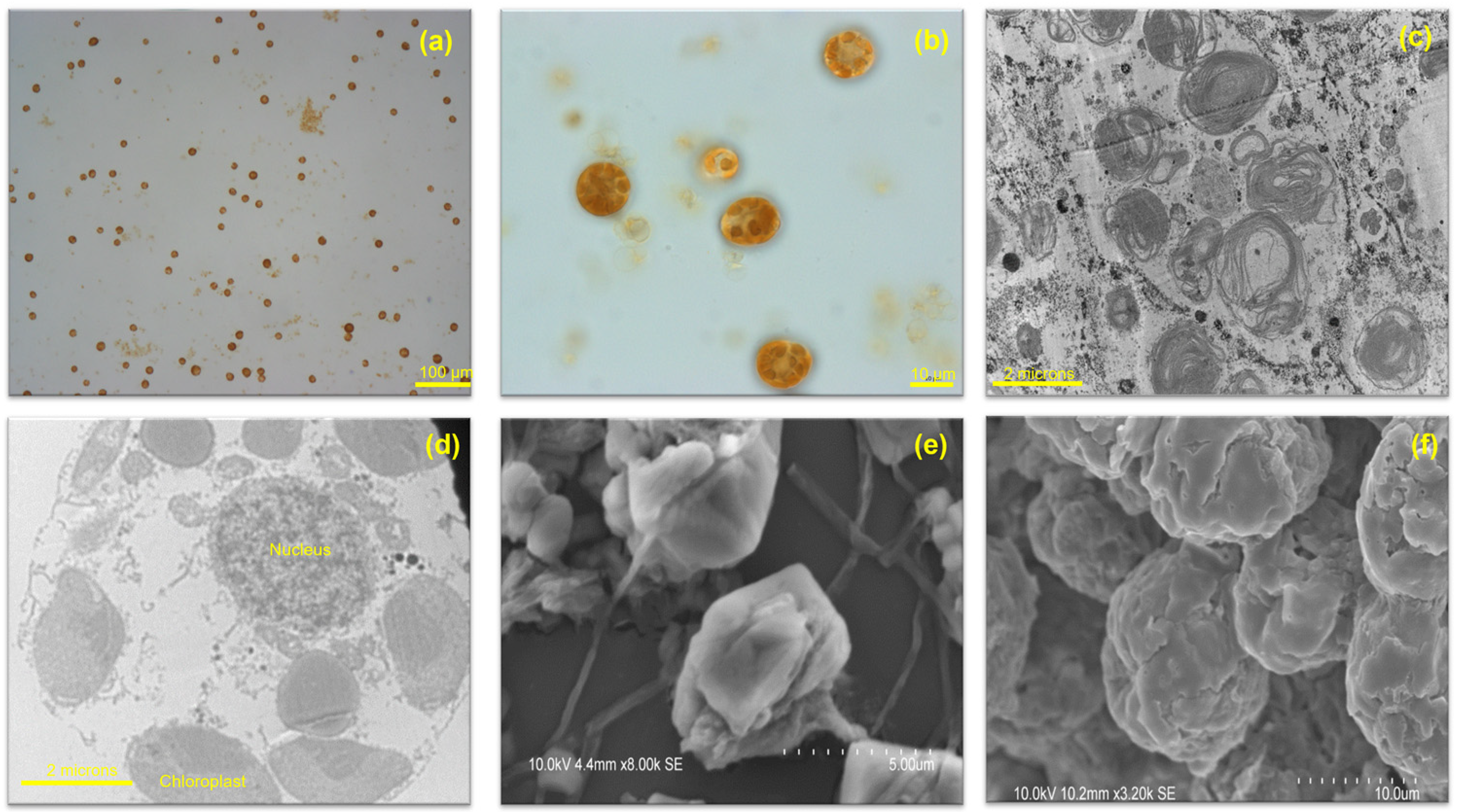
H. akashiwo (Y.Hada) Y.Hada ex Y.Hara & M.Chihara, formerly named H. carterae or Olisthodiscus luteus, is a species with the following characteristics: 12–34 μm in length and 8–10 μm in thickness, pleomorphic, with its shape changing from spheroidal to ovoid or oblong. Contingent upon culture condition and cell age, it can have 10–25 greenish-brown discoid chloroplasts in the peripheral region of each cell, with main pigments of chlorophyll a+c with fucoxanthin. H. akashiwo has two flagella: the anterior flagellum is used for swimming and movement, and the posterior flagellum is rigid [1,5,24,25], Figure 2.
Growth phases, irradiance, duration of photoperiod light phase, diurnally, and nutrient availability can affect the number of chloroplasts per cell [24]. Before daybreak, Heterosigma cells migrate vertically from the bottom layers of their environment to the surface water, and then they migrate downhill just before dusk [6,26]. The cells swim spirally [24]. Under stress, H. akashiwo cells form benthic vegetative cysts [5,27,28]. The cysts are coated with mucilaginous layers and contain a variety of other particles including sand grains, mud, and pieces of diatom frustule [27]. Multiple mucocysts act as discharge sites surrounding the chloroplast. Glycocalyx, a mucus layer, with 1–2 μm thickness covering the motile cells, contains acidic, complex carbohydrates, and a neutral protein–carbohydrate complex of glycoproteins. The function of this layer is still unknown [24]. H. akashiwo is a euryhaline and eurythermal cell with the ability to withstand a salinity range from 2 to more than 50 and a temperature range from less than 5 °C to more than 30 °C [24].

3. The Physiological Effects of Temperature, Light, and Salinity on H. akashiwo

A wide range of environmental drivers can modify physiological processes, growth, and bloom development in a marine ecosystem. H. akashiwo’s growth rate, toxin production, and even pigment composition are directly influenced by various environmental factors such as temperature, light, salinity, pH, and different concentrations of nutrients [12,13,16,26,27]. As it was mentioned before, H. akashiwo is a euryhaline and eurythermal microorganism which can tolerate a broad range of salinity (2 to more than 50), and temperature (less than 5 and more than 30 °C) [24]. The cell can germinate under dim light and can tolerate high light intensity [26].
H. akashiwo isolated from the Seto Inland Sea, Japan showed the highest growth at 25 °C, and the highest toxin production at 20 °C. The lowest growth for this strain was observed at 10 °C and 15 °C, while the highest growth was detected at 25 °C and 30 °C; however, cells were more toxic at lower temperatures. The highest toxin production was detected at 200 μmol photons m−2 s−1 but with a poor growth rate. This condition is in contrast with growth at 100 μmol photons m−2 s−1 which resulted in low toxin production with high growth [12].
Six different strains of H. akashiwo isolated from different geographical regions in the United States, Europe, and New Zealand presented similar responses to temperature, light, and salinity. All different strains showed the highest growth rate at 23 °C with 100 μmol photons m−2 s−1 light which is almost similar to the growth results from the Japanese strain. The optimum growth rate was also obtained at salinities of 20 and 35, while there was no growth at a salinity of 5 [16]. In addition, it was reported that temperature acts as an important factor for cyst survival and germination for these strains. Germination was not observed at 5 °C, was low at 10 °C, increased at 15 °C, and reached its highest level at 25 °C [16].
As a euryhaline species, salinity plays a major role in the growth and toxicity of H. akashiwo [29]. Lower salinity imposes toxin production and affects the morphological condition and motility of H. akashiwo [13]. The optimum growth rate for the Japanese strain was obtained at a salinity of 25 while the highest toxin production was observed at a salinity of 20 [13]. The intensity of H. akashiwo algal bloom in Saudi coastal waters negatively correlated with salinity over a narrow range (26.3–34.20). H. akashiwo cells failed to flourish in salinity over 40 [30]. Similar results regarding the salinity effect on the growth and toxicity of H. akashiwo isolated from North America coastal waters were observed by Strom et al. [29] and Ikeda et al. [31]. Each study reported higher cell toxicity at the lowest salinity level used in their studies.
Employing a design-of-experiment (DOE) approach is an efficient and accurate method to investigate the simultaneous response in the presence of two or more factors and showed the maximum growth rate of H. akashiwo to occur at 25 °C, a salinity of 20.5, and a light intensity of 200 μmol photons m−2 s−1 [32].
Salinity presented a direct influence on the growth rate and toxin yields of the two other strains of harmful raphidophytes: Chattonella antiqua and C. marina. These two strains showed a maximum toxicity at a salinity of 25 [33].
The broad halotolerant may be a defensive adaptation as a refuge from predation using the following mechanisms: the difference between H. akashiwo and some of its protistan predators in salinity tolerance, a reduction in feeding rate of the protist or an increase of H. akashiwo toxicity against protist predators in low salinity, and strong ability to acclimate to lower salinity as physiological and behavioural plasticity in H. akashiwo [29]. To reduce the stress associated with increased turgor pressure caused by a reduction in salinity level, the H. akashiwo cell membrane alters its permeability [31]. The lack of a thick cell wall in H. akashiwo [27], Figure 2, suggests low elastic modulus (Ɛ) values [31] associated with the elasticity of the cell wall that maintain turgor pressure without damage to the cell structure [34]. A strong correlation was reported between cell permeability and cytotoxicity of H. akashiwo isolated from the west coast of the USA [31]. Both responses increased as salinity decreased from 32 to 10 [31]. The DOE approach presented the maximum cell permeability at a temperature of 15 °C, salinity of 5, and light intensity of 30 μmol photons m−2 s−1 [32]. The interaction between light intensity and temperature showed a significant impact on H. akashiwo’s cell permeability [32].

4. Nutrients Impact

As a result of urban, agricultural, and industrial development in coastal areas around the world, where half of the world’s human population resides, huge amounts of nutrient inputs lead to the enhancement of phytoplankton growth and biomass accumulation, known as eutrophication. In many cases, eutrophication further progresses in hypoxia, fish and shellfish kills, and losses of higher plant and animal habitat [35]. The structure of phytoplankton communities and the metabolites they produce, including toxins and allelopathic compounds, are in direct relation with nutrient concentrations, ratios, and speciation in their environment [19,36].
A strong positive correlation between algal bloom intensity and the accumulation of macronutrients, such as NH4, NO3, and PO4, was detected for the H. akashiwo algal bloom in Saudi coastal waters [30]. The growth and bloom of H. akashiwo may be initiated or maintained by any of the three nitrogen substrates (ammonium (NH4+), nitrate (NO3), and urea (CH4N2O) in the following order in N uptake: NH4+ > NO3 > urea [37,38]. There is little expression of toxicity when cells are phosphate limited; cells are somewhat more toxic when growth is nitrogen limited [37], and even more toxic when cell growth is iron limited [39]. H. akashiwo growth rate and cell density under low phosphorus concentration treatment were 17–21% and 41% less than the cells that grew under high phosphorus concentration treatment, respectively, which implies that phosphorus is a major parameter for H. akashiwo bloom formation [15].
Micronutrients have also been implicated in the regulation of toxicity. Runoff water is a major source of micronutrients such as iron and vitamin B12 in English Bay, Vancouver [2]. During the period of maximum runoff, the bloom of H. akashiwo was reported in this area [2], and it was concluded that the increase in vitamin B12 alleviated a vitamin deficiency that was impeding growth. A substantial proportion of examined phytoplankton require exogenous vitamin B12 for their growth and this source seems to be bacteria, representing an important and unsuspected symbiosis [40]. However, in marine ecosystems, the distributions and dynamics of dissolved vitamin B12 have not received appropriate attention with regard to HAB dynamics.

5. Climate Change and Global Warming

Anthropogenic activities are having profound effects on the global environment [41]. The atmospheric and ocean surface temperatures have been increasing, resulting in melting ice and icebergs and a subsequent rise in the ocean level [42]. The average increase in temperature between 1880 to 2012 for combined land and ocean surface has been recorded at 0.85 (0.65 to 1.06) °C. However, the average increase in ocean surface temperature between 1971 and 2010, was 0.11 (0.09 to 0.13) °C per decade. The substantial glacier mass loss globally, along with ocean thermal expansion from warming has raised sea levels since the early 1970s and stratified surface waters [42]. In addition, an increase in temperature can alter seasonal patterns by making summers longer and shifting fall and spring timings [36]. Mid- and high-latitude regions are anticipated to be influenced excessively by these changes [19,36,42].
In addition, since 1750, the atmospheric levels of carbon dioxide, methane, and nitrous oxides have grown remarkably [43]. CO2 has increased from 280 ppm in pre-industrial times in the late eighteenth century to 417.51 ppm in November 2022 [44], mostly due to fossil fuel combustion and deforestation, and is predicted to increase to ~750 ppm by 2100 [42]. Approximately half of this atmospheric CO2 has been absorbed by the oceans over the past 200 years [45,46] and dissolved in surface waters, resulting in ocean acidification by lowering the oceanic hydrogen ion concentration (pH) [42]. Since the beginning of the industrial era, this uptake of CO2 has led to a reduction of 0.1 units in surface seawater pH, corresponding to a 26 % increase in the concentration of hydrogen ions [42,46].
Climate change and global warming can affect both species selection and phytoplankton population dynamics [19,36]. Growth [47,48], germination [49], motility [50,51], nutrient uptake, photosynthesis and other physiological processes [52,53,54], metabolic rates [19,36], and toxicity [47] of planktonic systems globally will be influenced by climate change pressures.
H. akashiwo growth, maximum population density, carrying capacity, and primary productivity rates may be elevated by future high CO2 regimes and temperature [48,55]. Employing the DOE approach showed that warmer temperatures and elevated levels of CO2 will likely increase the potential growth rate and biomass yield but not necessarily the toxicity of the fish-killing flagellate H. akashiwo [56].
The motility behaviour of H. akashiwo is another parameter that may be affected by the elevation of CO2 under future conditions [51]. An increase in swimming speed was detected by increasing pCO2 by 8.2% when pCO2 shifted from 280 ppm to 750 ppm [51].
H. akashiwo growth may benefit excessively from a future increase in CO2 and temperature as proved by laboratory experiments carried out under future environmental conditions, and scientific reports in the past decades [48,51,55]. For example, the population density of H. akashiwo has increased during recent decades in various locations. In the Skidaway estuary, Georgia, USA, the annual mean concentration of H. akashiwo increased from 94–127 cells mL−1 in 1986–1987 to 552–670 cells mL−1 in 2007–2008 [23].

6. H. akashiwo Cyst Formation

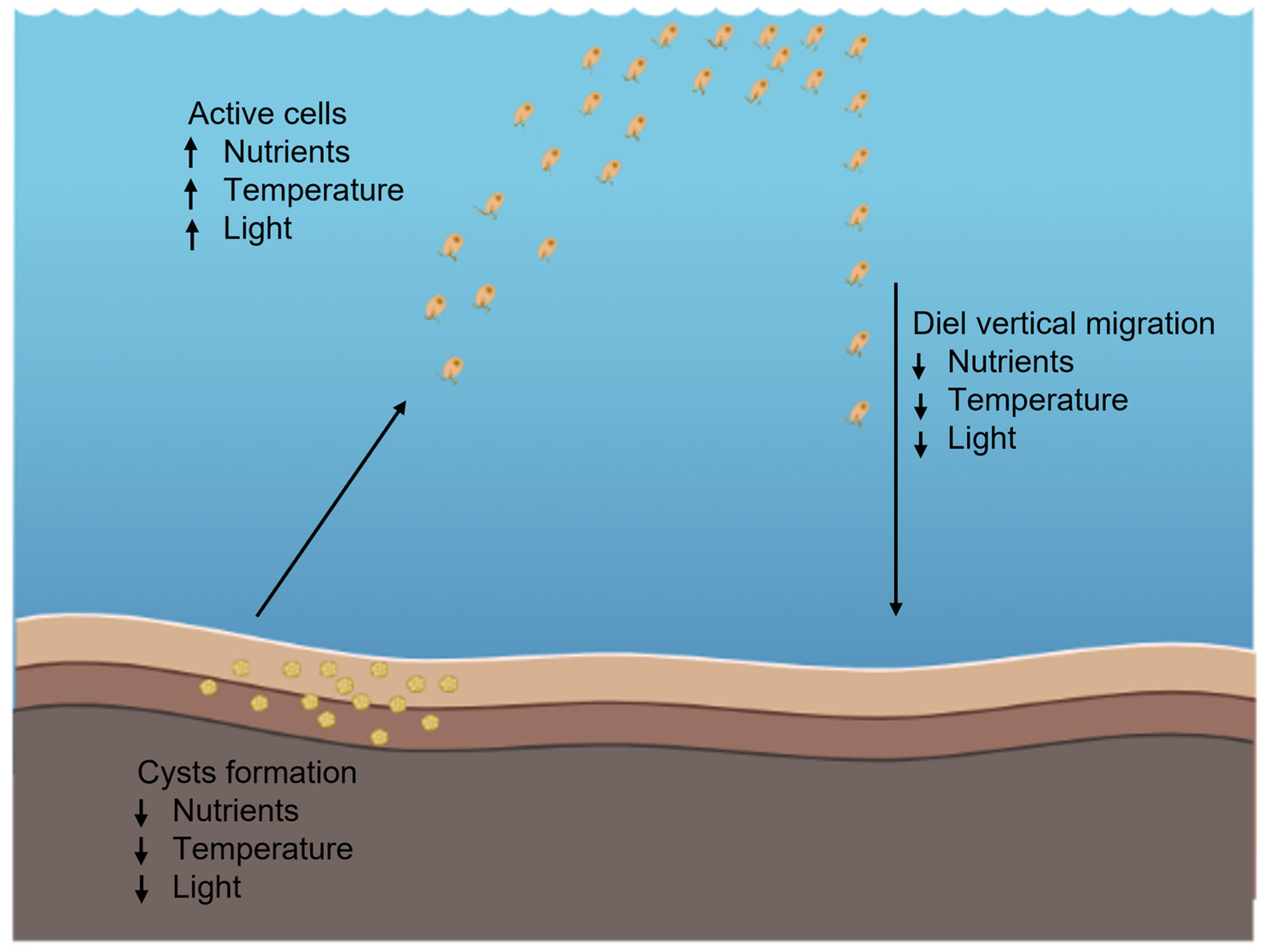
Many harmful algae exhibit an active motile stage and a benthic vegetative cyst stage [26,28]. As a result of stress, such as nutrient limitation, reduced temperature, and light, these rigid cysts form and sink to the seabed where they can survive for years in ocean sediments [5,10,28,57,58]. These cysts are immovable, close to 10 μm in diameter, spherical in shape, surrounded by mucilaginous materials of sediments, and have a yellow-greenish colour [27,57].
Environmental characteristics such as temperature, light, and nutrients are the main factors affecting germination and transfer from benthic vegetation cysts to the active cell phase. The active cell phase is followed by an increase in metabolic activity, cell division, and upward movement to the water surface. The rapid transition could form harmful algal blooms [10,16,26].
H. akashiwo germinates quickly, transferring from a benthic cyst to an active cell [26]. Temperature and light are the key factors affecting germination. A water temperature of 15 °C and dim light have been reported as the threshold for H. akashiwo cyst germination [10,16,26,59,60], Figure 3.
H. akashiwo showed no or very slow growth at 11 °C; nevertheless, its cysts survived at 11 °C in the dark for 650 days. Cyst survival decreased to 165 days in the dark at 25 °C [59]. The cyst cells contain a significantly lower total lipid content compared to active cells. For a successful transition between these two stages, fatty acid composition plays a crucial role [26]. During this transition and under certain environmental conditions such as an abrupt change in temperature, salinity, biogenic elements content, and illumination, polyunsaturated fatty acids (PUFAs) are required to form photosynthetic membranes [61] and modify cellular and organelle membranes [62]. Neutral lipid reservoirs provide the required energy for algal motility [26].
In contrast, nutrient depletion and a decrease in irradiance with depth may be possible triggers to force the cells towards the benthic environment in search of new nutrients or induce vegetative resting cyst formation [63]. H. akashiwo revealed greater toxicity under nutrient depletion stress [64] during the late stationary growth phase [28]. Within individual communities or blooms, cysts with toxic characteristics can form if different potential toxicity components exist in a population, and therefore, the chance of a harmful algal bloom increases. Cells with a high sinking rate display the highest toxicity in comparison with positively buoyant populations [28].

7. H. akashiwo Fish-Killing Mechanisms

Every year, massive mortality of fish occurs in coastal waters around the world because of algal blooms of raphidophyte H. akashiwo, resulting in huge financial losses [65,66]. However, the toxin(s) and mechanism(s) of fish-killing by H. akashiwo and other ichthyotoxic raphidophytes are still unclear. The following procedures are some of the toxicological mechanisms for fish killing presented by different researchers, Figure 4:
  • Asphyxiation by covering fish gills and physical damage as a result of extreme mucous secretion [4].
  • Asphyxiation and gill tissue damage due to reactive oxygen species (ROS) production, such as superoxide, hydrogen peroxide, and hydroxyl radicals [67,68,69].
  • Cardiac disorders and gill injury as a result of organic toxin production, known as brevetoxin-like neurotoxins [12,13,70].
  • Hemolytic compounds that lyse red blood cells [39,71].
Each of these mechanisms will be discussed in the following sections.
Figure 4. Framework for physiological activities associated with a fish kill by H.akashiwo. After attachment to the gill of the fish, algal cells embed into gill tissues using exoenzymes. The presence of the algal cells stimulates mucus secretion, followed by the algal cells producing neurotoxins, and reactive oxygen species that cause asphyxiation in the fish. In addition, hemolysin compounds can lyse the erythrocytes to scavenge nitrogen and iron. This image was created with BioRender (https://biorender.com/ (accessed on 22 January 2023)).
Microbiolres 14 00012 g004

7.1. Mucous Secretion

One of the listed fish-killing mechanisms is a mucous secretion produced by some marine algae which has been associated with fish kills worldwide, resulting in asphyxiation by physical clogging and mechanical obstruction of gill perfusion [4].
The produced algal toxin by C. antiqua caused osmoregulatory disorders on the gill primary lamella and removed the mucus layer from the young yellowtail fish gills [72]. It was then followed by mucous secretion stimulation leading to hyaline degeneration of the cell membrane and consequently cell loss and, therefore, disruption in gas distribution and finally fish death. A similar mechanism in the presence of H. akashiwo was responsible for the massive chinook salmon killing in Big Glory Bay, Stewart Island, New Zealand in 1989 [4]. The histopathological test was performed on chinook salmon from this bloom and uncommon changes were observed in their gills and intestines. Algal toxins damaged the gill lamella, reduced blood flow, and increased oxygen, carbon dioxide, and ammonia diffusion distance, which caused asphyxiation and osmotic shock [4]. On the other hand, H. akashiwo’s toxin was concentrated in the intestine as the water was absorbed by the intestine [4].
Mucous secretion in yellowtail gills was stimulated with oxygen radical production by Chattonella and the destruction of the gas exchange capacity of the gills by shunting respiratory water current away from the lamellae [73]. In normal growth conditions, superoxide anions (O2) and hydrogen peroxide (H2O2) are produced and released by C. marina and H. akashiwo. O2 production is stimulated in the presence of some mucous compounds such as lectins. Excessive mucus on the surface of gill lamella may interfere with O2 transfer resulting in asphyxia in yellowtail fish [68].
In the presence of 100 μg mL−1 fish mucus prepared from the skin and gills of yellowtail, red sea bream, and Japanese flounder, a 4- to 6-fold increased level of O2 was detected in both H. akashiwo and C. marina, which may suggest that the common substances present in the skin and gill mucus may be responsible for stimulating these two different raphidophytes [74]. Similar results were observed in the presence of C. marina on short-necked clam (Ruditapes philippinarum). The results indicated that the contact between C. marina and gill tissue led to simultaneous glycocalyx discharge from C. marina with increased O2 production causing massive mucous secretion from the gills and suffocation in the end [75].
Gill damage was reported as a potential cause of fish mortality [76]. Due to algal toxin, the gas exchange capacity is reduced in the gill level, and therefore, epithelial separation of gill filaments occurs, resulting in asphyxiation [76].

7.2. Reactive Oxygen Species (ROS) Production

Reactive oxygen species (ROS) production such as superoxide (O2), hydrogen peroxide (H2O2), and hydroxyl radicals (OH) is another mechanism that has been suggested for fish mortality in the presence of H. akashiwo [69,74,76,77,78,79,80]. These molecules are unstable, with a short half-life that can cause death by damaging DNA, RNA, and proteins when they react with other molecules [81].
O2 and H2O2 are generated in the cell plasma membranes as a result of NADH oxidation activity. The NADH oxidation activity is directly related to the presence of iron ions which is crucial for ROS generation [68,82]. The membrane of the epithelial cells of the secondary lamellae is the first target of the produced ROS by H. akashiwo, which is followed by edema in gill lamellae [67].
Under normal growth conditions H. akashiwo, C. marina, and some other strains of Raphidophyceae class are able to produce and release O2 and H2O2 [68,74]. C. marina cells are able to produce greater amounts of O2 and H2O2 due to their larger cell size [68,83]. As a result of a smaller cell size, H. akashiwo produces a lower level of cellular superoxide but with a high potential to produce a greater level of total superoxide through the high cell biomass achieved during a bloom [83]. The production rate of O2 and H2O2 in H. akashiwo depends on the cell concentration [67]. ROS is hardly detectable at the cell concentrations less than 104 cells mL−1, but when cell density reaches a concentration higher than 104 cells mL−1, greater levels of O2 and H2O2 are released [67]. H. akashiwo produced up to 7.6 pmol min−1 104 cells−1 hydrogen peroxide which led to an H2O2 extracellular concentration of ~0.5 μmol l−1 [69]. The temperature and light intensity did not show a significant influence on hydrogen peroxide production; nevertheless, when the iron quantity was reduced to 10−9 mol l−1, the H2O2 production increased from 3.9 to 6.9 pmol min−1 104 cells−1 [69]. Iron is a redox metal that reduces H2O2 to produce hydroxy radicals (OH) [78]. Consequently, it was concluded that the production of extracellular hydrogen peroxide occurs through a metabolic pathway that is not directly linked to photosynthesis. The results of a study on the influence of hydrogen peroxide on the mortality of vertebrate cell lines (human embryonic kidney and rat osteosarcoma) and the invertebrate Artemia salina (brine shrimp) indicated that the cell toxicity was induced at hydrogen peroxide concentrations greater than 10−4 M. On the other hand, comparing the H2O2 induced toxicity between vertebrate cell lines and marine invertebrates indicated that marine invertebrate cells were not as sensitive to hydrogen peroxide as vertebrate cells [78].
On the other hand, the mucus production from the skin and gill tissue of different fish species was reported to have a stimulating effect on the increased amount of O2 in H. akashiwo and C. marina [84]. However, H2O2, which is the most stable ROS, did not reveal any increase in the hemolytic activity of H. akashiwo and erythrocyte lysis [39].
Similar results were observed for other raphidophyte species, such as C. marina, in the presence of reactive oxygen species (ROS) which were responsible for fish and shellfish gill tissue damage and mortality [75,85]. This strain is able to produce 100 times more reactive oxygen species compared to other algal species [86]. The highest level of O2 production was observed at the exponential growth phase of C. marina with cell concentrations of more than 103 cells mL−1.
On the other hand, the excessive generation of O2 was related to the presence of iron ions [85,86]. The first physiological disorder observed in fish after exposure to C. marina was a decrease in oxygen partial pressure of arterial blood [68]. The ventilation volume and velocity of C. marina gills increased when the dissolved oxygen decreased [86]. The discharge of glycocalyx from C. marina increased the O2 production, possibly resulting in a massive ROS-mediated mucous secretion from the gills tissue which caused suffocation [68,74,75].
Small amounts of free fatty acids (FFA) are toxic to fish [86]. In the presence of superoxide and 0.2 mg l−1 polyunsaturated fatty acid eicosapentaenoic acid (EPA), an LT50 of 83 min was obtained, which is equal to either 4 mg l−1 of EPA or around 103 cells mL−1 of Chattonella culture as a toxic level for fish [86].
Different reasons for gill damage as a result of FFAs and ROS have been suggested, including the reduction of respiratory capacity because of gill membrane separation, reduction of blood pH due to FFAs or superoxide absorption leading to suppressed respiratory and/or osmoregulatory capacity, and reduction of osmoregulatory capacity as a result of the destruction of the chloride cells [86].
Nutrient deficiency (nitrogen, phosphorus) has a negative effect on ROS production by C. antiqua during the cell growth phase [79]. ROS (O2 and H2O2) production in C. antiqua was higher in the daytime, which suggests that it has a correlation with the photosynthetic process [79]. A wide range of N:P ratios, salinity, and light and darkness cycle did not show a significant influence on ROS production, and ROS production was not directly related to the growth rate of C. marina whereas high pH and iron limitation affected the ROS production in this strain [80].

7.3. Toxin Production

Neurotoxin production is another fish-killing mechanism caused by polycyclic polyether brevetoxins (PbTx) which leads to gill damage or cardiac disorders [70,87,88]. Brevetoxins contain nine different components which are based on two different ladder-liker lipophilic carbon structural backbones, PbTx-2 (brevetoxin B) and PbTx-1 (brevetoxin A) [88]. Other toxins are derived from these two groups [88], Figure 5. The consumption of brevetoxin by humans is responsible for a neurological disease called neurotoxic shellfish poisoning (NSP), which causes a malfunction of voltage-gated sodium channels in nerve cells [89]. Other food poisoning syndromes are caused by different algal toxins, especially neurotoxins, and impact human health through the consumption of contaminated shellfish, coral reef fish, and finfish, or through water or aerosol exposure are as follows: paralytic shellfish poisoning (PSP), amnesic shellfish poisoning (ASP), diarrheic shellfish poisoning (DSP), ciguatera fish poisoning (CFP), and azaspiracid shellfish poisoning (ASP) [90].
H. akashiwo was able to produce four different neurotoxic compounds, HaTx-I, HaTx-IIa, HaTx-IIb, and HaTx-III, which corresponded to brevetoxin components, PbTx-2, PbTx-9, PbTx-3, and oxidized PbTx-2, respectively, using high-pressure chromatography (HPLC) technique [70]. Among these toxins, HaTx-III had the highest (1.30 pg cell−1) and HaTx-IIa the lowest (0.05 pg cell−1) concentration of toxin in this algal culture. Fish death as a result of paralysis occurred when the cell density of H. akashiwo was more than 120,000 cells mL−1. In addition to cell density, cell size, age of culture, temperature, salinity, and light intensity are some other factors that may influence toxin production in H. akashiwo [12,13,70]. It was reported that low salinity stimulated brevetoxin production and the lowest toxin production was observed at 30 °C [12,13]. Moreover, rupturing H. akashiwo cells enhanced the ichthyotoxic effect of this raphidophyte, meaning that the toxin(s) is located inside the cellular compartments [91].
Fibrocapsa japonica was reported to produce the highest amount of toxin in the middle of the exponential phase and the lowest level in the stationary phase. F. japonica produced five different toxins, the concentration of which fluctuated based on cell age and the growth phase of the culture. No toxicity was observed until the culture cell density reached 4.1 × 10³ cells mL−1 [92].
Neurotoxin compounds created by Chatonella red tide in coastal waters in Delaware, USA, caused a massive fish kill in 2000 [93]. The histological studies showed severe damage to fish gills in the presence of C. marina [94]. The initial toxicity for C. marina was recorded at 700 cells mL−1 [95]. It was observed that when the fish were exposed to C. marina, their heartbeat decreased very fast and consequently reduced the oxygen uptake through the gills due to limited blood flow resulting in gill damage and cardiac disorder [87,95]. The depolarization of the vagal nerve of the fish is reported as a reason for heartbeat reduction in the presence of the neurotoxin produced by C. marina [87].

7.4. Hemolytic Activity

The last introduced cause for fish killing by toxic algae is the hemolytic and hemagglutinating compounds [30,39,96,97,98]. Unfortunately, little is known about the mechanism of hemolysis, but it was reported that the hemolytic activity of raphidophytes is directly related with environmental conditions such as light, salinity, and nutrients [39,98].
The highest hemolytic activity in H. akashiwo was observed when the cell was damaged [39]. On the one hand, the most hemolytic activity was detected in the exponential growth phase when the cells had the maximum activity. The hemolytic agents were active in the presence of adequate light. On the other hand, the ultrasonically ruptured cell cultures of H. akashiwo indicated powerful hemolytic activity in comparison with intact cell cultures, which suggested that the hemolytic compounds were located in certain intracellular compartments [39,56].
In the bloom area of H. akashiwo in Saudi coastal waters, hemolytic activity increased when the number of cells in the culture declined and reached the maximum level when the bloom began to collapse [30].
The maximum hemolytic activity had a direct relation with low salinity and high nutrient concentrations; however, nutrient limitation did not induce hemolytic activity [30]. The DOE approach showed that the highest hemolytic activity was detected at a temperature of 15 °C, a salinity of 10, and a CO2 level at 400 p.p.m., while there was an interaction between temperature and CO2 level that can significantly influence the hemolytic activity of H. akashiwo [56].
F. japonica, another raphidophyte species, resulted in a wide range of EC50 between 0.4 and 1.9 × 10⁴ cells mL−1 [99]. The hemolytic activity of F. japonica was only 7% of its activity in the presence of light and minimum salinity conditions of the test [96,98]. No significant difference in hemolytic activity was observed between the exponential and stationary phases. However, in the stationary phase, these cells showed less motility, and under P and/or C limitations in this stage they were able to produce sticky aggregates [97].
Some of the hemolytic compounds were isolated and characterized as polyunsaturated fatty acids (PUFAs) in nature [96,97,98]. Various techniques such as NMR, LC–ESI–MS, ESI–MS–MS, IR, GC–MS, and GC–HRMS were employed to identify the chemical characteristics of three hemolytic compounds, Fj1, Fj2, and Fj3, isolated from F. japonica and categorized as polyunsaturated fatty acids [98]. Gel-filtration chromatography was used to isolate the toxins from cell-free methanol extract of C. marina, followed by further purification procedures via thin-layer chromatography (TLC) and high-performance liquid chromatography (HPLC) [100]. The results showed that the hemolytic compounds isolated from C. marina were also categorized as fatty acids such as polyunsaturated fatty acids [100,101].

8. Summary

H. akashiwo is a golden-brown dinoflagellate with a significant potential to create harmful algal blooms and generate massive fish kills, resulting in a significant economic loss per bloom. H. akashiwo cell size varies between 12–34 μm in length and the shape changes from spheroidal to ovoid or oblong. Each cell contains 10–25 greenish-brown discoid chloroplasts in the peripheral region of each cell.
This raphidophyte is known as a euryhaline and eurythermal microorganism, which means it can tolerate a broad range of salinity and temperature. However, under unpleasant environmental conditions and for protection and survival, cells form cysts and sink into the sediment. Although scientists still debate about toxin production and mechanisms, four different mechanisms for fish kill were proposed: mucus secretion, ROS production, toxin production, and hemolytic activity. This knowledge help researchers to find better solutions for protection of fish industries and reduce economic losses due to the presence of harmful algal blooms.

Funding

This research received no external funding.

Acknowledgments

The author thanks C.G. Trick for his guidance.

Conflicts of Interest

The author declares no conflict of interest.

References

  1. Hara, Y.; Chihara, M. Heterosigma akashiwo morphology, ultrastructure and taxonomy of the Raphidophycean alga Heterosgima akashwio. Bot. Mag. Tokyo 1987, 100, 151–163. [Google Scholar] [CrossRef]
  2. Taylor, F.J.R.; Haigh, R. The Ecology of fish-killing blooms of the chloromonad flagellate Heterosigma in the Strait of Georgia and adjacent Waters. In Toxic Phytoplankton Blooms in the Sea; Elsevier: New York, NY, USA, 1993; pp. 705–710. [Google Scholar]
  3. Smayda, T.J. Harmful Algal Bloom Communities in Scottish Coastal Waters: Relationship to Fish Farming and Regional Comparisons—A Review Paper 2006/3. Scottish Executive, Scottish Environmental Protection Agency SEPA. Available online: https://www.webarchive.org.uk/wayback/archive/20160122225400/http://www.gov.scot/Publications/2006/02/03095327/16 (accessed on 22 January 2023).
  4. Chang, F.H.; Anderson, C.; Boustead, N.C. First record of a Heterosigma (Raphidophyceae) bloom with associated mortality of cage-reared salmon in Big Glory Bay, New Zealand. N. Z. J. Mar. Freshw. Res. 1990, 24, 461–469. [Google Scholar] [CrossRef]
  5. O’Halloran, C.; Silver, M.W.; Holman, T.R.; Scholin, C.A. Heterosigma akashiwo in central California waters. Harmful Algae 2006, 5, 124–132. [Google Scholar] [CrossRef]
  6. Yamoehi, S.; Abe, T. Mechanisms to initiate a Heterosigma akashiwo red tide in Osaka Bay. II. Diel vertical migration. Mar. biol. 1984, 261, 255–261. [Google Scholar] [CrossRef]
  7. Nagasaki, K.; Tarutani, K.; Yamaguchi, M. Growth characteristics of Heterosigma akashiwo virus and its possible use as a microbiological agent for red tide control. Appl. Environ. Microbiol. 1999, 65, 898–902. [Google Scholar] [CrossRef] [PubMed] [Green Version]
  8. Yu, J.; Yang, G.; Tian, J. The Effects of the harmful alga Heterosigma akashiwo on cultures of Schmackeria Inopinus (Copepoda, Calanoida). J. Sea Res. 2010, 64, 287–294. [Google Scholar] [CrossRef]
  9. Hershberger, P.K.; Rensel, J.E.; Matter, A.L.; Taub, F.B. Vertical distribution of the chloromonad flagellate Heterosigma carterae in columns: Implications for bloom development. Can. J. Fish. Aquat. Sci. 1997, 2234, 2228–2234. [Google Scholar] [CrossRef]
  10. Rensel, J.E.; Haigh, N.; Tynan, T.J. Fraser river sockeye salmon marine survival decline and harmful blooms of Heterosigma akashiwo. Harmful Algae 2010, 10, 98–115. [Google Scholar] [CrossRef]
  11. Livingston, R.J. Phytoplankton bloom effects on a gulf estuary: Water quality changes and biological response. Ecol. Appl. 2007, 17, 110–128. [Google Scholar] [CrossRef]
  12. Ono, K.; Khan, S.; Onoue, Y. Effects of temperature and light intensity on the growth and toxicity of Heterosigma akashiwo (Raphidophyceae). Aquac. Res. 2000, 31, 427–433. [Google Scholar] [CrossRef]
  13. Haque, S.M.; Onoue, Y. Effects of salinity on growth and toxin production of a noxious phytoflagellate, Heterosigma akashiwo (Raphidophyceae). Bot. Mar. 2002, 45, 356–363. [Google Scholar] [CrossRef]
  14. Flores-Leñero, A.; Vargas-Torres, V.; Paredes-Mella, J.; Norambuena, L.; Fuenzalida, G.; Lee-Chang, K.; Mardones, J.I. Heterosigma akashiwo in Patagonian Fjords: Genetics, growth, pigment signature and role of PUFA and ROS in ichthyotoxicity. Toxins 2022, 14, 577. [Google Scholar] [CrossRef]
  15. Yang, A.; Bellerby, R.G.J.; Wang, Y.; Li, X. Growth and nutrient uptake characteristics of Heterosigma akashiwo (Raphidophyceae) under nitrogen and phosphorus concentrations in the East China Sea. Water 2021, 13, 3166. [Google Scholar] [CrossRef]
  16. Martínez, R.; Orive, E.; Laza-Martínez, A.; Seoane, S. Growth response of six strains of Heterosigma akashiwo to varying temperature, salinity and irradiance conditions. J. Plankton Res. 2010, 32, 529–538. [Google Scholar] [CrossRef]
  17. Engesmo, A.; Strand, D.; Gran-Stadniczeñko, S.; Edvardsen, B.; Medlin, L.K.; Eikrem, W. Development of a QPCR assay to detect and quantify ichthyotoxic flagellates along the Norwegian coast, and the first Norwegian record of Fibrocapsa japonica (Raphidophyceae). Harmful Algae 2018, 75, 105–117. [Google Scholar] [CrossRef] [PubMed]
  18. Bornman, E.; Adams, J.B.; Strydom, N.A. Algal blooms of Heterosigma akashiwo and mugilidae gill alterations. Estuar. Coast. 2022, 45, 1674–1687. [Google Scholar] [CrossRef]
  19. Wells, M.L.; Karlson, B.; Wulff, A.; Kudela, R.; Trick, C.; Asnaghi, V.; Berdalet, E.; Cochlan, W.; Davidson, K.; De Rijcke, M.; et al. Future HAB science: Directions and challenges in a changing climate. Harmful Algae 2020, 91. [Google Scholar] [CrossRef] [PubMed]
  20. Mardones, J.I.; Paredes-Mella, J.; Flores-Leñero, A.; Yarimizu, K.; Godoy, M.; Artal, O.; Corredor-Acosta, A.; Marcus, L.; Cascales, E.; Pablo Espinoza, J.; et al. Extreme harmful algal blooms, climate change, and potential risk of eutrophication in Patagonian Fjords: Insights from an exceptional Heterosigma akashiwo fish-killing event. Prog. Oceanogr. 2022, 210, 102921. [Google Scholar] [CrossRef]
  21. Guiry, G.M.; Guiry, M. AlgaeBase. World-Wide Electronic Publication, National University of Ireland, Galway. 2022. Available online: https://www.algaebase.org (accessed on 21 December 2022).
  22. Throndsen, J. The planktonic marine flagellates. In Identifying Marine Phytoplankton; Tomas, C.R., Ed.; Academic Press: San Diego, CA, USA, 1997; pp. 591–729. [Google Scholar]
  23. Verity, P.G. Expansion of Potentially Harmful Algal Taxa in a Georgia Estuary (USA). Harmful Algae 2010, 9, 144–152. [Google Scholar] [CrossRef]
  24. Smayda, T.J. Ecophysiology and bloom dynamics of Heterosigma akashiwo (Raphidophyceae). In Physiology Ecology of Harmful Algal Blooms; Anderson, D.M., Cembella, A.D., Hallegraeff, G.M., Eds.; Springer-Verlag: Berlin, Germany, 1998; pp. 113–132. [Google Scholar]
  25. Bowers, H.A.; Kempton, J.W.; Lewitus, A.J.; Oldach, D.W. Raphidophycea [chadefaud ex silva] systematics and rapid identification: Sequence analyses and real-time PCR assay. J. Phycol. 2006, 1348, 1333–1348. [Google Scholar] [CrossRef] [PubMed] [Green Version]
  26. Tobin, E.D.; Grünbaum, D.; Patterson, J.; Cattolico, R.A. Behavioral and physiological changes during benthic-pelagic transition in the harmful alga, Heterosigma akashiwo: Potential for rapid bloom formation. PLoS ONE 2013, 8, 1–15. [Google Scholar] [CrossRef] [PubMed] [Green Version]
  27. Imai, I.; Itakura, S.; Itoh, K. Cysts of the red tide flagellate Heterosigma akashiwo, Raphidophyceae, found in bottom sediments of Northern Hiroshima Bay, Japan. Nippon Suisan Gakkaishi 1993, 59, 1669–1673. [Google Scholar] [CrossRef] [Green Version]
  28. Powers, L.; Creed, I.F.; Trick, C.G. Sinking of Heterosigma akashiwo results in increased toxicity of this harmful algal bloom species. Harmful Algae 2012, 13, 95–104. [Google Scholar] [CrossRef]
  29. Strom, S.L.; Harvey, E.L.; Fredrickson, K.A.; Menden-Deuer, S. Broad salinity tolerance as a refuge from predation in the harmful raphidophyte alga Heterosigma akashiwo (Raphidophyceae). J. Phycol. 2013, 49, 20–31. [Google Scholar] [CrossRef]
  30. Mohamed, Z.A.; Al-Shehri, A.M. The Link between shrimp farm runoff and blooms of toxic Heterosigma akashiwo in Red Sea coastal waters. Oceanologia 2012, 54, 287–309. [Google Scholar] [CrossRef] [Green Version]
  31. Ikeda, C.E.; Cochlan, W.P.; Bronicheski, C.M.; Trainer, V.L.; Trick, C.G. The effects of salinity on the cellular permeability and ichthyotoxicity of Heterosigma akashiwo. J. Phycol. 2016, 53, 745–760. [Google Scholar] [CrossRef]
  32. Mehdizadeh Allaf, M.; Trick, C.G. Multiple-stressor design-of-experiment (DOE) and one-factor-at-a-time (OFAT) observations defining Heterosigma akashiwo growth and cell permeability. J. Appl. Phycol. 2019, 31, 3515–3526. [Google Scholar] [CrossRef]
  33. Haque, S.M.; Onoue, Y. Variation in toxin compositions of two harmful raphidophytes, Chattonella antiqua and Chattonella marina, at different salinities. Environ. Toxiol. 2001, 17, 113–118. [Google Scholar] [CrossRef]
  34. Philip, J.R. The osmotic cell, solute diffusibility, and the plant water cconomy. Plant Physiol. 1958, 33, 264–271. [Google Scholar] [CrossRef] [Green Version]
  35. Paerl, H.W.; Hall, N.S.; Peierls, B.L.; Rossignol, K.L. Evolving paradigms and challenges in estuarine and coastal eutrophication dynamics in a culturally and climatically stressed world. Estuar. Coast. 2014, 37, 243–258. [Google Scholar] [CrossRef]
  36. Wells, M.L.; Trainer, V.L.; Smayda, T.J.; Karlson, B.S.O.O.; Trick, C.G.; Kudela, R.M.; Ishikawa, A.; Bernard, S.; Wulff, A.; Anderson, D.M.; et al. Harmful algal blooms and climate change: Learning from the past and present to forecast the future. Harmful Algae 2015, 49, 68–93. [Google Scholar] [CrossRef] [PubMed] [Green Version]
  37. Herndon, J.; Cochlan, W.P. Nitrogen utilization by the raphidophyte Heterosigma akashiwo: Growth and uptake kinetics in laboratory cultures. Harmful Algae 2007, 6, 260–270. [Google Scholar] [CrossRef]
  38. Ji, N.; Zhang, Z.; Huang, J.; Zhou, L.; Deng, S.; Shen, X.; Lin, S. Utilization of various forms of nitrogen and expression regulation of transporters in the harmful alga Heterosigma akashiwo (Raphidophyceae). Harmful Algae 2020, 92, 101770. [Google Scholar] [CrossRef] [PubMed]
  39. Ling, C.; Trick, C.G. Expression and standardized measurement of hemolytic activity in Heterosigma akashiwo. Harmful Algae 2010, 9, 522–529. [Google Scholar] [CrossRef]
  40. Croft, M.T.; Lawrence, A.D.; Raux-Deery, E.; Warren, M.J.; Smith, A.G. Algae acquire vitamin B12 through a symbiotic relationship with bacteria. Nature 2005, 438, 90–93. [Google Scholar] [CrossRef] [PubMed]
  41. Steffen, W.; Grinevald, J.; Crutzen, P.; McNeill, J. The anthropocene: Conceptual and historical. Phil. Trans. R. Soc. A 2011, 369, 842–867. [Google Scholar] [CrossRef]
  42. IPCC. Climate Change: The physical science basis. In Working Group I Contribution to the Fifth Assessment Report of the Intergovernmental Panel on Climate Change; Stocker, T.F., Qin, D., Plattner, G.K., Tignor, M.M.B., Allen, S.K., Boschung, J., Nauels, A., Xia, Y., Bex, V., Midgley, P.M., Eds.; Cambridge University Press: Cambridge, UK; New York, NY, USA, 2013; p. 1535. [Google Scholar]
  43. Moore, S.K.; Trainer, V.L.; Mantua, N.J.; Parker, M.S.; Laws, E.A.; Backer, L.C.; Fleming, L.E. Impacts of climate variability and future climate change on harmful algal blooms and human health. Environ. Heal. A Glob. Access Sci. 2008, 7, 1–12. [Google Scholar] [CrossRef] [Green Version]
  44. NOAA. Trends in Atmospheric Carbon Dioxide–Mauna Loa. US Department of Commerce, National Oceanic and Atmospheric Administration. 2022. Available online: https://gml.noaa.gov/ccgg/trends/ (accessed on 22 January 2023).
  45. Caldeira, K.; Wickett, M.E. Anthropogenic carbon and ocean ph. Nature 2003, 425, 365. [Google Scholar] [CrossRef] [Green Version]
  46. Raven, J.; Caldeira, K.; Elderfield, H.; Hoegh-Guldberg, O.; Liss, P.; Riebesell, U.; Shepherd, J.; Turley, C.; Watson, A. Ocean acidification due to increasing. Coral Reefs 2005, 12/05, 68. [Google Scholar]
  47. Moore, S.K.; Mantua, N.J.; Hickey, B.M.; Trainer, V.L. Recent trends in paralytic shellfish toxins in Puget Sound, relationships to climate, and capacity for prediction of toxic events. Harmful Algae 2013, 8, 463–477. [Google Scholar] [CrossRef]
  48. Xu, D.; Zhou, B.; Wang, Y.; Ju, Q.; Yu, Q.; Tang, X. Effect of CO2 enrichment on competition between Skeletonema costatum and Heterosigma akashiwo. Chin. J. Oceanol. Limnol. 2010, 28, 933–939. [Google Scholar] [CrossRef]
  49. Itakura, S.; Yamaguchi, M. Morphological and physiological differences between the cysts of Alexandrium catenella and A. tamarense (Dinophyceae) in the Seto Inland Sea, Japan. Plankt. Biol. Ecol. 2005, 52, 85–91. [Google Scholar]
  50. Kamykowski, D.; Mccollum, S.A. The temperature acclimatized swimming speed of selected marine dinoflagellates. J. Plankton Res. 1986, 8, 275–287. [Google Scholar] [CrossRef]
  51. Kim, H.; Spivack, A.J.; Menden-deuer, S. pH alters the swimming behaviors of the raphidophyte Heterosigma akashiwo: Implications for bloom formation in an acidified ocean. Harmful Algae 2013, 26, 1–11. [Google Scholar] [CrossRef]
  52. Beardall, J.; Raven, J.A. The potential effects of global climate change on microalgal photosynthesis, growth and ecology. Phycologia 2004, 43, 26–40. [Google Scholar] [CrossRef]
  53. Raven, J.A.; Geider, R.J. Temperature and algal growth. New Phytol. 1988, 110, 441–461. [Google Scholar] [CrossRef]
  54. Marinov, I.; Doney, S.C.; Lima, I.D. Response of ocean phytoplankton community structure to climate change over the 21st century: Partitioning the effects of nutrients, temperature and light. Biogeosciences 2010, 7, 3941–3959. [Google Scholar] [CrossRef] [Green Version]
  55. Fu, F.X.; Zhang, Y.; Warner, M.E.; Feng, Y.; Sun, J.; Hutchins, D.A. A comparison of future increased CO2 and temperature effects on sympatric Heterosigma akashiwo and Prorocentrum minimum. Harmful Algae 2008, 7, 76–90. [Google Scholar] [CrossRef]
  56. Mehdizadeh Allaf, M. The Effect of Multiple Environmental Stressors on the Growth and Toxicity of the Red Tide Alga Heterosigma Akashiwo. Ph.D. Thesis, Western University, London, ON, Canada, 2018. Available online: https://ir.lib.uwo.ca/etd/5799/ (accessed on 22 January 2023).
  57. Itakura, S.; Nagasaki, K.; Yamaguchi, M.; Imai, I. Cyst formation in the red tide flagellate Heterosigma akashiwo (Raphidophyceae). J. Plankton Res. 1996, 18, 1975–1979. [Google Scholar] [CrossRef]
  58. Camacho, F.G.; Rodríguez, J.G.; Mirón, A.S.; García, M.C.C.; Belarbi, E.H.; Chisti, Y.; Grima, E.M. Biotechnological significance of toxic marine dinoflagellates. Biotechnol. Adv. 2007, 25, 176–194. [Google Scholar] [CrossRef]
  59. Imai, I.; Itakura, S. Importance of cysts in the population dynamics of the red tide flagellate Heterosigma akashiwo (Raphidophyceae). Mar. Biol. 1999, 133, 755–762. [Google Scholar] [CrossRef]
  60. Kok, J.W.K.; Leong, S.C.Y. Decline and recovery in cell population densities of Heterosigma akashiwo (Raphidophyceae) as a novel bloom driver for the species. J. Trop. Ecol. 2021, 37, 91–97. [Google Scholar] [CrossRef]
  61. Solovchenko, A.E. Physiological role of neutral lipid accumulation in eukaryotic microalgae under stresses. Russ. J. Plant. Physl. 2012, 59, 167–176. [Google Scholar] [CrossRef]
  62. Cohen, Z.; Khozin-Goldberg, I.; Adlerstein, D.; Bigogno, C. The role of triacylglycerol as a reservoir of polyunsaturated fatty acids for the rapid production of chloroplastic lipids in certain microalgae. Biochem. Soc. Trans. 2000, 28, 740–743. [Google Scholar] [CrossRef] [PubMed]
  63. Yamochi, S. Mechanisms for outbreak of Heterosigma akashiwo red tide in Osaka Bay, Japan—Part 3. Release of vegetative cells from bottom mud. J. Oceanogr. Soc. Jpn. 1984, 40, 343–348. [Google Scholar] [CrossRef]
  64. Honjo, T. Overview of bloom dynamics and physiological ecology of Heterosigma akashiwo. In Toxic phytoplankton blooms in the sea. Proceedings of the 5th international conference on toxic marine phytoplankton, Newport, RI, USA, 28 October–1 November 1991; Elsevier: New York, NY, USA, 1993; pp. 33–41. [Google Scholar]
  65. Twiner, M. Azaspiracid shellfish poisoning: A review on the chemistry, ecology, and toxicology with an emphasis on human health impacts. Mar. Drugs 2008, 6, 39–72. [Google Scholar] [CrossRef]
  66. Kegel, J.U.; Del Amo, Y.; Costes, L.; Medlin, L.K. Testing a microarray to detect and monitor toxic microalgae in Arcachon Bay in France. Microarrays 2013, 2, 1–23. [Google Scholar] [CrossRef] [Green Version]
  67. Yang, C.Z.; Albright, L.J.; Yousif, A.N. Oxygen-radical-mediated effects of the toxic phytoplankter Heterosigma carterae on juvenile rainbow trout Oncorhynchus mykiss. Dis. Aquat. Organ. 1995, 23, 101–108. [Google Scholar] [CrossRef] [Green Version]
  68. Oda, T.; Nakamura, A.; Shikayama, M.; Kawano, I.; Ishimatsu, A.; Muramatsu, T. Generation of reactive oxygen species by raphidophycean phytoplankton. Chem. Pharm. Bull. 1997, 61, 1658–1662. [Google Scholar]
  69. Twiner, M.J.; Trick, C.G. Possible physiological mechanisms for production of hydrogen peroxide by the ichthyotoxic flagellate Heterosigma akashiwo. Plankton Res. 2000, 22, 1961–1975. [Google Scholar] [CrossRef] [Green Version]
  70. Khan, S.; Arakawa, O.; Onoue, Y. Neurotoxins in a toxic red tide of Heterosigma akashiwo (Raphidophyceae) in Kagoshima Bay, Japan. Aquac. Res. 1997, 28, 9–14. [Google Scholar] [CrossRef]
  71. Van Wagenen, J.; Holdt, S.L.; De Francisci, D.; Valverde-Pérez, B.; Plósz, B.G.; Angelidaki, I. Microplate-based method for high-throughput screening of microalgae growth potential. Bioresour. Technol. 2014, 169, 566–572. [Google Scholar] [CrossRef]
  72. Shimada, M.; Imahayashi, T.; Ozaki, H.S.; Murakami, T.H.; Toyoshima, T.; Okaichi, T. Effects of sea bloom, Chattonella antiqual, on gill primary lamellae of the young yellowtail, Seriola quinqueradiata. Acta Histochem. Cytochem. 1983, 16, 232–244. [Google Scholar] [CrossRef] [Green Version]
  73. Ishimatsu, A.; Sameshima, M.; Tamura, A.; Oda, T. Histological analysis of the mechanisms of Chattonella induced hypoxemia in yellowtail. Fish. Sci. 1996, 62, 50–58. [Google Scholar] [CrossRef] [Green Version]
  74. Oda, T.; Nakamura, A.; Okamoto, T.; Ishimatsu, A.; Muramatsu, T. Lectin-induced enhancement of superoxide union production by red tide phytoplankton. Mar. Biol. 1998, 131, 383–390. [Google Scholar] [CrossRef]
  75. Kim, D.; Kumamoto, O.; Lee, K.S.; Kuroda, A.; Fujii, A.; Ishimatus, A.; Oda, T. Deleterious effect of Chattonella marina on short-necked clam (Ruditapes philippinarum); possible involvement of reactive oxygen species. J. Plankton Res. 2004, 26, 967–971. [Google Scholar] [CrossRef]
  76. Carrasquero, R. Role of associated bacteria in Heterosigma carterae toxicity to salmonids. Aquat. Toxicol. 1999, 45, 19–34. [Google Scholar] [CrossRef]
  77. Kim, D.; Nakamura, A.; Okamoto, T.; Komatsu, N.; Oda, T.; Iida, T.; Ishimatsu, A.; Muramatsu, T. Mechanism of superoxide anion generation in the toxic red tide phytoplankton Chattonella marina: Possible involvement of NAD(P)H oxidase. Biochim. Biophys. Acta Gen. Subj. 2000, 1524, 220–227. [Google Scholar] [CrossRef]
  78. Twiner, M.J.; Dixon, S.J.; Trick, C.G. Toxic effects of Heterosigma akashiwo do not appear to be mediated by hydrogen peroxide. Limnol. Oceanogr. 2001, 46, 1400–1405. [Google Scholar] [CrossRef] [Green Version]
  79. Kim, D.; Watanabe, M.; Nakayasu, Y.; Kohata, K. Changes in O2 and H2O2 production by Chattonella antiqua during diel vertical migration under nutrient stratification. Aquat. Microb. Ecol. 2005, 39, 183–191. [Google Scholar] [CrossRef]
  80. Liu, W.; Au, D.W.T.; Anderson, D.M.; Lam, P.K.S.; Wu, R.S.S. Effects of nutrients, salinity, pH and light:dark cycle on the production of reactive oxygen species in the alga Chattonella marina. J. Exp. Mar. Bio. Ecol. 2007, 346, 76–86. [Google Scholar] [CrossRef]
  81. Hallegraeff, G.; Dorantes-Aranda, J.J.; Mardones, J.; Seger, A. Review of progress in our understanding of fish-killing microalgae: Implications for management and mitigation. In Marine and Fresh-Water Harmful Algae, Proceedings of the 17th International Conference on Harmful Algae, Florianópolis, Brazil, 9–14 October 2016; International Society for the Study of Harmful Algae and Intergovernmental Oceanographic Commission of UNESCO 2017: Florianópolis, Brazil, 2016; pp. 148–153. [Google Scholar]
  82. Marshall, J.A.; Nichols, P.D.; Hallegraeff, G.M. Chemotaxonomic survey of sterols and fatty acids in six marine raphidophyte algae. J. Appl. Phycol. 2002, 14, 255–265. [Google Scholar] [CrossRef]
  83. Marshall, J.; de Salas, M.; Oda, T.; Hallegraeff, G.M. Superoxide production by marine microalgae. Mar. Biol. 2005, 147, 533–540. [Google Scholar] [CrossRef]
  84. Nakamura, A.; Okamoto, T.; Komatsu, N.; Ooka, S.; Oda, T.; Ishimatsu, A.; Muramatsu, T. Fish mucus stimurates the generation of superoxide anion by Chattonella marina and Heterosigma akashiwo. Fish. Sci. 1998, 64, 866–869. [Google Scholar] [CrossRef] [Green Version]
  85. Kawano, I.; Oda, T.; Ishimatsu, A.; Muramatsu, T. Inhibitory effect of the iron chelator desferrioxamine (desferal) on the generation of activated oxygen species by Chattonella marina. Mar. Biol. 1996, 126, 765–771. [Google Scholar] [CrossRef]
  86. Marshall, J.A.; Nichols, P.D.; Hamilton, B.; Lewis, R.J.; Hallegraeff, G.M. Ichthyotoxicity of Chattonella marina (Raphidophyceae) to damselfish (Acanthochromis polycanthus): The synergistic role of reactive oxygen species and free fatty acids. Harmful Algae 2003, 2, 273–281. [Google Scholar] [CrossRef]
  87. Endo, M.; Onoue, Y.; Kuroki, A. Neurotoxin-induced cardiac disorder and its role in the death of fish exposed to Chattonella marina. Mar. Biol. 1992, 112, 371–376. [Google Scholar] [CrossRef]
  88. Baden, D.G.; Bourdelais, A.J.; Jacocks, H.; Michelliza, S.; Naar, J. Natural and derivative brevetoxins: Historical background, multiplicity, and effects. Environ. Health Perspect. 2005, 113, 621–625. [Google Scholar] [CrossRef] [PubMed]
  89. Poli, M.A.; Mende, T.J.; Baden, D.G. Brevetoxins, unique activators of voltage-sensitive sodium channels, bind to specific sites in rat brain synaptosomes. Mol. Pharmacol. 1986, 30, 129–135. [Google Scholar] [PubMed]
  90. Wang, D.-Z. neurotoxins from marine dinoflagellates: A brief review. Mar. Drugs 2008, 6, 349–371. [Google Scholar] [CrossRef]
  91. Mehdizadeh Allaf, M.; Trick, C. Yeast cell as a bio-model for measuring the toxicity of fish-killing flagellates. Toxins 2021, 13, 821. [Google Scholar] [CrossRef]
  92. Khan, S.; Arakawa, O.; Onoue, Y. Neurotoxin production by a chloromonad Fibrocapsa japonica (Raphidophyceae). J. World Aquac. Soc. 1996, 27, 254–263. [Google Scholar] [CrossRef]
  93. Bourdelais, A.J.; Tomas, C.R.; Naar, J.; Kubanek, J.; Baden, D.G. New fish-killing alga in coastal delaware produces neurotoxins. Environ. Health Perspect. 2002, 110, 465–470. [Google Scholar] [CrossRef] [PubMed] [Green Version]
  94. Endo, M.; Sakai, T.; Kuroki, A. Histological and histochemical changes in the gills of the yellowtail Seriola quinqueradiata exposed to the raphidphycean flagellate Chattonella marina. Mar. Biol. 1985, 87, 193–197. [Google Scholar] [CrossRef]
  95. Dorantes-Aranda, J.J.; Waite, T.D.; Godrant, A.; Rose, A.L.; Tovar, C.D.; Woods, G.M.; Hallegraeff, G.M. Novel application of a fish gill cell line assay to assess ichthyotoxicity of harmful marine microalgae. Harmful Algae 2011, 10, 366–373. [Google Scholar] [CrossRef]
  96. Endo, M.; Foscarini, R.; Kuroki, A. Electrocardiogram of a marine fish, Pagrus major, exposed to red tide plankton, Chattonella marina. Mar. Biol. 1988, 97, 477–481. [Google Scholar] [CrossRef]
  97. De Boer, M.K.; Tyl, M.R.; Vrieling, E.G.; Van Rijssel, M. Effects of salinity and nutrient conditions on growth and haemolytic activity of Fibrocapsa japonica (Raphidophyceae). Aquat. Microb. Ecol. 2004, 37, 171–181. [Google Scholar] [CrossRef] [Green Version]
  98. Fu, M.; Koulman, A.; Van Rijssel, M.; Lützen, A.; De Boer, M.K.; Tyl, M.R.; Liebezeit, G. Chemical characterisation of three haemolytic compounds from the microalgal species Fibrocapsa japonica (Raphidophyceae). Toxicon 2004, 43, 355–363. [Google Scholar] [CrossRef]
  99. de Boer, M.K.; Tyl, M.R.; Fu, M.; Kulk, G.; Liebezeit, G.; Tomas, C.R.; Lenzi, A.; Naar, J.; Vrieling, E.G.; van Rijssel, M. Haemolytic activity within the species Fibrocapsa japonica (Raphidophyceae). Harmful Algae 2009, 8, 699–705. [Google Scholar] [CrossRef]
  100. Kuroda, A.; Nakashima, T.; Yamaguchi, K.; Oda, T. Isolation and characterization of light-dependent hemolytic cytotoxin from harmful red tide phytoplankton Chattonella marina. Comp. Biochem. Physiol. C Toxicol. Pharmacol. 2005, 141, 297–305. [Google Scholar] [CrossRef] [PubMed]
  101. Landsberg, J.H.; Landsberg, J.H. The effects of harmful algal blooms on aquatic organisms the effects of harmful algal blooms on aquatic organisms. Rev. Fish. Sci. 2002, 10, 113–390. [Google Scholar] [CrossRef]
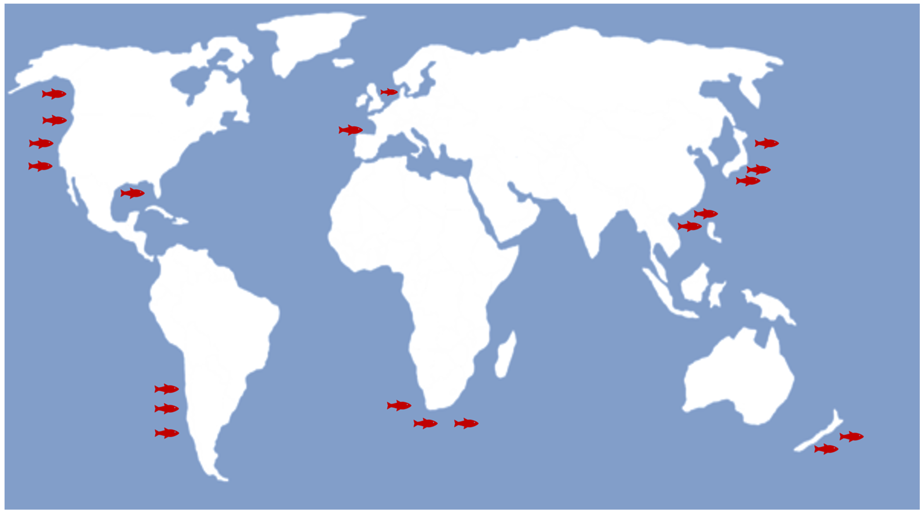
Figure 1. Worldwide distribution of H. akashiwo. The fish icons reveal the sites where harmful algal blooms with fish mortality were reported.
Microbiolres 14 00012 g001
Figure 2. Light microscopy (a,b), transmission electron micrograph (TEM) (c,d) and scanning electron micrograph (SEM) (e,f) of H. akashiwo. The images were taken by Zeiss Imager Z1 Upright, Philips 420 Transmission Microscope, and Philips CM10 Transmission Microscope, respectively.
Microbiolres 14 00012 g002
Figure 3. Schematic representation of H. akashiwo cyst formation. Light, temperature, and nutrients are key parameters to form benthic vegetative cyst or transfer cells into active stage. This image was created with BioRender (https://biorender.com/(accessed on 22 January 2023)).
Microbiolres 14 00012 g003
Figure 5. Brevetoxin structures. Modified from [88].
Microbiolres 14 00012 g005
Table 1. Hetrosigma classification [21].
EmpireEukaryota
KingdomChromista
PhylumOchrophyta
ClassRaphidophyceae
OrderChattonellales
FamilyChattonellaceae
GenusHeterosigma
Disclaimer/Publisher’s Note: The statements, opinions and data contained in all publications are solely those of the individual author(s) and contributor(s) and not of MDPI and/or the editor(s). MDPI and/or the editor(s) disclaim responsibility for any injury to people or property resulting from any ideas, methods, instructions or products referred to in the content.