Article

The Emergence of Carbapenem-Resistant Gram-Negative Bacteria in Mizoram, Northeast India

by
Vanlalruati S. C. Ralte
1,
Archana Loganathan
2,
Prasanth Manohar
3,
Christine Vanlalbiakdiki Sailo
4,
Zothan Sanga
5,
Lalremruata Ralte
6,
John Zothanzama
4,
Sebastian Leptihn
3,7,
Ramesh Nachimuthu
2,* and
Nachimuthu Senthil Kumar
4,*
1
Civil Hospital Lunglei, Lunglei 796701, India
2
Antibiotic Resistance and Phage Therapy Laboratory, School of Bio-Sciences and Technology, Vellore Institute of Technology, Vellore 632014, India
3
Zhejiang University-University of Edinburgh Institute, School of Medicine, Zhejiang University, Haining 314400, China
4
Department of Biotechnology, Mizoram University, Aizawl 796004, India
5
Department of Health and Family Welfare, Directorate of Health Services, Aizawl 796009, India
6
Department of Microbiology, Synod Hospital, Durtlang 796025, India
7
Infection Medicine, Biomedical Sciences, Edinburgh Medical School, College of Medicine and Veterinary Medicine, The University of Edinburgh, 1 George Square, Edinburgh EH8 9JZ, UK
*
Authors to whom correspondence should be addressed.
Microbiol. Res. 2022, 13(3), 342-349; https://doi.org/10.3390/microbiolres13030027
Submission received: 4 May 2022 / Revised: 17 May 2022 / Accepted: 20 May 2022 / Published: 22 June 2022

Abstract

:
(Background) Numerous reports on carbapenem resistance in different parts of India have been published, yet there are insufficient studies on the prevalence of antibiotic resistance in the northeast region of the country. This study evaluated the emergence of carbapenem resistance in the clinical isolates collected in Mizoram. (Results) A total of 141 Gram-negative clinical isolates were collected from the two hospitals, including the Civil Hospital in Lunglei and the Synod Hospital in Aizawl. The isolates include Escherichia coli (n = 62, 43.9%), Klebsiella spp. (n = 43, 30.4%), Pseudomonas aeruginosa (n = 9, 6.3%), Serratia marcescens (n = 3, 2.1%), Proteus mirabilis (n = 2, 1.4%), Shigella spp. (n = 4, 2.8%), Enterobacter spp. (n = 6, 4.2%) and Acinetobacter spp. (n = 12, 8.5%). The isolates were found to be resistant to meropenem (11%), colistin (48%), tigecycline (25%) and cefotaxime (50%). A total of four E. coli and one Shigella sonnei encoded the blaOXA-48-like gene. The blaCTX-M-1 gene was detected in 13 isolates, of which eight were E. coli, two Shigella flexneri, and one isolates each of K. pneumoniae, K. oxytoca and Shigella sonnei, respectively. (Conclusion) Carbapenem-resistant Enterobacteriaceae are common among other parts of India, despite limited access to antibiotics, the emergence of resistance in the northeastern region is worrying.

1. Introduction

Antimicrobial resistance in bacterial pathogens is a widespread and critical challenge in clinical treatment resulting in high morbidity and mortality rates globally [1]. Gram-negative bacteria can result in severe complications or even treatment failure when caused by multi-drug resistant (MDR) strains that are increasingly common around the globe. Carbapenems, a highly effective class of broad-spectrum antibiotics usually reserved for MDR infections, are increasingly deployed, and as a result, resistance among Gram-negative bacteria has now become a worldwide concern [2]. Carbapenems are transpeptidase inhibitors that inhibit the peptide crosslinking during cell wall synthesis leading to autolytic activity (cell death) [3]. The basis of Carbapenem resistance mechanisms is either acquired via horizontal gene transfer or is based on intrinsic mechanisms, which include mutations of the target site, enzymatic degradation of the antimicrobial compound, and changes in the efflux pump mechanisms. Among these, enzymatic degradation is the most prevailing mechanism [4]. Molecular mechanisms of carbapenemase enzymes have been proposed and are being classified as class A, class B and class D. Class A types are more often found in Enterobacteriaceae but rarely encountered in P. aeruginosa, which covers SME, NMC, KPC, IMI, GES-type of enzymes. Class B is found in Acinetobacter species, P. aeruginosa, and Enterobacteriaceae and includes VIM, SPM, GIM, and IMP-type enzymes. Class D includes the OXA-type, which is again more common among Acinetobacter [5].
The distribution of carbapenem-resistant Enterobacteriaceae is increasing, but the availability of data is limited in many countries. For lactose non-fermenters, the recorded rate of carbapenem resistance appears to be substantially higher than for lactose fermenters globally [6]. The virulence, as well as the pathogenicity of the carbapenem-resistant bacterial strains, were found to vary in the country in which a person is infected [7,8]. The epidemiological distribution of carbapenemase producers varies vastly. In Asian sub-continents, blaOXA-48-like and blaNDM-types are more common, while blaKPC and other blaOXA-types are recorded quite often in the European countries. In northern America, half of the carbapenem-resistant Enterobacteriaceae (CRE) is found to be carbapenemase-producing, while blaNDM, blaVIM and blaOXA-48-like are less prevalent. In comparison to other Asian countries, the rate of carbapenem resistance was found to be significantly lower in India. Yet, in hotspots in India, an average of 13% of cases were registered as CRE [9].
In the northeast regions, in the states of Mizoram, Tripura, Nagaland and Meghalaya, the prevalence of carbapenem resistance were found to be below 5% due to less developed healthcare and the comparably reduced amounts of carbapenems used in these regions [10]. Based on this information, this study was conducted to assess the emergence of Gram-negative bacteria in Mizoram that are carbapenem-resistant, as well as to detect the presence of carbapenemase genes in the clinical isolates collected from two hospitals in the region.

2. Materials and Methods

2.1. Collection of Bacterial Isolates

A total of 141 Gram-negative bacterial isolates were obtained from Civil Hospital Lunglei (Lunglei District) as well as Synod Hospital Aizawl (Aizawl District), Mizoram, between 2018 and 2019. The isolates were transported as cryoprotectant stocks to the Antibiotic Resistance and Phage therapy research Laboratory at VIT, Vellore. All the isolates were sub-cultured and stored at −20 °C. The bacterial identification was performed using the VITEK identification system and the isolates were further confirmed by 16S rRNA identification using the universal primers 27F and 1492R. PCR products were Sanger sequenced (Eurofins Genomics Ltd., India) and analyzed using the NCBI-BLAST tool. The blast hit was analyzed against the available data from the NCBI database and the sequences were deposited in NCBI-GenBank [11].

2.2. Minimal Inhibitory Concentration (MIC)

A test to determine the MIC was performed for all the isolates using meropenem, cefotaxime, colistin and tigecycline (Sigma Aldrich, India). Briefly, Muller Hinton No. 2 cation-controlled broth (Hi-Media, India) was prepared and 100 μL of the broth was added to the 96 well-microtiter plates. The antibiotic to be tested was diluted by adding 100 μL of the respective antibiotic ranging from 0.25 μg/mL to 256 μg/mL. The bacterial culture at 0.5 McFarland standards (approx. 1 × 10⁷–108 CFU/mL) was prepared and 5 μL was added to each well. The plates were incubated at 37 °C for 18 h. The results were recorded and interpreted according to CLSI guidelines (CLSI, 2019) [11].

2.3. DNA Isolation

Bacterial DNA was isolated from the clinical isolates following the boiling lysis method as described in our previous study [11]. Briefly, 10 colonies were inoculated into sterile distilled water and boiled for 10 min (temp: 95 °C) to break open the cells. The mixture was allowed to cool down to room temperature and centrifuged at 8000× g for 5 min and the supernatant containing the DNA was used as a template for PCR analysis.

2.4. Screening of Beta-Lactamase Genes

The resistance genes screening was performed on both chromosomal and plasmid DNA. Chromosomal DNA was isolated by the boiling lysis method (above), while plasmid DNA was isolated using the Plasmid DNA isolation kit (Hi-Media, India). Carbapenem resistance genes blaNDM, blaOXA-48-like, blaKPC, blaIMP, blaVIM, blaDIM, blaBIC, blaGIM, blaSIM and blaAIM and cefotaxime resistance genes, CTX-M group of genes: blaCTX-M-1, blaCTX-M-2, blaCTX-M-9, blaCTX-M-26 and blaCTX-M-8 were screened using PCR [12,13]. The integrase genes intI1, intI2 and intI3 were amplified (n = 16) by multiplex PCR using the primers described in a previous study [14]. The clonality of the bacterial species was determined (n = 141) by ERIC-PCR (enterobacterial repetitive intergenic consensus) using ERIC2 primers as described by Versalovic et al. [15]. ERIC-PCR results were analyzed by PyElph software (Python-based gel images analyzer) [16]. In brief, a DNA gel image was generated by photographing it with UV gel Doc. The DNA banding patterns (gel image) were inserted into PyElph software in JPEG format. The ERIC-PCR patterns obtained were interpreted and compared as described by Bilung et al. [17]. For cluster studies by ERIC-PCR, a dendrogram was generated using the unweighted pair group average (UPGMA) against a 1 kb DNA marker (HiMedia, Mumbai, India). The dendrogram was drawn for the entire genus separately and when the band similarity was greater than 80%, the clones were treated as related. The strains with carbapenem resistance genes are highlighted in red (Supplementary Figures S1–S7).

2.5. Identification of Carbapenemase Producers

The Modified Hodge test (MHT) was performed to detect the carbapenemase production in the resistant isolates. Briefly, an indicator organism, E. coli ATCC 25922, and the test bacteria were inoculated in MH broth and the turbidity was adjusted to 0.5 McFarland standard. Then, the indicator organism was plated onto Mueller–Hinton agar plates and the plates were allowed to dry for 10 min. Meropenem antibiotic disks (10 mcg) were placed at the centre of the agar plate. The test bacteria were streaked as a straight line from the edge of the disk towards the edge of the plate. The plates were incubated at 37 °C for 18 h. The results were interpreted according to the CLSI guideline [18].

2.6. Synergy Testing

The synergistic effect of the combination of two antibiotics was analyzed by the checkerboard method. The synergy was assessed using the two last-resort antibiotics, colistin and meropenem. Briefly, the test bacteria were diluted to 0.5 McFarland standard. Both the antibiotics were diluted in separate microtiter plates and combined into a single plate. In brief, meropenem (A) was diluted vertically from column A to H (1 μg/mL to 256 μg/mL) in 96 well microtiter plates marked as A and colistin (B) was diluted horizontally in rows from 1 to 12 in 96 well microtiter plate marked as B (1 μg/mL to 256 μg/mL). The diluted antibiotic from plate A was transferred to plate B onto their respective wells. The selected test bacteria were inoculated and the plates were incubated at 37 °C for 18 h. The synergistic effect was measured by calculating the Fractional Inhibitory Concentration (FIC) index of the antibiotic alone and in combination [19]. The FIC was calculated by a universal mathematical expression, FIC = (FIC A/=FIC B), where FIC A = (MIC of the antibiotic A in combination/MIC of the antibiotic A alone) and FIC B = (MIC of the antibiotic B in combination/MIC of the antibiotic B alone). Based on the value obtained at FIC, the results are interpreted as synergy (FIC ≤ 0.5), indifference (2 > FIC ≥ 0.5), and antagonistic (FIC ≥ 2).

3. Results

3.1. Bacterial Isolates and Carbapenem Resistance

The collected 141 non-repetitive Gram-negative clinical strains of bacteria included E. coli (n = 62, 43.9%), Klebsiella spp. (n = 43, 30.4%), P. aeruginosa (n = 9, 6.3%), Serratia marcescens (n = 3, 2.1%), Proteus mirabilis (n = 2, 1.4%), Shigella spp. (n = 4, 2.8%), Enterobacter spp. (n = 6, 4.2%) and Acinetobacter spp. (n = 12, 8.5%). The MIC results showed that 16/141 (11.3%) were resistant to meropenem, 68/141 (48.2%) were resistant to colistin, 35/141 (24.8%) to tigecycline and 71/141 (50.3%) to cefotaxime. MIC50 and MIC90 values for meropenem were 0.12 μg/mL, for tigecycline MIC50 and MIC90 value were 0.5 μg/mL and 0.25 μg/mL, for cefotaxime MIC50 and MIC90 value were 128 μg/mL and 0.12 μg/mL, for colistin MIC50 and MIC90 value were 16 μg/mL and 8 μg/mL. For none of these isolates we detected carbapenemase production by MHT. The isolates that showed co-resistance to colistin and meropenem was 4 (2.8%), tigecycline and meropenem was 9 (6.3%) and cefotaxime and meropenem was 12 (8.5%) (Figure 1).

3.2. Molecular Studies

Of the 141 isolates, five carried blaOXA-48-like and 13 encoded the blaCTX-M-1 gene. One isolate each of E. coli and Shigella sonnei was identified to carry both the blaOXA-48-like and the blaCTX-M-1 gene. In total, 16/141 isolates carried resistance genes, blaOXA-48-like and blaCTX-M-1 (Table 1). The blaOXA-48-like (5/141) carriers were E. coli (n = 4) and Shigella sonnei (n = 1). The isolates with blaCTX-M-1 (13/141) were E. coli (n = 8), Shigella flexneri (n = 2), K. pneumoniae (n = 1), K. oxytoca (n = 1) and Shigella sonnei (n = 1). Other beta-lactamase genes such as blaKPC, blaVIM, blaBIC, blaGIM, blaDIM, blaSIM and blaAIM were absent. None of the isolates had plasmid-borne resistance genes. A total of 16 isolates that carried resistance genes were identified at the species level and the sequences were deposited in GenBank.
Integron gene screening showed the presence of intl1 in 13 isolates and interestingly, all the isolates were blaCTX-M-1 positive (Table 1). The two isolates, E. coli and S. sonnei, that carried both blaCTX-M-1 and blaOXA-48-like genes were positive for intl1. The ERIC-PCR dendrogram demonstrates that the blaCTX-M-1 carrying carbapenem-resistant E. coli strains (EC46, EC48 and EC60) are closely related. Other strains of E. coli that carried blaOXA-48-like appear not related to each other. The ERIC-PCR results are shown as dendrograms in Supplementary Figures S1–S7.

3.3. Combinational Activity of Colistin-Meropenem

The effect of antibiotic combinations on the bacterial cells was studied by the checkerboard method using colistin and meropenem. Isolates that showed resistance to both meropenem and colistin were chosen for this experiment. Four K. pneumoniae isolates (KP1, KP2, KP11 and KP12) were selected. The selected isolates had meropenem MIC of >128 μg/mL, while colistin MIC was 4 μg/mL (KP 1, KP2 and KP11) and 128 μg/mL (KP 12). All four isolates showed a reduced MIC during combinational studies. The FIC value for KP1, KP2, KP11 and KP12 were 1 (indifference), 0.5 (synergy), 1 (indifference) and 50 (antagonistic), respectively.

4. Discussion

Our findings illustrate the emergence of carbapenem resistance among Gram-negative bacteria in the northeastern region of India. The occurrence of 11.3% meropenem resistance, previously uncommon in the Mizoram region, is an indicator of the spread of resistance across India and possibly also globally. In addition, the higher prevalence of resistance to other last-resort antibiotics is highly concerning; we found resistance to colistin in more than 48% of the isolates, while almost a quarter of the strains are resistant to tigecycline.
In India, the prevalence of carbapenem-resistant bacterial infections is extensive, especially in the Southern and Northern regions, where the population density is high [20]. This is the first study to report the distribution of carbapenem resistance among Gram-negative bacteria from clinical samples collected from the two districts of Mizoram. The most common carbapenemase genes reported in the Indian isolates were blaNDM and blaOXA [20], though other beta-lactamase genes such as blaIMP, blaSIM, blaVIM and blaAIM, also contribute to carbapenem resistance. In this study, only the blaOXA-48-like carbapenemase gene was detected in the chromosomal DNA of E. coli and Shigella sonnei. To the best of our knowledge, this is the first report of a carbapenemase gene in Shigella sonnei. A recent report from northeast India showed a low level of around 5% of carbapenem resistance, especially in the Enterobacteriaceae [21]. One of the previous studies in this region showed the presence of the blaNDM-1 gene in K. pneumoniae and its association with insertion sequence (IS) elements [22]. In this study, integrase (intl1) genes were detected in E. coli, Klebsiella spp., and Shigella spp., which carried beta-lactamase genes. Their role in disseminating resistance remains to be investigated. Though plasmid-borne genes were reported to disseminate resistance among the bacterial community, none of the isolates had plasmid-borne resistance genes in this study. Possibly, the genetic information might be acquired by transduction/infection by temperate phages; recently, it was shown that around 5% of prophage sequences in the nosocomial pathogen Acinetobacter baumannii encode antimicrobial resistance genes, including blaOXA23 and blaNDM-1 [23]. A recent study in the northeast region of Assam reported the presence of 18.9% of carbapenem-resistant Enterobacteriaceae [24]. The absence of carbapenemase production (detected by MHT) in the isolates carrying the blaOXA-48-like gene indicated that they were not efficient carbapenemase producers (or did not produce the enzyme at all). This implies that the strains are resistant to some other beta-lactamases.
Extended-spectrum cephalosporin-resistant isolates (n = 71) were also identified in this study. The common beta-lactamase genes found in the northeastern regions include blaCTX-M-15, blaCTX-M-3, SIM and VIM [22,25]. The presence of blaCTX-M-1 in E. coli and K. pneumoniae in this study is one of the first reports in the Mizoram region. To add to this report, two E. coli isolates were found to carry both the blaCTX-M-1 and blaOXA-48-like genes. This is a rather alarming finding for the northeastern region of India because it indicates faster dissemination of antibiotic resistance genes had occurred among the bacterial population. A study conducted by the state referral hospital in Falkawn, Mizoram, showed the emergence of a large number of MDR strains in this region [10]. Our study, which focused on two areas, Lunglei and Aizawl, found a high incidence of antibiotic resistance in Gram-negative bacteria to the last-resort antibiotics carbapenem, colistin and tigecycline. This indicates that resistance is disseminating to the northeastern parts of India, where the population is not dense. Possibly, the extensive use of colistin and other antibiotics in farming may contribute to the increasing numbers of resistant isolates as the resistance can spread from animal to human pathogens. In our study, carbapenem resistance was high among the E. coli, K. pneumoniae and Shigella spp. The discovery of Metallo-β-lactamases (MBL) and OXA-type carbapenemase in the clinical isolates of Gram-negatives are significant because the majority of the isolates are resistant to almost all -lactams and mostly involved in horizontal gene transfer [26,27]. A clonal similarity (ERIC-PCR) was not found between most of the resistant isolates, which is an indication that the resistance is currently just developing.
Clinical therapeutic options to treat carbapenem/colistin-resistant infections are mostly combinational therapies. Interestingly, the combination of two last-resort antibiotics, colistin and meropenem, showed a synergistic effect against four MDR K. pneumoniae. Although earlier studies showed synergy of the colistin-meropenem combination [28], this is one of the first against the clinical isolates collected from Mizoram. These results again support the combinational activity of two antibiotics against resistant bacterial pathogens. This study highlights the emergence of carbapenem-resistant bacteria in Mizoram, northeast India, and is a clear indication for immediate surveillance of AMR in this region. A high frequency of colistin, tigecycline, cefotaxime, and carbapenem resistance in association with beta-lactamase genes is a matter of concern to public health, not only in India but globally.

5. Conclusions

In this study, the emergence of carbapenem resistance and the high level of resistance developed towards cefotaxime, colistin and tigecycline are reported from the northeastern region of India, Mizoram, for the first time. A detailed investigation of the antibiotic susceptibility pattern and the exact cause of antibiotic resistance with the geographical location are some of the most crucial data to combat AMR. The present study showed the increased prevalence of carbapenem-resistant Gram-negative bacteria among clinical isolates from Lunglei and Aizawl regions covering the disseminating resistance genes, blaOXA-48-like and blaCTX-M-1 in northeast India. Similar to other studies from different parts of the world, this study also confirms the emergence of MDR bacteria against last-resort antibiotics with multiple resistance genes. This study contributes to the development of new strategies counteracting or limiting the spread of drug resistance in the northeastern region of India, the whole country and the world.

Supplementary Materials

The following supporting information can be downloaded at: https://www.mdpi.com/article/10.3390/microbiolres13030027/s1, Figure S1: Dendrogram of Enterobacter spp. (EB) based on ERIC-PCR; Figure S2: Dendrogram of Shigella spp. based on ERIC-PCR. Ssp1—Shigella spp.; SS—Shigella sonnei; SF—Shigella flexneri; Figure S3: Dendrogram of Klebsiella pneumoniae (KP) based on ERIC-PCR; Figure S4: Dendrogram of E. coli (EC) based on ERIC-PCR; Figure S5: Dendrogram of Acinetobacter spp. based on ERIC-PCR; Figure S6: Dendrogram of Pseudomonas aeruginosa (PA) based on ERIC-PCR; Figure S7: Dendrogram of Serratia marcescens (SM) based on ERIC-PCR.

Author Contributions

Conceptualization, V.S.C.R., C.V.S., Z.S., S.L. and L.R.; methodology, A.L., P.M. and R.N.; validation, A.L., P.M., R.N. and N.S.K.; formal analysis, A.L., V.S.C.R., C.V.S. and Z.S.; investigation, A.L., S.L. and R.N.; resources, V.S.C.R., C.V.S., J.Z., Z.S. and L.R.; writing—original draft preparation, P.M. and A.L.; writing—review and editing, R.N., S.L. and N.S.K.; supervision, R.N. and N.S.K.; project administration, A.L., P.M. and R.N.; funding acquisition, N.S.K. All authors have read and agreed to the published version of the manuscript.

Funding

This research was funded by DBT, New Delhi, Govt. of India for the Advanced State Biotech Hub at Mizoram University; grant number BT/PR23092/NER/95/589/2017.

Institutional Review Board Statement

The study was conducted in accordance with the Declaration of Helsinki, and approved by the Institutional Review Board (or Ethics Committee) of Vellore Institute of Technology for studies involving humans.

Informed Consent Statement

Not applicable.

Data Availability Statement

All the data that support the findings of this study have been deposited in GenBank under the accession numbers: MW600382, MW600383, MW600387, MW600385, MW600381, MW600380, MW60039, MW600384, MW600394, MW600392, MW600386, MW600389, MW588017, MW600393, MW600390 and MW600388.

Acknowledgments

The authors would like to thank the Vellore Institute of Technology (VIT, Vellore) for providing the ‘VIT Seed Grant’.

Conflicts of Interest

The authors declare no conflict of interest.

References

  1. Leptihn, S. Welcome back to the Pre-Penicillin Era. Why we desperately need new strategies in the battle against bacterial pathogens. Infect. Microb. Dis. 2019, 1, 33. [Google Scholar] [CrossRef]
  2. Georgios, M. Carbapenem Resistance: Overview of the Problem and Future Perspectives. Ther. Adv. Infect. Dis. 2016, 3, 15–21. [Google Scholar] [CrossRef] [Green Version]
  3. Francis, C.; Donkor, E. Carbapenem Resistance: A Review. Med. Sci. 2017, 6, 1. [Google Scholar] [CrossRef] [Green Version]
  4. Fatma, E. Carbapenem-Resistant Enterobacteriaceae. Med. J. Islamic World Acad. Sci. 2017, 25, 6–11. [Google Scholar] [CrossRef]
  5. Aslam, B.; Rasool, M.; Muzammil, S.; Siddique, A.B.; Nawaz, Z.; Shafique, M.; Zahoor, M.A.; Binyamin, R.; Waseem, M.; Khurshid, M.; et al. Carbapenem resistance: Mechanisms and drivers of global menace. In Pathogenic Bacteria; Kırmusaoğlu, S., Bhardwaj, S.B., Eds.; IntechOpen: London, UK, 2020. [Google Scholar] [CrossRef] [Green Version]
  6. Patrice, N.; Poirel, L. Epidemiology and Diagnostics of Carbapenem Resistance in Gram-Negative Bacteria. Clin. Infect. Dis. 2019, 69 (Suppl. 7), S521–S528. [Google Scholar] [CrossRef] [Green Version]
  7. Vanegas, J.M.; Parra, O.L.; Jiménez, J.N. Molecular Epidemiology of Carbapenem Resistant Gram-Negative Bacilli from Infected Pediatric Population in Tertiary-Care Hospitals in Medellín, Colombia: An Increasing Problem. BMC Infect. Dis. 2016, 16, 463. [Google Scholar] [CrossRef] [Green Version]
  8. Garg, A.; Garg, J.; Kumar, S.; Bhattacharya, A.; Agarwal, S.; Upadhyay, G.C. Molecular Epidemiology & Therapeutic Options of Carbapenem-Resistant Gram-Negative Bacteria. Indian J. Med. Res. 2019, 149, 285. [Google Scholar] [CrossRef]
  9. Brink, A.J. Epidemiology of carbapenem-resistant Gram-negative infections globally. Curr. Opin. Infect. Dis. 2019, 32, 609–616. [Google Scholar] [CrossRef]
  10. Lalhmangaihzuali, F.E.; Varte, Z.; Laldinmawii, G. Antibiotic Resistance Pattern of Uropathogens in Urinary Tract Infections in Children at State Referral Hospital, Falkawn, Mizoram, India. Int. J. Contemp. Pediatrics 2018, 5, 2108–2113. [Google Scholar] [CrossRef] [Green Version]
  11. Ramesh, N.; Prasanth, M.; Ramkumar, S.; Suresh, M.; Tamhankar, A.J.; Gothandam, K.M.; Karthikeyan, S.; Bozdogan, B. Colistin Susceptibility of Gram-Negative Clinical Isolates from Tamil Nadu, India. Asian Biomed. 2016, 10, 35–39. [Google Scholar] [CrossRef]
  12. Doyle, D.; Peirano, G.; Lascols, C.; Lloyd, T.; Church, D.L.; Pitout, J.D. Laboratory Detection of Enterobacteriaceae That Produce Carbapenemases. J. Clin. Microbiol. 2012, 50, 3877–3880. [Google Scholar] [CrossRef] [PubMed] [Green Version]
  13. Nachimuthu, R.; Kannan, V.R.; Bozdogan, B.; Krishnakumar, V.; Pandian, K.S.; Manohar, P. CTX-M-type ESBL-mediated resistance to third-generation cephalosporins and conjugative transfer of resistance in Gram-negative bacteria isolated from hospitals in Tamil Nadu, India. Access Microbiol. 2020, 3, 000142. [Google Scholar] [CrossRef] [PubMed]
  14. Kargar, M.; Mohammadalipour, Z.; Doosti, A.; Lorzadeh, S.; Japoni-Nejad, A. High Prevalence of Class 1 to 3 Integrons among Multidrug-Resistant Diarrheagenic Escherichia Coli in Southwest of Iran. Osong Public Health Res. Perspect. 2014, 5, 193–198. [Google Scholar] [CrossRef] [PubMed] [Green Version]
  15. Versalovic, J.; Schneider, M.; De Bruijn, F.J.; Lupski, J.R. Genomic Fingerprinting of Bacteria Using Repetitive Sequence-Based Polymerase Chain Reaction. Methods Mol. Cell. Biol. 1994, 5, 25–40. [Google Scholar]
  16. Pavel, A.B.; Vasile, C.I. PyElph—A Software Tool for Gel Images Analysis and Phylogenetics. BMC Bioinform. 2012, 13, 1–6. [Google Scholar] [CrossRef] [Green Version]
  17. Bilung, L.M.; Pui, C.F.; Su’ut, L.; Apun, K. Evaluation of BOX-PCR and ERIC-PCR as Molecular Typing Tools for Pathogenic Leptospira. Dis. Markers 2018, 2018, 1351634. [Google Scholar] [CrossRef] [Green Version]
  18. Pasteran, F.; Gonzalez, L.J.; Albornoz, E.; Bahr, G.; Vila, A.J.; Corso, A. Triton Hodge Test: Improved Protocol for Modified Hodge Test for Enhanced Detection of NDM and Other Carbapenemase Producers. J. Clin. Microbiol. 2016, 54, 640–649. [Google Scholar] [CrossRef] [Green Version]
  19. Costa, R.A.; Ortega, D.B.; Fulgêncio, D.L.; Costa, F.S.; Araújo, T.F.; Barreto, C.C. Checkerboard Testing Method Indicates Synergic Effect of Pelgipeptins against Multidrug Resistant Klebsiella Pneumoniae. Biotechnol. Res. Innov. 2019, 3, 187–191. [Google Scholar] [CrossRef]
  20. Manohar, P.; Shanthini, T.; Ayyanar, R.; Bozdogan, B.; Wilson, A.; Tamhankar, A.J.; Nachimuthu, R.; Lopes, B.S. The Distribution of Carbapenem- And Colistin-Resistance in Gram-Negative Bacteria from the Tamil Nadu Region in India. J. Med. Microbiol. 2017, 66, 874–883. [Google Scholar] [CrossRef]
  21. Chellapandi, K.; Dutta, T.K.; Sharma, I.; De Mandal, S.; Kumar, N.S.; Ralte, L. Prevalence of Multi Drug Resistant Enteropathogenic and Enteroinvasive Escherichia Coli Isolated from Children with and without Diarrhea in Northeast Indian Population. Ann. Clin. Microbiol. Antimicrob. 2017, 16, 49. [Google Scholar] [CrossRef]
  22. Mukherjee, S.; Bhattacharjee, A.; Naha, S.; Majumdar, T.; Debbarma, S.K.; Kaur, H.; Dutta, S.; Basu, S. Molecular Characterization of NDM-1-Producing Klebsiella Pneumoniae ST29, ST347, ST1224, and ST2558 Causing Sepsis in Neonates in a Tertiary Care Hospital of North-East India. Infect. Genet. Evol. 2019, 69, 166–175. [Google Scholar] [CrossRef] [PubMed]
  23. Loh, B.; Chen, J.; Manohar, P.; Yu, Y.; Hua, X.; Leptihn, S. A Biological Inventory of Prophages in A. baumannii Genomes Reveal Distinct Distributions in Classes, Length, and Genomic Positions. Front. Microbiol. 2020, 11, 579802. [Google Scholar] [CrossRef] [PubMed]
  24. Sharma, M.; Chetia, P.; Puzari, M.; Neog, N.; Borah, A. Menace to the Ultimate Antimicrobials among Common Enterobacteriaceae Clinical Isolates in Part of North-East India. BioRxiv 2019. [Google Scholar] [CrossRef]
  25. Kar, B.; Sharma, M.; Peter, A.; Chetia, P.; Neog, B.; Borah, A.; Pati, S.; Bhattacharya, D. Prevalence and Molecular Characterization of β-Lactamase Producers and Fluoroquinolone Resistant Clinical Isolates from North East India. J. Infect. Public Health 2021, 14, 628–637. [Google Scholar] [CrossRef] [PubMed]
  26. Walther-Rasmussen, J.; Høiby, N. OXA-Type Carbapenemases. J. Antimicrob. Chemother. 2006, 57, 373–383. [Google Scholar] [CrossRef] [PubMed] [Green Version]
  27. Lee, K.; Kim, C.K.; Yong, D.; Jeong, S.H.; Yum, J.H.; Seo, Y.H.; Docquier, J.D.; Chong, Y. Improved Performance of the Modified Hodge Test with MacConkey Agar for Screening Carbapenemase-Producing Gram-Negative Bacilli. J. Microbiol. Methods 2010, 83, 149–152. [Google Scholar] [CrossRef] [PubMed]
  28. Manohar, P.; Shanthini, T.; Ekta, P.; Mahesan, J.B.; Gothandam, K.M.; Bozdogan, B.; Ramesh, N. Colistin-Resistant Klebsiella Pneumoniae: Prevalence of Integrons and Synergistic out Turn for Colistin-Meropenem. Arch. Clin. Infect. Dis. 2018, 13, e55099. [Google Scholar] [CrossRef] [Green Version]
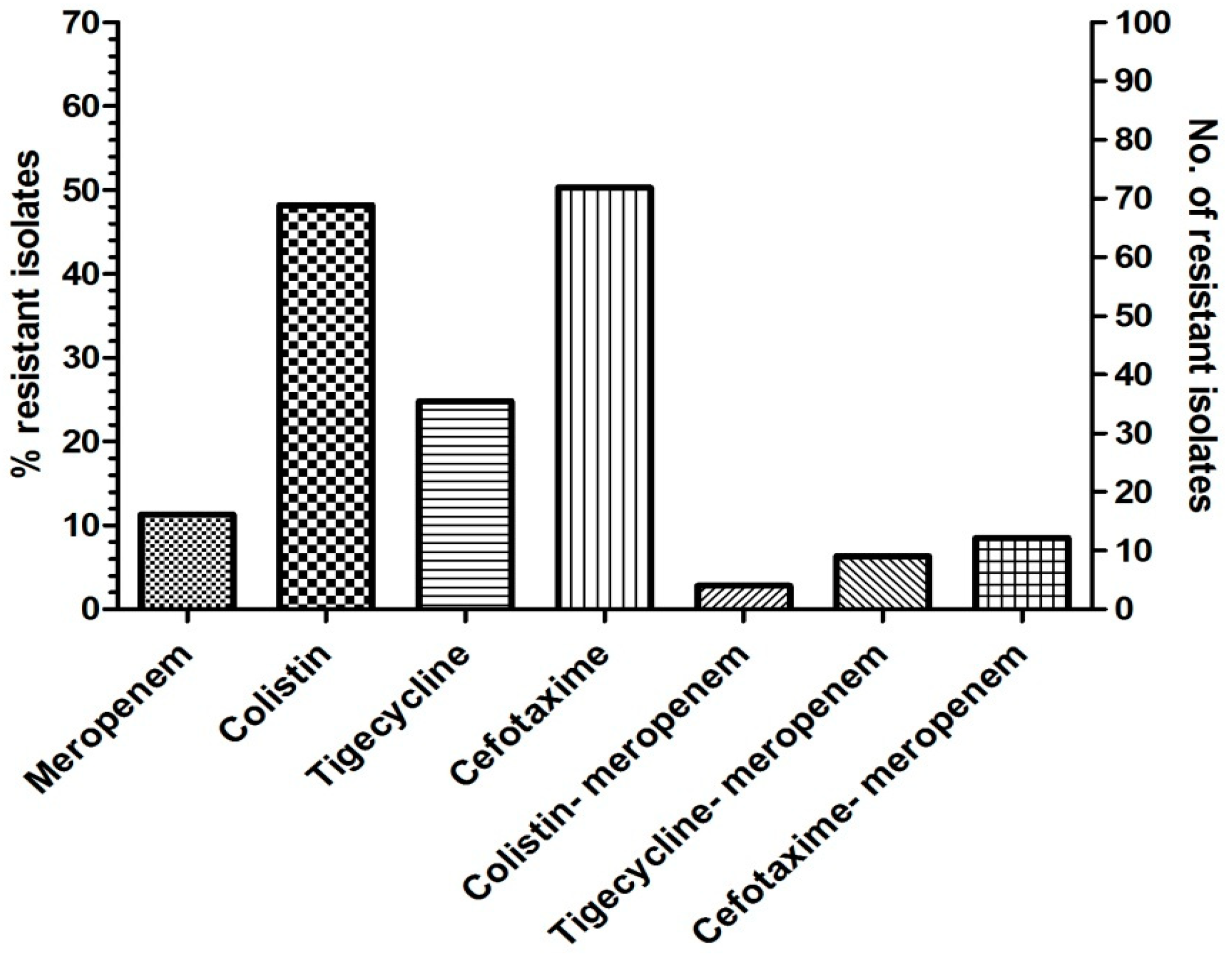
Figure 1. The distribution of antibiotic-resistant isolates among the Gram-negative bacterial isolates. A total of 141 Gram-negative bacteria were investigated in this study.
Microbiolres 13 00027 g001
Table 1. The distribution of beta-lactamase genes among clinical Gram-negative bacteria is investigated in this study.
S.noIsolate IDBacteriaMICResistance GeneInt Gene
ColTigMerCef
1EC 2E. coli1464>128blaOXA-48-like-
2EC 20E. coli2<0.1232>128blaOXA-48-like-
3EC 15E. coli1<0.12128>128blaOXA-48-like-
4SS 1Shigella sonnei2<0.12128>128blaOXA-48-like and
CTX-M-1
Intl1
5EC 17E. coli2<0.1232>128blaOXA-48-like and
CTX-M-1
Intl1
6SF 1Shigella flexneri24>128>128CTX-M-1Intl1
7EC 4E. coli20.50.5>128CTX-M-1Intl1
8SF 2Shigella flexneri2<0.12<0.12>128CTX-M-1Intl1
9EC 8E. coli1<0.12<0.12>128CTX-M-1Intl1
10EC 12E. coli20.5<0.124CTX-M-1Intl1
11EC 13E. coli20.5<0.12>128CTX-M-1Intl1
12KO 1K. oxytoca160.5<0.12>128CTX-M-1Intl1
13KP 37K. pneumoniae8<0.12<0.12>128CTX-M-1Intl1
14EC 47E. coli4<0.12<0.12>128CTX-M-1Intl1
15EC 49E. coli80.5<0.12<0.12CTX-M-1Intl1
16EC 61E. coli20.12<0.12>128CTX-M-1Intl1
Mer—Meropenem; Col—Colistin; Tig—Tigecycline; Cef—Cefotaxime; EC—E. coli; SF—Shigella flexneri; KO—Klebsiella oxytoca; SS—Shigella sonnei; KP—Klebsiella pneumoniae. The number in bold represents resistance by MIC. Bold numerals represent resistant isolates.
Publisher’s Note: MDPI stays neutral with regard to jurisdictional claims in published maps and institutional affiliations.